REVIEW
Left ventricular apical diseases
Silvia Cisneros&Ricardo Duarte&Gabriel C. Fernandez-Perez&Daniel Castellon& Julia Calatayud&Iñigo Lecumberri&Eneritz Larrazabal&Berta Irene Ruiz
Received: 18 October 2010 / Revised: 15 January 2011 / Accepted: 1 April 2011 / Published online: 18 April 2011 #European Society of Radiology 2011
Abstract There are many disorders that may involve the left ventricular (LV) apex; however, they are sometimes difficult to differentiate. In this setting cardiac imaging methods can provide the clue to obtaining the diagnosis. The purpose of this review is to illustrate the spectrum of diseases that most frequently affect the apex of the LV including Tako-Tsubo cardiomyopathy, LV aneurysms and pseudoaneurysms, apical diverticula, apical ventric-ular remodelling, apical hypertrophic cardiomyopathy, LV non-compaction, arrhythmogenic right ventricular dysplasia with LV involvement and LV false tendons, with an emphasis on the diagnostic criteria and imaging features.
Keywords Cardiac aneurysm . Left ventricular remodelling . Left ventricular hypertrophy . Tako-Tsubo
cardiomyopathy . Isolated non-compaction of the ventricular myocardium
Introduction
The left ventricle (LV) is affected by many diseases with different clinical and morphological features. Within this broad spectrum, a subset of heterogeneous diseases is characterised as preferentially affecting the LV apex. Generally LV assessment begins with echocardiography because of its wide availability and because it is fast, low-priced, portable and robust in experienced hands. However, because of intrinsic or technical limitations (limited field of vision, inadequate acoustic window, inability to definitively depict the endocardial border, especially the anterolateral
Electronic supplementary material The online version of this article (doi:10.1007/s13244-011-0091-6) contains supplementary material, which is available to authorized users.
S. Cisneros (*)
:
I. Lecumberri:
E. Larrazabal:
B. I. Ruiz Department of Radiology, Hospital Basurto,Avd. Montevideo, nº18, 48013 Bilbao, Vizcaya, Spain e-mail: [email protected]
I. Lecumberri
e-mail: [email protected]
E. Larrazabal
e-mail: [email protected]
B. I. Ruiz
e-mail: [email protected]
R. Duarte
Department of Radiology, Centro Hospitalar de Gaia, Rua Conceição Fernandes - Vilar de Andorinho, 4430–502 Vila Nova de Gaia, Portugal e-mail: [email protected]
G. C. Fernandez-Perez
Department of Radiology, HNS Sonsoles. Avila, Juan Carlos I s/n,
05001 Avila, Spain
e-mail: [email protected]
D. Castellon
:
J. Calatayud Department of Radiology, Povisa, C/ Salamanca nº 5,36211 Vigo, Galicia, Spain
D. Castellon
e-mail: [email protected]
J. Calatayud
free wall of the left ventricle in the parasternal short-axis view and the apex) [1,2] and technical artefacts (reverber-ations, near-field artefacts), in such cases further investiga-tion, usually with magnetic resonance (MR) imaging, which is the method of choice for the diagnosis of myocardial diseases, may be necessary. MR imaging is less dependent on the operator and is not subject to acoustic window limitations [3]. Due to their high spatial and temporal resolution, MR and CT can clearly visualise the apex of the LV, but CT is not yet recommended as the method of primary choice in the case of suspected left ventricular apical disease. Cine MR imaging with the steady-state free precession pulse sequence can offer the advantages of multiplanar imaging, complete coverage of the entire myocardium without obliquity, and excellent soft-tissue contrast between the myocardial border and the blood pool [4, 5]. In addition, the status of myocardial blood flow can be assessed by using adenosine stress cardiac MR imaging. Delayed enhancement MR imaging techniques can provide unique information for tissue characterisation, specifically for the identification of myocardial fibrosis or scarring [6,7]. CT has also emerged
as a novel technique for evaluating cardiac morphology and function, as well as the coronary artery. Compared with the spatial resolution of MR imaging, that of multidetector CT is usually higher [8]. Therefore, cardiac multidetector CT can offer anatomical and functional information about the cardiac chambers and a high-quality non-invasive coronary angiography [9,10]. Multi-detector CT would be more appropriate in those cases for which there are specific requests to exclude coronary artery disease and in those with contraindications for MR imaging, such as the patient having a pacemaker [11].
Therefore, radiologists play an increasing role in the evaluation of LV apical abnormalities, thus requiring a greater awareness and familiarity with this diseases.
The aim of this paper is to illustrate the spectrum of diseases that most frequently affect the apex of the LV, with an emphasis on the diagnostic criteria and imaging features.
Normal findings
Myocardial thinning (one of the principal signs of myocar-dial injury) is an important observation in patients suspected of having had ischaemic insults to the heart. However, thinning of the LV apex has been described by anatomists as a normal feature. With the advent of motion-gated cross-sectional cardiac imaging, normal apical thin-ning can be quite noticeable, especially on computed tomography (CT) in which the spatial resolution is submillimetre [12,13].
Ischaemia-related diseases
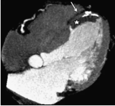
Magnetic resonance imaging has increasing potential in the non-invasive evaluation of patients with myocardial infarc-tion and subsequent complicainfarc-tions. The low interobserver variability and the high interstudy reproducibility of MR imaging for quantifying LV volume, wall thickening and mass make it an attractive imaging technique for follow-up Fig. 1 Patient with antecedent of myocardial infarction.
Contrast-enhanced CT (CECT) shows a small amount of contrast medium outside the ventricular wall due to a pseudoaneurysm (white arrow). Note that the calcifications formed as a result of necrosis define the ventricular wall
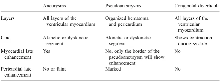
Aneurysms Pseudoaneurysms Congenital diverticula
Layers All layers of the ventricular myocardium
Organized hematoma and pericardium
All layers of the ventricular myocardium Cine Akinetic or dyskinetic
segment
Akinetic or dyskinetic segment
Shows contraction during systole Myocardial late
enhancement
Yes No, only the border of the pseudoaneurysm will show enhancement
No
Pericardial late enhancement
No or faint Marked No
studies in patients with ischaemic coronary artery disease and LV remodelling after infarction [14–16] . Moreover, MR imaging can clearly localise the site of the aneurysm, pseudoaneurysm or diverticula, is capable of distinguishing among pericardium, thrombus and myocardium, and delayed enhancement MR imaging helps in the differenti-ation between a true aneurysm and a pseudoaneurysm [17].
Apical aneurysm and pseudoaneurysm
Pseudoaneurysms or false aneurysms are defined as a rupture of the myocardium that is contained by pericardial adhesions. A pseudoaneurysm usually represents a rare complication of myocardial infarction (Fig. 1, Video 1and2), but it may also occur after cardiac surgery, chest trauma and endocarditis [18]. They can occur in different locations, but the purpose of this paper is to describe those arising from the apex of the LV. The wall of a false aneurysm is composed of organised haematoma and pericardium (Table1). Pathological examination shows fibrous tissue and lacks the myocardial elements that are usually seen in the wall of true aneurysms [18].
Conversely, a true aneurysm, which occurs in 5–10% of patients with acute myocardial infarction, contains the endocardium, epicardium and a predominantly fibrous tissue that has replaced myocardium in its wall [17]. An important difference is the lower rupture potential of a true aneurysm compared with a pseudoaneurysm. Surgical repair is the treatment of choice for pseudoaneurysms, while true aneurysms, in the absence of other surgical indications (i.e. refractory angina pectoris, congestive heart failure, systemic embolisation or refractory arrhythmia) are treated medically [18,19]. Cine MR and cine CT imaging
reveal, in both aneurysms and pseudoaneurysms, an akinetic/dyskinetic segment in the LV wall [18, 19]. A typical feature of pseudoaneurysm is that it has a narrow ostium connecting them to the ventricle. In most cases, the maximal width of the ostium is less than the maximal parallel internal diameter [18].
True aneurysms will show late enhancement, which indicates that their wall is formed by scar tissue secondary to infarcted myocardial muscle. Owing to the fact that their wall is composed only of pericardium, pseudoaneurysm
Fig. 2 True aneurysm in a patient with myocardial infarction. CECT in the four-chamber axis demonstrates a large and thinning area of akinesia in the anteroseptal-apical wall (arrow). A mural thrombus is also observed in this aneurysm (asterisk)
Fig. 3 Apical ventricular remodelling in a patient with myocardial infarction antecedent. Magnetic resonance imaging demonstrates left ventricular (LV) dilation (white arrow) with distortion of its geometry resulting in impairment in systolic function (EF: 28%)
sacs do not show late enhancement; however, their border will show enhancement, indicating a perianeurysmal in-farcted area [17, 18]. There is often marked delayed enhancement of the pericardium in pseudoaneurysms, and it is uncommon or faint in true aneurysm, with a sensitivity
and specificity of 100% and 83%, respectively [18]. Associated mural thrombus is common in both aneurysms and pseudoaneurysms (Fig. 2and Video3) [17,18].
Apical diverticula
Apical diverticulum is a finger-like contractile pouch with connection to the ventricle [19]. It is important to differentiate apical diverticula from aneurysm and pseudoa-neurysm. Ventricular diverticula are congenital, often asymptomatic and are usually incidentally found while performing diagnostic imaging procedures for other reasons [20]. The wall of the diverticulum contains all layers of the ventricular myocardium with preserved myocardial archi-tecture [20]. Contrary to a LV aneurysm, this diverticulum shows contraction during systole, because it contains a muscular wall [19,20]. Delayed enhancement is not seen.
In children, apical diverticula can be associated with Cantrell’s syndrome, which is a rare syndrome characterised by a partial sternal cleft, anterior abdominal wall defects, anterior diaphragmatic defect and intracardiac defects of which the ventricular septal defect is the most common and Table 2 Relative merits of each non-invasive imaging technique for
the assessment of hypertrophic cardiomyopathy (HCM)
Factor assessed Echocar-diographya
Multi-detector CT
MR imaging
LV volume +++ ++ ++++
Ejection fraction +++ +++ ++++ LV filling
pressure
+++ - ++
Dynamic obstruction
+++ + +++
IschemialCFR + - ++
Tissue
characterisation
++ + ++++
—The number of pluses indicates the relative usefulness of the modality. -= modality is not useful, CFR = coronary flow reserve, PA = pulmonary artery.aData in this column are from Nagueh and Mahmarian [7]. Table adapted from [3]
is invariably present. LV diverticulum is present in 20% to 50% of these cases [19].
Apical ventricular remodelling
Cardiac remodelling refers to the change in size, shape and function of the heart after injury to the ventricles [16,21,
22]. The injury is typically due to large acute myocardial infarction depending on anterior descending coronary artery occlusion. However, there are other non-ischaemic causes (i.e. chronic pressure or chronic volume overload and genetically determined cardiomyopathy) [21]. Myocardial infarction promotes transformation of both infarcted and non-infarcted regions, which leads to global LV dilation, referred to as “ventricular remodelling”. It is a dynamic process, starting in the acute phase with myocardial thinning and lengthening of the infarcted area, progressing to LV dilatation (Fig. 3) and hypertrophy of the non-infarcted area [21, 22]. Dysfunction with increased sphericity is common, as is the deposition of myocardial fat [23] and the development of apical thrombus (Fig. 4
and Video4) [21].
Apical hypertrophic cardiomyopathy
Hypertrophic cardiomyopathy (HCM) is a primary and genetic myocardial disease characterised by a hypertro-phied, non-dilated LV in the absence of another systemic or cardiac disease (e.g. systemic hypertension, aortic valve stenosis) capable of producing LV hypertrophy [1,24].
There are several distinctive involvement patterns, one variant being the apical hypertrophic cardiomyopathy (AHC). Unlike usual HCM, AHC possesses typical clinical features and has a relatively good prognosis. However, it seems that the clinical course is less benign in western countries than in Japan [9].
Cardiac MR imaging and multidetector CT are capable of providing clinically useful information for the accurate diagnosis of HCM, as well as for the determination of clinical management strategies. However, MR imaging can allow better characterisation of the pattern and distribution of LV hypertrophy in HCM than in CT. Cardiac MR imaging should be considered as the reference standard for establishing a diagnosis of HCM when the results from echocardiography are inconclusive or are suspected of being false-negative findings. Moreover, cardiac MR imaging is strongly recommended as a powerful imaging technique for differentiating HCM from other cardiomyopa-thies in the differential diagnosis (Table2) [3].
The diagnosis is primarily based on the demonstration of localised apical hypertrophy, defined as an end-diastolic LV
apical wall thickness greater than 15 mm or a ratio comparing apical LV and basal LV wall thicknesses of≥1.3–1.5 [25].
More subjective criteria for the diagnosis of AHC include: obliteration of the LV apical cavity in systole, failure to identify a normal progressive reduction in LV wall thickness towards the apex [25] and apical aneurysm formation with delayed enhancement [25,26]. The forma-tion of apical aneurysm is thought to be due to ischaemia, which results from reduced capillary density, hyperplasia of the arterial media, increased perivascular fibrosis and myocardial bridging. This process usually occurs in the presence of normal epicardial coronary arteries [25].
Patchy delayed enhancement in a mid-wall location is present in most patients with HCM (Fig. 5) [25].
There are three types of apical HCM: (1) true apical form (“spade-like” configuration), (2) involvement of the apex and symmetric hypertrophy of the four ventricular wall segments, and (3) involvement of the apex with asymmetric involvement of the wall segments (“non-spade” apical HCM) [27].
Table 3 Most frequent primary and secondary cardiomyopathies [4]
Primary cardiomyopathies (mostly confined to the heart)
Secondary cardiomyopathies (cardiac involvement is a part of systemic disease)
Genetic: Infiltrative:
Hypertrophic Amyloidosis Arrhythmogenic
Non-compaction
Mixed: Storage:
Dilated Hemochromatosis
Restricted
Acquired: Toxicity:
Inflammatory Drugs
Tako-Tsubo Chemical agents Perip artum
Endocrine: Diabetes mellitus Hyperthyroidism Hypothyroidism Hyperparathyroidism Pheochromocytoma Acromegaly
Autoimmune/collagen: Systemic lupus erythematosis Dermatomyositis
Tako-Tsubo cardiomyopathy
Transient LV apical ballooning syndrome or Tako-Tsubo cardiomyopathy (TSC) is a primary (Table3) [24], acquired and reversible cardiomyopathy that usually affects post-menopausal women and is often provoked by a stressful event or physical stress. This disorder shows clinical features practically indistinguishable from acute myocardial infarction in the absence of significant coronary artery stenosis (which can be demonstrated by a normal CT coronarography) with systolic dysfunction that is completely reversible within a few days or weeks [24,28].
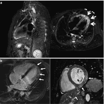
The MR findings are characterised by LV myocardial oedema with a diffuse and transmural distribution better visualised on the STIR T2-weighted sequences as high signal intensity (Figs.6,7) that matches with wall-motion abnormalities seen on cine-MRI. Hypokinesis or akinesis in the mid- and apical segments (although it can affect the basal segments alone) of the LV causes“apical ballooning” with compensatory basal segment hyperkinesis, taking the name of Tako-Tsubo from the morphology of LV in the ventriculography, which is similar to the Japanese pot used to capture octopuses. These wall motion abnormalities can produce a dynamic obstruction in the LV outflow (LVOT)
seen as a “jet” in the LVOT plane and also mitral regurgitation [28].
Tako-Tsubo cardiomyopathy and acute myocardial infarc-tion (Fig.8) can be clinically indistinguishable. Nevertheless, the differential diagnosis of the disorders can be suspected by the oedema location in the apical and mid-segments of Fig. 6 A 72-year-old woman with Tako-Tsubo cardiomyopathy who
was seen in the emergency room for acute chest pain, raised cardiac enzymes and ST elevation segments. Conventional coronarography did not show significant epicardial stenosis. a STIR T2-weighted
images in the short-axis view show diffuse oedema in apical segments with transmural location. Note how the mid and especially the basal segments are spared.bDelayed enhancement sequence shows normal and viable myocardium ruling out necrosis in the LV wall
LV that does not correlate with a vascular territory (Table4) [29], and TSC has rarely been reported to cause late enhancement. Wall-motion abnormalities extend beyond the region supplied by one coronary artery. This feature is characteristic of Tako-Tsubo cardiomyopathy, whereas wall-motion abnormalities observed in acute myocardial infarc-tion are often more localised. Acute myocarditis can also be clinically similar to TSC (acute chest pain, elevated cardiac enzymes levels and ECG changes), but has a more diffuse, heterogeneous distribution of the oedema (Fig.9), often with sub-epicardial affectation that may be accompanied by pericardial enhancement [28].
Another characteristic feature of TSC is the lack of hypoperfusion on the first-pass imaging and at rest and of late enhancement on the delayed contrast sequence (Fig.6) in the LV segments where there is STIR spotted oedema, which allows the differentiation from anterior ST elevation myocardial infarction. A rare but serious complication is the thrombus formation in the LV. The LV wall thickness is normal [28].
In combination with clinical history and catheter-isation findings, cardiac MR imaging can be useful in supporting the diagnosis and monitoring functional
recovery [30]. MRI is also useful in differentiating Tako-Tsubo cardiomyopathy from myocardial infarction and myocarditis. Although coronary CT angiography is not indicated in the initial evaluation of patients with Tako-Tsubo cardiomyopathy, reports are emerging of the use of coronary CT angiography in the subsequent evaluation of patients with TSC [31].
Left ventricular non-compaction
LV non-compaction (LVNC) may be an isolated finding (isolated left ventricular non-compaction) or may be associated with other congenital heart anomalies (left ventricular non-compaction). It is a congenital cardiomy-opathy [24] characterised by a thickened wall with multiple prominent ventricular trabeculations and deep intertrabecular recesses (sinusoids) in communication with the ventricular cavity, resulting in systolic and diastolic ventricular dysfunc-tion. Non-compaction involves predominantly the apical portion of the LV chamber, with or without right ventricular involvement, due to an arrest in the normal embryogenesis [32–34].
Fig. 8 A 68-year-old man with acute myocardial infarction. STIR T2-weighted images show high signal intensity (white arrow) due to ventricular oedema that has a vascular distribution involving segments irrigated by the left anterior descending artery
Tako-Tsubo AMI Myocarditis
Hyperintensity distribution in T2-STIR
Transmural or diffuse Transmural or subendocardial
Medium or subepicardic
Location Apical and mid ventricular segments without vascular distribution
Vascular distribution
Patchy, without vascular distribution
Hyperintensity durability in T2-STTR
2 weeks More than 2 weeks
Because both the left and right ventricles normally have trabeculations, there is always the risk of over-diagnosis of this disease. It is therefore essential to be thorough while establishing the relationship between compacted and non-compacted myocardium when the diagnosis of this entity is being made. Using MRI, the most appropriate criteria would be a ratio between non-compacted and compacted areas greater than 2.3 (Fig.10) in a diastolic long-axis view [35].
Although ventricular non-compaction can appear similar to ventricular diverticula or prominent trabeculations (while virtually always <3 in number in normal variants), diagnosis of ventricular non-compaction usually requires the finding of more than three deep intertrabecular recesses in one imaging plane apically from the insertion of the papillary muscles [20, 32, 36]. False tendons, dilated cardiomyopathy, endocardial fibroelastosis and cardiac Fig. 9 A 25-year-old man with
acute myocarditis.aSTIR T2-weighted sequence planned in the vertical long axis and four-chamber view showing oedema with a different form of Tako-Tsubo disease. The location of the oedema is diffuse but af-fecting all LV planes; also, the appearance of the oedema is patched.bDelayed enhance-ment sequence in the four-chamber view and post-contrast balance sequence in the short-axis view showing typical en-hancement in the mid- and sub-epicardial LV wall (arrows)
metastases are other important differential diagnostic con-siderations [34,36].
Although echocardiography has been the diagnostic test of choice for non-compaction, other techniques have been used for the diagnosis, including CT and MRI [34]. Echocardiography may not visualise the apical region optimally, leading to underestimation of the degree of left ventricular non-compaction [37]. MRI provides good correlation with echo for the localisation and extent of non-compaction, and is useful in cases of poor echocardio-graphic image quality. In addition, the demonstration of differences in MRI signal intensity in non-compacted myocardium may help identify substrates for potentially lethal arrhythmias [34].
Arrhythmogenic right ventricular dysplasia
Arrhythmogenic right ventricular dysplasia (ARVD) is an uncommon form of inheritable cardiomyopathy [24] and
is characterised by progressive loss of myocytes and fibrofatty replacement of the right ventricular myocardium [24,38–41].
The diagnosis of ARVD is based on established criteria determined by a task force comprising the European Society of Cardiology and the International Society and Federation of Cardiology [42].
The diagnosis of ARVD is based on the presence of major and minor criteria (Table 5) [39, 42]. These criteria include, in addition to personal and family history, 12-lead ECG, echocardiography, right ventricular angiography, cardiac magnetic resonance imaging and computerised tomography [24, 39]. Although these criteria are specific, they lack sensitivity and have never been validated, in part because there is no single definitive means of making the diagnosis [39]. Endomyocardial biopsy from the right ventricular free wall is a sensitive diagnostic marker when fibrofatty infiltration is associated with surviving strands of myocytes [24] and is the preferred method for making the diagnosis [39]. Experience with
Table 5 Diagnostic criteria for arrhythmogenic right ventricular dysplasia (ARVD)
Diagnostic criteria
I. Global and/or regional dysfunction and structural alterations
•Major
—Severe dilatation and reduction of right ventricular ejection fraction with no (or only mild) left and ventricular impairment
—Localized right ventricular aneurysms(akineticor dyskinetic areas with diastolic bulging) —Severe segmental dilatation of the right ventricle
•Minor
—Mild global right ventricular dilatation and/or ejection fraction reduction with normal left ventricle —Mild segmental dilatation of the right ventricle
—Regional right ventricular hypokinesia II. Tissue characterisation
of walls
•Major
—Fibrofatty replacement of myocardium at endomyocardial biopsy III. Repolarisation
abnormalities
•Minor
—Inverted T waves in right precordial leads (V2 and V3) (people >12 years old; in absence of right bundle branch block)
IV. Depolarisation of conduction abnormalities
•Major
—Epsilon waves or localised prolongation (>110 ms) of the QRS complex in right precordial leads (V1—V3)
•Minor
—Late potentials (signal-averaged ECG) V. Arrhythmias •Minor
—Left bundle branch block type ventricular tachycardia (sustained or nonsustained; ECO, Holter, exercise testing)
—Frequent ventricular extrasystoles on Holter (>1,000/24 h) VI. Family history •Major
—Familial disease confirmed at necropsy or surgery
•Minor
—Familial history of premature sudden death (at <35 years) due to suspected right ventricular cardiomyopathy —Familial history (clinical diagnosis based on present criteria)
CT in the diagnosis of ARVD is limited, but it has been used in the detection of morphological abnormalities [38,
43, 44] and wall motion abnormalities, particularly in patients with implantable defibrillators [43]. MRI is established as the imaging technique of choice in the assessment of ARVD [39,44].
Reports of left ventricular involvement have led to the recognition of ARVD as a diffuse disease of the heart muscle affecting both ventricles [40,41,45, 46]. There is evidence of LV involvement with fibrofatty replacement, chamber enlargement and myocarditis in up to 75% of patients [24]. Focal left ventricular dyskinesia can also be present in the setting of fatty infiltration within the left ventricle [39]. Late left ventricular enhancement has also
been described in patients with ARVD [39,47]. In addition, these changes can definitely affect the LV apex.
Left ventricular false tendons
Besides the normal papillary muscles and chordae tendinae, in a minority of the population, false tendons or false chordae occur as normal variants in the LV [48,49]. They might be single or multiple, usually thin, but sometimes very prominent, leading to confusing echocardiograms. They extend from the interventricular septum to the LV free wall (Fig. 11), or rarely, from the septum to the papillary muscle [36, 48, 50]. The LV false tendons are seen with considerable frequency in the echocardiographic evaluation of patients referred for having clinically innocent heart murmurs. They have a fibrous or fibromuscular structure (the thinner the tendon is, the higher fibrous component they have, and at greater thickness, a greater fibromuscular component) [48].
Apico-aortic conduit
As an infrequent finding radiologists could discover left apical devices secondary to iatrogenic manoeuvres that involve the ventricular apex; one of them is the implantation of an LV apico-aortic conduit (AAC) (Fig.12), which is an alternative approach for patients with aortic stenosis and conditions that either preclude a median sternotomy or present a high risk of aortic cross-clamping and aortotomy [51, 52]. It has three components: an outflow graft to the descending aorta, a valved conduit and an elbow apical connector. An apico-aortic conduit decreases the LV apico-aortic pressure gradient, preserves or improves ventricular function, and maintains
Fig. 12 Apico-aortic bypass is an alternative surgical option to aortic valve replacement for patients with a porcelain aorta or LV outflow tract obstruction. It is formed by a graft positioned in the ventricular apex with a valve into the conduit and anas-tomosed to the thoracic aorta (arrows)
normally distributed blood flow through the systemic and coronary circulation [53].
Conclusions
The diagnosis of diseases affecting the apex of the LV can be challenging. This is mainly caused by the technical limitations of echocardiography. CT and MRI are non-invasive techniques that, because of their high temporal and spatial resolution, are valuable in assess-ing the apex of the LV, overcomassess-ing the technical limitations of echocardiography and contributing to the differential diagnosis (i.e. apical diverticula, aneurysms and pseudoaneurysms).
References
1. Maron BJ (2002) Hypertrophic cardiomyopathy: a systematic review. JAMA 287(10):1308–1320
2. Prasad K, Atherton J, Smith GC, McKenna WJ, Frenneaux MP, Nihoyannopoulos P (1999) Echocardiographic pitfalls in the diagnosis of hypertrophic cardiomyopathy. Heart 82(suppl 3): III8–III15
3. Chun EJ, Choi SI, Jin KN, Kwag HJ, Kim YJ, Choi BW, Lee W, Park JH (2010) Hypertrophic cardiomyopathy: assessment with MR imaging and multidetector CT. Radiographics 30(5):1309– 1328
4. Pennell DJ, Sechtem UP, Higgins CB et al (2004) Clinical indications for cardiovascular magnetic resonance (CMR): con-sensus panel report. Eur Heart J 25(21):1940–1965
5. Lima JA, Desai MY (2004) Cardiovascular magnetic resonance imaging: current and emerging applications. J Am Coll Cardiol 44 (6):1164–1171
6. Maron MS, Maron BJ, Harrigan C et al (2009) Hypertrophic cardiomyopathy phenotype revisited after 50 years with cardio-vascular magnetic resonance. J Am Coll Cardiol 54(3):220–228 7. Nagueh SF, Mahmarian JJ (2006) Noninvasive cardiac imaging in
patients with hypertrophic cardiomyopathy. J Am Coll Cardiol 48 (12):2410–2422
8. Yoshida M, Takamoto T (1997) Left ventricular hypertrophic patterns and wall motion dynamics in hypertrophic cardiomyop-athy: an electron beam computed tomographic study. Intern Med 36(4):263–269
9. Ghersin E, Lessick J, Litmanovich D, Engel A, Reisner S (2006) Comprehensive multidetector CT assessment of apical hypertro-phic cardiomyopathy. Br J Radiol 79:e200–e204
10. Juergens KU, Wessling J, Fallenberg EM, Mönnig G, Wichter T, Fischbach R (2000) Multislice cardiac spiral CT evaluation of atypical hypertrophic cardiomyopathy with a calcified left ventricular thrombus. J Comput Assist Tomogr 24(5):688–690
11. Williams TJ, Manghat NE, McKay-Ferguson A, Ring NJ, Morgan-Hughes GJ, Roobottom CA (2008) Cardiomyopathy: appearances on ECG-gated 64-detector row computed tomogra-phy. Clin Radiol 63(4):464–474
12. Ferencik M, Abbara S, Hoffmann U, Cury RC, Brady TJ, Achenbach S (2004) Left ventricular thin-point detection using multidetector spiral computed tomography. Am J Cardiol 93 (7):949–951
13. Johnson KM, Johnson HE, Dowe DA (2009) Left ventricular apical thinning as normal anatomy. J Comput Assist Tomogr 33 (3):334–337
14. Globits S, De Marco T, Schwitter J et al (1997) Assessment of early left ventricular remodelling in orthotopic heart transplant recipients with cine magnetic resonance imaging: potential mechanisms. J Heart Lung Transplant 16:504–510
15. Semelka RC, Tomei E, Wagner S et al (1990) Inter-study reproducibility of dimensional and functional measurements between cine magnetic resonance studies in the morphologically abnormal left ventricle. Am Heart J 119:1367–1373
16. Sabed M, Watzinger N, Krombach GA et al (2002) Left ventricular remodeling after infarction: sequential MR imaging with oral nicorandil therapy in rat model. Radiology 224:830–837 17. Kumbasar B, Wu KC, Kamel IR, Lima JAC, Bluemke DA (2002) Left ventricular true aneurysm: diagnosis of myocardial viability shown on MR imaging. AJR Am J Roentgenol 179:472–474 18. Konen E, Merchant N, Gutierrez C et al (2005) True versus false
left ventricular aneurysm: differentiation with MR imaging-initial experience. Radiology 236:65–70
19. Marijon E, Ou P, Fermont L (2006) Diagnosis and outcome in congenital ventricular diverticulum and aneurism. J Thorac Cardiovasc Surg 131:433–437
20. Srichai MB, Hecht EM, Kim DC, Jacobs JE (2007) Ventricular diverticula on cardiac CT: more common than previously thought. AJR Am J Roentgenol 189:204–208
21. Sutton MGJ, Sharpe N (2000) Left ventricular remodeling after myocardial infarction: pathophysiology and therapy. Circulation 101:2981–2988
22. Visser CA (2003) Left ventricular remodelling after myocardial infarction: importance of residual myocardial viability and ischaemia. Heart 89:1121–1122
23. Goldfarb JW, Roth M, Han J (2009) Myocardial fat deposition after left ventricular myocardial infarction: assessment by using MR Water-Fat separation imaging. Radiology 253:65–73 24. Maron B, Towbin J, Thiene G et al (2006) Contemporary
definitions and classification of the cardiomyopathies: an American Heart Association scientific statement from the Council on Clinical Cardiology, Heart Failure and Transplanta-tion Committee; quality of care and outcomes research and functional genomics and translational biology interdisciplinary working groups; and council on epidemiology and prevention. Circulation 113:1807–1816
25. Hansen MW, Merchant N (2007) MRI of hypertrophic cardiomy-opathy: part I, MRI appearances. AJR Am J Roentgenol 189:1335– 1343
26. Zenovich AG, Lesser JR, Casey SA, Maron BJ (2004) Hypertro-phic cardiomyopathy with apical aneurysm. Circulation 110:e450 27. Bogaert J, Taylor AM (2005) Nonischemic myocardial disease. In: Bogaert J, Dymarkowski S, Taylor AM (eds) Clinical cardiac MRI. Springer, Berlin, pp 217–263
28. Fernandez GC, Aguilar JA, Tardaguila G et al (2010) Takotsubo cardiomyopathy. Assessment with cardiac MRI. AJR Am J Roentgenol 195:W139–W145
29. Assomull RG, Lyne JC, Keenan N et al (2007) The role of cardiovascular magnetic resonance in patients presenting with chest pain, raised troponin, and unobstructed coronary arteries. Eur Heart J 28:1242–1249
30. Cummings KW, Bhalla S, Javidan-Nejad C, Bierhals AJ, Gutierrez FR, Woodard PK (2009) A pattern-based approach to assessment of delayed enhancement in nonischemic cardiomyop-athy at MR imaging. Radiographics 29:89–103
32. Monserrat L (2008) Miocardiopatía no compactada: una enfermedad en busca de criterios. Rev Esp Cardiol 61(2):112–115
33. Hughes SE, McKenna WJ (2005) New insights into the pathology of inherited cardiomyopathy. Heart 91:257–264
34. Weiford BC, Subbarao VD, Mulhern KM (2004) Noncompaction of the ventricular myocardium. Circulation 109:2965–2971 35. Petersen SE, Selvanayagam JB, Wiesmann F et al (2005) Left
ventricular non-compaction: insights from cardiovascular magnet-ic resonance imaging. J Am Coll Cardiol 46:101–105
36. Stöllberger C, Finsterer J (2006) Pitfalls in the diagnosis of left ventricular hypertrabeculation/non-compaction. Postgrad Med J 82:679–683
37. Alhabshan F, Smallhorn JF, Golding F, Musewe N, Freedom RM, Yoo SJ (2005) Extent of myocardial noncompaction: comparison between MRI and echocardiographic evaluation. Pediatr Radiol 35:1147–1151
38. Kimura F, Sakai F, Sakomura Y, Fujimura M, Ueno E, Matsuda N et al (2002) Helical CT features of arrhythmogenic right ventricular cardiomyopathy. Radiographics 22:1111–1124 39. Murphy DT, Shine SC, Cradock A et al (2010) Cardiac MRI in
arrhythmogenic right ventricular cardiomyopathy. AJR Am J Roentgenol 194(4):W299–W306
40. Pasquale CG, Heddle WF (2001) Left sided arrhythmogenic ventricular dysplasia in siblings. Heart 86:128–130
41. Corrado D, Fontaine G, Marcus FI et al (2000) Arrhythmogenic right ventricular dysplasia/cardiomyopathy: need for an interna-tional registry. Study group on arrhythmogenic right ventricular dysplasia/cardiomyopathy of the working groups on myocardial and pericardial disease and arrhythmias of the European Society of Cardiology and of the Scientific Council on Cardiomyopathies of the World Heart Federation. Circulation 101:E101–E106 42. McKenna WJ, Thiene G, Nava A et al (1994) Diagnosis of
arrhythmogenic right ventricular dysplasia/cardiomyopathy. Task force of the working group myocardial and pericardial disease of the european society of cardiology and of the scientific council on cardiomyopathies of the International Society and Federation of Cardiology. Br Heart J 71:215–218
43. Bomma C, Dalal D, Tandri H et al (2007) Evolving role of multidetector computed tomography in evaluation of arrhythmo-genic right ventricular dysplasia/cardiomyopathy. Am J Cardiol 100:99–105
44. Arda K, Ciledag N, Kacmaz F, Tufekcioglu O, Sereflisan Y (2006) Arrhythmogenic right ventricular dysplasia; radiologic findings of the left ventricle: a case report and review of the literature. Indian J Radiol Imaging 16(1):131–134
45. Corrado D, Basso C, Thiene G et al (1997) Spectrum of clinicopathologic manifestations of arrhythmogenic right ventric-ular cardiomyopathy/dysplasia: a multicenter study. J Am Coll Cardiol 30:1512–1520
46. Horimoto M, Akino M, Takenaka T, Igarashi K, Inoue H, Kawakami Y (2000) Evolution of left ventricular involvement in arrhythmogenic right ventricular cardiomyopathy. Cardiology 93:197–200
47. Sen-Chowdhry S, Prasad SK, Syrris P et al (2006) Cardiovascular magnetic resonance in arrhythmogenic right ventricular cardio-myopathy revisited: comparison with task force criteria and genotype. J Am Coll Cardiol 48:2132–2140
48. Malouf J, Gharzuddine W, Kutayli F (1986) A reappraisal of the prevalence of left ventricular false tendons in children and adults. Br Heart J 55:587–591
49. Casta A, Wolf WJ (1986) Left ventricular bands (false tendons): echocardiographic and angiocardiographic delineation in children. Am Heart J 111(2):321–324
50. Gullace G, Yuste P, Letouzey JP et al (1987) Aspetti ecocardiog-rafici dei falsi tendini intraventricolari. G Ital Cardiol 17:318–328 51. Vassiliades TA Jr (2003) Off-pump apicoaortic conduit insertion for high-risk patients with aortic stenosis. Eur J Cardiothorac Surg 23:156–158
52. Crestanello JA, Zehr KJ, Daly RC et al (2004) Is there a role for the left ventricle apical-aortic conduit for acquired aortic stenosis? J Heart Valve Dis 13:57–62

![Table 3 Most frequent primary and secondary cardiomyopathies [4]](https://thumb-us.123doks.com/thumbv2/123dok_us/871079.2080930/5.595.305.544.366.711/table-frequent-primary-secondary-cardiomyopathies.webp)