IJPCDR
ORIGINAL RESEARCH
Clinical Comparative Evaluation of Envelope Flap and
Triangular Flap in Lower Third Molar Surgery: A Prospective
Study
Kritant Bhushan1, Rajnish Sahu2, Anchal Mudgal3
ABSTRACT
Objective: The main objective of the study is to compare the efficacy of envelop flap and triangular flap in lower third molar surgery and to evaluate the effect of flap design on operating time, accessibility and ease of suturing in lower third molar surgery, post-operative complications after lower third molar removal, and periodontal health of the second molar.
Study Design: Twenty individuals with age group 18–35 years, with bilateral impacted lower third molars were the study sub-jects and divided into two groups - Group (A) - those in which envelop flap was used and Group (B) - those in which triangu-lar flap was used.
Result: The results of our study suggested that flap design has an influence in post-operative complications of third molar surgery. Envelop flaps had a better short-term outcome on post-operative swelling and trismus, while triangular flaps allowed for an early return to pre-operative probing depth around the second molar. Intraoperatively it was also found that envelop flap is easier to perform and suture than the tri-angular flap.
Conclusion: The decision about which flap to use for third molar surgery in young patients should be according to sur-geon’s preference, taking into consideration the patient’s needs and oral hygiene.
Keywords: Triangular flap, Envelope flap, Trismus
How to cite this article: Bhushan K, Sahu R, Mudgal A. Clinical Comparative Evaluation of Envelope Flap and Triangular Flap in Lower Third Molar Surgery: A Prospective Study. Int J Prev Clin Dent Res 2018;5(2):59-65.
Source of support: Nil Conflicts of interest: None
1,2,3Oral and Maxillofacial Surgeon
1Oral and Maxillofacial Surgeon, Military Dental Centre, Bhopal, Madhya Pradesh, India
2Oral and Maxillofacial Surgeon, Dental and Facial Plastic Centre, Kanpur, Uttar Pradesh, India
3Oral and Maxillofacial Surgeon, CHL Hospital, Indore, Madhya Pradesh, India
Corresponding Author: Dr. Kritant Bhushan, Oral and Maxillofacial Surgeon, Bhopal, Military Dental Centre, Madhya Pradesh, India. e-mail: [email protected]
INTRODUCTION
The term impaction is of Latin origin from impetus; it means organ/structures have been prevented from assuming its normal position due to an abnormal mechanical condition.[1] Third molars are present in 90%
of the population with 33% having at least one impacted third molar. In most of situations, it results in recurrent pericoronitis, caries to an adjacent tooth, cyst, etc. Due to these, surgical removal of the third molar is one of the most frequently performed procedures in the oral and maxillofacial surgery.[2-4] Surgical removal of impacted
third molar may be associated with a variety of com-plications such as pain, swelling, trismus, and wound infection. There are different variables in the surgery which influences the post-operative complications, flap designing being one of such variables.[2,3] Flap designs
are not only important to allow optimal visibility and access to the impacted third molar but also for subse-quent healing of the surgically created defects. Hence, to minimize the post-operative discomfort, various flaps have been designed, among them are envelope, triangu-lar, marginal, paramarginal, standard Ward’s, modified Ward’s, and comma incision.[4-8]
Envelope flaps have no release incisions and the ease of access to tooth to be extracted depends on the length of mesial extension of the sulcular incision, which can if necessary extend up to the second premo-lar.[7] Triangular flaps involve a buccal releasing
inci-sion, which can be positioned mesially or distally to the second molar beside the papilla.[7] Each flap has its own
merits and demerits. Although the choice of flap has remained predominantly a surgeon’s preference, there are various studies comparing the flaps with conflicting results. Hence, we found a need for a study to compare two commonly used flaps in third molar surgeries. Our study aims at comparing the envelope flap with trian-gular flap related to the post-operative pain, swelling, trismus, and periodontal health of the second molar.
METHODOLOGY
into two groups:
• Group (A) - those in which envelope flap was used. • Group (B) - those in which triangular flap was used.
In every patient, one side was Group A and the other was Group B. Impacted third molars were selected ran -domly for the flap design.
A standard pro forma was used to collect necessary information regarding each case after inclusion. The patients were informed about the study, and necessary consent was taken from them. All necessary pre-oper-ative, intraoperpre-oper-ative, and post-operative photographic records were maintained for these patients, and all treatments were performed on an outpatient basis.
Inclusion Criteria
The following criteria were included in this study:
• Age group between 18 and 35 years.
• Bilateral impacted lower third molars and to have a similar degree of surgical difficulty (as per Warfe’s difficulty assessment index) requiring similar surgi -cal techniques.
• The patient should be healthy and without any sig -nificant medical diseases that may compromises healing.
Exclusion Criteria
The following criteria were excluded from this study:
• Immuno-suppressed patients like patient with
uncontrolled diabetes mellitus.
• Impacted molars with pathology and periapical
infection.
Following Standard Parameters were used in Both the Types of Surgery
• All the patients were treated using 2% lignocaine
HCL with adrenaline in 1:100000 concentrations
(Lignox 2% - Warren).
• Both right and left impacted molars were treated by
the same surgeon.
• Incision was given with B.P. blade no. 15.
• Sutures were given with round body needle 3–0 black silk (Lifeline) after surgery.
• Removal of the third molar of other side was done
after 1 month.
• Same medications were given postoperatively after
removal of both third molars.
• Cap Amoxicillin 500 mg thrice daily for 5 days. (If allergic, tab Cefixime 200 mg BD for 5 days) - Tab aceclofenac 100 mg twice daily for 5 days (if aller
-gic, tab paracetamol 500 mg TID) postoperatively
Clinical Parameters
Various pre-operative, intraoperative, and post-op-erative parameters were used to evaluate the study subjects. They were:
Pre-operative assessment
• Opening of mouth with Vernier Caliper (interincisal distance) to compare it with post-operative mouth
opening.
• Facial measurement with thread to compare with
post-operative swelling. It was measured from cor-ner of mouth to attachment of earlobe following the bulge of cheek and the distance from outer canthus of the eye to angle of the mandible.
• Periodontal health by measuring pocket depth using
William’s periodontal probe to compare it with post-operative periodontal health.
• Difficulty level of impacted third molar using
Wharf’s difficulty index.
Intraoperative assessment
Operating time
• Time taken from the time of incision till the comple -tion of the final suture.
• Accessibility and ease of suturing in lower third
molar surgery.
Post-operative assessment
Postoperatively patient was evaluated for:
Pain
It was evaluated using visual analog scale (VAS) of
10 cm size, in which endpoints are indicated with “no pain” to “unbearable pain.”
Swelling
The facial swelling was determined by measuring the distance from corner of mouth to attachment of earlobe following the bulge of cheek and the distance from outer canthus of the eye to angle of the mandible.[7]
Trismus
Opening of mouth after removal of impacted third molar will be evaluated by measuring the distance between incisal edges of upper and lower central incisors using Vernier’s Caliper.[6,7] The patients were evaluated for
Envelope flap and triangular flap in lower third molar surgery
IJPCDR
Periodontal health
• It was checked with William’s periodontal probe,
with millimeter marking by measuring pocket depth.
• It was checked from free gingival margin to bottom
of pocket on mesiobuccal, midbuccal, distobuccal, mesiolingual, midlingual, distolingual, and distal aspect of second molar.[7]
• It was compared with pre-operative pocket depth. Postoperatively it will evaluated after 24 h, 1 week,
1 month, and 3 months.
Procedure for Surgical Removal of Impacted Lower Third Molars
Anesthesia
Classical inferior alveolar nerve block technique by Halstead, long buccal nerve block.
Incision
1. Group A: For envelope flap, the incision started on
the ascending ramus, following the center of the third molar shelf to the distobuccal surface of the second molar and then extended as a sulcular inci-sion to the mesiobuccal corner of the first molar.[7,9] 2. Group B: For triangular flap, the incision started on
the ascending ramus, following the center of the third molar shelf to the distobuccal surface of the second molar and then extended as a sulcular incision up to the midpoint of the buccal sulcus of the second molar, followed by an oblique vestibular extension.[7,9]
Bone removal
Bone removal was done by buccal guttering technique
and was performed using rotary instruments with proper cooling. Maximum care was taken to preserve the alveolar bone on the buccal side.[7,9,10]
RESULTS
An unpaired t-test was used to compare the mean oper-ating time between two incisions. The mean
operat-ing time for envelope flap (49.6 min) was less than the
mean operating time required for the triangular flap
(51.3 min), but this difference was not statistically sig
-nificant (P = 0.368) [Table 1].
The pain was evaluated with the help of VAS, with 0 = no pain and 10 = worst pain. An unpaired t-test was used to compare the mean pain scores between two incisions. The post-operative pain values in all the four post-operative visits were almost same in both envelope flap and triangular flap. There was no statistically sig-nificant difference present between pain scores of both
flaps at different post-operative days. The pain score was gradually decreasing from 1st post-operative day to
15th post-operative day [Table 2].
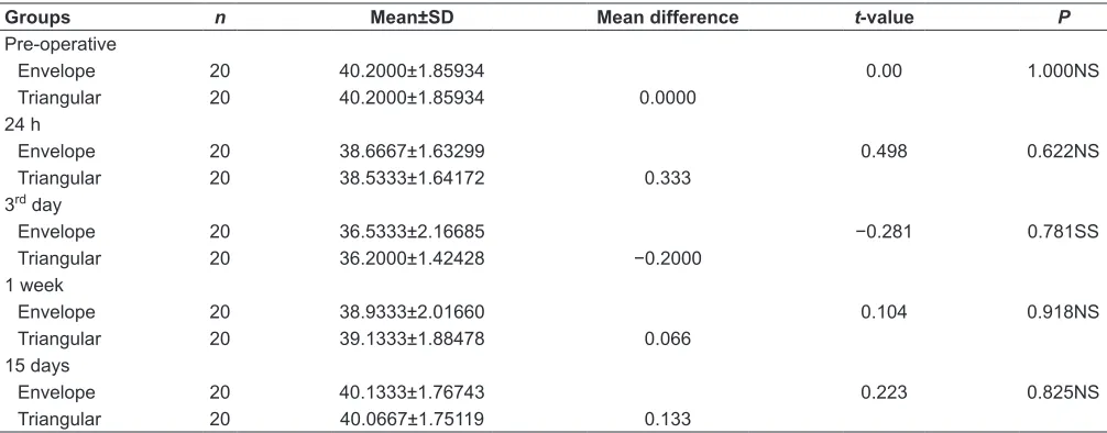
An unpaired t-test was used to compare the mean trismus scores between two incisions. It was observed that there was no significant difference present between two incisions on 1st, 1 week, and 15th post-operative day.
However, there was statistically significant difference present on the third post-operative day. The mean inter-incisal opening on the third post-operative day for enve-lope flap group was 36.53 mm while for triangular flap group was 36.2 mm. The difference was 3.33 mm which indicates that triangular flap group has more trismus on the 3rd post-operative day [Table 3].
An unpaired t-test was used to compare the mean swelling scores between two incisions. It was observed that there was no significant difference present between two incisions on 1st, 7th, and 15th post-operative day.
However, there was statistically significant difference present on the third post-operative day. The mean swell-ing measurement on the third post-operative day for envelope flap group was 21.57 mm while for triangular flap group was 22.13 mm. The difference was 0.563 mm, which indicates that triangular flap group has more swelling on the 3rd post-operative day [Table 4].
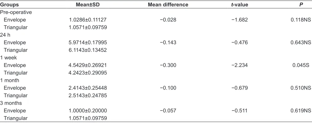
An unpaired t-test was used to compare the mean pocket depth scores between two incisions. It was observed that there was no significant difference pres-ent between two incisions on 1st, 1 month, and 3 months
post-operative period. However, there was statistically significant difference present on the 7th post-operative
day. The mean pocket depth on the 7th post-operative
day for envelope flap group was 4.54 mm while for tri-angular flap group was 4.242 mm. The difference was 0.3 mm, which indicates that envelope flap group has more pocket dept on the 7th post-operative day [Table 5].
DISCUSSION
There are different variables in the third molar surgery which influences the post-operative complications, flap designing being one of such variables. Flap designs are not only important to allow optimal visibility and access to the impacted third molar but also for subsequent healing of the surgically created defects.[11-15] Among
different flaps used for third molar surgery, envelope and triangular flaps are most commonly used.[14,16]
Envelope flaps have no release incisions and the ease of access to the tooth to be extracted depends on the length of mesial extension of the sulcular incision, which can if necessary extend up to the second premo-lar.[7] In our study, it was extended up to the first molar.
the second molar beside the papilla.[7] In our study, it
was positioned mesially to the second molar beside the papilla.
Intraoperatively both flaps were checked for acces-sibility of tooth and ease of suturing. It was found that envelope flap is easier to perform and suture than the triangular flap. This is probably because of more suturing involved due to releasing incision and difficulty in the repositioning of triangular flap. It has been reported that flap designing influences the post-operative complications in third molar sur-gery.[16-30] In our study, we have compared the
enve-lope flap with triangular flap related to the post-oper-ative pain, swelling, trismus, and periodontal health of the second molar.
In our study, visual analog scale was used for the assessment of pain. There was no statistically significant difference present between pain scores of envelop flap and triangular flap at different post-operative days.
In our study, the mean operating time for envelope
flap (49.6 min) was less than the mean operating time required for triangular flap (51.3 min), but this differ
-ence was not statistically significant (P = 0.368). When
mean trismus scores were compared between two inci-sions, it was observed that there was no significant dif-ference present between two incisions on 1st, 1 week, and
15th post-operative day. However, there was statistically
significant difference present on the third post-operative day. The mean interincisal opening on the third post-op-erative day for envelope flap group was 36.53 mm while
Triangular 20 51.3333±4.41858
SD: Standard deviation
Table 2: Comparison of mean pain scores in the triangular flap and envelop flap at different post-operative days
Groups n Mean±SD Mean difference t‑value P
24 h
Envelope 20 6.6000±0.73679 −0.775 0.445NS
Triangular 20 6.8000±0.67612 −0.20
3rd day
Envelope 20 3.9333±0.88372 −0.774 0.442NS
Triangular 20 4.1333±0.63994 −0.20
1 week
Envelope 20 1.2667±0.70373 1.402 0.172NS
Triangular 20 0.9333±0.59362 0.33
15 days
Envelope 20 0.0667±0.25820 1.00 0.326NS
Triangular 20 0.0000±0.00000 0.066667
SD: Standard deviation
Table 3: Comparison of mean trismus scores (in mm) in triangular flap and envelope flap at different post-operative days
Groups n Mean±SD Mean difference t‑value P
Pre-operative
Envelope 20 40.2000±1.85934 0.00 1.000NS
Triangular 20 40.2000±1.85934 0.0000
24 h
Envelope 20 38.6667±1.63299 0.498 0.622NS
Triangular 20 38.5333±1.64172 0.333
3rd day
Envelope 20 36.5333±2.16685 −0.281 0.781SS
Triangular 20 36.2000±1.42428 −0.2000
1 week
Envelope 20 38.9333±2.01660 0.104 0.918NS
Triangular 20 39.1333±1.88478 0.066
15 days
Envelope 20 40.1333±1.76743 0.223 0.825NS
Triangular 20 40.0667±1.75119 0.133
Envelope flap and triangular flap in lower third molar surgery
IJPCDR
for triangular flap group was 36.2 mm. The difference was 3.33 mm, which indicates that triangular flap group has more trismus on the 3rd post-operative day.
It has been suggested that triangular mucoperiosteal flaps induce inflammation in the muscles of mastica-tion and it is possible that muscle irritamastica-tion induced by hematoma forming when the periosteum is incised for the anterior releasing incision, is more likely with this design.[31-42] In contrary to our results Kirk et al. and
Nageshwar found no significant difference in mouth opening between the two flap designs; explaining their findings on the grounds that the distal incision, which follows the same course in both flap designs, is similar.[42]
The third parameter compared to our study was swelling. It was observed that there was no significant difference present between two incisions on 1st, 7th, and
15th post-operative day. However, there was statistically
significant difference present on the third post-op-erative day. The mean swelling measurement on the third post-operative day for envelope flap group was 21.57 mm while for triangular flap group was 22.13 mm. The difference was 0.563 mm which indicates that trian-gular flap group has more swelling on the 3rd
post-op-erative day.
Triangular flaps were associated with significantly greater measures of facial swelling after surgery on the 3rd post-operative day, but the difference lost
sta-tistical difference when measured on the 7th
post-oper-ative day. This finding is in accordance with the out-come of Kirk et al. and Z.H. Baqain et al., but contrary to those of Sandhu et al.who reported that post-operative swelling is related to operating time and not because of flap design.[25] However, in our study, there was
no significant correlation between duration of surgery
and post-operative swelling (P > 0.05). It is likely that Table 4: Comparison of mean swelling scores (in mm) in the triangular flap and envelope flap at different post-operative days
Groups n Mean±SD Mean difference t‑value P
Pre-operative
Envelope 20 20.3567±0.399 −0.013 −0.096 0.924NS
Triangular 20 20.3700±0.358
24 h
Envelope 20 20.6833±0.324 −0.260 −0.274 0.786NS
Triangular 20 20.9433±0.371
3rd day
Envelope 20 21.5733±0.327 −0.563 −3.041 0.05 SS
Triangular 20 22.1367±3.711
1 week
Envelope 20 20.4633±0.385 0.053 0.401 0.692NS
Triangular 20 20.4100±0.342
15 days
Envelope 20 20.3833±0.359 0.070 0.538 0.595 NS
Triangular 20 20.3133±0.353
SD: Standard deviation
Table 5: Comparison of mean pocket depth scores (in mm) in the triangular flap and envelope flap at different post-operative days
Groups Mean±SD Mean difference t‑value P
Pre-operative
Envelope 1.0286±0.11127 −0.028 −1.682 0.118NS
Triangular 1.0571±0.09759
24 h
Envelope 5.9714±0.17995 −0.143 −0.476 0.643NS
Triangular 6.1143±0.13452
1 week
Envelope 4.5429±0.26921 −0.300 −2.234 0.045S
Triangular 4.2423±0.29095
1 month
Envelope 2.4143±0.25448 −0.100 −0.679 0.510NS
Triangular 2.5143±0.24785
3 months
Envelope 1.0000±0.20000 −0.057 −0.511 0.619NS
Triangular 1.0571±0.09759
in the buccal tissues.
In our study, all operations were performed by a sin-gle surgeon, under similar operating conditions, using same instruments. Therefore, patient’s compliance bias was eliminated, and all other possible factors and surgical procedures were kept as constant as possible, presenting flap design as the sole independent factor to determine the severity of outcome variables. The results of our study suggested that flap design has an influ-ence on accessibility and ease of suturing during lower third molar removal and it also influences post-opera-tive complications of third molar surgery. Envelop flaps had a better outcome in terms of ease of suturing and on post-operative swelling and trismus, while triangular flaps allowed for an early return to pre-operative prob-ing depth around the second molar. However, there
were no differences in the long term (1 month and more)
in both the flaps with respect to periodontal health.
SUMMARY AND CONCLUSION
The aim of this prospective clinical study was to com-pare the efficacy of envelope flap and triangular flap in lower third molar surgery and effect of flap design in post-operative complications of third molar surgery.
There was statistically significant difference pres-ent in post-operative trismus and swelling on the 3rd post-operative day. Triangular flap group had more
swelling and trismus on the 3rd post-operative day than
envelope flap group. While periodontal health was bet-ter in triangular flap group on the 7th post-operative day than envelope flap group (P > 0.05), but there were no
significant differences found in intraoperative time and pain scores of both the groups.
The results of our study suggested that flap design has an influence in post-operative complications of third molar surgery. Envelope flaps had a better short-term outcome on post-operative swelling and trismus, while triangular flaps allowed for an early return to a pre-operative probing depth around the second molar. Intraoperatively it was also found that envelope flap is easier to perform and suture than the triangular flap.
Therefore, the decision about which flap to use for third molar surgery in young patients should be accord-ing to surgeon’s preference, takaccord-ing into consideration the patient’s needs and oral hygiene.
REFERENCES
1. Osunde OD, Adebola RA, Saheeb BD. A comparative study
of the effect of suture-less and multiple suture techniques on inflammatory complications following third molar surgery. Int J Oral Maxillofac Surg 2012;41:1275-9.
3. Dicus C, Blakey GH, Faulk-Eggleston J, Hoverstad E, Offenbacher S, Phillips C, et al. Second molar periodontal inflammatory disease after third molar removal in young adults. J Oral Maxillofac Surg 2010;68:3000-6.
4. Moss KL, Mauriello S, Ruvo AT, Offenbacher S, White RP Jr., Beck JD, et al. Reliability of third molar probing measures
and the systemic impact of third molar periodontal pathol-ogy. J Oral Maxillofac Surg 2006;64:652-8.
5. Cetinkaya BO, Sumer M, Tutkun F, Sandikci EO, Misir F.
Influence of different suturing techniques on periodon-tal health of the adjacent second molars after extraction of impacted mandibular third molars. Oral Surg Oral Med Oral
Pathol Oral Radiol Endod 2009;108:156-61.
6. Koyuncu BO, Çetingül E. Short-erm clinical outcomes of
two different flap techniques in impacted mandibular third
molar surgery. Oral Surg Oral Med Oral Pathol Oral Radiol
2012;13:565-9.
7. Monaco G1, Daprile G, Tavernese L, Corinaldesi G,
Marchetti C. mandibular third molar removal in young patients: An evaluation of 2 different flap designs. J Oral Maxillofacial Surg 2009;67:15-21.
8. Goldsmith SM, De Silva RK, Tong DC, Love RM. Influence of a Pedicle Flap Design on Acute Postoperative Sequelae after Lower Third Molar Removal. Dunedin, New Zealand:
Department of Oral Diagnostic and Surgical Sciences, University of Otago; 2011.
9. Kirtiloğlu T, Bulut E, Sümer M, Cengiz I. Comparison of
2 flap designs in the periodontal healing of second molars after fully impacted mandibular third molar extractions. J Oral Maxillofac Surg 2007;65:2206-10.
10. Quee TA, Gosselin D, Millar EP, Stamm JW. Surgical removal
of the fully impacted mandibular third molar. The influence of flap design and alveolar bone height on the periodontal
status of the second molar. J Periodontol 1985;56:625-30. 11. Kirk DG, Liston PN, Tong DC, Love RM. Influence of two
different flap designs on incidence of pain, swelling, tris-mus, and alveolar osteitis in the week following third molar
surgery. Oral Surg Oral Med Oral Pathol Oral Radiol Endod
2007;104:e1-6.
12. Rakprasitkul S, Pairuehvej V. Mandibular third molar
surgery with primary closure and tube drain. Int J Oral Maxillofac Surg 1997;26:187-90.
13. Suarez-Cunqueiro MM, Gutwald R, Reichman J, Otero-Cepeda XL, Schmelzeisen R. Marginal flap versus
paramarginal flap in impacted third molar surgery: A
pro-spective study. Oral Surg Oral Med Oral Pathol Oral Radiol
Endod 2003;95:403-8.
14. Rosa AL, Carneiro MG, Lavrador MA, Novaes AB Jr.
Influence of flap design on periodontal healing of sec-ond molars after extraction of impacted mandibular third
molars. Oral Surg Oral Med Oral Pathol Oral Radiol Endod
2002;93:404-7.
15. Jakse N, Bankaoglu V, Wimmer G, Eskici A, Pertl C. Primary
wound healing after lower third molar surgery: Evaluation
of 2 different flap designs. Oral Surg Oral Med Oral Pathol Oral Radiol Endod 2002;93:7-12.
16. Pasqualini D, Cocero N, Castella A, Mela L, Bracco P. Primary
Envelope flap and triangular flap in lower third molar surgery
IJPCDR
Int J Oral Maxillofac Surg 2005;34:52-7.
17. Hashemi HM, Beshkar M, Aghajani R. The effect of suture -less wound closure on postoperative pain and swelling
after impacted mandibular third molar surgery. Br J Oral
Maxillofac Surg 2012;50:256-8.
18. UStün Y, Erdogan O, Esen E, Karsli ED. Comparison of the
effects of 2 doses of methylprednisolone on pain, swelling, and trismus after third molar surgery. Oral Surg Oral Med
Oral Pathol Oral Radiol Endod 2003;96:535-9.
19. Yuasa H, Sugiura M. Clinical postoperative findings after removal of impacted mandibular third molars: Prediction of
postoperative facial swelling and pain based on
preopera-tive variables. Br J Oral Maxillofacial Surg 2004;42:209-14. 20. Poeschl PW, Eckel D, Poeschl E. Postoperative prophylactic
antibiotic treatment in third molar surgery – a necessity? J Oral Maxillofac Surg 2004;62:3-8.
21. Clauser C, Barone R. Effect of incision and flap reflection on
postoperative pain after the removal of partially impacted mandibular third molars. Quintessence Int 1994;25:845-9.
22. Karaca I, Simşek S, Uğar D, Bozkaya S. Review of flap design
influence on the health of the periodontium after
mandib-ular third molar surgery. Oral Surg Oral Med Oral Pathol Oral Radiol Endod 2007;104:18-23.
23. Dolanmaz D, Esen A, Isik K, Candirli C, Antalya K, Ankara T. Effect of 2 flap designs on postoperative pain and swelling after impacted third molar surgery Oral Surg Oral
Med Oral Pathol Oral Radiol 2012;27:203-7
24. García AG, Sampedro FG, Rey JG, Vila PG, Martin MS.
Pell-gregory classification is unreliable as a predictor of
diffi-culty in extracting impacted lower third molars. Br J Oral
Maxillofac Surg 2000;38:585-7.
25. Baqain ZH, Al-Shafii A, Hamdan AA, Sawair FA. Flap design
and mandibular third molar surgery: A split mouth random-ized clinical study. Int J Oral Maxillofac Surg 2012;41:1020-4. 26. Sandhu A, Sandhu S, Kaur T. Comparison of Two Different
Flap Designs in the Surgical Removal of Bilateral Impacted Mandibular Third Molars. Amritsar, Punjab - 143006, India: Department of Oral and Maxillofacial Surgery. SGRD Institute of Dental Sciences and Research; 2010.
27. Polat HB, Ozdemir H, Ay S. Effect of different mouth rinses
on third molar surgery-related oral malodor. Oral Surg Oral
Med Oral Pathol Oral Radiol Endod 2008;105:e1-8.
28. Guralnick W. Third molar surgery. Br Dent J 1984;156:389.
29. Nageshwar. Comma incision for impacted mandibular third molars. J Oral Maxillofac Surg 2002;60:1506-9.
30. Kan KW, Liu JK, Lo EC, Corbet EF, Leung WK. Residual
periodontal defects distal to the mandibular second molar 6-36 months after impacted third molar extraction. J Clin
Periodontol 2002;29:1004-11.
31. Neelkandhan RS, Wadhwani P, Prasad S, Devadoss P.
Influence of incisions on post operative complications in impacted third molar removal: Comma incision versus stan-dard incisions. J Oral Maxillofac Surg 2005;4:124-7.
32. Filho JR, Silva ED, Camargo IB. The influence of cryotherapy
on reduction of swelling, pain and trismus after third-molar extraction. J Am Dent Assoc 2005;136:774-8.
33. Susarla SM, Dodson TB. Estimating third molar extraction
difficulty: A comparison of subjective and objective factors. J Oral Maxillofac Surg 2005;63:427-34.
34. Danda KA, Tatiparthi MK, Narayanan V, Siddareddi A. Influence of primary and secondary closure of surgical wound after impacted mandibular third molar removal on postoperative pain and swelling – A comparative and split mouth study. Oral Maxillofac Surg 2010;68:309-12.
35. Alistair W, Berwick AW. Alternative method of flap reflec
-tion. Br Dent J 1966;20:295-6.
36. Lekholm U, Grondahl HG. Influence of mandibular third
molars on related supporting tissues. Int J Oral Surg 1973;2:137-42.
37. Wood GD, Bronco JA. A comparison of three method of mea -suring maximal opening of mouth. Oral Surg 1979;37:175-7.
38. Halmos DR, Ellis E 3rd, Dodson TB. Mandibular third molars
and angle fractures. J Oral Maxillofac Surg 2004;62:1076-81.
39. Sanchis JM, Saez U, Penarrocha M, Gay C. Tetracycline com-pound placement to
40. Krausz A, Machtie E. Effect of lower third molar extraction on attachment level and alveolar bone height of the adjacent second molar. Int J Oral Maxillofac Surg 2005;34:756-60.
41. Gool AV, Bosch JJ, Boering G. Clinical consequences of com -plaints and complications after third molar removal. Int J Oral Surg 1977;6:29.
42. Howe LG. Minor Oral Surgery. 3rd ed. Bristol: John Wright