
R E S E A R C H
Open Access
Transgenerational effects of
polychlorinated biphenyls: 1. Development
and physiology across 3 generations of rats
Jan A. Mennigen
†, Lindsay M. Thompson
†, Mandee Bell, Marlen Tellez Santos and Andrea C. Gore
*Abstract
Background:Polychlorinated biphenyls (PCBs) are persistent organic environmental contaminants and known endocrine-disrupting chemicals (EDCs). Previous studies demonstrated that developmental exposure to the weakly estrogenic PCB mixture Aroclor 1221 (A1221) in Sprague-Dawley rats altered sexual development, adult
reproductive physiology and body weight. The current study tested the hypothesis that prenatal A1221 exposure not only disrupts these endpoints within an exposed individual’s (F1generation) lifespan, but may also affect subsequent generations (F2-F3).
Methods:We treated pregnant female rats on embryonic days (E) 16 and E18 with A1221 (1 mg/kg), estradiol benzoate (50μg/kg, positive estrogenic control), or vehicle (3% DMSO in sesame oil, negative control). Endpoints related to sexually dimorphic developmental trajectories of reproductive and developmental physiology were measured, and as adults, reproductive endocrine status was assessed, in the F1, F2, and F3generations.
Results:Significant effects of transgenerational EDCs were found for body weight and serum hormones. The A1221 descendants had significantly higher body weight in the F2-maternal lineage throughout postnatal development, and in F3-maternal lineage animals after weaning. In females, generation- and lineage-specific effects of exposure were found for serum progesterone and estradiol. Specifically, serum progesterone concentrations were lower in F2-A1221 females, and higher in F3-A1221 females, compared to their respective F2- and F3-vehicle counterparts. Serum estradiol concentrations were higher in F3-A1221 than F3-vehicle females. Reproductive and adrenal organ weights, birth outcomes, sex ratio, and estrous cycles, were unaffected. It is notable that effects of A1221 were only sometimes mirrored by the estrogenic control, EB, indicating that the mechanism of action of A1221 was likely via non-estrogenic pathways.
Conclusions:PCBs caused body weight and hormonal effects in rats that were not observed in the directly exposed F1offspring, but emerged in F2and F3generations. Furthermore, most effects were in the maternal lineage; this may relate to the timing of exposure of the F1fetuses at E16 and 18, when germline (the future F2 generation) epigenetic changes diverge in the sexes. These results showing transgenerational effects of EDCs have implications for humans, as we are now in the 3rd generation since the Chemical Revolution of the mid-twentieth century, and even banned chemicals such as PCBs have a persistent imprint on the health of our descendants.
Keywords:Polychlorinated biphenyl (PCB), Reproduction, Estradiol, Progesterone, Corticosterone, Body weight, Endocrine-disrupting chemical (EDC), Transgenerational, Sex difference
* Correspondence:[email protected] †Equal contributors
Division of Pharmacology and Toxicology, College of Pharmacy, University of Texas at Austin, 107 W Dean Keeton, C0875, Austin, TX 78712, USA
Background
Polychlorinated biphenyls are a class of persistent or-ganic pollutants consisting of 209 specific congeners that were heavily used by industry for decades before being banned in the late 1970s [1]. Today, due to their envir-onmental persistence and high capacity for biomagnifi-cation in food chains, PCBs remain detectable in most wildlife and humans [2, 3]. Both experimental and epi-demiological lines of evidence support endocrine-disrupting properties of specific PCB congeners and mixtures, with well-described consequences on repro-ductive [4–6] and metabolic [7,8] physiology. More spe-cifically, the weakly estrogenic PCB mixture Aroclor 1221 (A1221) disrupted sexual development and led to sex-specific effects on adult reproductive physiology and its neuroendocrine regulation, potentially through estro-genic mechanisms [9–11]. For body weight, recent stud-ies have suggested obesogenic effects of A1221 that, in contrast, do not point to an estrogenic mode of action [12,13].
Most work on EDCs has focused on their direct effects in animals exposed during early life development (F1
gen-eration). However, evidence over the last decade has shown intergenerational effects of endocrine-disrupting chemicals (EDCs) in the F2generation, and
transgenera-tional effects in F3 generations and beyond [14–18],
al-though little of this work was on PCBs [4,19]. In addition, the question of transmission of disrupted traits across generations has been seldom studied in head-to-head comparisons of maternal vs. paternal lineages, which would likely lead to very different phenotypic outcomes due to sex differences in the timing of epigenetic program-ming [20].
In the current study, we sought to determine effects of the PCB mixture, A1221, on the F1, F2, and F3
genera-tions of maternally- and paternally-derived lineages. The A1221 mixture has a relatively short half-life compared to most PCBs [21,22] allowing us to target the exposure to a specific developmental window in exposed individ-uals. We administered the treatments during the begin-ning of the critical period of brain sexual differentiation, and conducted work on both females and males to en-able us to discern sex differences in direct and inherited responses to environmental EDCs. This experimental approach is translationally relevant [23], given that the human generational time is 20–30 years; therefore, we are in the 3rd human generation following the chemical revolution in the mid-twentieth century. Indeed, living in a contaminated world in which animal and human species are incidentally exposed to persistent man-made chemicals such as PCBs even in remote areas, it be-comes paramount to understand if and how physiology across development and generations is affected by envir-onmental contaminants [24].
Methods
Experimental design and animal husbandry
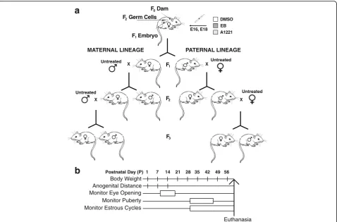
Female and male Sprague-Dawley rats were purchased from Harlan (Houston, TX), and all animal procedures were conducted in compliance with protocols approved by the Institutional Care and Use Committee at the University of Texas at Austin, and following NIH guidelines. Rats were housed in a colony room with controlled temperature (22 °C) and partially reversed light cycle (12:12 dark:light, lights on at 2400 h). Virgin females were mated with sexually-experienced males. The day following successful mating, as indicated by a sperm-positive vaginal smear, was termed embryonic day 1 (E1). During pregnancy, rats (termed F0) were exposed to one of three
treat-ments, given on E16 and E18 to target the beginning of the period of sexual differentiation of the rat brain [25–27]. The treatments were: 1 mg/kg PCB mixture Aroclor 1221 (A1221, i.p.); 50 μg/kg estradiol benzoate (EB, s.c.) as a positive control for the estrogenic effects of A1221; or a negative vehicle control (3% DMSO in sesame oil; half of these animals received i.p., and half s.c., injections). The A1221 PCB mixture is lightly chlorinated, resulting in a relatively short biological half life in the range of days [21,22]. The treatment dosages were based on prior work conducted in the Gore lab, which elicited developmental and adult neuroendocrine effects on reproductive physiology at multiple levels of biological organization [4,5,9,10]. The day of parturition was referred to as postnatal day 0 (P0), and litters were culled to 3 males and 3 females per group or as close as possible until weaning, after which animals were housed in male- and female-specific sibling tri-ads. Upon reaching sexual maturity, one exposed male and female offspring (F1) per litter were used as
foun-ders of paternal and maternal lineages of all treatment groups (F2), and mated with untreated adult rats. One
F2male from each paternal litter, and one female from
each maternal litter were subsequently mated with un-treated adult rats to extend paternal and maternal lin-eages into the F3 generation. Figure 1 summarizes the
transgenerational experimental design (Fig. 1a), and an experimental timeline of individual measurements and sampling protocols (Fig. 1b). Sample sizes and groups are in Table 1.
To provide breeder males with sexual experience, female stimulus rats were purchased as young adults from Harlan, and ovariectomized under isoflurane anesthesia. Stimulus animals were not subjected to any of the EDC treatments, although the ovariecto-mized females were primed with estradiol plus pro-gesterone to induce receptivity. In addition, breeding male and female partners (untreated with EDCs or
vehicle) were purchased to generate the F2 and F3
Developmental endpoints
On P1, the day after birth, pups were weighed and their anogenital distance (AGD) was measured using a digital microcaliper. The anogenital index (AGI) was calculated (AGI = AGD divided by the cube root of body weight) as it enables proper sexing of neonates and also serves as an indicator of masculinization or feminization [28]. Characteristics of birth outcomes (number of male and female pups and sex ratio were determined; they were unaffected by treatment (Additional file 1: Figure S1). Litters were culled as follows: the three males and
females with AGIs closest to the median of the same sex and litter were kept to achieve equal litter size and sex ratio. All litters, including controls, were culled to a total of 6 pups (3 male and 3 female) to ensure that sex ratio was equal, to avoid possible confounding effects of a biased sex ratio, and to allow us to attain adequate stat-istical power based on a power analysis. Litter was con-sidered as a variable in analyses (below). Along with being measured on P1, AGI was monitored weekly on P7 and 14 (Fig. 1b). Age at eye opening was assessed before weaning, and time of puberty was assessed after
DMSO EB A1221 E16, E18
F Dam F Germ Cells
F Embryo 0 2
1
F 1
F 2
F 3 Untreated
Untreated
Untreated
Untreated
MATERNAL LINEAGE PATERNAL LINEAGE
a
X X
X X
b
Postnatal Day (P) 1 7 14 21 28 35 42 49 56Body Weight Anogenital Distance Monitor Eye Opening Monitor Puberty Monitor Estrous Cycles
Euthanasia
Fig. 1aThe transgenerational experimental design and breeding strategy of the study.bThe developmental timeline and endpoints for individual animals. Abbreviations: E: embryonic day of age
Table 1Groups and sample sizes. Individual animal numbers per group are indicated, with numbers of litters in parentheses
Generation Females Males
F1 DMSO: 41 (14)
EB: 32 (11) A1221: 35 (12)
DMSO: 39 (14) EB: 32 (11) A1221: 36 (12)
Generation and lineage Maternal Paternal Maternal Paternal
F2 DMSO:35 (12)
EB: 32 (11) A1221:32 (11)
DMSO: 38 (14) EB: 30 (11) A1221: 35 (12)
DMSO: 34 (12) EB: 33 (11) A1221: 30 (11)
DMSO: 38 (14) EB: 32 (11) A1221: 34 (12)
F3 DMSO:33 (11)
EB: 30 (11) A1221:31 (11)
DMSO: 39 (14) EB: 32 (11) A1221: 30 (11)
DMSO: 32 (11) EB: 30 (11) A1221: 27 (11)
weaning in females (day of vaginal opening) and males (day of preputial separation). Body weight was recorded weekly from P1–56 (Fig.1b).
Monitoring of estrous cycles
Following puberty in females, daily vaginal smears were taken to track estrous cycles by vaginal cytology under a light microscope. Based on these data, several parame-ters were determined: The number of completed estrous cycles from puberty to age at P56, average length of es-trous cycles in days, and percentage of elongated (dur-ation >5d) or irregular (aberrant transitions) cycles were determined. No significant treatment effects were found on these endpoints (Additional file2: Figure S2).
Tissue collection
Within each generation (F1-F3), animals were euthanized
by rapid decapitation 2–3 h before lights-out.
To minimize the possible confound of the estrous cycle on hormone concentrations, females were euthanized on proestrus as close to P60 as possible, between P58–63, and for each female, a male littermate was euthanized on the same day. Thus all rats were euthanized from P58–63. Trunk blood was collected, allowed to clot, and
serum was separated via centrifugation (1500×g for
5 min). Tissues and sera were stored at −80 °C until use. Dams of all generations were euthanized between 1 and 2 weeks after pups were weaned in order to examine the uterus for the number of implantation sites, and to calculate the number of resorptions based on the differ-ence between numbers of pups born and implantation sites. No effects of treatment on resorptions was ob-served (Additional file1: Figure S1).
Hormone assays
Serum samples were used to determine concentrations of circulating estradiol, progesterone, testosterone, and corticosterone using previously validated commercial kits [12, 13, 29]. The assays and their characteristics were: estradiol, Beckman #DSL-4800, range 5–750 pg/
ml, sensitivity 2.2 pg/ml; progesterone, Cayman
#582601, range 7.8–1000 pg/ml, sensitivity 10 pg/ml;
testosterone, MP Biomedical #07189102, range 0.1–
10 ng/ml, sensitivity 0.03 ng/ml; and corticosterone, MP Biomedical #07120102, range 25–1000 ng/ml, sensitivity 7.7 ng/ml. Final sample sizes for individual treatment groups within sex and lineage weren = 17–25 for estra-diol, n = 8–10 for progesterone, n = 10 for testosterone
(males only, as the assay is not sensitive enough to measure testosterone in females) and n = 10 for cortico-sterone. All assays were run according to the manufac-turers’protocols.
Statistical analyses and graphs
The identity of treatment groups and sample numbers were coded throughout the study, in order to ensure that for experimenters were blind to the treatment in the analyses of all endpoints. For all datasets, a custom R-based, generalized extreme studentized deviate (ESD) test was used to detect outliers, limited to a maximum of two per group. Following the validation of normality and homoscedasticity in groups using Shapiro-Wilk and Levene’s test, respectively, data were analyzed using a general linear model approach. A two-way ANOVA was used for F1data, with sex and treatment as factors, and
litter as random factor. For data from F2and F3animals,
a three-way ANOVA, with sex, lineage, and treatment as main factors, and litter as random factor, was used. When data were non-parametric after standard trans-formation procedures, Kruskal-Wallis analysis was used for individual factors. Repeated measures tests (split-plot ANOVAs) were used for longitudinal datasets (body weight and AGI), using time as factor within groups. In cases of repeated developmental data measurements, Greenhouse-Geisser corrections for violations of spher-icity (assessed by Mauchly’s test), were used where ap-propriate. Data that revealed significant effects of treatment or treatment interactions with other analyzed factors were further analyzed using Sidak’s post-hoc test, in order to determine global treatment differences or treatment differences within interacting factors. In cases where the data did not satisfy the assumptions for para-metric analyses, post-hoc analysis was conducted using Dunn’s test. Post-hoc comparisons of significant treat-ment effects in repeated measures ANOVAs were
ana-lyzed using Sidak’s post-hoc comparisons. While
descriptive statistics and outlier identification was con-ducted using a custom R scripts, general linear model analyses were conducted using SPSS Version 24 (IBM, New York, NY, USA). Graphs were plotted using Prism Version 7 (Graphpad, LaJolla, CA, USA). For the sake of presentation, detailed statistical outcomes (F-, H- andp -values, and degrees of freedom) are described only for significant treatment differences (p< 0.05); however all detailed model parameters can be accessed in Additional file3: Figure S3.
Results
Prenatal A1221 effects on body weight
Body weight was monitored weekly from P1–56, and
60
BW (g)
F1
40
20
1 7 14 21
60
BW (g)
F2
40
20
1 7 14 21
60
BW (g)
F3
40
20
1 7 14 21
1 7 14 21
1 7 14 21
Body Weight P1-21 - Females Body Weight P28-56 - Females
240
BW (g)
200 160 120 80
28 35 42 49 56
240
BW (g)
200 160 120 80
28 35 42 49 56
240
BW (g)
200 160 120 80
28 35 42 49 56
28 35 42 49 56
28 35 42 49 56 DMSO EB A1221
Maternal Paternal Maternal Paternal
Maternal Paternal Maternal Paternal
D,E<A D,E<A
D,E<A D,E<A
DMSO,EB<A1221
F1
F2
F3
D,E <A
E<A E<A
DMSO,EB<A1221
D,E <A
D,E<A D,E<A
D,E<A
b
a
Fig. 2Developmental profiles of female body weight are shown for pre-weaning rats from P1–21 (a), and for adolescents from P28–56 (b), across the three generations. Data were analyzed by repeated measure ANOVAs, and significant overall treatment effects between groups (p< 0.05) are indicated within the panels below the graphs. In cases of significantly different treatment, lineage, and time interactions, specific differences for each time point were resolved by Sidak-adjusted post-hoc comparisons, indicated above individual time points. For the latter, the treatment groups are abbreviated as D, DMSO; E, EB; A, A1221
BW (g)
1 7 14 21
BW (g)
BW (g)
Body Weight P1-21 - Males Body Weight P28-56 - Males
BW (g)
28 35 42 49 56
BW (g)
Maternal Paternal Maternal Paternal
Maternal Paternal Maternal Paternal
60 40 20
240
160
80
80 320
1 7 14 21 60
40 20 80
1 7 14 21 60
40 20 80
1 7 14 21
1 7 14 21
28 35 42 49 56 240
160
80 320
28 35 42 49 56 240
160
80 320
BW (g)
F1
F2
F3
F1
F2
F3
28 35 42 49 56
28 35 42 49 56 DMSO EB A1221
D,E<A
D,E<A D,E<A DMSO,EB<A1221 D,E<A
D,E<A D,E<A
D,E<A D,E<A
D,E <A
E<A E<A
DMSO,EB<A1221
a
b
previously [13]. In F1 animals, no significant treatment
effects were observed (Additional file 3: Figure S3). Lineage-dependent treatment effects were detected in F2
animals (F2.83, 3616= 7.77, p< 0.01), and Sidak’s post-hoc
test revealed an overall significant increase in body weight in the A1221 group compared to DMSO and EB groups in maternal lineage descendants, irrespective of sex (p< 0.05). An interaction of treatment and lineage was also observed over time (F2.83, 3616= 3.41, p< 0.01),
and Sidak-adjusted post-hoc comparisons of lineage-dependent treatment effects at individual time points re-vealed that in the maternal lineage of the F2 generation
(both males and females), ancestral A1221 treatment significantly increased body weight compared to DMSO and EB at P1 and for all time points following P28 (p< 0.05). In F3animals, a significant interaction of treatment
and lineage was observed over time (F2.83, 3466= 3.48, p <
0.01), and Sidak-adjusted post-hoc comparisons of lineage-dependent treatment effects at individual time points revealed that in the maternal lineage of the F3
gen-eration, ancestral A1221 treatment significantly increased
bodyweight compared to DMSO and EB (p< 0.05) from P42 to P56.
Prenatal A1221 exposure had few effects on
developmental milestones of eye opening and puberty
Age at eye opening (Fig. 4a) was sexually dimorphic in F1(H1,215= 4.49, p < 0.05) and F2(H1,403= 3.92, p < 0.05),
but not F3 animals (H1,385= 3.24, p> 0.05). Prenatal
treatment did not affect postnatal age of eye opening in F1animals (H2,215= 1.17, p > 0.05). In F2rats, eye
open-ing exhibited a significant difference between treatments (H2,202= 6.89, p< 0.01), with a delay in EB-lineage
ani-mals compared to A1221 (p< 0.05). F3 animals did not
exhibit significant treatment effects on age at eye open-ing (H2,385= 2.653, p > 0.05).
Markers of the onset of puberty (vaginal opening in fe-males, preputial separation in males; Fig. 4b) occurred significantly earlier in females compared to males in all generations (F1: H1,215= 659, p< 0.01; F2: H1,403= 1422,
p< 0.01; F3: H1,385= 1181; p < 0.01). No treatment effects
were found in F1 animals (H1,215= 0.78; p > 0.05). In F2
F1
F2
F3
Eye Opening
Age (days)
Age (days)
Age (days)
A1221<EB
MAT PAT MAT PAT
MAT PAT MAT PAT FEMALES MALES
A1221
MAT PAT
MAT MAAT PATPAT FEMALES
<EB
MAT PAT
MAT MAT PATPAT
MALES
Puberty
EB<A1221
MAT PAT MAT PAT
MAT PAT MAT PAT FEMALES MALES
EB<A
MAT PAT
MAT PAT
FEMALES
A1221
MAT PAT
MAT PAT MALES
A
A1221<EB
DMSO EB A1221
b
a
F1
F2
F3
A. AGI - Females
AGI
B. AGI - Males
DMSO EB A1221
1 7 14 1 7 14 1 7 14 1 7 14
1 7 14 1 7 14 1 7 14 1 7 14
4 1 7 1 4 1 7 1 1.8 1.4 1.0 2.2 1.8 1.4 1.0 2.2 1.8 1.4 1.0 2.2 2.8 2.4 2.0 3.2 2.8 2.4 2.0 3.2 2.8 2.4 2.0 3.2 AGI AGI EB<DMSO E,A<D E,A<D EB<DMSO E,A<D E,A<D
Maternal Paternal Maternal Paternal
Maternal Paternal Maternal Paternal
F1
F2
F3
AGI - Females
AG
I
1 7 14 1 7 14
1 7 14 1 7 14
1 7 14
1.8 1.4 1.0 2.2 1.8 1.4 1.0 2.2 1.8 1.4 1.0 2.2 A GI A G I EB<DMSO E,A<D E,A<D Maternal Paternal Maternal Paternal
a
b
AGI - Males
1 7 14 1 7
DMSO EB A1221
14
1 7 14 1 7 14
1 7 14
2.8 2.4 2.0 3.2 2.8 2.4 2.0 3.2 2.8 2.4 2.0 3.2 EB<DMSO E,A<D E,A<D Maternal Paternal Maternal Paternal
Fig. 5Anogenital index (AGI), calculated as [anogenital distance /3√body weight] is shown for females (a) and males (b) of the three generations. Data were analyzed using repeated measures ANOVAs. Significant overall treatment effects between groups (p < 0.05) are indicated within the panels below the graphs. In cases of significantly different main effects of treatment between groups, specific differences for each time point were resolved by Sidak-adjusted comparisons, indicated above individual time points
F1
F2
F3
Ovary Testis Uterus Adrenal
FEMALES MALES 0.06 0.08 0.02 0.04 0 0.06 0.08 0.02 0.04 0 0.06 0.08 0.02 0.04 0 1.0 1.5 0.5 0 1.0 1.5 0.5 0 1.0 1.5 0.5 0 4 6 2 0 4 6 2 0 4 6 2 0 0.03 0.04 0.01 0.02 0 0.03 0.04 0.01 0.02 0 0.03 0.04 0.01 0.02 0
MAT PAT MAT PAT MAT PAT MAT PAT MAT PAT
DMSO EB A1221
MAT PAT MAT PAT MAT PAT MAT PAT MAT PAT
MAT PAT MAT PAT MAT PAT MAT PAT MAT PAT MAT PAT FEMALES MAT PAT MAT PAT MALES
T MAT PAT MAT PAT
T
FEMALES MALES FEMALES
a
b
c
d
animals, there was a significant treatment effect on pu-berty (H2,202= 5.44, p < 0.01), attributable to EB rats
hav-ing significantly earlier puberty than A1221 rats. In the F3
generation, puberty onset was also affected by treatment (H1,385= 4.427, p< 0.05), with EB rats displaying later
puberty compared to A1221 treated rats (p < 0.05).
Anogenital index was affected by EB in F1, and by A1221 and EB in F3maternal lineage descendants
The expected strong sexual dimorphism of AGI (Fig.5a, b) was found in all 3 generations (F1: F1,215 = 226, p <
0.01; F2: F1,202= 1054, p < 0.01; F3: F1,385= 11,013, p <
0.01). Treatment significantly affected F1generation AGI
(F2,324= 9.55, p < 0.01), with the EB rats having smaller
AGIs than their DMSO counterparts (p< 0.05). In F3
an-imals, we found treatment effects to be lineage-specific, as evidenced by a significant interaction of both factors (F2,385= 3.37, p < 0.05). Sidak’s post-hoc test reveals the
DMSO group had an overall higher AGI compared to the A1221 group within the maternal lineage (p < 0.05). A significant interaction of treatment and lineage was also evident over time in F3animals (F1155, 0.257= 3.571,
p < 0.05), and Sidak-adjusted post-hoc comparisons re-vealed significantly lower AGI in EB and A1221 groups (p < 0.05) compared to DMSO group animals in the ma-ternal F3lineage at P7 and P14.
A1221 treatment did not affect adult endocrine tissue weights
The gonadal, uterine, and adrenal indices were calcu-lated by normalizing organ weight to body weight. There was no main effect of treatment, nor any interaction ef-fects, on any of these organ indices in any generation (Fig. 6). The expected sexual dimorphism in the adrenal index was found throughout the generations (female > male). Detailed statistical analysis results are described in Additional file3: Figure S3.
Ancestral A1221 exposure altered estradiol and progesterone concentrations in females only, in a generation- and lineage-dependent manner
The expected sex differences were found for the steroid hormones estradiol, progesterone, and corticosterone in all generations (Additional file 3: Figure S3). For serum estradiol concentrations (Fig. 7a), there were no effects of treatment in females of the F1or F2generation.
How-ever, in F3females, a significant interaction of treatment
and sex was found (F2,129= 5.14,p< 0.05), irrespective of
lineage. Post-hoc analysis revealed that in F3 females,
serum estradiol was significantly higher in A1221 com-pared to DMSO and EB rats (p < 0.05).
For serum progesterone concentrations (Fig. 7b), the expected sexual dimorphism was found (female > male). F1 animals were not affected by treatment. F2 animals
a
b
c
d
had a significant treatment effect dependent on sex (F2,55= 3.517, p < 0.05), with female A1221 rats having
lower progesterone concentrations than DMSO controls (p < 0.05), irrespective of lineage. In F3animals, a
signifi-cant treatment effect dependent on lineage and sex (F1,58= 3.23, p < 0.05, Fig.7b) was found. In the maternal
lineage females, serum progesterone was significantly higher in A1221 compared to DMSO and EB descen-dants (p < 0.05). There was no treatment effect in the F3
-paternal lineage females. In males, treatment did not affect serum progesterone concentrations in any gener-ation (Additional file3:Figure S3).
Serum concentrations of testosterone in males (Fig.7c; female testosterone could not be measured due to limits of assay detection), and corticosterone in both sexes (Fig. 7d), were not affected by treatment. The expected sexual dimorphism of corticosterone (females > males) was confirmed in this study (Additional file 3: Figure S3), and was consistent with the larger adrenal indices in the females (Fig.6d).
Discussion
Exposures to endocrine-disrupting chemicals (EDCs) during critical periods of development have been linked to changes in reproductive, metabolic, and other endo-crine functions later in life, consistent with the“
develop-mental origins of health and disease” (DOHAD)
hypothesis [30–32]. While much work has focused on
outcomes in the exposed F1 generation,
inter-generational (F2) and transgenerational (F3) effects are
beginning to be demonstrated for low-dose EDC expo-sures [16,17]. The expectation is that the phenotype will be unique to each generation due to the differential tim-ing of exposure [33,34], as follows. The F1generation is
exposed directly as embryos on E16 and E18, the begin-ning of the critical period of brain sexual differentiation in rats [25–27] and a life stage during which the entire body undergoes enormous developmental change. The F2generation is exposed as germ cells within the F1
em-bryos. During germline development, epigenetic modifi-cations of DNA demethylation and remethylation, histone modifications, and non-coding RNAs take place [35–37]. This epigenetic programming could lead to phenotypic changes in the F2 generation that are likely
to be quite different from that of their F1 progenitors.
Furthermore, the timing of demethylation and remethy-lation is not identical in the sexes [20,38–40], leading to the expectation of sexually-dimorphic transgenerational effects. For the F3 generation, any exposure effects are
truly transgenerational [33, 41], and must be inherited via germline or context-dependent (e.g. maternal behav-ior) mechanisms [42]. Again, the expectation is that the F3 generation will differ from both F1 and F2
genera-tions; but that subsequent (F4 and beyond) generations
would better phenocopy the F3generation due to
herit-able epigenetic programming. This current study was our initial step to thoroughly document the inter- and transgenerational phenotype; a companion study will focus on specific neuroendocrine molecular outcomes.
One of our most interesting findings was a small but significant and consistent increase in body weight in A1221-lineage rats, restricted to the maternal-lineage F2
and F3animals. This effect was not mimicked in the
EB-descendants, suggesting a maternally transmitted,
estrogen-independent mode-of action of A1221. While in the F2generation this effect was detectable as early as
P1, and maintained through adulthood, in the F3
gener-ation the onset of this body weight difference did not take place until post-weaning. The dramatically different diet and energetic demands of an individual pre- vs. post-weaning may account for the adolescent onset; rat pups continue to nurse through P21 and begin to eat chow at ~P18 in our colony. After P21, rats are limited entirely to the chow diet. Presumably, differences in the mode of transmission of the effect across generations ac-count for the differential phenotypes in F2and F3
gener-ation rats. It is notable that we did not find any body weight effect in the F1generation, which is inconsistent
with our previous studies showing increased body weight in F1-A1221 rats [12, 13]. We are unsure of the reason
for this discrepancy, but small variances in environmen-tal housing conditions, food lots, or other factors such as the microbiota that change the rats’external and in-ternal environment must be considered.
The finding of increased body weight is consistent with the “obesogen hypothesis,”which suggests that en-vironmental factors including EDCs are contributing fac-tors to the increasing incidence of obesity and metabolic syndrome [43–46]. Increased adipogenesis, disruption of insulin and glucose regulation [47–50], and specific al-terations in lipid metabolism [51,52], have also been re-ported in association with PCBs and other EDCs. Furthermore, a transgenerational phenotype was shown for another class of EDCs, tributyltin [16] and a mixture of EDCs in plastics [14].
phenotype and its maternal lineage-specific transmis-sion. In this context, investigations into a potential role of sex-specific effects on primordial germ cell (PGC) de- and re-methylation dynamics are especially war-ranted, as female embryonic PGCs, at least in the mice, remain demethylated for longer (E16.5) than male embry-onic PGCs (E13.5) [38]. Thus, while comparative DNA methylation dynamics have not been described for rat PGCs, the increases in body weight specific to offspring of the maternal lineage may be a consequence of the tem-poral overlap of A1221 exposure and the onset of DNA remethylation in female, but not male PGCs.
As a whole, the current model provides an environ-mentally realistic and timely opportunity to consider the integration of different hypotheses of the etiology of obesity and metabolic disease over generational time-frames, namely, the obesogen hypothesis [43–46], the DOHAD hypothesis [30–32], and the life-style hypoth-esis, the latter postulating diet and sedentary lifestyles as important factors of metabolic disease [54]. This is par-ticularly relevant in order to delineate the interaction of ancestral burdens of previously unregulated contami-nants, such as PCBs, and their interaction with add-itional environmental challenges to energy homeostasis. Although the body weight phenotype of the F2 and F3
generation was an unexpected finding of current work and the study was not designed to get at metabolic mechanisms, this is something we hope to pursue in the future.
Although there were small effects of A1221 and EB across generations on the timing of vaginal opening/pre-putial separation, and on anogenital index, the most in-teresting effects were on serum hormone concentrations in adulthood. In F2females, serum progesterone
concen-trations were significantly lower in A1221 than control rats, regardless of lineage, while conversely, and specific to the F3-maternal lineage females, serum progesterone
in A1221 rats were increased compared to DMSO and EB. For serum estradiol, F3 A1221 females had
signifi-cantly higher concentrations compared to DMSO con-trols, irrespective of lineage. Together, these findings reveal that circulating sex steroids have unique alter-ations in females of the F2and F3generations descended
from the A1221 lineage. Consistent with this present finding, a previous study from our lab [4] demonstrated significantly reduced circulating progesterone concentra-tions in F2females compared to DMSO controls. It is also
interesting that the hormonal phenotype did not emerge until the F2(progesterone) or F3(estradiol, progesterone)
generations. Although further research is needed to pro-vide a mechanistic understanding, we speculate that the exposure of the F2generation as germ cells affected the
programming of gonadal or neuroendocrine cells respon-sible for hormone synthesis and release, and that through
germline transmission was manifested as an emergent phenotype in the F3generation.
Conclusions
As a whole, our study shows that ancestral exposure to A1221 caused sex- and lineage-specific inter- and trans-generational effects. It is not surprising that the sexes had a unique phenotype, as the mechanisms by which hormones affect brain and somatic development, the timing of germline development, and epigenetic mecha-nisms such as DNA methylation, histone modifications, and small non-coding RNAs, differ in timing between the sexes [20, 39, 40]. In a companion study, we are evaluating expression of suites of genes in the brains of these animals in order to evaluate neuroendocrine func-tional and underlying molecular changes that may con-tribute to the body weight and hormonal outcomes.
These results have important implications for humans. We are now in the 3rd generation since the Chemical Revolution of the mid-twentieth century, and even banned chemicals such as PCBs have a persistent im-print on the health of our descendants. There is no doubt that the same epigenetic mechanisms that are re-sponsible for transgenerational effects in rodents are conserved in humans [55]. While best-studied for dietary factors (over- or under-nutrition), the heritability of the propensity to develop metabolic or endocrine disease across generations has been reported in humans, albeit mainly only up to the F2generation [56,57]. Moreover,
the population of humans descending from women ex-posed in utero to the pharmaceutical estrogen, diethyl-stilbestrol, has been followed to the F2generation, with
increased reproductive abnormalities such as hypospa-dias in sons [58] and birth defects in both sexes, espe-cially heart conditions [59] reported. Those populations are being followed, with results on the F3 generation
forthcoming. Thus, ancestral exposure to environmental chemicals and pharmaceuticals has the potential to affect the propensity for health and disease for genera-tions to come.
Additional files
Additional file 1:Figure S1.Birth outcomes are shown for: (A) litter
size, (B) resorption numbers, determined by counting implantation sites on the uterus post-mortem and comparing to the number of live births, and (C) sex ratio (male: female). Individual litter datapoints are shown for each bar. Data were analyzed by Kruskal-Wallis test. No significant differ-ences were detected. (EPS 1304 kb)
Additional file 2:Figure S2.Estrous cyclicity was evaluated daily
Additional file 3:Figure S3.General linear model analysis results of developmental, morphometric and endocrine endpoints analyzed in this study. (PDF 133 kb)
Abbreviations
A1221:Aroclor 1221; AGD: Anogenital distance; AGI: Anogenital index; DMSO: Dimethylsulfoxide (vehicle control); DOHAD: Developmental origins of health and disease; E: Embryonic day of age; EB: Estradiol benzoate; EDC: Endocrine-disrupting chemical; ER: Estrogen receptor; P: Postnatal day of age; PCB: Polychlorinated biphenyl
Acknowledgements N/A.
Funding
Funding for all aspects of this research was provided by National Institutes of Health (NIEHS) grants 1RC1 ES018139 and 1RO1 ES023254.
Availability of data and materials
Datasets used in this study will be made available upon request.
Authors’contributions
JAM organized and conducted the statistical analyses of the datasets, wrote the first draft of the manuscript, created the figures, and during the revision process re-analyzed the statistics and edited the manuscript; LMT was project manager for experimental work including breeding the generations of rats, scheduling procedures, collecting tissues, and organizing data; MB and MT performed essential experimental and procedural work; ACG had the idea for the project, wrote and managed the grants that funded it, and was the principle investigator responsible for oversight of the work; she also exten-sively edited figures and the manuscript itself. All authors read and edited drafts of the manuscript and approved the final version for submission.
Ethics approval and consent to participate
All animal procedures were conducted in compliance with protocols approved by the Institutional Care and Use Committee at the University of Texas at Austin, following NIH guidelines.
Consent for publication N/A.
Competing interests
The authors declare that they have no competing interests.
Publisher’s Note
Springer Nature remains neutral with regard to jurisdictional claims in published maps and institutional affiliations.
Received: 4 August 2017 Accepted: 8 February 2018
References
1. McFarland VA, Clarke JU. Environmental occurrence, abundance, and potential toxicity of polychlorinated biphenyl congeners: considerations for a congener-specific analysis. Environ Health Perspect. 1989;81:225–39. 2. Meeker JD, Maity A, Missmer SA, Williams PL, Mahalingaiah S, Ehrlich S, et al.
Serum concentrations of polychlorinated biphenyls in relation to in vitro fertilization outcomes. Environ Health Perspect. 2011;119(7):1010–6. 3. Quinn CL, Wania F, Czub G, Breivik K. Investigating intergenerational
differences in human PCB exposure due to variable emissions and reproductive behaviors. Environ Health Perspect. 2011;119(5):641–6. 4. Steinberg RM, Walker DM, Juenger TE, Woller MJ, Effects of perinatal
polychlorinated biphenyls on adult female rat reproduction: development, reproductive physiology, and second generational effects. Biol Reprod. 2008; 78(6):1091–101.
5. Dickerson SM, Cunningham SL, Gore AC. Prenatal PCBs disrupt early neuroendocrine development of the rat hypothalamus. Toxicol Appl Pharmacol. 2011;252(1):36–46.
6. Han L, Hsu WW, Todem D, Osuch J, Hungerink A, Karmaus W. In Utero exposure to polychlorinated biphenyls is associated with decreased
fecundability in daughters of Michigan female fisheaters: a cohort study. Environ Health. 2016;15(1):92.
7. Wahlang B, Falkner KC, Gregory B, Ansert D, Young D, Conklin DJ, et al. Polychlorinated biphenyl 153 is a diet-dependent obesogen that worsens nonalcoholic fatty liver disease in male C57BL6/J mice. J Nutr Biochem. 2013;24(9):1587–95.
8. Ghosh S, Murinova L, Trnovec T, Loffredo CA, Washington K, Mitra PS, et al. Biomarkers linking PCB exposure and obesity. Curr Pharm Biotechnol. 2014; 15(11):1058–68.
9. Walker DM, Goetz BM, Gore AC. Dynamic postnatal developmental and sex-specific neuroendocrine effects of prenatal polychlorinated biphenyls in rats. Mol Endocrinol. 2014;28(1):99–115.
10. Walker DM, Kirson D, Perez LF, Molecular GAC. Profiling of postnatal development of the hypothalamus in female and male rats. Biol Reprod. 2012;87:1–12. 11. Topper VY, Walker DM, Gore AC. Sexually dimorphic effects of gestational
endocrine-disrupting chemicals on microRNA expression in the developing rat hypothalamus. Mol Cell Endocrinol. 2015;414:42–52.
12. Reilly MP, Weeks CD, Topper VY, Thompson LM, Crews D, Gore AC. The effects of prenatal PCBs on adult social behavior in rats. Horm Behav. 2015;73:47–55. 13. Gillette R, Reilly MP, Topper VY, Thompson LM, Crews D, Anxiety-like
behaviors in adulthood are altered in male but not female rats exposed to low dosages of polychlorinated biphenyls in utero. Horm Behav. 2016;87:8–15.
14. Manikkam M, Tracey R, Guerrero-Bosagna C, Skinner MK. Plastics derived endocrine disruptors (BPA, DEHP and DBP) induce epigenetic transgenerational inheritance of obesity, reproductive disease and sperm epimutations. PLoS One. 2013;8(1):e55387.
15. Anway MD, Cupp AS, Uzumcu M, Skinner MK. Epigenetic transgenerational actions of endocrine disruptors and male fertility. Science. 2005;308:1466–9. 16. Chamorro-Garcia R, Sahu M, Abbey RJ, Laude J, Pham N, Blumberg B.
Transgenerational Inheritance of increased fat depot size, stem cell reprogramming, and hepatic steatosis elicited by prenatal exposure to the obesogen tributyltin in mice. Environ Health Perspect. 2013;121(3):359–66. 17. Susiarjo M, Xin F, Bansal A, Stefaniak M, Li C, Simmons RA, et al. Bisphenol A
exposure disrupts metabolic health across multiple generations in the mouse. Endocrinology. 2015;156(6):2049–58.
18. Wolstenholme JT, Goldsby JA, Rissman EF. Transgenerational effects of prenatal bisphenol A on social recognition. Horm Behav. 2013;64(5):833–9. 19. Walker DM, Gore AC. Transgenerational neuroendocrine disruption of
reproduction. Nat Rev Endocrinol. 2011;7(4):197–207.
20. Reik W, Dean W, Walter J. Epigenetic reprogramming in mammalian development. Science. 2001;293:1089–93.
21. Matthews HB, Anderson MW. The distribution and excretion of 2,4,5,2′,5′ -pentachlorobiphenyl in the rat. Drug Metab Dispos. 1975;3(3):211–9. 22. Tanabe S, Nakagawa Y, Tatsukawa R. Absorption efficiency and biological
half-life of individual chlorobiphenyls in rats treated with Kanechlor products. Agric Biol Chem. 1981;45:717–26.
23. Polychlorinated CDO. Biphenyls and human health. Int J Occup Med Environ Health. 1998;11(4):291–303.
24. Crews D, Gore AC. Life imprints: living in a contaminated world. Environ Health Perspect. 2011;119(9):1208–10.
25. Davis EC, Popper P, Gorski RA. The role of apoptosis in sexual differentiation of the rat sexually dimorphic nucleus of the preoptic area. Brain Res. 1996;734:10–8. 26. DeVries GJ, Forger N, McCarthy MM. Cellular mechanisms of brain sexual
differentiation. In: Pfaff DW, Arnold A, Etgen A, Fahrbach S, Moss R, Rubin R, editors. Hormones, Brain and Behavior. New York: Academic Press; 2008. 27. Arnold AP, Gorski RA. Gonadal steroid induction of structural sex differences
in the central nervous system. Ann Rev Neurosci. 1984;7:413–42. 28. Vandenbergh JG, Huggett CL. The anogenital distance index, a predictor of
the intrauterine position effects on reproduction in female house mice. Lab Anim Sci. 1995;45(5):567–73.
29. Walker DM, Juenger TE, Gore AC. Developmental profiles of neuroendocrine gene expression in the preoptic area of male rats. Endocrinology. 2009;150: 2308–16.
30. Barker DJP. The developmental origins of adult disease. Eur J Epidemiol. 2003;18:733–6.
31. Heindel JJ, Balbus J, Birnbaum L, Brune-Drisse MN, Grandjean P, Gray K, et al. Developmental origins of health and disease: integrating environmental influences. Endocrinology. 2015;156(10):3416–21.
33. Skinner MK. What is an epigenetic transgenerational phenotype? Reprod Toxicol. 2008;25:2–6.
34. Xin F, Susiarjo M, Bartolomei MS. Multigenerational and transgenerational effects of endocrine disrupting chemicals: a role for altered epigenetic regulation? Semin Cell Dev Biol. 2015;43:66–75.
35. Murray EK, Hien A, de Vries GJ, Forger NG. Epigenetic control of sexual differentiation of the bed nucleus of the stria terminalis. Endocrinology. 2009;150(9):4241–7.
36. McCarthy MM, Auger AP, Bale TL, De Vries GJ, Dunn GA, Forger NG, et al. The epigenetics of sex differences in the brain. J Neurosci. 2009;29(41):12815–23. 37. Rodgers AB, Morgan CP, Leu NA, Bale TL. Transgenerational epigenetic
programming via sperm microRNA recapitulates effects of paternal stress. Proc Natl Acad Sci U S A. 2015;112(44):13699–704.
38. Seisenberger S, Andrews S, Krueger F, Arand J, Walter J, Santos F, et al. The dynamics of genome-wide DNA methylation reprogramming in mouse primordial germ cells. Mol Cell. 2012;48(6):849–62.
39. Abe M, Tsai SY, Jin SG, Pfeifer GP, Szabo PE. Sex-specific dynamics of global chromatin changes in fetal mouse germ cells. PLoS One. 2011;6(8):e23848. 40. Messerschmidt DM, Knowles BB, Solter D. DNA methylation dynamics
during epigenetic reprogramming in the germline and preimplantation embryos. Genes Dev. 2014;28(8):812–28.
41. Walker DM, Gore AC. Epigenetic impacts of endocrine disruptors in the brain. Front Neuroendocrinol. 2017;44:1–26.
42. Crews D, McLachlan JA. Epigenetics, evolution, endocrine disruption, health, and disease. Endocrinology. 2006;147:S4–10.
43. Baillie-Hamilton PF. Chemical toxins: a hypothesis to explain the global obesity epidemic. J Altern Complement Med. 2002;8(2):185–92. 44. Neel BA, Brady MJ, Sargis RM. The endocrine disrupting chemical
tolylfluanid alters adipocyte metabolism via glucocorticoid receptor activation. Mol Endocrinol. 2013;27(3):394–406.
45. Naville D, Pinteur C, Vega N, Menade Y, Vigier M, Le Bourdais A, et al. Low-dose food contaminants trigger sex-specific, hepatic metabolic changes in the progeny of obese mice. FASEB J. 2013;27(9):3860–70.
46. Grun F, Blumberg B. Perturbed nuclear receptor signaling by environmental obesogens as emerging factors in the obesity crisis. Rev Endocr Metab Disord. 2007;8(2):161–71.
47. Alonso-Magdalena P, Morimoto S, Ripoll C, Fuentes E, Nadal A. The estrogenic effect of bisphenol-A disrupts pancreaticβ-cell function in vivo and induces insulin resistance. Environ Health Perspec. 2006;114:106–12. 48. Ruzzin J, Petersen R, Meugnier E, Madsen L, Lock EJ, Lillefosse H, et al.
Persistent organic pollutant exposure leads to insulin resistance syndrome. Environ Health Perspect. 2010;118(4):465–71.
49. Midtbo LK, Ibrahim MM, Myrmel LS, Aune UL, Alvheim AR, Liland NS, et al. Intake of farmed Atlantic salmon fed soybean oil increases insulin resistance and hepatic lipid accumulation in mice. PLoS One. 2013;8(1):e53094. 50. Alonso-Magdalena P, Vieira E, Soriano S, Menes L, Burks D, Quesada I, et al.
Bisphenol a exposure during pregnancy disrupts glucose homeostasis in mothers and adult male offspring. Environ Health Perspect. 2010;118(9): 1243–50.
51. Arsenescu V, Arsenescu RI, King V, Swanson H, Cassis LA. Polychlorinated biphenyl-77 induces adipocyte differentiation and proinflammatory adipokines and promotes obesity and atherosclerosis. Environ Health Perspect. 2008;116(6):761–8.
52. Boucher MP, Lefebvre C, Chapados NA. The effects of PCB126 on intra-hepatic mechanisms associated with non alcoholic fatty liver disease. J Diabetes Metab Disord. 2015;14:88.
53. Desai M, Jellyman JK, Han G, Beall M, Lane RH, Ross MG. Maternal obesity and high-fat diet program offspring metabolic syndrome. Am J Obstet Gynecol. 2014;211(3):237. e231–237.e213
54. Jacobs DR Jr. Fast Food and sedentary lifestyle: a combination that leads to obesity. Am J Clin Nutr. 2006;83(2):189–90.
55. Martos SN, Tang WY, Wang Z. Elusive inheritance: transgenerational effects and epigenetic inheritance in human environmental disease. Prog Biophys Mol Biol. 2015;118(1–2):44–54.
56. Bygren LO, Tinghog P, Carstensen J, Edvinsson S, Kaati G, Pembrey ME, et al. Change in paternal grandmothers' early food supply influenced cardiovascular mortality of the female grandchildren. BMC Genet. 2014;15:12.
57. Veenendaal MV, Painter RC, de Rooij SR, Bossuyt PM, van der Post JA, Gluckman PD, et al. Transgenerational effects of prenatal exposure to the 1944-45 Dutch famine. BJOG. 2013;120(5):548–53.
58. Klip H, Verloop J, van Gool JD, Koster ME, Burger CW, van Leeuwen FE. Hypospadias in sons of women exposed to diethylstilbestrol in utero: a cohort study. Lancet. 2002;359(9312):1102–7.
59. Titus-Ernstoff L, Troisi R, Hatch EE, Palmer JR, Hyer M, Kaufman R, et al. Birth defects in the sons and daughters of women who were exposed in utero to diethylstilbestrol (DES). Int J Androl. 2010;33(2):377–84.
• We accept pre-submission inquiries
• Our selector tool helps you to find the most relevant journal
• We provide round the clock customer support
• Convenient online submission
• Thorough peer review
• Inclusion in PubMed and all major indexing services
• Maximum visibility for your research
Submit your manuscript at www.biomedcentral.com/submit


![Fig. 5 Anogenital index (AGI), calculated as [anogenital distance / 3√ body weight] is shown for females (a) and males (b) of the threegenerations](https://thumb-us.123doks.com/thumbv2/123dok_us/9137971.1907396/7.595.54.540.402.689/anogenital-index-calculated-anogenital-distance-weight-females-threegenerations.webp)

