University of New Orleans University of New Orleans
ScholarWorks@UNO
ScholarWorks@UNO
University of New Orleans Theses and
Dissertations Dissertations and Theses
Summer 8-6-2013
Approaches for Improved Positional Proteomics
Approaches for Improved Positional Proteomics
Yanjie Jiang
University of New Orleans, [email protected]
Follow this and additional works at: https://scholarworks.uno.edu/td Part of the Analytical Chemistry Commons
Recommended Citation Recommended Citation
Jiang, Yanjie, "Approaches for Improved Positional Proteomics" (2013). University of New Orleans Theses and Dissertations. 1715.
https://scholarworks.uno.edu/td/1715
This Dissertation is protected by copyright and/or related rights. It has been brought to you by ScholarWorks@UNO with permission from the rights-holder(s). You are free to use this Dissertation in any way that is permitted by the copyright and related rights legislation that applies to your use. For other uses you need to obtain permission from the rights-holder(s) directly, unless additional rights are indicated by a Creative Commons license in the record and/ or on the work itself.
University of New Orleans
ScholarWorks@UNO
University of New Orleans Theses and Dissertations Dissertations and Theses
07-11-2013
Approaches
for
Improved
Positional
Proteomics
1
Yanjie Jiang
Department of Chemistry-UNO, [email protected]
Follow this and additional works at: http://scholarworks.uno.edu/td
1
Recommended Citation
Yanjie Jiang, "Approaches for Improved Positional Proteomics" (2013). University of New Orleans Theses and Dissertations.
Paper .
Approaches for Improved Positional Proteomics
A Dissertation
Submitted to the Graduate Faculty of the University of New Orleans
in partial fulfillment of the requirements for the degree of
Doctor of Philosophy in
Chemistry
By
Yanjie Jiang
Bachelor of Science. Nankai University, P. R. China, 1996
Master of Science. Dalian Institute of Chemical Physics, CAS, P. R. China, 1999 Master of Science. University of New Orleans, LA, USA, 2004
II
Acknowledgement
I would like to express my deepest appreciation to all those who provided
me the possibility to complete this thesis. I would like to thank my advisor Dr.
Richard Cole for his guidance and encouragement during this work. I owe my
biggest thank you to Dr. Jonathan Lansing for his constructive advices in the
process of this study and his tremendous help in polishing the writing. I would also
like to thank the committee member Dr. Mark Trudell and Dr. Yang Cai for taking
time reviewing and evaluating this study.
I am grateful to Dr. James Madsen for and Dr.Victor Farutin for their
collaboration. I also want to thank Dr. Rick Sachleben and Dr. John Schaeck for
their valuable discussion on chemistry reaction. I would like to thank my
colleagues who supported me one way or the other.
I thank the support I received from Momenta Pharmaceuticals while
conducting this research. I thank the graduate committee at department of
chemistry in University of New Orleans to accept me in the graduate program.
This achievement is not possible without the support of my family. This is
III
Table of Contents
Abstract ... XI
Chapter 1. Introduction ... 1
1.1 Introduction of Proteomics ... 1
1.2 Isobaric Labeling in Proteomics ... 2
1.3 Positional Proteomics ... 5
1.3.1 Positional Proteomics approach for N-terminal analysis ... 8
1.3.2 Positional Proteomics approach for C-terminal analysis ... 17
1.3.3 Positional Proteomics approach for both N and C-terminal analysis ... 21
1.3.4 Thesis overview ... 23
1.4 References ... 24
Chapter 2 Experiments toward a streamlined workflow for positional proteomics ... 33
2.1 Overview of method ... 33
2.2 Consideration for development ... 33
2.3 Optimization ... 34
2.3.1 Guanidination reaction ... 34
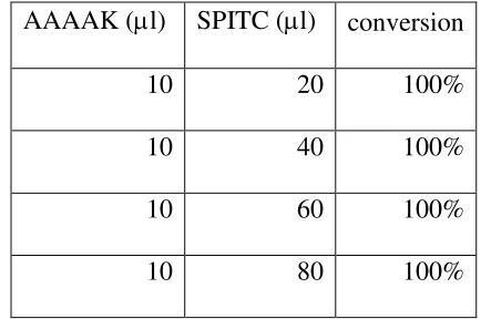
2.3.2 SPITC labeling ... 36
2.3.3 Isolation/Purification ... 37
2.3.4 Trypsin digestion ... 39
IV
2.3.6 Mass spectrometer selection ... 42
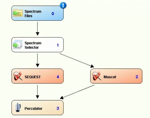
2.3.7 Database searching ... 43
2.4 Summary ... 44
2.5 References ... 44
Chapter 3 High Fidelity Approach for Proteomic Scale Enrichment and Identification of N-termini ... 47
3.1 ABSTRACT ... 47
3.2 INTRODUCTION ... 48
3.3 MATERIALS AND METHODS ... 51
3.3.1 Materials ... 51
3.3.2 Protein denaturation, reduction and alkylation ... 51
3.3.3 Guanidination ... 51
3.3.4 SPITC modification of N-termini ... 52
3.3.5 Trypsin digestion ... 52
3.3.6 N-terminal enrichment by ERLIC ... 53
3.3.7 LC-MS/MS analysis ... 54
3.3.8 Peptide identification ... 54
3.4 RESULTS AND DISCUSSION ... 55
3.4.1 Overall workflow description ... 55
3.4.2 Chemical modification of free amine groups in proteins ... 59
V
3.4.4 Effectiveness of N-terminal enrichment ... 62
3.4.5 Tandem mass spectra comparison between CID and HCD ... 65
3.4.6 N-termini analysis of E. coli cell lysate ... 67
3.4.7 Comparison of bound and flow-through fractions of E. coli cell lysate .. 75
3.5 CONCLUSIONS ... 81
3.6 REFERENCES ... 81
Chapter 4 iTRAQ Labeling of N-terminal Amines in Complex Samples and Its Application in Protease Substrate Degradomics ... 85
4.1 Abstract ... 85
4.2 Introduction ... 86
4.3 Experiment ... 89
4.3.1 Materials ... 89
4.3.2 Reduction and alkylation ... 89
4.3.3 Guanidination ... 89
4.3.4 AspN-digestion ... 90
4.3.5 iTRAQ labeling ... 90
4.3.6 Trypsin digestion ... 91
4.3.7 N-terminal enrichment by NHS-activated agarose resin ... 91
4.3.8 LC-MS/MS ... 92
4.4 Results and Discussion ... 94
VI
4.4.2 E. coli cell lysate results using the proposed workflow... 96
4.5 Summary ... 99
4.6 References ... 100
Chapter 5 Summary ... 104
References ... 106
Appendix ... 108
VII
List of Figures
Figure 1.1 Scheme for chemical labeling of ICAT and iTRAQError! Bookmark
not defined.5
Figure 1.2 Illustration of MS and MS/MS of iTRAQ labeled peptide ... Error!
Bookmark not defined.5
Figure 1.3 Typical positional proteomics workflowError! Bookmark not defined.7
Figure 1.4 Scheme for N-TAIL workflow ... Error! Bookmark not defined.13
Figure 1.5 Scheme for COFRADIC for N-terminal peptides Error! Bookmark not
defined.16
Figure 1.6 scheme of proteome wide C-termini analysis by Overall lab ... Error!
Bookmark not defined.20
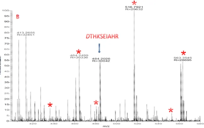
Figure 2.1 Comparison between before (A) and after (B) enrichment by NHS activated agarose resin spin column for BSA sample. N-terminal peptides (SPITC-DTHK(guandinyl)SEIAHR m/z= 484.203+). ... Error! Bookmark not defined.39
Figure 2.2 Workflow for database searching ... Error! Bookmark not defined.43
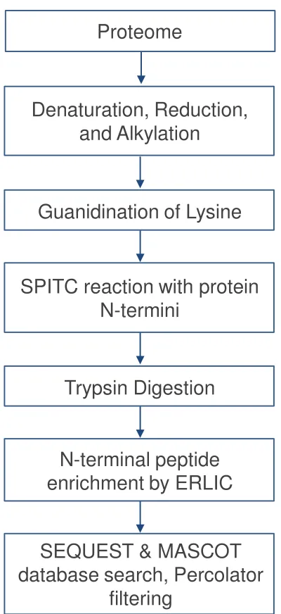
Figure 3.1 Flow chart for N-terminal identification by SPITC modification and ERLIC enrichment. ... Error! Bookmark not defined.54
Figure 3.2 Schematic illustration for the guanidination reactionError! Bookmark
not defined.58
Figure 3.3 Schematic illustration of SPITC modificationError! Bookmark not
defined.58
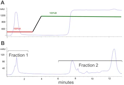
Figure 3.4 Fraction collections for trypsin digested E.coli cell lysate using electrostatic repulsion hydrophilic interaction (ERLIC) chromatography; (A) UV
detection at 215 nm and (B) UV detection at 280 nm.Error! Bookmark not
VIII
Figure 3.5 MS/MS spectra for BSA peptide SPITC-DTHK(guandinyl)SEIAHR m/z= 484.203+ by (A) CID and (B) HCD. SPITC is attached to N-terminal ions labeled in red. ... Error! Bookmark not defined.64
Figure 3.6 MS/MS spectra for N-terminal peptides without guanidinyl lysine; (A) sequence MNIIEANVATPDAR, M1-SPITC protein ID P61714
(6,7-dimethyl-8-ribityllumazine synthase OS=Escherichia coli) and (B) sequence
AVTNVAELNALVER, A2-SPITC protein ID P0A9Q7 ( Aldehyde-alcohol dehydrogenase OS=Escherichia coli). SPITC is attached to N-terminal ions labeled in red. ... Error! Bookmark not defined.66
Figure 3.7 MS/MS spectra of N-terminal peptide containing guanidinyl lysine; (A) AGK(guanidinyl)AGVEVDDR protein ID P0A9P0 (dihydrolipoyl dehydrogenase
OS=Escherichia coli) (B) VVINK(guanidinyl) DTTTIIDGVGEEAAIQGR
protein ID P0A6F5 (60 kDa chaperonin OS=Escherichia coli ) and (C) LDMLNEELSDK(guanidinyl )ER protein ID P63284(Chaperone protein ClpB OS=Escherichia coli). SPITC is attached to N-terminal ions labeled in red. ... Error!
Bookmark not defined.67
Figure 3.8 MS/MS spectra for SPITC peptide ELVTAAKLGGGDPDANPR (protein ID P0A8A0; (A) containing unmodified lysine and (B) containing guanidinyl Lysine. ... Error! Bookmark not defined.69
Figure 3.9 MS/MS spectra for N-terminal peptides of protein P0A850 (Trigger factor OS=Escherichia coli); (A) P0A850from1to13, (B) P0A850from244to255, and (C) P0A850from322to334. SPITC is attached to N-terminal ions labeled in red. ... Error! Bookmark not defined.71
IX
fraction as labeled with SPITC on N-terminii. Areas are approximately proportional to the counts shown in the figures. .... Error! Bookmark not defined.74
Figure 3.11 Sums of PSMs for all proteins (A) in flow-through fraction and (B) those in bound fraction that were identified as N-terminal SPITC labeled. In each case, proteins were split into two groups those that were also present in the other set (N-terminal SPITC in bound fraction or all proteins in flow-through fraction respectively) or only present in the given set. Proteins present in both sets tend to
have higher PSM sums, suggesting their higher abundance.Error! Bookmark not
defined.75
Figure 3.12 fractions of PSMs assigned to peptides starting from different ranges of positions in original protein sequences. ... Error! Bookmark not defined.77
Figure 4.1 Scheme for N-terminal Proteolytic Peptides by iTRAQ labeling.... Error!
Bookmark not defined.92
Figure 4.2 iTRAQ labeled N-terminal peptides (a) iTRAQ-MFPEYR from protein P0ACW6 (Uncharacterized protein YdcH OS=Escherichia coli), position 1-6 (b) iTRAQ-DVQVFTR from protein P0A8V2(DNA-directed RNA polymerase
subunit beta OS=Escherichia coli), position 930-936.Error! Bookmark not
X
List of Tables
Table 2.1 Guanidination optimization ... Error! Bookmark not defined.35
Table 2.2 Evaluation of SPITC reaction conditionsError! Bookmark not
defined.36
XI
Abstract
Positional proteomics is emerging as an attractive technique to characterize
protein termini, which play important biological roles in cells. Even with the
advances in past decades, there still are areas for improvement. This thesis focuses
on improving data quality and assignment confidence in positional proteomics.
A novel workflow was designed for the large-scale identification of protein
N-terminal sequences. 4-sulfophenyl isothiocyanate (SPITC) is used for N-termini
sulfonation; Upon higher energy collisional dissociation (HCD), SPITC peptides in
electrospray ionization ESI generate predominately y-type ion series; such
simplification of spectra enables the identification of N-termini with high fidelity.
The presence of b1 + SPITC product ions upon HCD furthers the confidence for
N-terminal identifications. Secondly, sulfonated N-N-terminal peptides possess one
negative charge site at low pH, which was exploited to enrich the SPITC modified
N-terminal peptides by electrostatic repulsion hydrophilic interaction (ERLIC)
chromatography. Such enrichment process allows both termini enriched and
N-termini deficient fractions to be collected and analyzed by LC-MS/MS. This
method was applied to an E. coli cell lysate, identifying approximately 350
N-terminal peptides (85% represented neo-N-termini from protein degradation and
XII
274 distinct E.coli proteins, 224 of which were also identified in the analysis of
flow-through fractions from internal peptides.
Another approach we took to boost the identification confidence is by
exploiting iTRAQ (isobaric tag for relative and absolute quantitation) in the
positional proteomics workflow. This approach allows for multiplexed comparison
between different samples, and thus is well-suited for degradadomics analyses
where degraded samples are compared to control samples. Both control and
protease treated sample are labeled by different tags which allows direct
comparison of protein N-termini with neo-N-termini. In addition, samples are
analyzed duplicate by labeling with two tags, aiming for quick validation of
peptides by internal replicates. In this study, Asp-N digested E.coli cell lysate is
taken as a model system. A total of 500 N-terminal peptides, corresponding to 370
proteins, were identified with high confidence in one experiment, with 87% of
those proteolytic products matching the expected protease digestion specificity,
validating the assignment accuracy of this approach.
1
Chapter 1.
Introduction
1.1 Introduction of Proteomics
Proteomics is the large-scale study of proteins particularly their structures
and functions.1,2 Proteomics is the next step in the study of biological systems after
genomics and transcriptomics, since proteins are vital parts of living organisms as
the main components of the physiological metabolic pathways of cells. Mass
spectrometry (MS) has increasingly become a key technology for protein sequence
analysis in the past decade, especially for complex mixtures, due to significant
advances in instrumentation, sample preparation techniques and data interpretation
algorithms.
Large scale, “bottom-up” (also called “shotgun”) characterization of cellular
proteomes is the most widely adapted method in proteomics study.3,4,5,6,7,8,9,10 A
typical workflow for high-throughput bottom-up characterization is composed of
protein extraction from cells, enzymatic digestion, LC-MS/MS analysis and
database searching which correlate MS/MS spectra with sequence and ultimately
the parent proteins.
Proteome systems typically encompass thousands of individual components
present in concentrations ranging over several orders of magnitude;11 therefore, to
2
generally used for fractionation prior to MS analysis.12 , 13 The combination of
online/offline strong cation exchange (SCX) with reversed-phase (RP)
chromatography is the most widely used MDLC due to the good orthogonality of
the methods.14,15,16 Peptides are separated by charge in SCX and by hydrophobicity
in RP. The use of high concentrations of salt in SCX may cause problems in
downstream analysis; therefore, online desalting of the collected fractions from
SCX with the trap column is the common practice,17 even with the drawback of a
high tendency for autosampler blockage and quick deterioration of the trap
column. Extensive separation of components aids in the protein coverage; it is now
possible to identify more than 10,000 proteins from human cells.18,19
1.2 Isobaric Labeling in Proteomics
A major aspect of proteome research is quantitative proteomics aiming at
measuring relative changes in proteins expressed in cells or tissues of different
states, e.g. healthy versus disease state.20,21,22,23,24 Measurement of relative changes
is simplified when two or more analytes can be labeled distinctly, combined, and
analyzed as a mixture. Labeling schemes based on isotopic labeling, in which all
labeled versions of a single peptide co-elute from LC, are especially powerful, as
the relative abundance for this peptide corresponds directly to the relative signal
3
has been made to develop stable isotopic labeling methods to facilitate downstream
MS analysis for direct comparison.25,26,27,28
Isotopic labeling with stable isotopes is a well-known method for "tagging"
specific proteins. Such metabolic labeling is applicable to cell culture, e.g., through
growth in isotope-labeled media (e.g., 15N media29,30). Another approach is stable
isotope labeling by essential amino acids in cell culture (SILAC)31,32 which relies
upon addition of intact isotopically-labeled amino acids. The most commonly used
stable isotope-encoded amino acids are 13C6-lysine or 13C6-arginine.33,34,35,36 The
biggest advantage of SILAC compared to other isotope labeling techniques is that
the SILAC technique offers minimum technical variations in sample processing
due to the fact that the isotope is introduced into the cell culture, the earliest
possible sample mixing stage.
Most in vitro labeling techniques are based on the formation of a covalent
bond between the labeling reagent and the specific functional groups in
polypeptides. Isotope-coded affinity tag (ICAT) is among the first of such
applications reported in 1999.37 ICAT consists of three elements: an iodoacetamide
group to modify cysteine residues, an isotopically coded linker, and a biotin tag for
the affinity isolation of labeled proteins/peptides (refer to figure 1.1). The process
starts with ICAT labeling, followed by trypsin digestion, then, the ICAT labeled
4
reduction of sample complexity. The concept is innovative and widely accepted.38
However, the method provides limited coverage over the proteome, as the cysteine
content in proteins is fairly low giving poor coverage of the digested peptides, and
many proteins have no cysteine.
Among all the isotopic labeling techniques, the iTRAQ (isobaric tags for
relative and absolute quantification) method shows significant advantages.39,40,41,42
The iTRAQ reagent reacts with proteolyzed peptides to form an NHS ester
derivative with primary amino groups. Differentially labeled peptides appear as
single peaks in MS scans due to the isobaric mass design of the iTRAQ reagent
(refer to figure 1.1); such multiplication of peptide abundance results in
improvement of sensitivity. When subjected to MS/MS, the isotope encoded
reporter ions provide relative quantitative information on peptides and ultimately
on proteins (refer to figure 1.2). In a complex mixture, iTRAQ samples subjected
to independent data acquisition in LC-MS/MS have a tendency to allow
identification of only high abundance proteins in a traditional proteomics
workflow. Both 4-plex and 8-plex versions of iTRAQ are commercially available.
In this thesis, effort has been made to incorporate iTRAQ with positional
Figure 1.1 Scheme for chemical labeling of ICAT and iTRAQ
Figure 1.2 Illustration of MS and MS/MS of iTRAQ labeled peptide
1.3 Positional Proteomics
The concept of positional proteomics is that a protein can be identified by a
single, position-defined peptide, with the two most obvious positional locations
5
Scheme for chemical labeling of ICAT and iTRAQ
Illustration of MS and MS/MS of iTRAQ labeled peptide
Positional Proteomics
The concept of positional proteomics is that a protein can be identified by a
defined peptide, with the two most obvious positional locations
Scheme for chemical labeling of ICAT and iTRAQ
Illustration of MS and MS/MS of iTRAQ labeled peptide
The concept of positional proteomics is that a protein can be identified by a
6
within every protein being the N- and C-termini.43 Sample complexity is
dramatically reduced in N-terminal or C-terminal enriched samples of proteolytic
digests. There are two driving forces in this field to motivate the advance of this
technology. One is the proteome annotation,44 where the termini play important
roles in protein function, mutation and post translational modification.45 ,46 The
other is the development of degradomics,47,48 which focuses on the elucidation of
protease substrate and cleavage sites. The newly generated termini after protease
treatment are called neo-N-termini or neo-C-termini, in order to differentiate them
from mature (innate) protein termini.
To identify terminal peptides, the normal proteomics workflow is no longer
suitable due to the fact that the terminal peptides are buried in the sea of internal
tryptic peptides. The sample complexity makes it difficult to select and detect the
terminal peptides during MS/MS acquisition. Therefore, terminal peptide
enrichment prior to MS analysis is essential in such workflow development.
Enrichment greatly simplifies the proteome by using single terminal peptides for
protein identification, which increases dynamic range and proteome coverage for
low abundant proteins.
It is essential to differentiate the termini peptides from the internal peptide in
positional proteomics, since the same functional groups that define the protein
proteolyzed peptides. In addition, primary amine and carboxylic groups are
present in the side chains of lysine and acidic amino acid residues, respectively.
The following paragraphs summarize strategi
positional proteomics.
Positional proteomics workflow can be roughly divided into the following
modules shown in figure 1.3: (1) labeling of termini, (2) proteolysis, (3)
enrichment, and (4) LC-MS/MS analysis.
Figure 1.3 Typical positional proteomics workflow
The enrichment process can be categorized as either
approach or a negative selection approach. The former modif
tag that enables targeted enrichment of
mixture. The latter takes advantage of the newly generated functional groups
(primary amine for N-termini and carboxylic acid for C
which can be conjugated to
enrichment of the targeted termini.
N- or C-terminal
labeling
Protein
mix Proteolysis Labeled
protein mix
7
proteolyzed peptides. In addition, primary amine and carboxylic groups are
present in the side chains of lysine and acidic amino acid residues, respectively.
The following paragraphs summarize strategies that have been applied in
Positional proteomics workflow can be roughly divided into the following
modules shown in figure 1.3: (1) labeling of termini, (2) proteolysis, (3)
MS/MS analysis.
Typical positional proteomics workflow
The enrichment process can be categorized as either a positive selection
negative selection approach. The former modifies the termini with a
tag that enables targeted enrichment of terminal peptides from the digested
advantage of the newly generated functional groups
termini and carboxylic acid for C-termini) after digestion,
which can be conjugated to another matrix and depleted, resul
enrichment of the targeted termini.
Proteolysis Enrichment
Labeled peptide mix
LC-MS/MS
Enriched labeled termini
proteolyzed peptides. In addition, primary amine and carboxylic groups are
present in the side chains of lysine and acidic amino acid residues, respectively.
es that have been applied in
Positional proteomics workflow can be roughly divided into the following
modules shown in figure 1.3: (1) labeling of termini, (2) proteolysis, (3)
positive selection
the termini with a
terminal peptides from the digested
advantage of the newly generated functional groups
termini) after digestion,
other matrix and depleted, resulting in the
8
1.3.1 Positional Proteomics approach for N-terminal analysis
1.3.1.1 Enrichment of protein N-termini by positive selection using
biotinylation
A method using biotinylation to positive select N-terminal peptides was
reported by Kuhn et al in 2003.49 This protein tag technology was later on adapted
and further developed by the Salvesen lab to identify protease substrates.50 In this
method, the side chains of lysine are first blocked by guanidination, and then free
alpha-amines (protein N-termini) are reacted with a disulfide-linked biotin
derivative (sulfosuccinimidyl 2-(biotinamido)-ethyl-1, 3-dithiopropionate
(sulfo-NHS-SS-biotin)). After trypsin digestion, the peptide mixture is incubated with
immobilized streptavidin beads, which retains the N-terminal peptide. The last step
of sample preparation is to elute the N-terminal peptide by reducing the disulfide
bond. This workflow was applied to study in vivo constitutive proteolysis in E.
coli, yeast, mouse and human samples and determine the specificity of methionine
aminopeptidases, signal peptidases and mitochondrial peptidases.
Another biotinylation protocol was reported by Wells et al. based on subtiligase
in 200851. An engineered variant of subtilisin called subtiligase was used, which
shows absolute selectivity for ligation of alpha-amines, forming a biotinylated
9
are then denatured, reduced and alkylated before trypsin digestion. Then, the
biotinylated N-terminal peptides are captured by immobilized streptavidin,
resulting in the separation from internal and C-terminal peptides. The N-terminal
peptides are eventually released by TEV digestion, with tagging of
Ser-Tyr-dipeptides at the N-termini as a signature for identification by LC-MS/MS. The
subtiligase method was validated by analysis of the proteome of living and
apoptotic Jurkat cells. It was reported that 333 unique cleavage sites in 282
proteins were identified after aspartic acid residues and were therefore linked to
caspase activity in etoposide treated apoptotic Jurkat cells. A separate study using
this technique combined with SILAC in cell culture to identify caspase-1 substrates
in vitro and in cell-based inflammation models was also reported.52
A third biotinylation method incorporated Edman degradation in the
workflow 53 . After protein is denatured, reduced and alkylated, phenyl
isothiocyanate (PITC) is used to block all primary amines in the proteome. Then,
trifluroacetic acid is used to break the peptide bond between the first and second
amino acid of PITC modified proteins, while PITC modified ε-amines (i.e. lysine
side chains) remain intact in this treatment. The next steps are similar to the
aforementioned method. The newly generated free α-amine is biotinylated with
sulfo-NHS-SS-biotin, followed by trypsin digestion. Streptavidin is used to trap
10
enriched N-terminal peptides in this case are one amino acid shorter than the true
N-termini. This approach was validated by a mixture of known proteins and
applied to characterize the constitutive N-terminal processing events in Jurkat
cells. Both known and new caspase substrates were identified in the
cisplatin-induced apoptosis of Jurkat cells.
A fourth biotinylation-related method in degradomics is called PICS
(proteomic identification of protease cleavage) developed by the Overall lab54.
This approach differs from the other approaches in that it is peptide centric and not
protein/substrate centric. First, proteins are digested by trypsin or endoproteinase
Glu-C, and then all primary amines are blocked by methylation. The peptide
mixture is treated as a library and incubated with a protease of interest.
Neo-N-terminal peptides are biotinylated and affinity-selected, followed by LC-MS/MS
analysis. This approach was applied to profile serine (thrombin, neutrophil
elastase, cathepsin G), aspartic (HIV-1 protease) and cysteine proteases (cathepsin
K, caspase-3, caspase-7).55
The advantage of positive selection using biotinylation is the high efficiency of
enrichment due to the high specificity between biotin-streptavidin. A drawback of
this type of technique may arise from amino acid bias during the biotinylation
reaction. For example, the secondary α-amino group of proline is less reactive
11
positive selection, mature proteins with acetylation, dimethylation or cyclization at
N-termini will not be detected.
1.3.1.2 Enrichment of protein N-termini by negative selection using
biotinylation
McDonald et al43 reported a protocol based on biotinylation for negative
selection in 2005. Here, all the primary amines in a proteome sample are acetylated
first, followed by trypsin digestion. The internal peptides and C-terminal peptides
containing an α-amino group at a newly generated N-termini are biotinylated and
removed by passing through immobilized streptavidin beads. The simplification
effect was shown in chicken skeletal muscle and E. coli cell lysate.
1.3.1.3 Enrichment of protein N-termini by negative selection using amine
reactive reagent
There are a few reports using amine reactive reagents to scavenge the newly
generated internal peptides after digestion, thus enriching N-terminal peptides. The
basic workflow is as follows: (1) The primary amines in proteins are acetylated or
dimethylated, followed by trypsin digestion. (2) The internal peptides and
C-terminal peptides are scavenged by amine reactive reagent and the flow through,
12
McDonald et al57 used NHS activated Sepharose beads to scavenge internal
peptides in 2006 and reported the identification of about 300 E. coli protein
N-termini.
Mikami and Takao used an isocyanate resin to capture internal peptides.58 The
performance of isocyanate resin was demonstrated by applying it to several peptide
mixtures, including proteolytic digests.
Kleifeld et al47 employed a self-synthesized dendritic polyglycerol aldehyde
polymer to capture tryptic and C-terminal peptides, which can be conveniently
removed by centrifugation. It is claimed that the binding of tryptic peptides of
such polymer is up to 2.5 mg peptide/ mg polymer, a more than ten-fold
improvement in capacity over amine reactive resins. This approach incorporated
with isotopic labeling under various formats is a valuable venue for N-terminal
positional proteomics research and it is further explained in the following section
of N-TAILS.
1.3.1.4 Protein N-terminal identification by N-terminal amine based
isotope labeling of substrates (N-TAILS)
A detailed and streamlined protocol of N-TAILS was described by the Overall
lab in 2011,59 based on previous development and applications by the same
13
N-terminal peptides and uses primary amine labeling-based quantification as the
discriminating factor. Labeling is versatile; the authors elaborate
dimethylation-TAILS, SILAC-TAILS and iTRAQ-TAILS in the current protocol. This method is
suited to many applications, including biochemical and cell culture analysis in
vitro as well as analysis of tissue samples from animal and human sources in vivo.
The TAILS workflow is composed of the following steps (refer to figure 1.4):
protein collection and proteolysis by the test protease; isotopic labeling and
primary amine blocking followed by tryptic digestion; negative selection by a high
efficiency polymer (dendritic polyglycerol aldehyde polymers); identification of
N-terminal peptides by LC-MS/MS; identification of protease substrates by the
sequence of the cleavage sites, or loss of cleaved natural N-terminal peptides.
To improve coverage, it is recommended to employ two or more digesting
14
Figure 1.4 Scheme for N-TAIL workflow 59
1.3.1.5 Enrichment of protein N-termini using phosphor tagging (PTAG)
and TiO2-based depletion
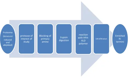
Mommen et al62 described a protocol to enrich protein N-terminal peptides
using phospho tagging (PTAG) and titanium dioxide (TiO2) affinity
chromatography. Primary amino groups in proteins are initially dimethylated with
formaldehyde, followed by digestion using trypsin, chymotrypsin and
endoproteinase Glu-C. The newly formed internal peptides are modified with the
PTAG reagent glyceraldhyde-3-phosphate in nearly perfect yields (>99%). The
resulting phosphopeptides are removed by binding onto TiO2 affinity column. This
method allowed identification of 753 N-terminal peptides, corresponding to 428
Proteome (denatured reduced and alkylated)
protease of interest of
study
Blocking of primary
amine
trypsin digestion
reaction with
HPA-ALD polymer
Ultrafiltration
15
proteins, in N. meningitides and 928 N-terminal peptides, corresponding to 572
proteins, in S. cerevisiae.
1.3.1.6 Enrichment of protein N-termini by combined fractional diagonal
chromatography (COFRADIC)
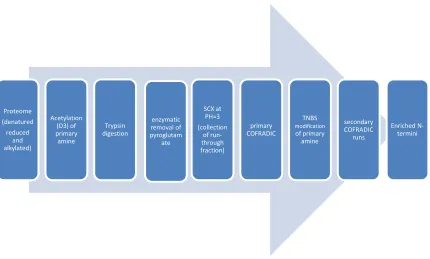
In 2003, the N-terminal combined fractional diagonal chromatography
(COFRADIC) technology introduced by the Gevaert lab63 was the first positional
proteomics technology by which N-terminal peptides were enriched by depleting
other peptides (refer to figure 1.5). This method progressed in the following
years64,65,66,67,68and the latest protocol was reported in 2011.69 The protocol can be
briefly described as the following: Before trypsin digestion, proteins undergo
denaturation, reduction and alkylation. Then, all primary amines are blocked by
trideutero-acetylation (to distinguish in vitro acetylation from in vivo acetylation)
so that trypsin digestion will produce only Arg-ending peptides. The peptide
mixture is incubated with glutamine cyclotransferases and pyroglutamyl amino
peptidases, respectively, to convert N-terminal glutamines into pyroglutamates and
remove these pyroglutamates from the peptide backbone. The above mixture is
then loaded onto an SCX cartridge at low pH, where N-terminal blocked peptides
are poorly retained and are collected in the flow through fraction since they carry
one less positive charge compared to internal peptides at low pH. This peptide
16
the above collected fractions are treated with 2,4,6-trinitrobenzenesulfonic acid
(TNBS), which reacts with the primary amine of any remaining internal peptides to
introduce a very hydrophobic trinitrophenyl group with these peptides. Each
treated fraction is put through the second RP-HPLC separation using identical
chromatographic conditions as in the first separation, where the N-terminal
peptides elute within the same time interval as during the primary run, however,
the TNBS modified internal and C-terminal peptides carrying a large hydrophobic
group elute much later in RP-HPLC, thus resulting in separation from the
neo-N-terminal and protein N-neo-N-terminal peptides.
Figure 1.5 Scheme for COFRADIC for N-terminal peptides 63
Proteome (denatured reduced
and alkylated)
Acetylation (D3) of primary amine
Trypsin digestion
enzymatic removal of pyroglutam
ate
SCX at PH=3 (collection
of run-through fraction)
primary COFRADIC
TNBS
modification
of primary amine
secondary COFRADIC
runs
17
The COFRADIC technology has been applied in various biological systems
and has been proved to be a reliable approach for protease-related studies. Stable
isotopes for labeling purpose are introduced in the protocol in biological
applications. Oxygen-18 was used to label peptides during trypsin digestion in a
human Jurkat cell culture; a total of 93 in vivo protease-processed sites in 71
proteins associated with Fas-induced apoptosis were identified. Oxygen-18
labeling together with COFRADIC was also used to map the proteolytic process in
anthracycline-induced acute myelogenous leukemia cell death.66
SILAC can also be incorporated into the COFRADIC workflow (refer to
figure 1.5), Arginine is chosen for SILAC labeling, since tryptic digestion
produces arginine-ending peptides. The control cells use regular arginine in cell
culture while the sample cells use 13C6 arginine in cell culture. Purified proteins in
the heavy labeled cells are treated with protease, and then combined with the
purified proteins in control cells. The following steps are the same as normal
COFRADIC. More than 800 cleavage sites in 332 human and 282 mouse
substrates for granzyme b were identified using SILAC combined with
COFRADIC.70
18
Generally speaking, C-terminal analysis is not as widely available as the
N-terminal analysis due to the formidable technical difficulties of selective activation
of carboxylic acids. Current methods of isolating C-terminal peptides are
predominantly affinity-based procedures. The application of mass spectrometry to
C-terminal analysis in the literature related to our topics is as follows:
1.3.2.1 Use anhydrotrypsin-lysine affinity to isolate the C-terminal
peptides
An elegant approach to isolate C-terminal peptides was reported in 2000.71
Basically, a sample is digested with endoprotease Lys-C, and then anhydrotrypsin
coupled onto agarose beads is applied to the digest sample. Anhydrotrypsin is a
catalytically inert variant of trypsin capable of binding peptides with C-terminal
lysine or arginine. Thus, the N-terminal and internal peptides are bound to
anhydrotrypsin beads; after centrifuging to get rid of beads, the supernatant only
contains the original C-terminal peptides for further analysis. However, this
method is not suitable for proteins ending with lysine or arginine (~ 84% of
proteins do not end with lysine or arginine). This approach lacks robustness; the
amount of anhydrotrypsin beads was adjusted for each of the proteins investigated.
The reason is that a small amount of anhydrotrysin beads is not sufficient to
19
binding of C-terminal peptides to the beads. For a complex system with a large
dynamic range, the integrity of the results cannot be ensured.
1.3.2.2 Use of DITC resin to isolate C-terminal peptides
In an approach described by Kuyama et al., a sample is initially digested with
endoprotease Lys-C, then TMPP modification is selectively applied to cap the
N-terminal amino group. The reagent for TMPP modification is
succinimidyloxycarbonylmethyl tris (2,4,6-trimethoxyphenyl) phosphonium
bromide (TMPP-Ac-Osu). p-Phenylenediisothiocyanate (DITC) resin is used to
scavenge the lysine-containing peptides from a Lys-C digest. Isolated C-terminal
peptides are then de novo sequenced using MALDI-MS/MS.72 This method has
been further optimized73 to use diisothiocyanate coupled glass beads. This method
is not suitable for proteins ending with lysine or arginine.
1.3.2.3 Use of polymer-based enrichment for C-terminal peptides
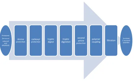
Overall et al74 reported a polymer-based enrichment approach to profile protein
C-terminal peptides. Briefly, the workflow is as follows (refer to figure 1.6):
protein thiol groups are reduced and alkylated, carboxyl groups of C termini,
aspartate and glutamate side chains are then protected by carbo-diimide-mediated
(1-ethyl-3-(3-dimethylaminopropyl) carbodiimide hydrochloride, EDC) and
20
derivatized proteins are then digested by trypsin. To prevent cross-reactivity,
peptide concatamerization or cyclization, free amines are protected with methyl
groups. The internal tryptic and N-terminal tryptic peptides are removed by
coupling with polyallylamine (MW ~56000, sigma) mediated with EDC.
Ultrafiltration is used to remove the polymer, retaining the enriched C-terminal
peptides in solution. LC-MS/MS follows to analyze the sample.
This approach is the first that allows proteome-wide C-terminal analysis. In the
same report, C-terminal amine-based isotope labeling of substrates (C-TAILS) was
also described, in which heavy isotope formaldehyde is used during both reductive
methylation steps. C-TAILS was tested using a Glu-C digested E. coli cell lysate
model system. The result showed that more than 90% of the peptides identified had
21
Figure 1.6 scheme of proteome wide C-termini analysis by Overall lab 74
1.3.3 Positional Proteomics approach for both N and C-terminal analysis
1.3.3.1 SCX to selectively enrich in vivo acetylated protein N-terminal and
C-terminal peptides
This method requires no chemical derivatization. The sample is trypsin digested
and SCX is run at pH 3 to fractionate the sample.44 The majority of C-terminal and
acetylated N-terminal peptides elute very early in the chromatogram, while internal
peptides elute much later. This separation is due to a lack of net positive charge in
both C-terminal and acetylated N-terminal peptides at pH 3, while internal peptides
have a net positive charge due to the unblocked amino terminus and the Lys or Arg
side chain. However, this separation will not work in the following two
circumstances: Proteome (denatured
reduced and alkylated)
Amine protection
carboxyl protection
tryptic digest
tryptic digestion
second amine protection
polymer
coupling filtration
Enriched C-terminal
22
o The C-terminal ends with lysine or arginine.
o Trypsin digestion has a missed-cleavage, resulting in peptides with
multiple basic residues.
In such circumstances, the terminal peptides will possess at least two free
amines, thus their charge properties change and they are no longer able to be
separated from the internal peptides. The author also pointed out that the selective
recovery of acetylated N-terminal and C-terminal peptides depends on the trypsin
digestion integrity.
1.3.3.2 Combined fractional Diagonal Chromatography (COFRADIC) for
simultaneous N and C terminal proteomics
In 2010, the Gevaert lab48 published a COFRADIC workflow for both N and
C- terminal peptide identification in a single sample preparation. This COFRADIC
workflow follows the procedures described in N-terminal COFRADIC, except that
after the primary fraction collection, peptides are reacted with an
N-hydroxy-succinimide ester of butyric acid, butyrylating the C-terminal peptides. Such
modification allows C-termini to elute 4-12 min after N-termini in secondary
RP-HPLC. The pooled N-terminal and C-terminal peptides are ready for LC-MS/MS
analysis. The authors used this COFRADIC based approach to study processing by
23
cell lysate is used as substrate pool for granzyme B, with 12C6 arginine cell lysate as
control. Thus, upon trypsin digestion, neo-N-terminal peptides can be
differentiated from the N-termini background of the cell lysate. C-termini are
differentiated by using N-hydroxysuccinimide (NHS) esters of 12C4 or 13C4 butyric
acid. In this study, a total of 1621 annotated N-termini and 760 annotated
C-termini were identified, with 334 neo-C C-termini resulting from granzyme B
processing and 16 neo-C termini resulting from carboxypeptidase A4 processing.
1.3.4 Thesis overview
With all the efforts and advances in positional proteomics, the inherent main
drawback of such techniques still needs to addressed, which is how to validate the
identified substrates due to the “one hit wonder” in such technique. The strength
of positional proteomics arise from the dramatically reduced complexity of
proteome samples, however, the strength turns into weakness if the question is
asked how you can validate the results. In this thesis, we propose an approach
which separates the enriched N-termini fraction for positional proteomics from the
peptide mixtures, while preserving the rest of the peptide mixture. Therefore, MS
analysis of the remaining peptide mixtures can serve as validation for the results of
positional proteomics. Such direct experimental validation is meaningful and
convincing considering both analyses use the same starting material. The above
24
N-termini and separate the SPITC modified peptides from others using
electrostatic repulsion hydrophilic interaction chromatography (ERLIC).
Moreover, the confidence of N-termini identification is further strengthened by
exploiting the unique fragmentation behavior of SPITC peptides, thus we conclude
that our approach offers high fidelity assignment of N-terminal peptides. This work
is presented in chapter 3.
Another attempt to simultaneously validate the results of positional
proteomics is by use of iTRAQ-4plex. In this study, both control and protease
treated samples are labeled by different tags which allows direct comparison of
protein N-termini with neo-N-termini. In addition, samples are analyzed in
duplicate by labeling with two tags (i.e. tag 116 and tag 117), aiming for quick
validation of peptides by internal replicates. A new workflow is designed which
incorporates iTRAQ into positional proteomics to study the specificity of protease.
This work is presented in chapter 4.
Experimental optimization is shown in chapter 2. A summary of study is
presented in chapter 5.
25
1 Anderson N.L.; Anderson N.G. Proteome and proteomics: new technologies, new concepts, and
new words. Electrophoresis 1998, 19 (11): 1853–61.
2 Blackstock W.P.; Weir M.P. Proteomics: quantitative and physical mapping of cellular
proteins. Trends Biotechnol. 1999, 17 (3), 121–7.
3 de Godoy, L. M. F.; Olsen, J. V.; Cox, J.; Nielsen, M. L.; Hubner, N. C.; Froehlich, F.;
Walther, T. C.; Mann, M. Nature 2008, 455, 1251-1254.
4 Cramer, Grant R.; Van Sluyter, Steve C.; Hopper, Daniel W.; Pascovici, Dana; Keighley,
Tim; Haynes, Paul A.; Proteomic analysis indicates massive changes in metabolism prior to the inhibition of growth and photosynthesis of grapevine (Vitis vinifera L.) in response to water deficit. BMC Plant Biology (2013), 13, 49.
5 Shimwell, N. J.; Bryan, R. T.; Wei, W.; James, N. D.; Cheng, K. K.; Zeegers, M. P.; Johnson,
P. J.; Martin, A.; Ward, D. G.; Combined proteome and transcriptome analyses for the discovery of urinary biomarkers for urothelial carcinoma. British Journal of Cancer (2013), 108(9), 1854-1861.
6 Alves, G.; Yu, Y.K.; Improving Peptide Identification Sensitivity in Shotgun Proteomics by
Stratification of Search Space. Journal of Proteome Research (2013), 12(6), 2571-2581.
7 Cantor, D.; Slapetova, I.; Kan, A.; McQuade, L. R.; Baker, M. S.; Overexpression of
αvβ6
integrin alters the colorectal cancer cell proteome in favor of elevated proliferation and a switching in cellular adhesion That Increases Invasion.
8 Yates, John R.; The Revolution and Evolution of Shotgun Proeomics for Large-Scale
Proteome Analysis. Journal of the American Chemical Society (2013), 135(5), 1629-1640.
9 Claassen, M.; Inference and validation of protein identifications. Molecular & Cellular
Proteomics (2012), 11(11), 1097-1104.
10 Vaudel, M.; Sickmann, A.; Martens, L.; Current methods for global proteome identification.
26
11 Teng, P.; Bateman, N.W; Hood, B.L. and Conrads T.P.; J. of proteome research 2010,
9(12), 6091-6100.
12 Di Palma, S.; Hennrich, M. L.; Heck, A. J. R.; Mohammed, S.; Recent advances in peptide
separation by multidimensional liquid chromatography for proteome analysis. Journal of Proteomics (2012), 75(13), 3791-3813.
13 Motoyama, A.; Yates, J. R.; Multidimensional LC Separations in Shotgun Proteomics.
Analytical Chemistry (Washington, DC, United States) (2008), 80(19), 7187-7193.
14 Fang, X.; Balgley B.; Wang W.; Park D.M.; Lee C.S.; comparison of multidimensional
shotgun technologies targeting tissue proteomics. Electrophoresis 2009, 30 (23), 4063-70.
15 Huang, E. L.; Orsat, V.; Shah, M. B.; Hettich, R. L.; VerBerkmoes, N. C.; Lefsrud, M. G.;
The temporal analysis of yeast exponential phase using shotgun proteomics as a fermentation monitoring technique. Journal of Proteomics (2012), 75(17), 5206-5214.
16 Kang, D.; Nam, H.; Kim, Y-S.; Moon, M. H.; Dual-purpose sample trap for on-line strong
cation-exchange chromatography/reversed-phase liquid chromatography/tandem mass spectrometry for shotgun proteomics. Application to the human Jurkat T-cell proteome. Journal of Chromatography A (2005), 1070(1-2), 193-200.
17 Unwin, R.D.; Griffiths J.R.; Whetton A.D.; Simultaneous analysis of relative protein
expression levels across multiple samples using iTRAQ isobaric tags with 2D nano LC-MS/MS. Nat Protoc. 2010, 5(9), 1574-1582.
18 Nagaraj N.; wisniewski J.R.; Geiger T.; Cox J.; Kircher M.; Kelso J.; Paabo S. and Mann M.;
Deep proteome and transcriptome mapping of a human cancer cell line. Mol Syst Biol 2011, 7, 1-8.
19 Beck M.; Schmidt a.; Malmstroem J.; Claassen M.; Or A.; Szymborska A.; Herzog F.; Rinner
27
20 Dihazi, H.; Prognosis markers for metastatic renal cell carcinoma: quantitative proteomics
approach. Expert Review of Proteomics (2013), 10(1), 21-24.
21 Dagley, L. F.; Emili, A.; Purcell, A. W.; Application of quantitative proteomics technologies
to the biomarker discovery pipeline for multiple sclerosis. Proteomics: Clinical Applications (2013), 7(1-2), 91-108.
22 Marcilla, M.; Albar, J. P.; Quantitative proteomics: A strategic ally to map protein
interaction networks. IUBMB Life (2013), 65(1), 9-16.
23 Holman, S. W.; Sims, P. F. G.; Eyers, C. E.; The use of selected reaction monitoring in
quantitative proteomics. Bioanalysis (2012), 4(14), 1763-1786.
24 Shi, T.; Su, D.; Liu, T.; Tang, K.; Camp, D. G.; Qian, W-J.; Smith, R. D.; Advancing the
sensitivity of selected reaction monitoring-based targeted quantitative proteomics. Proteomics (2012), 12(8), 1074-1092.
25 Kovanich, D.; Cappadona, S.; Raijmakers, R.; Mohammed, S.; Scholten, A.; Heck, A.J. R.
Applications of stable isotope dimethyl labeling in quantitative proteomics. Analytical and Bioanalytical Chemistry (2012), 404(4), 991-1009.
26 Christoforou, A. L.; Lilley, K. S.; Isobaric tagging approaches in quantitative proteomics:
the ups and downs. Analytical and Bioanalytical Chemistry (2012), 404(4), 1029-1037.
27 Arsova, B.; Kierszniowska, S.; Schulze, W. X.; The use of heavy nitrogen in quantitative
proteomics experiments in plants. Trends in Plant Science (2012), 17(2), 102-112.
28 Christoforou, A.; Lilley, K. S.; Taming the isobaric tagging elephant in the room in
quantitative proteomics. Nature Methods (2011), 8(11), 911-913.
29 Joost, W. G.; Bastiaan, B. J. T.; Jeroen, K.; Metabolic labeling of model organisms using
28
30 Rayavarapu, S.; Coley, W.; Cakir, E.; Jahnke, V.; Takeda, S.; Aoki, Y.; Grodish-Dressman,
H.; Jaiswal, K. J.; Hoffman, P. E.; Brown, J. K.; et al. Identification of Disease Specific Pathways Using in Vivo SILAC Proteomics in Dystrophin Deficient mdx Mouse. Molecular & cellular proteomics: MCP (2013), 12(5), 1061-73.
31 Munday, D. C.; Surtees, R.; Emmott, E.; Dove, B. K.; Digard, P.; Barr, J. N.; Whitehouse,
A.; Matthews, D.; Hiscox, J. A.; Using SILAC and quantitative proteomics to investigate the interactions between viral and host proteomes. Proteomics (2012), 12(4-5), 666-672.
32 Cox, J.; Matic, I.; Hilger, M.; Nagaraj, N.; Selbach, M.; Olsen, J. V.; Mann, M.; practical
guide to the MaxQuant computational platform for SILAC-based quantitative proteomics. Nature Protocols (2009), 4(5), 698-705.
33 Zanivan, S.; Krueger, M.; Mann, M.; In vivo quantitative proteomics: the SILAC mouse.
Methods in Molecular Biology (New York, NY, United States) (2011), 757(Integrin and Cell Adhesion Molecules), 435-450.
34 Austin, J. R.; Kuestner, E. R.; Chang, K. D.; Madden, R. K.; Martin, B. D.; SILAC
Compatible Strain of Pichia pastoris for Expression of Isotopically Labeled Protein Standards and Quantitative Proteomics. Journal of Proteome Research (2011), 10(11), 5251-5259.
35 Nie, A-Y.; Zhang, L.; Yan, G-Q.; Yao, J.; Zhang, Y.; Lu, H-J.; Yang, P-Y.; He, F-C.; In
Vivo Termini Amino Acid Labeling for Quantitative Proteomics. Analytical Chemistry (Washington, DC, United States) (2011), 83(15), 6026-6033.
36 Geiger, T.; Wisniewski, R. J.; Cox, J.; Zanivan, S.; Kruger, M.; Ishihama, Y.; Mann, M.;
Use of stable isotope labeling by amino acids in cell culture as a spike-in standard in quantitative proteomics. Nature Protocols (2011), 6(2), 147-157.
37 Gygi S.P.; Rist B.; Gerber S.A.; Turecek F.; Gelb M.H.; Aebersold R.; Quantitative analysis of
29
38 Aerbersold, R.; Gygi, P. S.; Griffin, J. T.; Han, K. M. D.; Yelle, J. M.; The isotope-coded
affinity tag reagent method for quantitative proteomics. American Genomic/Proteomic Technology (2001), 1(1), 22, 24, 26-27.
39 Wu, W. W., Wang, G., Baek, S. J. & Shen, R. F. Comparative study of three proteomic
quantitative methods, DIGE, cICAT, and iTRAQ, using 2D gel- or LC-MALDI TOF/TOF. J Proteome Res 5, 651-8 (2006).
40 Wiese S.; Reidegeld K.A.; Meyer H.E. and Warscheid B.; Protein labeling by iTRAQ: a new
tool for quantitative mass spectrometry in proteome research. Proteomics, 2007, 340-350.
41 Noirel, J.; Evans, C.; Salim, M.; Mukherjee, J.; Ow, S. Y.; Pandhal, J.; Pham, T. K.; Biggs,
A. C.; Wright, C. P.; Methods in quantitative proteomics: setting iTRAQ on the right track. Current Proteomics (2011), 8(1), 17-30.
42 Wang, W-S.; Liu, X-H.; Liu, L-X.; Jin, D-Y.; Yang, P-Y.; Wang, X-L.; Identification of
proteins implicated in the development of pancreatic cancer-associated diabetes mellitus by iTRAQ-based quantitative proteomics. Journal of Proteomics (2013), 84, 52-60.
43 McDonald, L. and Beynon R.J.; Positional proteomics: selective recovery and analysis of
N-terminal proteolytic peptides. Nature Methods, 2005 2(12), 955-957.
44 Dormeyer W.; Mohanned S.; Breukelen B.; Krijgsveld J. and Heck A.J.R.; Targeted analysis
of protein termini. Journal of proteome research 2007, 6, 4634-4645.
45 Ajami K.; Pitman M.R.; Wilson C.H.; Park J.; Menz R.I.; Starr A.E.; Cox J.H.; Abbott C.A.;
Overall C.M.; Gorrell M.D.; Stromal cell-derived factors 1alpha and 1beta, inflammatory protein-10 and interferon-inducible T cell chemo-attractant are novel substrates of dipeptidyl peptidase 8. FEBS lett 2008, 582, 819-825
46 McQuibban G.A.; gong J.H.; Wong J.P.; Wallace J.L.; Clark-Lewis I.; Overall C.M.; Matrix
30
47 Kleifeld O.; Doucet, A.; Keller, U.; Prudova, A.; Schilling, O.; Kainthan, R.; Starr, A.; Foster,
L.J.; Kizhakkedathu, J.; Overall, C.M.; Isotopic labeling of terminal amines in complex samples identifies protein N-termini and protease cleavage products 2010, Nature Biotech. 28(3), 281-288.
48 Van Damme P.; Staes A.; Bronsoms S.; Helsens K.; Colaert N.; Timmerman E.; Aviles F.X.;
Vandekerckhove J. and Gevaert K; Complementary positional proteomics for screening substrates of endo- and exoproteases; Nature Methods, 2010, 7(7) 512-515.
49 Kuhn K.; Thompson A.; Prinz T.; Muller J. et al.; Isolation of N-terminal protein sequence
tags fro cyanogen bromide cleaved proteins as a novel approach to investigate hydrophobic proteins. J. Proteome Res. 2003, 2, 598-609.
50 Timmer J.C.; Enoksson M.; Wildfang E.; Zhu W.; Igarashi Y.; Denault J-B.; Ma Y.; Dummitt
B.; Chang Y-H.; Mast A.E.; Eroshkin A.; Simth J.W.; Tao W.A. and Salvesen G.S.; Profiling constitutive proteolytic events in vivo.; Biochem. J., 2007, 407, 41-48.
51 Mahrus S.; Trinidad J.C.; Barkan D.T.; Sali A.; Burlingame A.L. and Wells J.A.; Global
sequencing of proteolytic cleavage sites in apoptosis by specific labeling of protein N termini. Cell, 2008, 134, 866-876.
52 Agard N.J.; Maltby D.; Wells J.A.; Inflammatory stimuli regulate caspase substrate profiles.
Mol. Cell Proteomics, 2010, 9, 880-893.
53 Xu G.; Shin S.B.Y. and Jaffrey S.R.; Global profileing of protease cleavage sites by
chemoselective labeling of protein N-termini. Proc Natl Acad Sci USA 2009, 106, 19310-19315.
54 Schilling O.; Overall C.M.; Proteome-derived, database-searchable peptide libraries for
identifying protease cleavage sites. Nat Biotechnol 2008, 26, 685-694
55 Schiling O.; Overall C.M.; Proteome-derived, database-searchable peptide libraries for
identifying proteoase cleavage sites. Nat. biotechnol. 2008, 26, 685-694.
56 Burger W.C.; Interference by carbonyl compounds in the trinitrobenzenesulfonic acid method
31
57 McDonald L.; Robertson D.H.; Hurst J.L.; Beynon R.J.; Positional proteomics: preparation of
amino-terminal peptides as a strategy for proteome simplification and characterization. Nat. Protoc. 2006, 1, 1790-1798.
58 Mikami T. and Takao T.; Selective isolation of N-blocked peptides by isocyanate-coupled
resin. Anal. Chem. 2007, 79, 7910-7915.
59 Kleifeld O.; Doucet A.; Prudova A.; auf dem Keller U.; Gioia M.; Kizhakkedathu J.N. and
Overall C.M.; Identifying and quantifying proteolytic events and the natural N terminome by terminal amine isotopic labeling of substrates. Nature protocol 2011, 6, 10, 1578-1611.
60Prudova A.; auf dem Keller U.; Butler G.S. and Overall C.M.; Multiplex N terminome analysis
of MMP-2 and MMP-9 substrate degradomes by iTRAQ-TAILS quantitative proteomics. Mol. Cell. Proteomics 2010, 9, 894-911.
61 Auf dem Keller U.; Prudova A.; Gioia M.; Butler G.S. and Overall C.M.; A statistic-based
platform for quantitative N terminome analysis and identification of protease cleavage products. Mol. Cell. Proteomics, 2010, 9, 912-927.
62 Mommen, G.P.M; Waterbeemd, B.V.D.; Meiring H.D.; Kersten, G.; Heck, A.J.R and Jong,
P.J.M; unbiased selective isolation of protein N-terminal peptides from complex proteome samples using phosphor tagging (PTAG) and TiO2-based depletion. MCP, published on June 22, 2012.
63 Gevaert K.; Goethals M.; Martens L.; Van Damme J.; Staes A.; Thomas G.R.;
Vandekerchhove J.; exploring proteomes and analyzing protein processing by mass spectrometric identification of sorted N-terminal peptides. Nat. Biotechnol. 2003, 21, 566-569.
64 Van Damme, P.; Martens, L.; Van Damme, J.; Hugelier, K.; Caspase-specific and non-specific
in vivo protein processing druing Fas-induced apoptosis. Nat. Methods 2005, 2, 771-777.
65 Staes A.; Van Damme P.; Helsens K.; Demol H. et al Improved recovery of
32
66Gausdal, G.; Gjertsen, B.T.; McCormack, E.; Van Damme, P. et al.; Abolition of stress induced
protein synthesis sensitizes leukemia cells to anthracycline –induced death. Blood 2008, 111, 2866-2877.
67 Impens, F.; Van Damme, P.; Demol, H.; Van Damme, J.; et al.; Mechanistic insight into
taxol-induced cell death. Oncogene 2008, 27, 4580-4591.
68 Arnesen T.; Van Damme P.; Rolevoda B.; Helsens K. et al; Proteomics analyses reveal the
evolutionary conservation and divergence of N-terminal acetyltransferases from yeast and humans. Proc. Natl. Acad. Sci. USA, 2009, 106, 8157-8162.
69 Staes A.; Impens F.; Van Damme P.; Ruttens B.; Goethals M.; Demol H.; Timmerman E.;
Vandederchhove J.; Gevaert K.; Selecting protein N-terminal peptides by combined fractional diagonal chromatography. Nat. Protoc. 2011, 6, 1130-1141.
70 Van Damme, P.; Maurer-Stroh S.; Plasman K.; van Durme J. et al.; Analysis of protein
processing by N-terminal proteomics reveals novel species-specific substrate determinants of granzyme B orthologs. Mol. Cell. Proteomics 2009, 8, 258-272.
71 Sechi S. and. Chait B.T.; A method to define the carboxyl terminal of proteins. Analytical
Chemistry 2000, 72, 3374-3378.
72 Hiroki Kuyama et al A simple and highly successful C-terminal sequence analysis of proteins
by mass spectrometry Proteomics 2008, 8, 1539-1550.
73 Sonomura, K. et al the specific isolation of C-terminal peptides of proteins through a
transamination reaction and its advantage for introducing functional groups into the peptide. Rapid Commun. Mass Spectrom. 2009, 23, 611-618.
74 Schilling O.; Barre O. Huesgen P.F. and Overall C.M.; proteome-wide analysis of protein
33
Chapter 2 Experiments toward a streamlined workflow for
positional proteomics
2.1 Overview of method
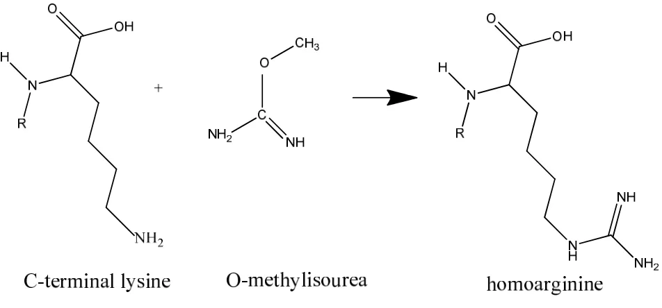
The workflow starts by converting lysine to homoarginine by guanidination,
followed by sulfonation of N-termini by 4-sulfophenyl isothiocyanate (SPITC).
After trypsin digestion, electrostatic repulsion hydrophilic interaction (ERLIC)
chromatography is used to enrich SPITC modified peptides. Both the flow-through
fraction (containing internal and C-terminal peptides) and eluted fraction
(containing SPTIC modified N-terminal peptides) are analyzed by LC-MS/MS. A
specialized N-terminal database with sequentially trimmed N-termini is used to
identify N-terminal peptides from degraded proteins.
2.2 Consideration for development
The two critical aspects for any positional proteomics workflow are terminal
labeling and enrichment. To identify minor species in a complex mixture, some
sort of enrichment for the minor species is required. There are various approaches
and efforts described for this topic in the literature as summarized in chapter 1.
N-terminal enrichment approaches can be categorized into three pathways, namely
chemically or enzymatically positive selection of N-termini,1,2 negative selection
34
exchange chromatography4 based on the charge differences among N-terminal ,
C-terminal and internal peptides. Both negative and positive selection approaches
were tested in our study, the results of which are described later in this chapter.
The second critical aspect is the manner of labeling of the protein termini.
Here we choose to use SPITC for labeling N-termini. SPITC derivatization is
predominantly applied to proteolytic peptides in the literature,5 so our development
effort was focused on modifying the protocol to make it work on complex mixtures
of intact proteins with high efficiency. Guanidination was performed prior to
SPITC, which converted lysine to homoarginine by blocking the side chain of
lysine. Therefore, SPITC derivatization only occurred at the N-terminal site and
was assigned as such during data interpretation.
Besides the two aspects mentioned above, the overall workflow was
streamlined, particularly with respect to protein/peptide purification steps.
2.3 Optimization
Experiments designed to optimize each step are presented in this section in
the same order as the steps in the workflow. These are: guanidination reaction,
SPITC reaction, protein purification, trypsin digestion, enrichment, LC-MS/MS
and database searching.
35
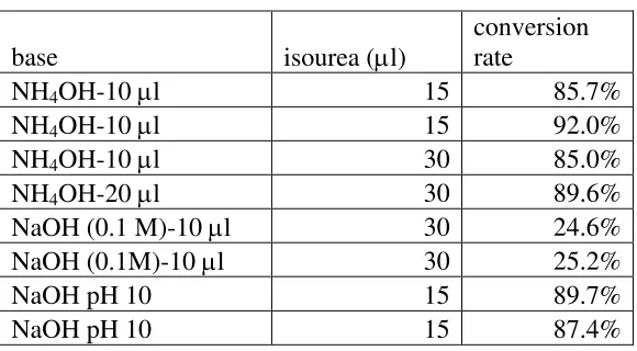
The purpose of guanidination is to selectively cap the side chain of lysine,
thus leaving only the N-terminal amine for the next step in the SPITC reaction. The
reaction is selective, except when glycine is the N-terminal amino acid; in such
cases the primary amine group of glycine can also be modified.6 The guanidination
reaction is performed as reported by Reilly et al.7
A standard peptide with the sequence
TNEIVEEQYTPQSLATLESVFQELGK (m/z 2952.4; m/z for guanidination
product is 2994.4) was used to monitor this reaction. The conversion rate was
computed as the area of m/z 2994.4 divided by the sum of the areas of m/z 2952.4
and m/z 2994.4 obtained from LC-MS. The concentration for isourea was 300 mM,
the starting material of peptide was 0.12 mM and only 1 µL was used for each
reaction. 10 µl NH4OH with 15 µL isourea is the recommended condition by the
kit vendor (Sigma Aldrich). The conversion was around 90%. Doubling the use of
isourea or hydroxide did not increase the conversion rate. Due to the downstream
SPITC derivatization of primary amines, the introduction of a large quantity of
ammonium ions is undesirable, so NH4OH was replaced with 10 µL of 0.1 M
NaOH. While this change in the base caused the conversion rate to drop
significantly to around 25%, further study determined that titrating the reaction
with NaOH to pH 10 produced a comparable conversion rate to that for the