R E S E A R C H
Open Access
Site-specific selection reveals selective
constraints and functionality of tumor
somatic mtDNA mutations
Deyang Li
1, Xiaohong Du
1, Xu Guo
1, Lei Zhan
2, Xin Li
1, Chun Yin
1, Cheng Chen
1, Mingkun Li
3, Bingshan Li
4,
Hushan Yang
5and Jinliang Xing
1*Abstract
Background:Previous studies have indicated that tumor mitochondrial DNA (mtDNA) mutations are primarily shaped by relaxed negative selection, which is contradictory to the critical roles of mtDNA mutations in tumorigenesis. Therefore, we hypothesized that site-specific selection may influence tumor mtDNA mutations.
Methods:To test our hypothesis, we developed the largest collection of tumor mtDNA mutations to date and evaluated how natural selection shaped mtDNA mutation patterns.
Results:Our data demonstrated that both positive and negative selections acted on specific positions or functional units of tumor mtDNAs, although the landscape of these mutations was consistent with the relaxation of negative selection. In particular, mutation rate (mutation number in a region/region bp length) in complex V and tRNA coding regions, especially in ATP8 within complex V and in loop and variable regions within tRNA, were significantly lower than those in other regions. While the mutation rate of most codons and amino acids were consistent with the expectation under neutrality, several codons and amino acids had significantly different rates. Moreover, the mutations under selection were enriched for changes that are predicted to be deleterious, further supporting the evolutionary constraints on these regions.
Conclusion:These results indicate the existence of site-specific selection and imply the important role of the mtDNA mutations at some specific sites in tumor development.
Background
The human mitochondrial genome is an about 16.5kbp circular mitochondrial DNA (mtDNA) that harbors a con-trol region of 1.1kbp and 37 genes that encode 2 rRNA, 22 tRNA, and 13 proteins of oxidative phosphorylation. Among them, 7 genes encode subunits of complex I (ND1–6andND4L); 1 gene encodes a subunit of complex III (CYTB); 3 genes encode subunits of complex IV (COX1–3); and 2 genes encode subunits of complex V (ATP6 and ATP8) [1]. Due to the lack of protective histones and efficient DNA repair system, the mutation frequency of mtDNA is about 10 times higher than that of nuclear DNA [2]. In the past decade, high-frequency
somatic mtDNA mutations have been reported in a variety of diseases, especially in cancers [3, 4].
A series of previous studies established a connection between mitochondrial dysfunctions caused by mtDNA mutations and tumor development and metastasis [5–10]. For example, Cruz-Bermúdez et al. showed that cybrids
con-taining mutations in ND1 (m.3460G > A), ND4
(m.11778G > A) and ND6 (m.14484 T > C) of mtDNA exhibit the enhanced tumorigenicity [5]; Ishikawa et al. revealed that the G13997A and 13885insC mutations in the coding region of ND6gene confer an increased metastatic potential in mouse P29 cell line that originated from Lewis lung carcinoma [6].
On the other hand, however, a handful of studies indi-cated that mtDNA mutations in tumors are primarily shaped by the relaxation of negative selection relative to germline, rather than positive selection [11–13]. For example, Coller et al. reported that random processes are * Correspondence:[email protected]
1State Key Laboratory of Cancer Biology and Experimental Teaching Center
of Basic Medicine, Fourth Military Medical University, 169 Changle West Road, Xi’an 710032, China
Full list of author information is available at the end of the article
sufficient to explain the incidence of homoplasmic mtDNA mutations in human tumors without the need of positive selection pressure [11]. Stafford et al. concluded that the re-laxation of negative selection shapes mtDNA mutation landscapes in cancer cells, as evidenced by the higher ratio of nonsynonymous to synonymous mutations among these mutations [12]. Michele Vidone et al. extracted mtDNA reads from WGS and WES data of glioblastoma multiforme with MToolBox [14] and found that the large majority of mtDNA mutations does not pass the prioritization filters and only a relatively limited burden of pathogenic muta-tions, which did not appear to determine a general impair-ment of the respiratory chain, is indeed carried by GBM [15]. Consistently, Liu et al. [13] reported that mtDNA mutations in esophageal cancer and other tumors are most likely shaped by the relaxation of negative selection, and pointed out that the observation of some level of positive selections [16] is likely due to sample mix-up or contamin-ation of exogenous DNA. Recently, two groups [17, 18] reported that most of missense mutations of mtDNA do not have distinct physiological advantages, indicating that these mutations are not under positive selection. In addition, sev-eral studies also concluded that mtDNA mutations are not related to the developments of most cases of lung can-cer [19] and head and neck squamous cell carcinoma [20]. All of these lines of evidence seem to be contradictory to the critical roles of mtDNA mutations in tumor develop-ment, leaving unclear whether somatic mtDNA mutations are drivers, or just bystanders, of tumor development.
The discrepancies among the findings of these various studies may partially be attributed to their limitations, such as small patient populations, incapacity of sequencing methods to detect low level of heteroplasmy, and neglect of replication-mediated mutation frequency bias. Another pos-sibility is that these evolution studies have mainly focused on the overall level of mtDNA mutations, but not on their level in specific regions or individual positions. In contrast, func-tional studies have mostly focused on individual mutations.
To shed more light on the subject, we aimed to explore whether site-specific natural selection might affect tumor somatic mtDNA mutations and, more importantly, their functions. We performed a systematic and comprehensive exploration of mtDNA mutations in various cancer types, and we developed the largest collection of mtDNA mutations in cancer to date using next generation sequen-cing (NGS) data generated in various sources, including The Cancer Genome Atlas (TCGA), International Cancer Genome Consortium (ICGC), other publications, as well as our own lab. We analyzed the mutations across the mito-chondrion genome as well as in different genomic regions and functional units (e.g., gene domains, codons, amino acids) to evaluate how natural selection shapes mtDNA mutation patterns both globally and locally. These compre-hensive analyses of mtDNA mutations, conducted at an
unprecedented scale, shed important novel insights on the roles of mtDNA mutations in tumorigenesis.
Methods
Data of somatic tumor mtDNA mutations from public resources and our lab
We identified 5423 somatic mtDNA mutations from whole genome sequencing (WGS) and whole exome se-quencing (WES) data reported in literature [17, 18, 21], as well as in TCGA (https://portal.gdc.cancer.gov/explora-tion?searchTableTab=mutations&ssmsTable_size=100) and ICGC (https://dcc.icgc.org/repositories) datasets. For pa-tients who were present in two or more datasets and sequenced by the same method (WES or WGS), the muta-tions were intersected; for patients who were present in two or more datasets and sequenced by different methods, the data from WGS were retained. Seven patients with an extremely large number of somatic mutations (≥13) were excluded from further analyses.
In addition, we used mtDNA capture sequencing approach to identify 104 somatic mutations from 54 colon cancer patients, 357 somatic mutations from 143 hepato-cellular carcinoma (HCC) patients, and 36 somatic mutations from 15 kidney cancer patients. All these muta-tions derived from tumor tissue in our lab were verified by parallel sequencing of the non-tumor tissue of the same patient. Considering the error rate of the Illumina sequen-cing platform, only the somatic mutations with ≥2% of variant allele fractions were counted.
Finally, we generated a combined dataset containing 26 tumor types, 3277 patients, and 5920 mutations, the most comprehensive tumor mtDNA mutation dataset to date (Additional file 1: Table S1 and Additional file 2: Table S2). Among them, there were 2465 mutation records with heteroplasmic level data from three publications. The heteroplasmic levels of 497 mutations from our lab data were calculated as the ratio of the mutant reads in all reads that cover the mutation positions. All mutations were annotated using Annovar software [22].
Germline variation data in natural human population
Germline variation data containing 4322 mtDNA single base substitutions in the natural human population was ex-tracted from phylotree (Build 17, 18 Feb 2016) [23]. The htm file of mtDNA tree Build 17 containing all mtDNA var-iations among populations to date was downloaded (http:// www.phylotree.org/builds/mtDNA_tree_Build_17.zip) and the base substitution information was extracted using a homemade python script. (Additional file 3: Table S3).
Recurrent rate analysis
Recurrent rate of mutation in a specific region was determined by formula:
F ¼ ΣR
L, in which F stands for mutation rate, R for
recurrent times of mutations in a position, and L for the length (bp) of regions. If the R value of a mutation was greater than 1 in patient group, this mutation was considered recurrent mutation.
Prediction of functional impacts of mutations in mRNA and tRNA coding regions
The functional impacts of mutations in mRNA coding regions were predicted according to Mitimpact 2.5 [24], and were classified as high, medium, low, or neutral impact. The functional impacts of mutations in tRNA coding regions were predicted, as described by Kondrashov et al. [18], and were classified as either benign or deleterious.
Simulation of mtDNA mutations
We now describe a process to simulate mtDNA mutations under neutrality as a reference. Since it is impossible to know the mutation rate of each base under neutrality, we assumed that the vast majority of the mtDNA mutations in tumors were derived under neutrality, and selection plays a negligible role in shaping the overall mutation pattern. Under this assumption, we utilized the mutations in the large-scale dataset we collected to calibrate neutral mutation rates. Specially, we calculated mutation frequency of each of the possible nucleotide base substitutions (12 types in total) based on the observed base substitutions in this dataset. That is, for each of the 12 base substitutions, we counted the number of occurrences observed in this dataset, to rep-resent the mutant frequency under neutrality; such esti-mates are expected to be robust given the large size of the collected mutation dataset. In simulating a mutation dataset under neutrality, we first stored all the calibrated base sub-stitution frequencies in a file (Additional file 4: Table S4), and then used a homemade python script to randomly simulate 12 types of base substitutions, according to the mutation frequencies stored in this file (Fig. 1b). The data thus simulated have the same distribution of mutations observed in the large-scale mutation dataset we collected, representing mutations derived under neutrality. We repeated this process N times to generate random replicates of mutations, and used the simulated datasets as a reference of neutrality to compare against the observed mutations in a dataset. Simulating scripts are available from https://github.-com/gadbee/Bioinformatics.
Statistical analysis
All statistical tests were performed using Graphpad Prism software (GraphPad Software, Inc.). Comparisons between groups with continuous variables and categor-ical variables were performed using Student’s t test, and then ANOVA and Chi Square test. Pearson’s correlation coefficient was used to evaluate the correlations among
related variables. All p-values were two-tailed. Residual was calculated by the difference between simulated data and observed data of a mutant codon or an amino acid. Stan-dardized residual was calculated by formula:δ¼ δ−δ=σ, in whichδ* stands for standardized residual,δfor residual of a certain data point,δ for the average of all residuals, andσ for the standard deviation of all residuals. Standardized residuals greater than 2 were considered significant deviation of observed values from simulated values.
Ethical statement
The study was approved by the Institutional Review Boards of FMMU and informed written consent was obtained from each patient. The experiment was conducted according to the Declaration of Helsinki.
Results
Characteristics of 5920 Single Base mtDNA mutations
A total of 5920 single base mutations from 3277 patients of 26 tumor types (Additional file 1: Figure S1) were collected from two databases (TCGA and ICGC), three previous publications, and our mtDNA sequencing data (see Materials and Methods and Additional file 2: Table S2). Similar to previous report [17], a high consistency among WGS, WES, and mtDNA capture data was observed by comparing the proportion of synonymous and nonsynon-ymous mutations (Additional file 1: Figure S1B). In addition, we also analyzed the correlation of base substitu-tion numbers or the amino acid substitusubstitu-tion number between WGS and WES data (Additional file 1: Figure S1C, D) or between WGS + WES and mtDNA capture data (Additional file 1: Figure S1E, F) and found a high consistency of substitution pattern among the data derived from different sequencing methods. These findings indi-cated that all data were correctly combined for further ana-lyses. A total of 5920 mutations included 816 synonymous, 2946 nonsynonymous, and 209 stop-gain/stop-loss muta-tions at mRNA coding regions and 1949 mutamuta-tions at tRNA, rRNA coding regions and non-coding region. The vast majority (5449, 92.04%) were transitions (Fig. 1a), and most (5447, 92.01%) mutations were located in coding re-gions (Fig. 1a). The mutations had an obvious bias towards specific base substitutions, with CH> TH followed by TL> CLhaving a much higher rate than others (Fig. 1b).
Fig. 1Characteristics of 5920 tumor somatic mtDNA single base mutations.aNumber and proportion of mutations in different regions.b
Number and proportion of nonsynonymous mutations, synonymous mutations, stoploss mutations and stopgain mutations.cNumber and proportion of transition and transversion.dNumber of base substitutions.eNumber of somatic mutations in a tumor patient.fAverage number of somatic mutations in carcinoma and sarcoma.gAverage number of somatic mutations per patient across 26 tumor types.hMutation densities (mutation number in a region/region bp length) in coding regions and non-coding regions.iNumber of the top 10 recurrent mutations. Mean ± SEM, t test was used
significantly lower than those in carcinomas (Fig. 1e, right). The mutation levels, available for 2962 mutations, ranged from 2% heteroplasmy to 100% homoplasmy. Of the
5920 somatic mutations, more than half (2951,
51.10%) were not registered in the mitomap database updated on 29 Dec 2016 [25].
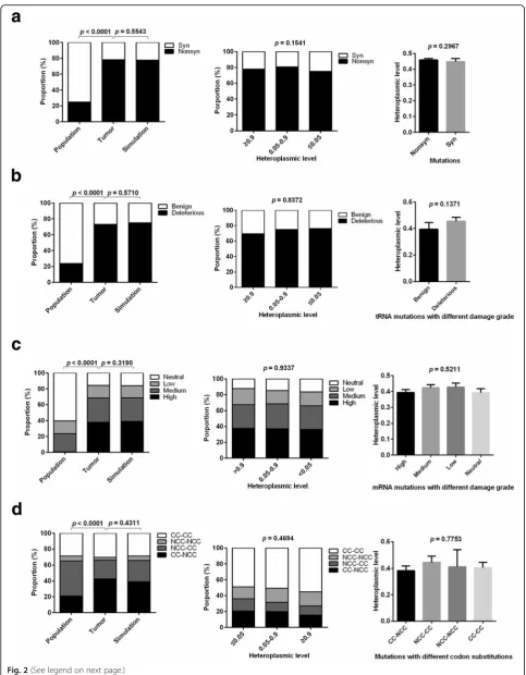
The relaxation of negative selection on tumor somatic mtDNA Single Base mutations
To evaluate the effects of natural selection on single base mutations, mutation patterns were analyzed in general population, tumor, and simulation data. Compared with general population data, tumor data showed a higher proportion of nonsynonymous mutations in mRNA-coding regions, more deleterious mutations in tRNA coding regions, higher impact on protein function, and more codons that cannot be completely complementary with anti-codon (Fig. 2a-d, left panel). These phenomena were consid-ered as a result of positive selection in previous studies [17]. Alternatively, another plausible model has been suggested that these tumor mutation patterns were shaped by muta-tion rate bias and relaxamuta-tion of negative selecmuta-tion [13, 17]. To further investigate whether the observed mutation patterns in the datasets that we developed are consistent with the relaxation of selection, we generated simulation data of mtDNA mutations under neutrality based on the mutation rate bias shown in Fig. 1b (see Methods). No significant differences existed between the observed muta-tions and the simulated data, supporting the effects of the relaxation of selection on mtDNA mutations at the genome scale (left panel of Fig. 2 a-d).
To rule out the possibility that mutations with different heteroplasmic levels are under different selective con-straints, we stratified the analyses by heteroplasmic levels (≤0.05, 0.05–0.9,≥0.9) and found that the same conclusion holds regardless of the heteroplasmy levels (Fig. 2 a-d, middle panel). Furthermore, we found that heteroplasmic levels were not significantly different between synonym-ous and nonsynonymsynonym-ous mutations, between deleterisynonym-ous and benign mutations in tRNA coding regions, among mutations with different damage grades in mRNA coding regions, and among mutations resulting in different codon substitutions (Fig. 2a-d, right panel). Taken together, our results indicate that single base mutations at the genome scale are not restricted by natural selection, which is consistent with previous reports [17, 18].
Functional unit-specific selection on tumor somatic mtDNA mutation
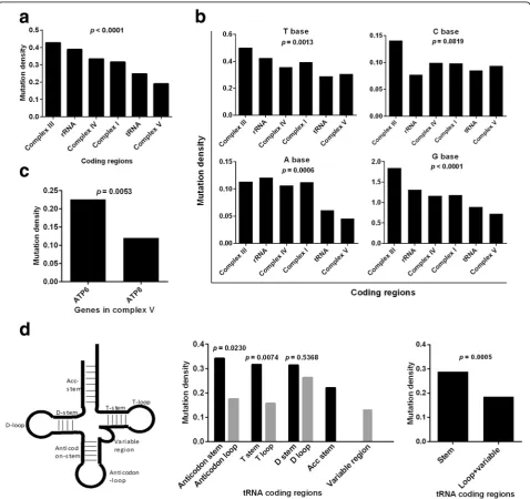
Previous studies demonstrated strong negative selection on truncating mtDNA mutations and tRNA anticodons [17, 18]. To further explore the effects of functional unit-specific natural selection on tumor mtDNA muta-tions, we compared the mutation rate (mutation number
in a region/region bp length) in different mtDNA coding regions. Interestingly, we found that the mutation dens-ities in complex V and tRNA coding regions were significantly lower than those in other regions (p< 0.0001, Fig. 3a). Considering the different mutation rates of 4 bases (Fig. 1b), the stratified analyses were performed. Our results indicated that the mutation rates of complex V and tRNA coding regions were the lowest among 6 subgroups when stratified by A, G and T bases (Fig. 3b). We further explored the mutation densities within the complex V and tRNA regions and found thatATP8has a lower mutation density than ATP6 in complex V region (p= 0.0053, Fig. 3c). The mutation densities in the loop/variable regions in tRNA were significantly lower than those in the stem regions (p= 0.0005, Fig. 3d, middle and right panels).
Site-preferred recurrence of tumor somatic mtDNA mutations
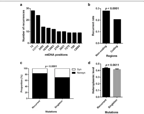
A total of 5920 mutations occurred at 3590 distinct posi-tions, among which 2384 were singletons and 3536 (59.73%) were recurrent. The recurrent mutations can be collapsed onto 1206 mtDNA positions with recurrence rate (i.e., num-ber of patients carrying the same mutation) ranging from 2 to 28. The 10 most recurrent mutation positions were listed in Fig. 4a. Furthermore, we found that non-coding regions had a higher probability of recurrent mutations than coding regions (p< 0.0001, Fig. 4b). The proportion of nonsynon-ymous mutations was higher in recurrent mutations than in singletons (p < 0.0001, Fig. 4c). The heteroplasmic levels of recurrent mutations tend to be higher than those of singletons, although the differences were not significant (p= 0.0611, Fig. 4d). These findings indicate that the recur-rence of tumor somatic mtDNA mutations is site-preferred, especially in non-coding region, and suggested the presence of positive selection of mtDNA mutations in tumors.
Codon-specific selection on tumor somatic mtDNA mutations
Fig. 2(See legend on next page.)
act on these specific mutant codons. In contrast, the observed numbers of mutant codon GAC (Asp) and AGC (Ser) were significantly lower than the simulated values (standardized residuals of −2.37 and −2.85, respectively), implying the possibility of negative selection of these
mutant codons (Fig. 5a, b). To investigate the robustness of the analysis, we carried out the same simulation 10 times and observed consistent patterns in all simulations (data not shown). To exclude the possibility that the outliers were influenced by the local sequence context of mutations, we
(See figure on previous page.)
Fig. 2The relaxation of negative selection on majority of tumor somatic mtDNA single base mutations.a,b,c,dProportion of synonymous and
nonsynonymous single base mutations in mRNA-coding regions, benign and deleterious in tRNA coding regions, mutations with different damage grades in mRNA coding regions, those resulting in different codon substitutions in population, tumor and simulation data (Left); Proportion only in tumor data stratified by heteroplasmic levels (Middle); Average heteroplasmic level in tumor data (Right). CC: codons completely complementary to anti-codons. NCC: codons not completely complementary to anti-codons. Left and middle: chi-square test was used. Right (a,b): t test was used; Right (c,d): ANOVA was used
Fig. 3Functional unit-specific selection on tumor somatic mtDNA mutations in coding regions.aMutation densities (mutation number in a region/region bp length) in different coding regions.bMutation densities of T, C, A, G bases in different coding regions.cMutation densities inATP6andATP8genes.d
Fig. 4Site-preferred recurrence of tumor somatic mtDNA mutations.aRecurrent times of top 10 mtDNA Positions.bRecurrent mutation rate, which was defined as the total recurrent times of mutations divided by bp length of region, in non-coding and coding regions. Chi-square test was used.cThe proportion of nonsynonymous and synonymous mutations in recurrent mutations and singletons.dThe heteroplasmic level of recurrent mutations and singletons
Fig. 5Codon-specific selection on tumor somatic mtDNA mutations.aThe correlation between numbers of mutation-caused codon from observed and simulated data. Red circles showed 4 codons which were significantly deviated from trend line.bResiduals and standardized residuals of four outliers in (a) were shown.cHeteroplasmic level of mutation-caused codons. Mean ± SEM, t test was used
re-analyzed the site-specific selection by examining the bases immediately 5′ and 3′ to the mutated bases. As shown in Additional file 1: Figure S3, significant correl-ation between observed and simulated mutant triplet-bases was observed (r= 0.9836, P< 0.0001). Moreover, three triplet-bases CAC, CAA and GAC with above−2 or 2 of standardized residuals were identified, suggesting that local sequence context may contribute to evolutional selection of specific mutations, although the underlying mechanism is largely unknown. Further analysis indicated that these three triplet-bases were not related with the selected codons shown in Fig. 5a. Specially, triplet-bases GAC in Additional file 1: Figure S3 and codon GAC in Fig. 5a were located on the different sides of trendline. Our results suggest that the outliers in Fig. 5a may not be determined by the context sequence. In addition, we also found that the average heteroplasmic levels of the base mutations in codon CCC and ACC were significantly higher than those in codon GAC and AGC (p= 0.0423, Fig. 5c), further supporting the existence of codon-specific selection on tumor mtDNA mutations.
Amino acid-specific selection on tumor somatic mtDNA mutations
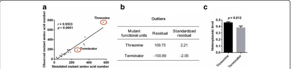
We further investigated the site-specific selection of mtDNA mutations at the amino acid level. Similarly, the number of each amino acid generated by mutant codons from observed or simulated data was determined. Once again, we found that the value from the simulated data was highly correlated with that from the observed data (r= 0.9533, p< 0.0001, Fig. 6a), further confirming the relaxation of negative selection for overall mutations. Nevertheless, the mutant amino acid Thr was signifi-cantly above the trend line (standardized residual of 2.21) and Ter (terminator) was significantly below the trend line (standardized residual of −2.05) (Fig. 6b). In addition, both amino acids Pro and Ser were also above and below trend line, respectively, although the devia-tions were not significant (standardized residuals of 1.23
and−0.89, respectively). We also observed that the aver-age heteroplasmic level of the base mutations in amino acids Thr was significantly higher than that in Ter (p= 0.012, Fig. 6c), which supports the notion that mutant amino acids are under specific selection. Notably, these findings align well with the selection pattern of codon mutations shown in Fig. 5.
Functionality of tumor somatic mtDNA mutations under site-specific selection
We investigated the predicted functionality of mutations in different genome regions and found that the proportion of mutations predicted to be highly deleterious was significantly different among complex III, IV, I, and V coding regions with the lowest value in complex V (p < 0.0001, Fig. 7a), suggest-ing the stricter negative selection on complex V, especially in ATP8 (p < 0.0001, Fig. 7b). We also observed that the pro-portions of deleterious mutations in loop and variable re-gions were significantly lower than those in stem rere-gions (p < 0.0001, Fig. 7c), indicating the possibility of stricter negative selection on loop and variable regions of tRNA. Moreover, we observed that the proportions of deleterious mutations in CCC and ACC codons and threonine were significantly greater than those in GAC and AGC codons and termin-ator, respectively (both p < 0.0001, Fig. 7d and e).
Discussion
Somatic mtDNA mutations have attracted significant atten-tion because of their high frequencies in cancers [3, 4]. Spe-cific mtDNA mutations have been implicated in tumor development and metastasis [5, 6]. However, an increasing number of recent studies have shown that tumor mtDNA mutations are under relaxed negative selection [13, 17, 18]. While the differing findings of these studies has raised con-troversy over the existence of specific functional mtDNA mutations, the indications of the relaxation of negative selec-tion on the majority of tumor mtDNA mutaselec-tions does not completely exclude the possibility of site-specific selection on individual mutations. To resolve these controversies, we
developed a comprehensive dataset of tumor mtDNA muta-tions and systemically analyzed the effects of site-specific selection on these mutations. We found most tumor mtDNA mutations are under the relaxed negative selection, which is highly consistent with previous studies [13, 17, 18]. We also noted that some specific regions are under stricter negative selection than other regions.
We further found that mutations in tRNA and complex V coding regions showed evidence of negative selection, especially inATP8gene coding regions of complex V and loop and variable regions of tRNA. In line with our findings, a previous study reported thatATP8gene seems to be protected from the relaxation of negative selection [26, 27]. Moreover, we found that the selective pattern on a region is related with the deleterious risk of mutations. This finding further confirms the fact that mutations in complex V and tRNA are under stricter negative selection, especially inATP8of complex V and variable/loop regions of tRNA, and partially explains the reason of evolutional constrains on these regions. This evolutional constraint implies that
the mutations disrupting the normal function of complex V and tRNA may confer an evolutionary disadvantage for tumors that predispose tumor to being negatively selected. Consistently, the important role of complex V in the etiology of tumor is also supported by experimental mouse data. For example, Petros et al. revealed that the prostate cancer cells with mutation T8993G inATP6gene generated larger tumor than wild-type cancer cells [28]. The essential role of the electron transport chain in aspartate synthesis [29] may be the reason that mtDNA mutations, especially those deleterious mutations, are negatively selected. The functional roles of mutations in complex V and tRNA in tumor need in-depth future investigations.
Recurrent mutations were analysed in this study. Generally, the recurrence of mutation in population tends to be the evidence of positive selection [30], which partially support our ratiocination. However, we cannot rule out such a possibility of negative selection on coding regions of mtDNA in tumors, which needs to be further explored in future studies.
Fig. 7Functionality of tumor somatic mtDNA mutations under site-specific selection.aProportion of mutations with high and medium impact in different coding regions.bProportion of mutations with high impact inATP6andATP8coding regions.cProportion of deleterious mutations in stem or loop and variable regions of tRNA.dProportion of mutations with high impact in mutation-caused codons with significant positive and negative selection.eProportion of mutations with high impact in mutation-caused threonine and terminator. Chi-square test was used
Our study also identified the specific codons and amino acids which tend to be under positive or negative selection. Our findings from selected codons and amino acids further supported the existence of site-specific selection of tumor mtDNA mutations and demonstrated that some selected mutations have potentially important functions in tumors. Moreover, our finding that the mutation-caused terminator was negatively selected is consistent with previous reports [17, 18], further supporting the reliability of our findings. Our analyses also showed that those codons that tend to be positively selected have a significantly higher pathogenic risk than those that tend to be negatively selected, and this may be the reason why these mutations are differentially selected. In consideration of the deleterious impact of stop-gain mutations, the negative selection on stop-stop-gain muta-tions enhanced the important role of mtDNA in tumors. The positive selections of codon CCC and ACC and amino acid Thr may imply the growth advantage they confer to tumor cells and needs to be further explored in the future.
In addition to the functional impact of the mutations in mRNA coding regions, the impacts on the mitochon-drial pool of free amino acids may be one of the reasons that some amino acids are differentially selected. Previ-ous study suggests that the concentration of free amino acids in mitochondria is positively correlated with the composition of mtDNA-encoded proteins [31]. There-fore, the mutation-caused composition changes of amino acids in mtDNA-encoded proteins may consume some free amino acids and increase the concentration of other free amino acids, which may influence the concentration of free amino acids and cause the unbalance of free amino acids pool in mitochondria. Considering the important roles of the concentration of free amino acids in tumor cells [29, 32–36], the changes in free amino acid concentration in mitochondria may influence the phenotype of tumor cells. This conjecture is supported by the observation that the codons/amino acids identi-fied to be selected in our study (Asp, Ser, Pro) overlap with the amino acids that are reported to influence the phenotype of tumor cells [34–36]. However, it is largely unknown about the cause-effect relationship between mtDNA mutation and the change of free amino acid concentration. On one hand, it is possible that the state of the amino acid pool may result in some codons/ amino acids to be selected for. On the other hand, the impacts of mutation on the mitochondrial pool of free amino acids may be one of the reasons that some amino acids are differentially selected. Therefore, the relation-ship between the amino acid pool and mtDNA mutation need to be further explored in future study.
Conclusions
Our results showed that the global landscape of tumor mtDNA mutations is shaped by the relaxation of
negative selection, and our detailed analysis also identi-fied regions and functional units that are under site-specific positive or negative selection, suggesting the extensive involvement of natural selection on mtDNA mutations during tumor development. The site-specific selection of tumor mtDNA mutations provides clues to identify driver mtDNA mutations of tumorigenesis and progression. More tumor mtDNA sequencing data and mechanistic experiments are needed to gain further in-sights into the roles of mtDNA mutations in tumors.
Additional files
Additional file 1: Figure S1.Figure S2.andTable S1.(DOCX 833 kb)
Additional file 2: Table S2.Information of 5920 tumor mtDNA base substitutions. (XLSX 488 kb)
Additional file 3: Table S3.Phylotree data annotation. (XLSX 224 kb)
Additional file 4: Table S4.All possible base substitution in mRNA coding regions of mtDNA. (XLSX 1519 kb)
Acknowledgements
This work was supported by the National Natural Science Foundation of China (grants 81320108021).
Authors’contributions
XJL designed the research. DXH collected and organized the public data. LDY, ZL and YC performed the“wet”experiments. LDY, DXH wrote the python and perl scripts. LDY, DXH, GX, LX and CC performed the statistical analysis. XJL wrote the manuscript. XJL, LMK, LBS, YHS, LDY edited the manuscript. All authors have read, revised and approved the final manuscript.
Competing interests
The authors declare no potential conflicts of interest.
Publisher’s Note
Springer Nature remains neutral with regard to jurisdictional claims in published maps and institutional affiliations.
Author details
1State Key Laboratory of Cancer Biology and Experimental Teaching Center
of Basic Medicine, Fourth Military Medical University, 169 Changle West Road, Xi’an 710032, China.2Department of Gastroenterology, Second Affiliated
Hospital of Harbin Medical University, Harbin 150086, China.3Fondation
Mérieux, 69002 Lyon, France.4Center for Human Genetics Research,
Department of Molecular Physiology and Biophysics, Vanderbilt University, Nashville, TN 37232, USA.5Division of Population Science, Department of
Medical Oncology, Sidney Kimmel Cancer Center, Thomas Jefferson University, Philadelphia, PA 19107, USA.
Received: 18 June 2017 Accepted: 16 November 2017
References
1. Taanman JW. The mitochondrial genome: structure, transcription, translation and replication. Biochim Biophys Acta. 1999;1410(2):103–23.
2. Tuppen HA, Blakely EL, Turnbull DM, Taylor RW. Mitochondrial DNA mutations and human disease. Biochim Biophys Acta. 2010;1797(2):113–28. 3. Cruz-Bermudez A, Vicente-Blanco RJ, Gonzalez-Vioque E, Provencio M,
Fernandez-Moreno MA, Garesse R. Spotlight on the relevance of mtDNA in cancer. Clin Transl Oncol. 2017;19(4):409–418.
4. Ye F, Samuels DC, Clark T, Guo Y. High-throughput sequencing in mitochondrial DNA research. Mitochondrion. 2014;17:157–63.
6. Ishikawa K, Takenaga K, Akimoto M, Koshikawa N, Yamaguchi A, Imanishi H, Nakada K, Honma Y, Hayashi J. ROS-generating mitochondrial DNA mutations can regulate tumor cell metastasis. Science. 2008;320(5876):661–4. 7. Wallace DC. Mitochondria and cancer: Warburg addressed. Cold Spring
Harb Symp Quant Biol. 2005;70:363–74.
8. Brandon M, Baldi P, Wallace DC. Mitochondrial mutations in cancer. Oncogene. 2006;25(34):4647–62.
9. Chatterjee A, Mambo E, Sidransky D. Mitochondrial DNA mutations in human cancer. Oncogene. 2006;25(34):4663–74.
10. He Y, Wu J, Dressman DC, Iacobuzio-Donahue C, Markowitz SD, Velculescu VE, Diaz LA Jr, Kinzler KW, Vogelstein B, Papadopoulos N. Heteroplasmic mitochondrial DNA mutations in normal and tumour cells. Nature. 2010; 464(7288):610–4.
11. Coller HA, Khrapko K, Bodyak ND, Nekhaeva E, Herrero-Jimenez P, Thilly WG. High frequency of homoplasmic mitochondrial DNA mutations in human tumors can be explained without selection. Nat Genet. 2001;28(2):147–50. 12. Stewart JB, Alaei-Mahabadi B, Sabarinathan R, Samuelsson T, Gorodkin J,
Gustafsson CM, Larsson E. Simultaneous DNA and RNA mapping of somatic mitochondrial mutations across diverse human cancers. PLoS Genet. 2015; 11(6):e1005333.
13. Reznik E, Miller ML, Senbabaoglu Y, Riaz N, Sarungbam J, Tickoo SK, Al-Ahmadie HA, Lee W, Seshan VE, Hakimi AA, et al. Mitochondrial DNA copy number variation across human cancers. elife. 2016;5:e10769.
14. Calabrese C, Simone D, Diroma MA, Santorsola M, Gutta C, Gasparre G, Picardi E, Pesole G, Attimonelli M. MToolBox: a highly automated pipeline for heteroplasmy annotation and prioritization analysis of human mitochondrial variants in high-throughput sequencing. Bioinformatics. 2014; 30(21):3115–7.
15. Vidone M, Clima R, Santorsola M, Calabrese C, Girolimetti G, Kurelac I, Amato LB, Iommarini L, Trevisan E, Leone M, et al. A comprehensive
characterization of mitochondrial DNA mutations in glioblastoma multiforme. Int J Biochem Cell Biol. 2015;63:46–54.
16. Zhidkov I, Livneh EA, Rubin E, Mishmar D. MtDNA mutation pattern in tumors and human evolution are shaped by similar selective constraints. Genome Res. 2009;19(4):576–80.
17. McMahon S, LaFramboise T. Mutational patterns in the breast cancer
mitochondrial genome, with clinical correlates. Carcinogenesis. 2014;35(5):1046–54. 18. Kondrashov FA. Prediction of pathogenic mutations in mitochondrially
encoded human tRNAs. Hum Mol Genet. 2005;14(16):2415–9.
19. Fang Y, Huang J, Zhang J, Wang J, Qiao F, Chen HM, Hong ZP. Detecting the somatic mutations spectrum of Chinese lung cancer by analyzing the whole mitochondrial DNA genomes. Mitochondrial DNA. 2015;26(1):56–60. 20. Liu SA, Jiang RS, Wang WY, Lin JC. Somatic mutations in the D-loop of
mitochondrial DNA in head and neck squamous cell carcinoma. Head Neck. 2015;37(6):878–83.
21. Larman TC, DePalma SR, Hadjipanayis AG, Cancer Genome Atlas Research N, Protopopov A, Zhang J, Gabriel SB, Chin L, Seidman CE, Kucherlapati R, et al. Spectrum of somatic mitochondrial mutations in five cancers. Proc Natl Acad Sci U S A. 2012;109(35):14087–91.
22. Wang K, Li M, Hakonarson H. ANNOVAR: functional annotation of genetic variants from high-throughput sequencing data. Nucleic Acids Res. 2010; 38(16):e164.
23. Ju YS, Alexandrov LB, Gerstung M, Martincorena I, Nik-Zainal S, Ramakrishna M, Davies HR, Papaemmanuil E, Gundem G, Shlien A, et al. Origins and functional consequences of somatic mitochondrial DNA mutations in human cancer. elife. 2014;3:e02935.
24. Castellana S, Ronai J, Mazza T. MitImpact: an exhaustive collection of pre-computed pathogenicity predictions of human mitochondrial non-synonymous variants. Hum Mutat. 2015;36(2):E2413–22. 25. West AP, Khoury-Hanold W, Staron M, Tal MC, Pineda CM, Lang SM,
Bestwick M, Duguay BA, Raimundo N, MacDuff DA, et al. Mitochondrial DNA stress primes the antiviral innate immune response. Nature. 2015; 520(7548):553–7.
26. Gasparre G, Romeo G, Rugolo M, Porcelli AM. Learning from oncocytic tumors: why choose inefficient mitochondria? Biochim Biophys Acta. 2011; 1807(6):633–42.
27. Pereira L, Soares P, Maximo V, Samuels DC. Somatic mitochondrial DNA mutations in cancer escape purifying selection and high pathogenicity mutations lead to the oncocytic phenotype: pathogenicity analysis of reported somatic mtDNA mutations in tumors. BMC Cancer. 2012;12:53.
28. Petros JA, Baumann AK, Ruiz-Pesini E, Amin MB, Sun CQ, Hall J, Lim S, Issa MM, Flanders WD, Hosseini SH, et al. mtDNA mutations increase tumorigenicity in prostate cancer. Proc Natl Acad Sci U S A. 2005; 102(3):719–24.
29. Birsoy K, Wang T, Chen WW, Freinkman E, Abu-Remaileh M, Sabatini DM. An essential role of the mitochondrial electron transport chain in cell proliferation is to enable Aspartate synthesis. Cell. 2015;162(3):540–51. 30. Zeng K, Fu YX, Shi S, Wu CI. Statistical tests for detecting positive selection
by utilizing high-frequency variants. Genetics. 2006;174(3):1431–9. 31. Ross-Inta C, Tsai CY, Giulivi C. The mitochondrial pool of free amino acids
reflects the composition of mitochondrial DNA-encoded proteins: indication of a post- translational quality control for protein synthesis. Biosci Rep. 2008; 28(5):239–49.
32. Sheen JH, Zoncu R, Kim D, Sabatini DM. Defective regulation of autophagy upon leucine deprivation reveals a targetable liability of human melanoma cells in vitro and in vivo. Cancer Cell. 2011;19(5):613–28.
33. Son J, Lyssiotis CA, Ying H, Wang X, Hua S, Ligorio M, Perera RM, Ferrone CR, Mullarky E, Shyh-Chang N, et al. Glutamine supports pancreatic cancer growth through a KRAS-regulated metabolic pathway. Nature. 2013; 496(7443):101–5.
34. Maddocks OD, Berkers CR, Mason SM, Zheng L, Blyth K, Gottlieb E, Vousden KH. Serine starvation induces stress and p53-dependent metabolic remodelling in cancer cells. Nature. 2013;493(7433):542–6.
35. Sullivan LB, Gui DY, Hosios AM, Bush LN, Freinkman E, Vander Heiden MG. Supporting Aspartate biosynthesis is an essential function of respiration in proliferating cells. Cell. 2015;162(3):552–63.
36. Loayza-Puch F, Rooijers K, Buil LC, Zijlstra J, Oude Vrielink JF, Lopes R, Ugalde AP, van Breugel P, Hofland I, Wesseling J, et al. Tumour-specific proline vulnerability uncovered by differential ribosome codon reading. Nature. 2016;530(7591):490–4.
• We accept pre-submission inquiries
• Our selector tool helps you to find the most relevant journal
• We provide round the clock customer support
• Convenient online submission
• Thorough peer review
• Inclusion in PubMed and all major indexing services
• Maximum visibility for your research
Submit your manuscript at www.biomedcentral.com/submit