88
Detection of Bacterial Pathogenicity by Using Various Techniques
of Biosensors
Dr. Minakshi
Assistant Professor, SP College of Education, Rewari
Abstract—There are various techniques which are utilized
by the Pathogenic Bacteria to cause various diseases in the humans. The Bacterial pathogenicity expresses a wide variety of particles that tie human body cells to expedite an assortment of diverse host reactions. The key to prevention of human health problems is the identification and detection of bacterial pathogenic detection.
The atomic methodologies used by microorganism to collaborate with the host may be novel to specific pathogens or saved over some numerous species. The traditional Bacterial pathogenic detection strategies could take up to seven or eight days to yield an answer but the research scholars now a days making their full efforts for the rapid development methods. A key to battling bacterial sickness is that the ID and characterization of these distinctive and latest methodologies. The accessibility of complete order groupings for bacterial pathogens including bioinformatics and biosensors can prompt vital developments to the present objective. In this paper I will give an overview of various methods used in the area of bacterial pathogenic detection as well as latest developments in this field like biosensors.
Keywords— Pathogen Detection, Biosensors, Antibiotic Resistance, Pattern Recognition, Amperometry; Immunosensors
I. INTRODUCTION
This paper was created to offer an overview about the industry of pathogen bacteria detection. First, the thing that is important of application and bacteria are presented on the basis of the literature that is scholastic the past two years. Upcoming, the analytical that is main will likely be described. These information covers skills which can be frequently genericweaknesses from each method. As soon as you can, details suchAs time per analysis and detection limitations would be offered. Future,the role of biosensors in this essential and industry this is really challengingare going to be addressed, together with types and that may be most important be covered. Presentbreakthroughs, for example the applications of magnetic beads andmicrosystems, will more than likely be highlighted.Acomprehensive literature study was done in terms ofpresent research. Because the literature pathogen that is regardingis vast, our research focuses just regarding the part this might be detection that is definitely analyticalQuantification and recognition, with a pay attention to biosensors.The meals industry will be the ongoing celebration that is main combined withpresence of pathogenic bacteria. The health that is publicof neglecting to identify bacteria which can be particular be fatal, combined withconsequences effortlessly end up in the news.The foodstuff
industry would be the ongoing celebration this is really main due to thepresence of pathogenic bacteria. The health this is public that is definitely generalof failing to identify bacteria which are specific be fatal, even thoughresults easily result in the headlines.
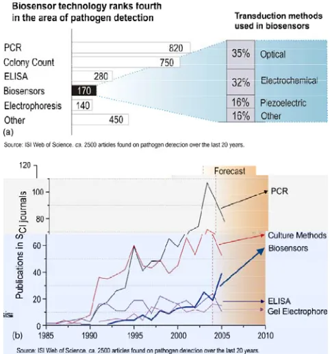
Biosensor technology comes with claims of similarly dependableleads to much time which can be reduced that will be perhaps why they are drawing a complete amount that is big of. However, there was nonethelessmuch striving to do before biosensors become a alternative that is real.Fig. 19(a) and (b) declare that biosensor technology may go quicklyahead of old-fashioned ELISA based techniques, and their potentialmarket (Alocilja and Radke, 2003) is very encouraging too.Many biosensors rely on either antibody which are certain DNAprobes to provide specificity. However, as Fig. 1 shows, the technologyis extremely split when it comes to detection modes.
89
the last twenty years. The fact that certain articles utilized more than one technique has been accounted for in order to make this graph.
II. VARIOUS EXISTING TECHNIQUES IN PATHOGEN DETECTION
A. Polymerase chain effect
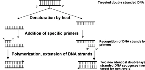
This is an acid amplification technology that is nucleic. It was developed within the mid 80s (Mullis et that is al) and it's also extremely commonly present in bacterial detection. Its on the basis of the isolation, amplification and quantification of a fast DNA that is sequence such as the targeted bacteria’s material that is hereditary. Types of different PCR methods developed for bacterial detection are: (i) real-time PCR (Rodr´ı guez-L´azaro et al., 2005), (ii) multiplex PCR (Jofr´e et al., 2005) and (iii) reverse transcriptase PCR (RT-PCR) (Deisingh, 2004). You can find methods PCR that is coupling that is coupling other methods such as for instance, as an example surface acousticwave sensor (SAW) (Deisingh, 2004) or wave that is evanescent (Simpson and Lim, 2005).
Fig. 2 illustrates the PCR method, consisting in a variety of Cycles of denaturation by temperature of the purifiedDNA and extracted, followed closely by an extension phase utilizing primers which can be certain a thermostable polymerization enzyme. Then each new doublestranded DNA will act as target for a brand name period that is brand new exponential amplification is therefore obtained. One of the PCR that is different, multiplex PCR is quite helpful as it permits the detection that is simultaneous of organisms permits the simultaneous detection of several organisms it by launching primers which can be various DNA that is amplify coding for specific genes of each stress that is microbial (Touron et al., 2005). Real-time PCR permits to have faster outcomes without means manipulation that is too much. This technique its detection in the fluorescent emission by a certain dye itself to your targeted amplicon since it attaches.
Fig. 2. Schematic representation of 1 PCR cycle place that is taking thermocycler
B. Tradition and colony methods that are counting
The culturing and plating technique could be the oldest detection that is microbial method and is still the detection technique that is standard. Nonetheless, other practices are crucial culturing practices are exceptionally time-consuming. when it comes to Campylobacter, 4–9 days are anticipated to get negative result that is bad between 14 and 16 times for
confirmation of a positive}Brooks et al., 2004). This really is a inconvenience in lots of commercial applications, specially within the meals sector. Different selective media are widely used to detect germs which can be specific species. They might include inhibitors (to help you to prevent or postpone the growth of non-targeted strains) or substrates that are particular simply the bacteria which can be targeted degrade or that confers a specific colour to your colonies that are growingrainbow agar from Salmonella detection (Fratamico, 2003)). Detection will be completed making use of optical practices, mainly by ocular examination.
C. Immunology-based methods
The entire world of immunology-based options for germs detection provides extremely effective analytical tools for a range that is wide of goals. For example, immune magnetic separation (IMS) (Mine, 1997; P´erez et al., 1998), a and/or pre concentration that is pre-treatment step, enables you to capture and draw out the targeted pathogen through the suspension that is microbial introducing antibody onto it (Gu et al., 2006). IMS can then be combined with just about any detection technique, e.g., optical, magnetic force microscopy, magneto resistance (Bead Array Countertop) (Baselt et al., 1998) and hall effect (Besse et al., 2002), and so on. Personalized derivatized beads that are magnetic being magnetic available The fundamental conspicuous of organizations, the absolute most conspicuous of from a wide range of from an array of organizations that could very well be Dynal. Beads of widely sizes that are ranging from as much as a couple of tens of microns) may be chosen with regards to the application. Whilst large beads may be used when it comes to measurement of intermolecular forces, smaller particles are perfect for the detection of tiny analytes where sensitiveness that is high is a must. Regarding germs being entire the use of beads within the Low micrometer range might provide the very best time. Then, a synopsis of current works biosensors that are using this field will probably be given. This overview aims to give a picture of existing varies technologies as well as methodologies.
III. BIOSENSORS IN PATHOGEN DETECTION
90 can be after biosensors according to their transduction
practices.
A. Biological recognition elements and immobilisation methods
There are three main classes of biological recognition elements that are used in biosensor applications. These are (i) enzymes, (ii) antibodies and (iii) nucleic acids. Within the detection of pathogenic bacteria, nevertheless, enzymes tend to work as labels instead of actual recognition that is microbial elements.
Enzymes may be used to label either antibodies (Ko and Grant, 2003) or DNA probes (Lucarelli et al., 2004) much in identical fashion as in an ELISA assay. Into the full instance of amperometric electrochemical) Biosensors labels that are enzymatic critical, because is supposed to be talked about below. More techniques which are advanced level operate without Labelling the recognition element, for instance the complete case of surface plasmon resonance (SPR), piezoelectric or biosensors which can be impedimetric (Guan et al., 2004). Because the usage of antibodies in biosensors happens to be more spread than compared to DNA probes, the parts being after primarily with antibody-based biosensors. This section addresses silver substrates only because of its Importance within the certain area of immunosensors and DNA probes, which form the foundation on most biosensors being bacterial. Fig. 3 programs the 3 most antibody that is regular channels, which are:
• Adsorption on gold. • The Avidin–biotin system.
• Self-assembled monolayers (SAMs). a) Adsorption on Gold
That is, undoubtedly the easiest, fastest and least reliable of this described techniques. Since it consists in the random accessory of the antibodies in the substrate, the orientation that is correct associated with the binding websites can't be controlled. The adsorption is non-specific and gratification that is biosensor seldom excellent (Tombelli and Mascini, 2000). Karyakin et al. (2000) reported a method antibody that is using whilst attaining a fair degree of performance. Fig. 3 describes the concepts of this method.
b) The Avidin–biotin system
This system is an easy and yet really way that is effective anchor biomolecules to an avidin surface that is coatedOuerghi et al., 2002). One of the most advantageous options that come with this functional system is that Although the affinity constant between biotin and avidin is rather high (ca. 10−15 mol−1 L), the bonding is of non-covalent nature, which allows for numerous washing and re-use for the sensing that is same device (Tombelli and Mascini, 2000).
Andrawback that is very important is the price that is high of reagents included. a glucose biosensor constructed on a few glucose that is avidin-biotinilated oxidase levels is proposed by Anzai et al. (1998).
c) SAMs
Self-assembled monolayers are acquired by immersion of a gold plate in an answer containing a surfactant that works a high purity solvent (Bain et al., 1989). The most instances which are popular those acquired by the immersion of silver in an ethanol solution containing disulphides or thiols (Su and Li, 2004). The packaging and depth for the monolayer that is formed dictated by the radical attached to the sulphide atom(s) (Vaughan et al., 1999). An important band of compounds utilized in the forming of SAMs is that integrated by alkanethiols.
B. Optical biosensors
They are the absolute most popular in bioanalysis, as a result of Their sensitivity and selectivity. Optical biosensors are developed for quick detection of contaminants (Willardson et al., 1998; Tschmelak et. that is al), toxins or medications (Bae et al., 2004) as well as pathogen bacteria (Baeumner et al., 2003). Recently, Surface and fluorescence plasmon resonance, SPR, based techniques have actually gained momentum because of their sensitivity.
91 immobilization; c5, clean and blockage of unreacted internet
sites that are active C6, sample c7 and addition, detection.
C. Electrochemical biosensors
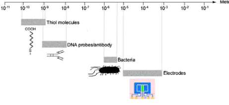
The unit are mainly based on the observation of current Or changes being prospective to interactions occurring at the sensor- test matrix program. Methods are categorized based on the parameter that is observed present (amperometric), possible (potentiometric) or impedance (impedimetric). Contrasted To practices which can be optical electrochemistry enables the analyst to use turbid samples, additionally the capital cost of equipment is much lower. On the other hand, electrochemical techniques present slightly more selectivity that is bound sensitivity than their optical counterparts. Fig. 4 compares the sizes of these different aspects of an biosensor that is electrochemical.
Fig. 4.Diagram representing the relative sizes regarding the right components integrating a biosensor.
IV. NEW TRENDS
More exotic approaches have been devised recently, such as the application of fractals theory to the analysis of biosensor data (Morris and Sadana, 2005). This kind of analysis not only enables the detection of pathogenic bacteria, but it also yields information about the binding and dissociation kinetics involved in the interaction of the pathogen with the biosensor surface. Although very powerful, this approach suffers from a very high degree of mathematical complexity. The combined use of micro- and nano-fabrication techniques in the area of biosensors holds great promise and different applications are beginning to crop up (Carrascosa et al., 2006; Murphy, 2006). Amongst the advantages of this smaller scale approach are: (a) the possibility of mass production and reduced unit costs, (b) it allows working with sample volumes in the range of nanolitres or less, which also implies that the cost of reagents is not too high, (c) micro-fluidics improve mixing rates and mass transport which is expected to result in much shorter analysis times, (d) the performance of multi-analyte analysis is enabled in the same device, which also shortens analysis time, and (e) because the volumes manipulated are so tiny, these devices provide more safety and they are more environmentally friendly.
Power consumption is extremely low and contamination associated to waste material may be easier to contain due to the
possibility to use tiny volumes and cartridge-like configurations. To the best of the authors’ knowledge, the first reports of bacterial detection at Microsystems dates back to the works of Bashir and co-workers (G´omez et al., 2001) in 2001. This work presents a microsystem capable of detecting listeria using impedance spectroscopy. Also in the same year, Woo and coworkers (Gau et al., 2001) reported the selective amperometric detection of E. coli (1000 cells; initial volume not quoted) in a very short time (40 min). It is interesting to note that both works rely on electrochemical and not optical detection.
V. CONCLUSIONANDFUTURESCOPE
Traditional pathogen detection techniques, although painful and sensitive sufficient, are often too sluggish to be of any use. Consequently, new practices are needed that meet or exceed their performance. On the Modern times, a complete large amount of effort moved in to the research and development of biosensors of the most extremely nature that is diverse however their performance is irregular and still requires enhancement. Optical techniques possibly provide better sensitivity than Electrochemical ones, but their complexity and cost makes them Ugly to get rid of users that are most. Electrochemical techniques, on one other hand, are easier to make use of but when it comes down to detecting pathogens, their performance remains not even close to adequate. In order to become appealing, biosensors first need to show that they truly are capable of reaching at the least the detection that is just like conventional practices (between 10 and 100 CFUmL−1). Next, They have to do this in a small fraction associated with the right time without overlooking cost.
Traditional pathogen detection techniques, although painful and sensitive sufficient, are often too sluggish to be of any use. Consequently, new practices are needed that meet or exceed their performance. On the Modern times, a complete large amount of effort moved in to the research and development of biosensors of the most extremely nature that is diverse however their performance is irregular and still requires enhancement. Optical techniques possibly provide better sensitivity than Electrochemical ones, but their complexity and cost makes them Ugly to get rid of users that are most. Electrochemical techniques, on one other hand, are easier to make use of but when it comes down to detecting pathogens, their performance remains not even close to adequate. In order to become appealing, biosensors first need to show that they truly are capable of reaching at the least the detection that is just like conventional practices (between 10 and 100 CFUmL−1). Next, They have to do this in a small fraction associated with the right time without overlooking cost.
92 Optical techniques possibly provide better sensitivity than
Electrochemical ones, but their complexity and cost makes them Ugly to get rid of users that are most. Electrochemical techniques, on one other hand, are easier to make use of but when it comes down to detecting pathogens, their performance remains not even close to adequate. In order to become appealing, biosensors first need to show that they truly are capable of reaching at the least the detection that is just like conventional practices (between 10 and 100 CFUmL−1). Next, They have to do this in a small fraction associated with the right time without overlooking cost.
REFERENCES
Alocilja, E.C., Radke, S.M., 2003. Biosens.Bioelectron. 18 (5–6), 841–846.
Barsoukov, E., Macdonald, J.R., 2005. Impedane Spectroscopy Theory, Experiment and Applications. John Wiley & Sons Ltd., Hoboken, New Jersey. Baselt, D.R., Lee, G.U., Natesan, M., Metzger, S.W., Sheehan, P.E., Colton, R.J.,1998.Biosens.Bioelectron. 13 (7–8), 731–739.
Bej, A.K., Mahbubani, M.H., Dicesare, J.L., Atlas, R.M., 1991. Appl. Environ.Microbiol. 57 (12), 3529–3534.
Bergveld, P., 2003. Sens. Actuators B Chem. 88 (1), 1–20.
Besse, P.A., Boero, G., Demierre, M., Pott, V., Popovic, R., 2002. Appl. Phys. Lett. 80 (22), 4199–4201.
Blais, B.W., L.J., Bosley, J., Martinez-Perez, A., 2004.Lett. Appl. Microbiol. 39, 516–522.
Brewster, J.D., Gehring, A.G., Mazenko, R.S., Van Houten, L.J., Crawford, C.J.,1996. Anal.Chem. 68, 4153–4159.
Brooks, B.W., J., D., L., L.-W., C., D., M., H., R. R., G., B.-S., 2004. Vet. Microbiol. 103, 77–84.
Carrascosa, L.G., Moreno, M., Alvarez, M., Lechuga, L.M., 2006.Trac-Trends Anal. Chem. 25 (3), 196–206.
Cooper, M.A., 2003. Anal.Bioanal. Chem. 377 (5), 834–842.
Croci, L., Delibato, E., Volpe, G., Palleschi, G., 2001.Anal.Lett. 34 (15), 2597–2607.
Daly P., C.T., Doyle S., 2002. Lett. Appl. Microbiol. 34, 222–226.
Deisingh A.K., T.M., 2004. J. Appl. Microbiol. 96, 419–429.
Dominguez, J.A., Matas, L., Manterola, J.M., Blavia, R., Sopena, N., Belda, F.J., Padilla, E., Gimenez, M., Sabria, M., Morera, J., Ausina, V., 1997. J. Clin.Microbiol. 35 (6), 1627–1629.
Fratamico, P.M., 2003. Mol. Cellular Probes 17, 215–221.
Gomez, R., Bashir, R., Bhunia, A.K., 2002. Sens. Actuators B Chem. 86 (2– 3),198–208.
G´omez, R., Bashir, R., Sarikaya, A., Ladisch, M.R., Sturgis, J., Robinson, J.S.,
Geng, T., Bhunia, A.K., Apple, H.L., Wereley, S., 2001. Biomed.Microdevices 3 (3), 201–209.
Grimnes, S., Martinsen, O., 2000. Bioimpedance and Bioelectricity Basics.Academic Press.
Gu, H.W., Xu, K.M., Xu, C.J., Xu, B., 2006. Chem. Commun. (9), 941–949. Guan, J.-G., Miao,Y.-Q., Zhang, Q.-J., 2004. J. Biosci. Bioeng. 97 (4), 219– 226.
Jofr´e A., M. B., Garriga, M., Hugas, M., Pla, M., Rodr´ıguez-L´azaro D., Aymerich, T., 2005.Food Microbiol. 22, 109–115.
Karyakin, A.A., Presnova, G.V., Rubtsova, M.Y., Egorov, A.M., 2000. Anal. Chem. 72, 3805–3811.
Katz, E., Willner, I., 2003. Electroanalysis 15 (11), 913–947.
Ko, S., Grant, S.A., 2003. Sens. Actuators B 96, 372–378.
Lagally, E.T., Scherer, J.R., Blazej, R.G., Toriello, N.M., Diep, B.A., Ramchandani, M., Sensabaugh, G.F., Riley, L.W., Mathies, R.A., 2004.Anal.Chem.76 (11), 3162–3170.
Lehtola, M.J., L.C.J., Keevil, C.W., 2005. J. Microbiol. Methods. 62, 211– 219.
Leonard, P., Hearty, S., Brennan, J., Dunne, L., Quinn, J., Chakraborty, T., O’kennedy, R., 2003. Enzyme Microb. Technol. 32, 3–13.
Leoni, E., Legnani, P.P., 2001. J. Appl. Microbiol. 90 (1), 27–33.
Leskel¨a T., T.-T.A., Kusnetsov, J., Neubauer, P., Breitstein, A., 2005. J. Microbiol.Methods. 62, 167–179.
Levi, K., Smedley, J., Towner, K.J., 2003. Clin.Microbiol. Infect. 9 (7), 754– 758.
Li, Y., Dick, W.A., Tuovinen, O.H., 2004. Biol. Fertility Soils 39 (5), 301– 311.
Lim, D.V., Simpson, J.M., Kearns, E.A., Kramer, M.F., 2005. Clin.Microbiol. Rev. 18 (4), 583–607.
Liu, H.S., Li, Y., Huang, X.X., Kawamura, Y., Ezaki, T., 2003.Microbiol. Immunol. 47 (11), 859–869.
Lucarelli, F.,Marrazza, G., Turner, A.P.F., Mascini, M., 2004. Biosens.Bioelectron. 19, 515–530.
Mansfield P.L., F. S. J., 2001.Food Microbiol. 18, 361–366. Mine, Y., 1997.J. Agric. Food Chem. 45, 3723–3727.
Mirsky, V.M., Riepl, M., Wolfbeis, O.S., 1997.Biosen.Bioelectron. 12 (9), 977–989.
Morris, B.A., Sadana, A., 2005. Sens. Actuators B Chem. 106 (2), 498–505. Muhhammad-Tahir, Z., Alocilja, E.C., 2003.Biosens.Bioelectron. 18, 813– 819.
Muhhammad-Tahir, Z., Alocilja, E.C., 2004.Biosyst. Eng. 88 (2), 145–151.
Mullis, K., Faloona, F., Scharf, S., Saiki, R., Horn, G., Erlich, H., 1986. Cold Spring Harbor Symposia on Quantitative Biology 51, 263–273.
Munoz, J., Jimenez, C., Bratov, A., Bartroli, J., Alegret, S., Dominguez, C., 1997. Biosens. Bioelectron. 12 (7), 577–585.
93
Ong, K.G., Grimes, C.A., Robbins, C.L., Singh, R.S., 2001. Sens. Actuators a-Phys. 93 (1), 33–43.
Ouerghi, O., Touhami, A., Jaffrezic-Renault, N., Martelet, C., Ben Ouada, H., Cosnier, S., 2002.Bioelectrochemistry 56, 131–133.
Radke, S.M., Alocilja, E.C., 2005. Biosens.Bioelectron. 20, 1662–1667.
Rodr´ıguez-L´azaro, D., D. A. M., Herrewegh, A., Pla, M., Cook, N., Ikonomopoulos, J., 2005.Int. J. Food Microbiol. 101, 93–104.
Simpson, J.M., Lim, D.V., 2005. Biosens.Bioelectron. 21 (6), 881–887.
Stevens, K.A., Jaykus, L.-A., 2004. Crit. Rev. Microbiol. 30 (1), 7–24.
Tims, T.B., Lim, D.V., 2003. J. Microbiol. Methods 55, 141–147.
Tombelli, S., Mascini, M., 2000.Anal.Lett. 33 (11), 2129–2151.
Touron, A., B.T., Pawlak, B., Petit, F., 2005. Res. Microbiol. 156, 541–553.
Tschmelak, J., Proll, G., Gauglitz, G., 2004. Anal.Chim.Acta 519 (2), 143– 146.
Vaughan, R.D., O’sullivan, C.K., Guilbault, G.G., 1999. Fresenius J. Anal. Chem. 364, 54–57.
Vaughan, R.D., O’sullivan, C.K., Guilbault, G.G., 2001. Enzyme Microb.Technol.
29, 635–638.
Willardson, B.M.,Wilkins, J.F., Rand, T.A., Schupp, J.M., Hill, K.K., Keim, P., Jackson, P.J., 1998. Appl. Environ. Microbiol. 64 (3), 1006–1012.
Willner, I., Katz, E., Willner, B., 1997. Electroanalysis 9 (13), 965–977.