_____________________________________________________________________________________________________
*Corresponding author: Email: [email protected];
www.sciencedomain.org
Radiologic Evaluation of Marginal Bone Loss in
Delayed vs. Immediate Loaded Implants –18 Month
Results of a Prospective Comparative Study
Anirudh Bhattacharya
1*and Vijay Sharma
21Department of Maxillofacial Surgery, Critical Care & Trauma Hospital, Jaipur, India.
2
Department of Maxillofacial Surgery, Aashirwad Hospital, Akola, India.
Authors’ contributions
This work was carried out in collaboration between both the authors. Author AB designed the study, wrote the protocol, and wrote the first draft of the manuscript. Author VS managed the literature searches and analyses of the study. Both authors read and approved the final manuscript.
Article Information
DOI: 10.9734/BJMMR/2015/17500 Editor(s): (1)Hai Ming Wong, The University of Hong Kong, 2/F, Prince Philip Dental Hospital, Hong Kong.
Reviewers: (1)Mark E Peacock, Oral Biology, Georgia Regents University, USA.
(2)Anonymous, Portugal. (3)Anonymous, Romania. Complete Peer review History:http://www.sciencedomain.org/review-history.php?iid=1114&id=12&aid=8893
Received 16th March 2015 Accepted 2nd April 2015 Published 20th April 2015
ABSTRACT
Aim: To compare the amount of bone loss (if any) at the mesial and distal sides in delayed and immediate loaded dental implants.
Study Design: Total 20 partially edentulous (anterior region of Maxilla) patients were randomized and equally divided into two groups. Group A received delayed loaded dental implants and group B received immediate loaded dental implants. Both the groups were monitored clinically and radiographically at 3, 6, 12 and 18 months.
Methodology: We included 20 patients (11 men, 09 women; age range 20-48 years) with single or multiple edentulous areas in mouth. Clinical as well as all routine hematological examinations were done. Radiographs were taken sequentially as required for 18 months.
Results: There was no significant statistical difference of bone loss mesially in both the groups at 3 (P=0.99) & 6 (P=0.25) months, but there was significant statistical difference of bone loss mesially seen in both the groups at 12 (P=0.03) & 18 (P=0.01) months. There was no significant statistical difference of distal bone loss in both the groups at 3 (P=0.22), 6 (P=0.38) and 12
(P=0.17) months, but there were significant statistical difference of distal bone loss seen in both the groups at 18 months (P=0.03). The bone loss was found more with the immediate loading type of implants at both mesial and distal sides of implant.
Conclusion: The immediate loading implants may provide a lot of benefits over conventional delayed loading implants but the bone loss at Crestal (Marginal) level is higher as compared to delayed loading implants when seen in maxillary anterior impants which should be considered whenever selecting any particular method of dental implantation.
Keywords: Dental implants; delayed loading; immediate loading; bone loss.
1. INTRODUCTION
Dental implants have, in many cases, become the treatment of choice for restoring missing teeth and have been documented to have a high degree of success. With implant therapy, the preparation of healthy teeth adjacent to the edentulous area can be avoided. An additional advantage to the implant restoration is the maintenance of the alveolar bone, which otherwise would undergo residual ridge resorption with most of the other restorative options complicating aesthetics.
The protocol established by Branemark stated a two stage surgical procedure in which implant was submerged during first surgical stage, maintaining a stress free period allowing it to heal and encouraging a direct bone-implant interface. The implant placed in that unloaded environment was kept for a minimum of 3 months in cases of mandible and 6 months in maxilla [1]. This was followed by second stage surgical procedure in which prosthetic abutment was connected. However this maneuver had some shortcomings like microgap between implant and abutment which eventually resulted in crestal bone loss and long extended edentulous period. The result of advances in research on implant design, materials, advanced imaging techniques (cone beam computed tomography scan) and novel loading protocols made it possible to shorten the treatment period and to avoid an edentulous condition encouraged the introduction of an immediate implant loading. In 1979 Ledermann revolutionized era of implant dentistry with successful immediate loading implants, in which implant is placed followed by prosthetic abutment connection and temporization in a single appointment [2]. This technique eliminated chances of microgap between implant and abutment which ruled out possibility of peri-implant bone loss as seen in delayed implant loading because one of the prerequisites for the successful placement of an implant is the presence of adequate bone
volume. Tarnow et al. [3] stated that a submerged implant, following the delivery of the prosthesis, will create circumferential or horizontal bone resorption of 1.3 to 1.4 mm. Grunder et al. [4] also stated that at least 2 mm of lateral alveolar bone must be present beyond the body of the implant to compensate for the effects of bone remodelling. If this amount of bone is not present, part or all of the facial or buccal bone plate will be lost after remodelling, with the subsequent risk of soft-tissue recession.
For the success in implant dentistry, we should ideally evaluate primary outcome of an implant-prosthetic complex as a whole. This can be achieved by evaluating success at the implant level, peri-implant soft tissue, prosthesis and level of patient’s satisfaction. The most important factor for the success of any implant is the bony coverage around it and any loss in that would be detrimental to implant’s health in long term [5]. The present study is done to evaluate the crestal bone loss at the mesial and distal side of implant in both delayed and immediate loading implants to assess which system has the least bone loss for a period of consecutive 18 months.
2. MATERIALS AND METHODS
2.1 Study Design
A total of 20 cases were selected for the study. The patients were randomly divided in two groups of 10 each. First Group (A) was planned for delayed implant loading & second Group (B) was planned for immediate implant loading. Adin Touareg- S(R) internal hex implants were used in all patients with varied lengths and diameter as required. All the patients were operated upon by a single surgeon.
2.1.1 Investigations
1. Routine blood investigations.
2.1.2 Inclusion criteria
1. Patients with single or multiple edentulous areas.
2. Anterior Maxilla partially edentulous. (Central and/or lateral incisors) 3. ASA Class I and relatively healthy ASA
class II patients.
4. Patients above 16 years of age. 5. Patients free of periodontal diseases. 6. Patients having sufficient amount of
residual alveolar ridge.
2.1.3 Exclusioncriteria
1. Patients unable to give or not willing to give informed consent.
2. ASA class III and class IV Category patients.
3. Patients who were pregnant, lactating or having habits of smoking, tobacco & betel nut chewing.
4. Patients on any drug which will compromise osseointegration Eg Cyclosporine etc.
2.2 Patient Groups
Group A patients were implanted and left submerged for a period of 3 months. After three months the implant was exposed and connected to abutment leading to final prosthesis.
Group B patients were implanted and were immediately at the same sitting connected to abutment and temporary crown was placed which was later replaced by a permanent crown.
All the patients in both groups received Porcelain fused metal crowns as their final prosthesis which were indigenously made in the ceramic lab of same hospital. As anatomic abutments with pre-formed shoulders margins were used, therefore no problem was encountered while seating of final prosthesis.
2.3 Statistical Analysis
Statistical analysis was carried out using SPSS v 16.0 statistical software.
Mann-whitney U test was used to compare bone loss between study & experimental groups at 3, 6, 12 & 18 months.
Repeated measures ANOVA with post-hoc boneferroni test was used for within group comparison of mean scores of bone loss at 3, 6, 12 & 18 months in both the groups.
2.4 Radiographic Analysis
Standardized intraoral radiographs were obtained using Intra oral sensor and position holding devices to eliminate manual errors of positioning at intervals of 3, 6, 12 and 18 months. The length of implant was measured on digital radiographs from the implant-abutment interface to the apex of implant. Next, the distance between the observed crestal bone level and the implant-abutment interface was measured at the mesial and distal implant surfaces. The actual implant length was known based on manufacturing standards. To adjust the measurements for magnification error the following equation was used to determine the corrected crestal bone levels.
Actual bone loss was calculated by formula:
Corrected crestal bone level = [(measured bone level) x (actual implant length /measured implant length)] [6]
3. RESULTS AND DISCUSSION
Comparisons of mean scores of mesial bone loss in both groups by Mann-whitney u test. All lengths in Millimetre (mm).
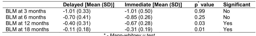
Mean scores of bone loss mesially in Group A was -1.01 (SD=0.33) at 3 months, -0.70 (SD=0.41) at 6 months, -0.40 (SD=31) 12 months and -0.11 (SD=0.18).
For Group B it was -1.01 (SD=0.50) at 3 months, -0.85 (SD=0.26) at 6 months, -0.67 (SD=0.28) at 12 months and -0.31 (SD=0.19) at 18 months.
There was no significant statistical difference of bone loss mesially in both the groups at 3 (p=0.99) & 6 (p=0.25) months, but there was significant statistical difference of bone loss mesially seen in both the groups at 12 (p=0.03) & 18 (p=0.01) months as shown in Table 1.
Comparisons of mean scores of distal bone loss in both groups by Mann-whitney u test.
Table 1. Bone loss mesially
Delayed [Mean (SD)] Immediate [Mean (SD)] p* value Significant
BLM at 3 months -1.01 (0.33) -1.01 (0.50) 0.99 No
BLM at 6 months -0.70 (0.41) -0.85 (0.26) 0.25 No
BLM at 12 months -0.40 (0.31) -0.67 (0.28) 0.03 Yes
BLM at 18 months -0.11 (0.18) -0.31 (0.19) 0.01 Yes
* - Mann-whitney u test
For Group B it was -0.62 (SD=0.57) at 3 months, -0.50 (SD=0.45) at 6 months, -0.28 (SD=0.30) at 12 months and -0.18 (SD=0.22) at 18 months.
There was no significant statistical difference of distal bone loss in both the groups at 3 (p=0.22), 6 (p=0.38) and 12 (p=0.17) months, but there were significant statistical difference of distal bone loss seen in both the groups at 18 months (p=0.03) as shown in Table 2.
Effect of time on bone loss on mesial side in both groups by repeated measures ANOVA summary shows statistically significant difference in Group A p >0.001 and in Group B p >0.01 (Table 3).
Effect of time on mesial bone loss in Group A
There was no significant statistical difference seen in Group A at 3 months (mean 1= -1.01) vs. 6 months (mean 2= -0.70) p value =0.23. But there was significant statistical difference seen in 3 months (mean 1= -1.01) vs. 12 months (mean 2 = -0.40) p value <0.01, in 3 months (mean 1= -1.01) vs. 18 months (mean 2 = -0.11) p value <0.001, in 6 months (mean 1= -0.70) vs. 12 months (mean 2 = -0.40) p value <0.01, in 6 months (mean 1= -0.70) vs. 18 months (mean 2 = -0.11) p value <0.01 & in 12 months (mean 1= -0.40) vs. 18 months (mean 2 = -0.11) p value <0.05 as shown in Table 4.
Effect of time on mesial bone loss in Group B
There was no significant statistical difference seen in Group B at 3 months (mean 1= -1.01) vs 6 months (mean 2= -0.85) p value =1 & at 3 months (mean 1= -1.01) vs 12 months (mean 2= -0.67) p value =0.12. But there was significant statistical difference seen in 3 months (mean 1= -1.01) vs. 18 months (mean 2 = -0.31) p value <0.05, in 6 months (mean 1= -0.85) vs.12 months (mean 2 = -0.67) p value <0.01, in 6 months (mean 1= -0.85) vs. 18 months (mean 2 = 0.31) p value <0.001, in 12 months (mean 1= -0.67) vs. 18 months (mean 2 = -0.31) p value <0.01 as shown in Table 5.
Effect of time on bone loss on distal side in both groups by repeated measures ANOVA summary
shows statistically significant difference in Group A p >0.001 and in Group B p >0.01 (Table 6).
Effect of time on distal bone loss in Group A
There was no significant statistical difference seen in Group A at 3 months (mean 1= -0.87) vs 6 months (mean 2= -0.65) p value =0.15 & at 6 months (mean 1= -0.65) vs 12 months (mean 2= -0.45) p value =0.37. But there was significant statistical difference seen in 3 months (mean 1= -0.87) vs. 12 months (mean 2 = -0.45) p value <0.05, in 3 months (mean 1= -0.87) vs. 18 months (mean 2 = -0.03) p value <0.01, in 6 months (mean 1= -0.65) vs. 18 months (mean 2 = 0.03) p value <0.01, in 12 months (mean 1= -0.45) vs. 18 months (mean 2 = -0031) p value <0.01as shown in Table 7.
Effect of time on distal bone loss in Group B
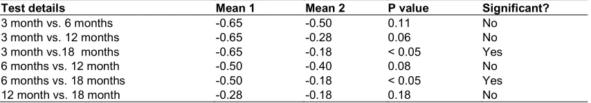
But there was no significant statistical difference seen in Group B in 3 months (mean 1= -0.65) vs.6 months (mean 2 = -0.50) p value <0.11, in 3 months (mean 1= -0.65) vs.12 months (mean 2 = 0.28) p value <0.06, in 6 months (mean 1= -0.50) vs. 12 months (mean 2 = -0.40) p value <0.08, in 12 months (mean 1= -0.28) vs. 18 months (mean 2 = -0.18) p value <0.18. There was significant statistical difference seen in group B at 3 months (mean 1= -0.65) vs 18 months (mean 2= -0.18) p value =0.05 & at 6 months (mean 1= -0.50) vs 18 months (mean 2= -0.18) p value =0.05 as shown in Table 8.
In our study mesial bone loss in delayed loading group was -1.22 to 0.00 and distal bone loss was -1.15 to -0.02 at the end of 18 months (95% Confidence interval).
In our study mesial bone loss in immediate loading group was -1.34 to -0.02 and distal bone loss was -0.91 to -0.02 at the end of 18 months (95% Confidence interval).
3.1 Discussion
Table 2. Bone loss distally
Delayed [Mean(SD)] Immediate [Mean(SD)] p* value Significant
BLD at 3 months -0.87 (0.45) -0.62 (0.57) 0.22 No
BLD at 6 months -0.65 (0.39) -0.50 (0.45) 0.38 No
BLD at 12 months -0.45 (0.31) -0.28 (0.30) 0.17 No
BLD at 18 months -0.03 (0.08) -0.18 (0.22) 0.03 Yes
* - Mann-whitney u test
Table 3. Effect of time on bone loss on mesial side
Repeated measures ANOVA summary Delayed Immediate
P value < 0.001 < 0.01
Statistically significant (P < 0.05)? Yes Yes
Table 4. Group A Mesial Bone loss comparison
Test details Mean 1 Mean 2 P value Significant?
3 month vs. 6 months -1.01 -0.70 0.23 No
3 month vs. 12 months -1.01 -0.40 < 0.01 Yes
3 month vs.18 months -1.01 -0.11 < 0.001 Yes
6 months vs. 12 month -0.70 -0.40 < 0.01 Yes
6 months vs. 18 months -0.70 -0.11 < 0.01 Yes
12 month vs. 18 month -0.40 -0.11 < 0.05 Yes
Table 5. Group B Mesial Bone loss comparison
Test details Mean 1 Mean 2 P value Significant?
3 month vs. 6 months -1.01 -0.85 1 No
3 month vs. 12 months -1.01 -0.67 0.12 No
3 month vs.18 months -1.01 -0.31 < 0.05 Yes
6 months vs. 12 month -0.85 -0.67 < 0.01 Yes
6 months vs. 18 months -0.85 -0.31 < 0.001 Yes
12 month vs. 18 month -0.67 -0.31 < 0.01 Yes
Table 6. Effect of time on bone loss on distal side
Repeated measures ANOVA summary Delayed Immediate
P value < 0.001 < 0.01
Statistically significant (P < 0.05)? Yes Yes
Table 7. Group A Distal bone loss comparison
Test details Mean 1 Mean 2 P value Significant?
3 month vs. 6 months -0.87 -0.65 0.15 No
3 month vs. 12 months -0.87 -0.45 < 0.05 Yes
3 month vs.18 months -0.87 -0.03 < 0.01 Yes
6 months vs. 12 month -0.65 -0.45 0.37 No
6 months vs. 18 months -0.65 -0.03 < 0.01 Yes
12 month vs. 18 month -0.45 -0.03 < 0.01 Yes
Table 8. Group B Distal bone loss comparison
Test details Mean 1 Mean 2 P value Significant?
3 month vs. 6 months -0.65 -0.50 0.11 No
3 month vs. 12 months -0.65 -0.28 0.06 No
3 month vs.18 months -0.65 -0.18 < 0.05 Yes
6 months vs. 12 month -0.50 -0.40 0.08 No
6 months vs. 18 months -0.50 -0.18 < 0.05 Yes
The patients demands to shorten the treatment period and to avoid an edentulous condition encouraged the introduction of non submerging of implant i.e immediate loading implant protocol [7]. The immediate loading of implants aim at a shorter treatment period with a stable and fixed long term interim restoration on the day of surgery [8]. This treatment option also aims at maintenance of the hard and soft tissues and reducing the waiting period.[9] In this technique abutment is attached at the time of implant placement, no microgap exist at or below the alveolar crest between the implant and restoration.
From several studies it has been proposed that marginal bone loss is more extensive around two stage implants as compared with one stage implants. The microgap between the implant and the abutment at the crestal level in two stage implants has been suggested to play a prominent role in the development of bone loss [10,11].
In our study, both the groups had randomized patients with partially edentulous anterior maxilla (central or lateral incisors) with age of edentulism being a minimum of 1 year and maximum 2 years. All patients received same manufacturer’s same version of implants, operated by a single surgeon. While giving prosthesis to multiple edentulous regions the rule followed was “1 implant 1 crown” so there were individual crowns for each implant and no bridges were given. The post operative radiological findings were monitored using a preset formula and the amount of bone loss (in mm.) was recorded using EasyDent(R) image management software. On comparing both the groups, bone loss mesially was not significant at 3 and 6 months but it was significant at 12 and 18 months.
When compared bone loss distally for both groups, it was significant only at 18 months.
In both the scenarios the significant bone loss was seen with immediate loading implants. Though the immediate loading implants have been shown to be highly successful, [12,13] but in our study it was found that submerged or delayed loading implants are better when the crestal bone loss is taken as criteria for the success of implants.
4. CONCLUSION
Mesial bone loss –
The crestal bone loss was near equal or statistically insignificant in both the groups
for first 3 and 6 months but at 12 and 18 months it was higher on mesial side of implant in the immediate loading group.
Distal Bone loss-
The crestal bone loss at 3, 6 and 12 months on the distal side of implant was equal but at 18 month it was higher on distal side of implant in the immediate loading group.
Though the immediate loading implants have many proven benefits over the conventional delayed loading implants like reduction in alveolar ridge resorption and overall treatment time, increased patient acceptance, quicker return of function, potentially superior soft tissue profile and reduced surgical trauma and ease of surgery but still the most important factor which is responsible for the success of implant treatment is circumferential bone around the implant. In our study it is proved that more bone resorption is evident in immediate loading cases observed for a period of 18 months as compared to delayed loading implants in terms of maxillary anterior implants.
Though a small study, it shows a definitive pattern of mesial and distal side of implant bone loss which may aid the implant surgeons to choose their method of placement and further studies relating the same.
CONSENT
All patients in this study gave their consent for the treatment procedure.
ETHICAL APPROVAL
It is not applicable.
COMPETING INTERESTS
This study is a part of another larger ongoing study which also includes other variables and factors carried out by the same authors and few other clinicians’.
REFERENCES
2. Romanos G, Froum S, Hery C, Cho SC, Tarnow D. Survival rate of immediately vs delayed loaded implants. J Oral Implantol. 2010;36(4):315-24.
3. Tarnow D, Elian N, Fletcher P, Froum S, Magner A, Cho SC, Salama M, Salama H, Garber DA. Vertical distance from the crest of bone to the height of the interproximal papilla between adjacent implants. J Periodontol. 2003;74(12):1785-8.
4. Grunder U, Gracis S, Capelli M. Influence of the 3-D bone-to-implant relationship on esthetics. Int J Periodontics Restorative Dent. 2005;25(2):113-9.
5. Guruprasada, Thapliyal GK, Pawar VR. A comparative analysis of periimplant bone levels of immediate and conventionally loaded implants. Med J Armed Forces India. 2013;69(1):41-7.
6. Papaspyridakos P, Chen CJ, Singh M, Weber HP, Gallucci GO. Success criteria in implant dentistry: a systematic review. J Dent Res. 2012;91(3):242-8.
7. Yoo RH, Chuang SK, Erakat MS, Weed M, Dodson TB. Changes in crestal bone levels for immediately loaded implants. Int J Oral Maxillofac Implants. 2006;21(2):253-61.
8. Abichandani SJ, Nadiger R. Maxillary immediate implant loading: A
comprehensive review. J Dent Implant. 2013;3:52-7.
9. Karthik K, Sivakumar, Sivaraj, Thangaswamy V. Evaluation of implant success: A review of past and present concepts. J Pharm Bioallied Sci. 2013;5(Suppl 1):S117-9.
10. Hermann JS, Buser D, Schenk RK, Cochran DL. A histometric evaluation of unloaded non-submerged and submerged implants in the canine mandible. J Periodontol. 2000;71(9):1412-24.
11. Heydenrijk K, Raghoebar GM, Meijer HJ, Stegenga B. Clinical and radiologic evaluation of 2-stage IMZ implants placed in a single-stage procedure: 2-year results of a prospective comparative study. Int J Oral Maxillofac Implants. 2003;18(3):424-32.
12. Bischof M, Nedir R, Szmukler-Moncler S, Bernard JP, Samson J. Implant stability measurement of delayed and immediately loaded implants during healing. Clin Oral Implants Res. 2004;15(5):529-39.
13. Castellon P, Block MS, Smith MB, Finger IM. Immediate loading of the edentulous mandible: delivery of the final restoration or a provisional restoration--which method to use? J Oral Maxillofac Surg. 2004;62(9 Suppl 2):30-40.
© 2015 Bhattacharya and Sharma; This is an Open Access article distributed under the terms of the Creative Commons Attribution License (http://creativecommons.org/licenses/by/4.0), which permits unrestricted use, distribution, and reproduction in any medium, provided the original work is properly cited.
Peer-review history:
The peer review history for this paper can be accessed here: