INVESTIGATION
The Importance of Conserved Features of Yeast
Actin-Binding Protein 1 (Abp1p): The Conditional
Nature of Essentiality
Bianca Garcia,* Elliott J. Stollar,†,1and Alan R. Davidson*,†,2 *Department of Molecular Genetics and†Department of Biochemistry, University of Toronto, Toronto, Ontario, Canada M5S 1A8
ABSTRACTSaccharomyces cerevisiaeActin-Binding Protein 1 (Abp1p) is a member of theAbp1family of proteins, which are in diverse organisms including fungi, nematodes, flies, and mammals. All proteins in this family possess an N-terminal Actin Depolymerizing Factor Homology (ADF-H) domain, a central Proline-Rich Region (PRR), and a C-terminal SH3 domain. In this study, we employed sequence analysis to identify additional conserved features of the family, including sequences rich in proline, glutamic acid, serine, and threonine amino acids (PEST), which are found in all family members examined, and two motifs, Conserved Fungal Motifs 1 and 2 (CFM1 and CFM2), that are conserved in fungi. We also discovered that, similar to its mammalian homologs,Abp1pis phosphorylated in its PRR. This phosphorylation is mediated by theCdc28pandPho85pkinases, and it protectsAbp1pfrom proteolysis mediated by the conserved PEST sequences. We provide evidence for an intramolecular interaction between the PRR region and SH3 domain that may be affected by phosphorylation. Although deletion of CFM1 alone caused no detectable phenotype in any genetic backgrounds or conditions tested, deletion of this motif resulted in a significant reduction of growth when it was combined with a deletion of the ADF-H domain. Importantly, this result demonstrates that deletion of highly conserved domains on its own may produce no phenotype unless the domains are assayed in conjunction with deletions of other functionally important elements within the same protein. Detection of this type of intragenic synthetic lethality provides an important approach for understanding the function of individual protein domains or motifs.
S
ACCHAROMYCES cerevisiae Acting-Binding Protein 1(Abp1p) was thefirst described member of a highly con-served family of actin-binding proteins (Drubinet al.1988) found in diverse organisms including fungi, worms, flies, and humans. The common features of these proteins are an N-terminal Actin Depolymerizing Factor Homology (ADF-H) domain (Lappalainenet al.1998), followed by a large, mainly unstructured central region including a Pro-Rich Region (PRR) and a C-terminal SH3 domain (Figure 1). The con-servation among the SH3 domains of these proteins is par-ticularly high (e.g., the human and yeast domains are 45% identical), and they recognize very similar target peptide
sequences (Stollaret al.2009). Given the high conservation and ubiquitous occurrence of Abp1 family members, these proteins undoubtedly fulfill a critical function, and investi-gating these functions is an important objective. In this work, we have used yeastAbp1pas a model to gain further insight into this family.
Abp1pwas originally identified as an actin-binding pro-tein by actin-affinity chromatography (Drubinet al.1988), and it has been shown to localize to cortical actin patches. Abp1p plays important roles in actin organization and en-docytosis. It binds to actin filaments, but not actin mono-mers, mainly through the ADF-H domain (Lappalainenet al. 1998, Goodeet al.2001), and also possesses two acidic motifs that are required for binding and activation of the Arp2/3 complex (Goodeet al.2001). The SH3 domain mediates bi-ologically relevant interactions with several other proteins involved in endocytosis, such asArk1p,Scp1p, andSjl2p(Lila and Drubin 1997; Faziet al.2002; Stefanet al.2005; Haynes
et al. 2007; Stollaret al. 2009). The mammalian homolog
of Abp1p(mAbp1), similar to the yeast Abp1p, also binds F-actin with its N-terminal actin-binding domain and is
Copyright © 2012 by the Genetics Society of America doi: 10.1534/genetics.112.141739
Manuscript received May 4, 2012; accepted for publication May 24, 2012 Supporting information is available online at http://www.genetics.org/content/ suppl/2012/06/01/genetics.112.141739.DC1
1Present address: Department of Physical Sciences, Eastern New Mexico University,
Station #33, Portales, NM 88130.
2Corresponding author: Department of Molecular Genetics and Microbiology,
involved in receptor-mediated endocytosis (Kessels et al. 2001; Mise-Omataet al.2003). The SH3 domain mediates protein–protein interactions with proteins involved in syn-aptogenesis, endocytosis, and cell motility (Kessels et al. 2001; Fenster et al. 2003; Hanet al. 2003; Cortesio et al. 2010). mAbp1p is recruited to dynamic actin structures (Kesselset al. 2000), and this localization is reminiscent of the localization of the yeast protein, which is found in cor-tical actin patches accumulating in the yeast bud but not at actin cables (Drubinet al.1988).
Although deletion of the yeastABP1gene does not result in slower growth, this deletion is synthetically lethal with deletions ofSAC6,SLA1, orSLA2, which also encode actin-associated components involved in endocytosis (Holtzman
et al. 1993). In addition, combined deletion of ABP1 and
PRK1, which encodes an actin patch-associated protein ki-nase, results in a temperature-sensitive phenotype (Cope
et al.1999). An interesting aspect ofAbp1pfunction is that
thein vivorequirements for its domains differ depending on
the genetic background in which the assay is carried out. For example, although the SH3 domain is required in all known ABP1-dependent genetic backgrounds (Lila and Drubin 1997; Hayneset al.2007), certain amino acid substitutions that partially decrease the affinity of this domain for its targets cause a marked reduction in viability only in the abp1Δsac6Δ and abp1Δsla2Δ backgrounds (Haynes et al. 2007). Surprisingly, deletion of the conserved ADF-H domain resulted in loss of viability in these same two backgrounds, but not in the abp1Δsla1Δ background (Quintero-Monzon
et al.2005), but the functional roles of the other regions of
et al.1999; Hanet al.2003; Lariveet al.2009; Boatenget al. 2012) and phosphorylated peptides fromAbp1phave been identified in several global studies of yeast protein phosphor-ylation (Ficarro et al.2002; Ubersaxet al. 2003; Chiet al. 2007; Holt et al.2009), a role for phosphorylation in the function ofAbp1pmight be expected.
In this study, we have analyzed the sequences of diverse Abp1 family members and identified previously unrecog-nized conserved motifs. By analyzing the effects of a variety of ABP1 deletion mutants assayed in several different ge-netic backgrounds, we have revealed functional roles for these conserved elements of Abp1p. We have also discov-ered thatAbp1pis phosphorylated in the same region as its mammalian counterparts. Our results highlight the impor-tance of conservation analysis and assessment of phenotypes under a variety of conditions for the elucidation of protein function.
Materials and Methods
Yeast strains and growth conditions
Yeast strains used in this study are listed in Table 1. ABP1 gene disruption to create the abp1Dsla2Dcoil1 strain was carried out by homologous recombination at the chromo-somal locus by standard PCR-based methods (Longtine
et al. 1998). Yeast cells were grown either in liquid YPD
complete medium (1% yeast extract, 2% bactopeptone, sup-plemented with 2% glucose) or in yeast nitrogen base (YNB) minimal medium supplemented with 2% glucose (SD), un-less otherwise stated. Tetrads were dissected using a Singer Instruments MSM manual micromanipulator. Yeast cells were transformed by using the lithium-acetate/SS carrier/ PEG method (Gietz and Woods 2002). Yeast cells expressing the analog-sensitive (as) alleles were grown in YPD media at 30° to exponential growth phase (0.5 OD600) and then split
into two equal volumes, one-half of which was allowed to grow without treatment and the second half of which was treated with CDK inhibitor 1NM-PP1 at 25mMfinal concentration.
To recover slow-growing double mutants that expressed mutant versions of Abp1p in combination with sla1 dele-tions, we used a similar strategy to the one described in
our previous work (Haynes et al. 2007), in which double mutants were isolated from the heterozygous diploid abp1Dsla1Dstrain by tetrad dissection and allowed to grow at room temperature. In the cases of abp1Dprk1D and
abp1Dsla2Dcoil1 strains, the double-mutant haploid strains
were transformed directly with the different genetic con-structs carrying mutant forms ofAbp1p.
Recombinant DNA manipulations
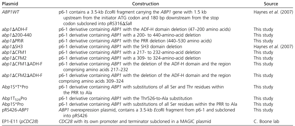
Plasmids used in this study are listed in Table 2. Plasmid p6-1 contains a 3.5-kb EcoRI fragment carrying the ABP1 gene with 1.5 kb upstream from the initiator ATG codon and 180 bp downstream from the stop codon subcloned into pRS316DSalI (Hayneset al.2007). This plasmid was used to generate yeast expression plasmids for wild-type (WT) and mutant versions of Abp1p using standard cloning techni-ques. All generated plasmids were sequenced to verify the integrity of the constructs.
Protein immunoprecipitation and Western blot analysis
Yeast-cell protein extracts were prepared from exponentially growing cells expressing WT or mutantAbp1pby lysing cells with glass beads (Lee et al.1998). Protein concentrations were measured using the BCA Protein Assay Reagent kit (Pierce Chemical, Rockford, IL). Protein extracts were sepa-rated on denaturing 10% SDS-PAGE using the Mini-Protean III system (BioRad). After electrophoresis, the proteins were transferred to nitrocellulosefilters and detected by Western blotting with a polyclonal chicken anti-Abp1p antibody (supplied by B. Goode) using enhanced chemiluminescence. ForAbp1pimmunoprecipitation, 1 mg of total protein ex-tracted from the BY263 yeast was incubated with 1 ml of Abp1SH3 mouse monoclonal antibody (18-A, produced by the University of Toronto monoclonal antibody facility) in a total volume of 100ml of lysis buffer [50 mM Tris–HCl (pH 7.5), 250 mM NaCl, 5 mM EDTA, 1 mM DTT, 0.1% NP-40, 50 mM NaF, and protease inhibitor cocktail (Sigma)]. After incubation at 4° for 1 hr, 15 ml of a 50% (v/v) Protein A-Sepharose slurry in lysis buffer was added and further incubated overnight at 4°. The resin was washed three times with 1 ml of wash buffer containing 50 mM Tris–HCl (pH Table 1 Yeast strains used in this study
Strain Genotype Source
Y263 MATatrp1D63 ura3-52 lys2-801 ade2-107 his3D200 leu2-D1 Measdayet al.(1994)
BY1009a BY263abp1D::Kan Hayneset al.(2007)
BY3002b a/aabp1D::Nat sla1D::Kan B. Andrew lab
abp1Dprk1D MATaabp1D::Nat prk1D::Kan Cross of BY1689 and BY2985 Hayneset al.(2007) RH3395 MATalys2 leu2 ura3 his3 bar1 sla2/end4::HIS3 end4D376-501::TRP1 Wespet al.(1997)
abp1Dsla2Dcoil1 MATalys2 leu2 ura3 his3 bar1 sla2/end4::HIS3 end4D376-501::TRP1 abp1::Nat
This study
BY4068b MATacdc28-as::Nat ura3 leu2 his3 met15 B. Andrew lab
BY4131b MATapho85-as::Hph ura3 leu2 his3 met15 B. Andrew lab
BY4129b MATacdc28-as::Nat pho85-as::Hph ura3 leu2 his3 met15CAN1+LYP1+ B. Andrew lab
aStrains are isogenic to the parent strain, BY263, an S288C derivative.
7.5), 250 mM NaCl, 5 mM EDTA, 1 mM DTT, and 0.1% NP-40 and then resuspended in SDS-gel loading buffer or phos-phatase reaction buffer [10 mM Tris–HCl (pH 7.5), 1 M NaCl, 10 mM MgCl2, 1 mM DTT, and 0.1 mM 4-(2-Aminoethyl)
benzenesulfonyl fluoride hydrochloride (AEBSF) (Sigma)]. Immune complexes were visualized by Western blotting with anti-Abp1pantibody. In some experiments, membranes were stripped of anti-Abp1pand reprobed with anti-tubulin antibody (Sigma).
In vivo labeling experiments
For overexpression of Abp1p, plasmid pRS426-ABP1 was transformed into strain BY263. Transformants were grown to mid-log phase in SD-URA, transferred to YPD for two gen-erations, and then shifted to YPD minus Pi and allowed to duplicate once. The cells were pelleted and concentrated 20-fold to afinal volume of 5 ml in YPD-Pi, and 1 mCi of [32P]
orthophosphate (Dupont/NEN) was then added. After 1 hr of incubation with shaking, cells were collected by centrifuga-tion, washed with 50 mM NaF, andflash-frozen.32P-labeled Abp1p was immunoprecipitated, subjected to SDS-PAGE, transferred to a nitrocellulose membrane, and detected by autoradiography. The nitrocellulose membrane was further analyzed by Western blotting.
Phosphatase treatment
Whole-cell extracts were obtained following the procedure described above using a modified lysis buffer [100 mM Tris (pH 8.0), 100 mM NaCl, 2 mM MnCl2, 10% glycerol, 1 mM
DTT, and protease inhibitor cocktail (Sigma)]. For phospha-tase treatment (Ho et al. 1999), 100 mg of total proteins were used and incubated for 30 min at 30°in a total volume of 20ml of lysis buffer in the presence of 2 mM MnCl2and
1600 U of l-phosphatase (New England Biolabs). Where indicated, the protease/phosphatase inhibitor (250 mM NaF,
10 mM EDTA, 4 mM sodium orthovanadate, 2 mM AEBSF) was added. The phosphatase reaction was stopped by addition of SDS sample buffer, and lysates were analyzed by Western blotting.
Spot dilution growth assays
To assay growth on solid media, yeast cells were grown overnight to saturation in selective media supplemented with 2% dextrose and the appropriate amino acids. Five microliters of 10-fold serial dilutions of equivalent cell concentrations (0.1 OD600nm) from each strain expressing
WT or mutant Abp1p variants was spotted onto minimal media lacking uracil (SD-URA). Growth differences were recorded following incubation of the plates for 48 hr at the indicated temperature. In some cases, 5 mM caffeine (Sigma) was added to emphasize growth defects. All growth assays were repeated at least three times, and one represen-tative experiment is shown in Figure 2.
Protein stability assays using cycloheximide
For analyses of protein stability, yeast cultures were grown in SD-URA to exponential growth phase (0.5 OD600nm) and
di-vided in two halves. One-half was allowed to continue growth, and 35 mg/ml of cycloheximide (Sigma) was added to the other. Subsequently, equal numbers of cells were collected after 4 hr by centrifugation and immediately frozen; degradation was stopped by the addition of 1 mM sodium azide. Proteins were extracted from whole-cell lysates, as described earlier, and analyzed by Western blotting using anti-Abp1pand anti-tubulin probes. Three independent experiments were carried out, and one representative experiment is shown in Figure 4D.
NMR spectroscopy
The extendedAbp1pSH3 domain construct, which included the SH3 domain and the 28 residues lying N-terminal to the Table 2 Plasmids used in this study
Plasmid Construction Source
ABP1WT p6-1 contains a 3.5-kbEcoRI fragment carrying theABP1gene with 1.5 kb upstream from the initiator ATG codon and 180 bp downstream from the stop codon subcloned into pRS316DSalI
Hayneset al.(2007)
abp1DADH-F p6-1 derivative containingABP1with the ADF-H domain deletion (47–200 amino acids) This study abp1D200-440 p6-1 derivative containingABP1with a 200- to 440-amino-acid deletion This study abp1DPR\R p6-1 derivative containingABP1with the PRR deletion (440–530 amino acids) This study
abp1DSH3 p6-1 derivative containingABP1with the SH3 domain deletion Hayneset al.(2007) abp1DCFM1 p6-1 derivative containingABP1with a 217- to 232-amino-acid deletion This study
abp1DCFM2 p6-1 derivative containingABP1with a 309- to 324-amino-acid deletion This study abp1DCFM1DADH-F p6-1 derivative containingABP1with the deletion of the ADF-H domain and the region
comprising amino acids 217–232
This study
abp1DCFM2DADH-F p6-1 derivative containingABP1with the deletion of the ADF-H domain and the region comprising amino acids 309–324
This study
Abp1S*T*Pro p6-1 derivative containingABP1with substitutions of all Ser and Thr residues within the PRR to Ala
This study
Abp1T526Pro p6-1 derivative containingABP1with the Thr526-to-Ala substitution This study Abp1S*Pro p6-1 derivative containingABP1with substitutions of all Ser residues within the PRR to Ala This study pRS426-ABP1 ABP1overexpression plasmid, contains a 3.5-kbEcoRI fragment from p6-1 and subcloned
into pRS426
This study
domain, was expressed as a thioredoxin fusion with a tobacco etch virus protease cleavage site and was purified as described previously (Stollaret al.2009). The1H-15N HSQC spectrum of
the extended construct was collected using standard methods
(Kayet al.1995) at 30°on a Varian 500 MHz spectrometer
equipped with pulsedfield gradients and a triple resonance probe with actively shielded z-gradients. Data were processed and analyzed using NMRPipe/NMRDraw (Delaglio et al. 1995) and NMRView (Johnson and Blevins 1994). Spectra were recorded in 50 mM sodium phosphate, 100 mM NaCl, and 1 mM EDTA (pH 7.0). Using the established assignments ofArk1ppeptide-bound and freeAbp1pSH3 domain (E. J. Stollar H. Lin, A. R. Davidson, and J. D. Forman-Kay, unpub-lished results), peptide titrations were used to monitor the movement of peaks and assign thefinal position of theAbp1p SH3 domain:PRR peptide complex resonances. Almost all of the peptide complexes were exchanging in the fast timescale regime facilitating this method of assignment. The 1H-15N
HSQC spectrum of the extended PRR-SH3 domain construct was essentially superimposable on that of the SH3 domain: PRR peptide complex; thus, further experiments to assign the peaks within this spectrum were unnecessary. Amide hydro-gen chemical shift differences were calculated as previously described (Stollaret al.2009).
Results
Conserved sequence features are found in the Abp1 family
To gain more insight into the functionally important regions of theAbp1protein family, we compared the sequence fea-tures of these proteins from diverse fungal and higher eukary-otic species. As seen in Figure 1A, all members of the family possess an N-terminal ADF-H domain and at least one C-terminal SH3 domain. In addition, a PRR containing at least 10% Pro is found upstream of the SH3 domains in all cases. Using the algorithm Epestfind (http://bioweb2.pasteur.fr/ docs/EMBOSS/epestfind.html), we were also able to detect sequences predicted with high PEST scores within the PRR regions of allAbp1phomologs examined. The prevalence of PEST sequences, which have been shown to mediate protein degradation (Rogers et al. 1986; Rechsteiner and Rogers 1996), within the whole Abp1 family has not been previ-ously reported.
within the same region (Figure 1A). Furthermore, examina-tion of the predicted helical regions of the fungal homologs revealed two highly conserved sequence motifs, Conserved Fungal Motifs 1 and 2 (CFM1 and CFM2) (Figure 1B). CFM1 lies just amino terminal to thefirst conserved predicted he-lix, and CFM2 lies within a predicted conserved helix (Figure 2A). These motifs are very conspicuous because they lie within an area of these proteins that is generally highly di-verse in sequence despite the conserved presence of predicted helices. The conservation of CFM1 (Figure 1B) is particularly striking since it is found even in very distantly relatedAbp1p orthologs from Basidiomycete species (Ustilago,Coprinopsis,
and Malassezia), which diverged from S. cerevisiae at least
500 million years ago (Taylor and Berbee 2006).
Deletion of the ADF-H domain reveals a functional role for CFM1
To assess the biological roles of the conserved features identified in Abp1p, we constructed plasmids expressing mutant versions of Abp1p with various regions deleted. The biological activity of these plasmid-expressed proteins was tested in several yeast genetic backgrounds in which Abp1pis required for viability under some conditions. These backgrounds includedabp1Dsla1D, which is unable to grow at 30°, but can be propagated at room temperature (Haynes
et al. 2007), andabp1Dprk1D, which is unable to grow at
37°(Copeet al.1999). We also utilized a strain combining abp1Δ with a partial deletion ofSLA2 (sla2Δcoil1), which displays impaired growth at 37°(Wespet al.1997). Trans-formation of these mutant strains with the plasmid express-ing wild-type ABP1 resulted in robust growth while cells containing an empty vector did not grow under the restric-tive conditions (Figure 2B), confirming the requirement for Abp1pin these strain backgrounds and growth conditions.
As has been previously observed (Hayneset al.2007), ex-pression ofAbp1placking its SH3 domain resulted in growth that was no better than vector alone in all conditions and in genetic backgrounds in whichAbp1pwas required. By contrast, deletion of the central region (residues 200–440) or PRR region (residues 440–530) caused no growth defects as com-pared to WT under any conditions. Deletion of residues 47–200 (abp1DADF-H), which includes the conserved ADF-H domain (residues 1–144), caused only a partial reduction in viability in theabp1Dsla1Dandabp1Dprk1Dbackgrounds, yet no growth was observed in abp1Dsla2Dcoil1 at 37°. These results are consistent with another study in which the ADF-H domain was found to be essential for viability in anabp1Dsla2D strain, but not in anabp1Dsla1Dstrain (Quintero-Monzonet al. 2005); however, theabp1Dprk1Dandabp1Dsla2Dcoil1were not tested in this previous study. We also tested a deletion in which both the ADF-H domain and central region were deleted (Δ47–440), but this mutant was very poorly expressed and meaningful growth data could not be obtained.
The presence of an ADF-H domain, which binds to actin, is a conserved feature of all Abp1phomologs; thus, it was surprising that this domain was not fully required for growth
under all conditions. Since a second actin-binding activity was identified in the putative helical region of mouseAbp1, we investigated the possibility that the CFM1 or CFM2 motifs (Figure 1), which are within the same region of yeast Abp1p, might play a functional role when the ADF-H do-main is deleted. As shown in Figure 2B, combining a deletion of CFM1 (D217–232) with a deletion of a fragment includ-ing the ADF-H (Δ47–200) domain resulted in a much greater reduction in growth than deletion of either region alone under the conditions where the ADF-H domain was not essential for growth. Importantly, the expression levels of all three of theseAbp1pdeletion mutants was very similar (Figure 2C). It should be noted that different stress condi-tions were used in these assays as compared to those de-scribed above because these conditions were found to best emphasize the effect of the CFM1 deletion. We did not ob-serve any reduction in viability when a deletion of the CFM2 motif (D309–324) was combined with an ADF-H domain deletion, suggesting that this region possesses a function distinct from that of the CFM1 region.
Yeast Abp1p is phosphorylated within the PRR
Since mAbp1 is phosphorylated (Larboletteet al.1999; Han
et al. 2003; Larive et al. 2009; Boateng et al. 2012) and
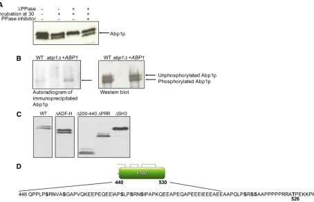
several phosphorylated peptides from yeast Abp1p have been identified in large-scale studies (Ficarro et al. 2002; Ubersaxet al.2003; Holtet al.2009), we sought to further investigate the possible phosphorylation ofAbp1p. Interest-ingly, Abp1p migrates as a double-protein band on SDS-PAGE gels (Drubinet al.1988). To determine whether the existence of this double band is due to phosphorylation, whole-yeast extracts were treated with l-phosphatase, and the electrophoretic mobility of the treated protein was eval-uated. As seen in Figure 3A, phosphatase treatment caused the disappearance of the band of faster mobility, implying that this band represents a phosphorylated form ofAbp1p. When phosphatase and phosphatase inhibitor were added to the reactions, the faster band remained. To confirm that this band contained phosphate, cells were grown in 32
P-orthophosphate, and immunoprecipitated Abp1p that had incorporated32P was visualized on SDS-PAGE gels by
expo-sure tofilm. It can be seen that a labeled band was observed in a WT extract, but not in an extract from an abp1Δstrain (Figure 3B). Furthermore, the labeled band was more intense in extracts of a strain carrying a plasmid that overexpressed Abp1p. Western blotting of the same gels demonstrated that the 32P-labeled bands corresponded to the faster-migrating
was confirmed by phosphatase treatment (Figure 3D, bot-tom). Since this result indicated that the phosphorylation site was located within the PRR, we mutagenized all Ser and Thr residues within this region (no Tyr residues were found in the PRR) as a means to identify the specific site of phosphoryla-tion. As expected, simultaneous replacement of all nine Ser and Thr residues within the PRR with Ala (S*T*PRR) abro-gated phosphorylation as detected by the absence of the faster-migrating Abp1p band. The remaining band, which comigrated with the upper band in the WT Abp1p extract, was not affected by addition of phosphatase. By substituting each Ser and Thr residue in the PRR individually, we found that only substitution of Thr526 (T526A) resulted in a loss of
phosphorylation. Thus, we conclude that phosphorylation of Thr526 causes the change in mobility ofAbp1p observed in SDS-PAGE. Although this site is clearly phosphorylated, the Thr526Ala substitution did not cause a detectable growth phenotype in any of theABP1-dependent strain backgrounds described above (data not shown). In addition, testing of a phosphomimetic Thr526Glu substitution in the abp1Δ/
prk1Δ and abp1Δ/sla2Δcoil1 backgrounds did not reveal
a growth phenotype (data not shown).
Cdc28p and Pho85p phosphorylate Abp1p
(K/R)], and this region was predicted with very high proba-bility to be phosphorylated by the yeast CDKs,Cdc28p, and Pho85p using the Predikin software (Saunders et al. 2008; Saunders and Kobe 2008). To determine whether Abp1p is a target of these CDKs, we assessed the level ofAbp1p phos-phorylation in yeast strains carrying inhibitor-sensitive alleles ofCDC28(cdc28-as) andPHO85(pho85-as). In these strains, kinase activity can be specifically inhibited by the addition of specific nucleotide analogs (Carrollet al.2001; Ubersaxet al. 2003), such as 1NM-PP1, which is the compound that we used here. As can be seen in Figure 4A, phosphorylation could still be detected in strains carrying thecdc28-asandpho85-as alleles on their own, but the cdc28-as/pho85-as double-mutant strain displayed no detectable phosphoylatedAbp1p. This result implies that both of these CDKs are able to phos-phorylate Abp1p so that Abp1p phosphorylation persists in the single-mutant strains. It was somewhat surprising that the cdc28-as/pho85-as double-mutant strain displayed no Abp1pphosphorylation even in the absence of inhibitor. Since the cdc28-asmutant is reduced by sixfold in overallin vitro activity in the absence of inhibitor (Bishop et al. 2000), we surmise that the loss ofAbp1pphosphorylation in the double-mutant strain is the result of simultaneous partial reduction in the activity of both kinases. We show here that thepho85-as mutant is definitely inhibited further by addition of 1NM-PP1 (Supporting Information,Figure S1), and the specific inhibi-tion of thecdc28-asmutant by this compound has been pre-viously demonstrated (Bishopet al.2000; Zimmermannet al. 2011).
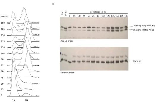
To directly test the ability ofCdc28p to phosphorylate Abp1p, a plasmid producing wild-type Cdc28p was intro-duced into thecdc28-as/pho85-asstrain. In this case, phos-phorylation of Abp1p was partially restored (Figure 4B), proving that the presence of Cdc28p can mediate phos-phorylation ofAbp1p. The incomplete restoration ofAbp1p phosphorylation in this strain background may be due to perturbed regulation of the plasmid-borne CDC28 gene or others factors specific to the double-mutant strain. Since many Cdc28p and Pho85p substrates are phosphorylated in a cell-cycle-dependent manner (Huang et al. 2007; Enserink and Kolodner 2010), we assessed the cell cycle dependence of Abp1pphosphorylation bya-factor growth synchronization experiments. Although the levels ofAbp1p phosphorylation varied with time after release of a-factor arrest, these changes did not correlate with cell cycle pro-gression as monitored by the changes in cellular DNA con-tent (Figure S2).
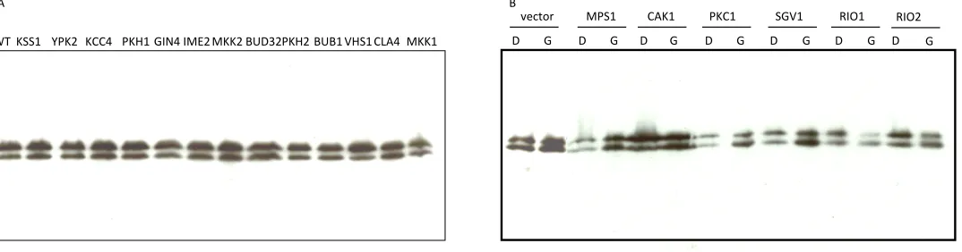
To determine whether any other yeast protein kinases are involved in Abp1p phosphorylation, strains bearing single deletions of known nonessential kinase-encoding genes were examined. We found that none of these 80 strains displayed any alteration in Abp1p phosphorylation as assessed by Western blotting of lysates of exponentially growing cells. Similarly, overexpression of 27 different es-sential kinases caused no change in the degree of Abp1p phosphorylation (Figure S3).
Novel role for the PRR and phosphorylation in Abp1p stabilization
when stationary phase was reached (Figure 4C). These data suggested that the phosphorylated form may be more stable. To directly test this idea, we monitored the degradation of Abp1pover time after inhibition of protein translation by the addition of cycloheximide. Strikingly, the unphosphorylated form of Abp1p was almost completely degraded after 4 hr of incubation in cycloheximide while little change in the level of the phosphorylated form was seen (Figure 4D, left). Con-sistent with this result, the Thr526Ala mutant, which cannot be phosphorylated, was also mostly degraded after 4 hr in cycloheximide (Figure 4D, center). These data demonstrate that the phosphorylation of Thr526 leads to stabilization ofAbp1p.
As described above, the presence of PEST sequences, which mediate protein degradation (Rogers et al. 1986; Rechsteiner and Rogers 1996), is a conserved feature of the PRRs of both fungal and higher eukaryoticAbp1p ortho-logs. To determine whether these PEST regions are required for the rapid degradation of unphosphorylated Abp1p, the effect of deletion of the PRR (DPRR) onAbp1pstability after inhibition of protein synthesis was analyzed. As is shown in Figure 4D (right), the ΔPRR mutant is resistant to proteol-ysis after addition of cycloheximide, supporting the impor-tance of the PEST sequences in mediating this degradation. It should be noted that the Abp1p phosphorylation site is within the region deleted in the ΔPRR mutant; thus, the increased stability of this mutant occurs in spite of its being unphosphorylated. In summary, these data indicate that pro-tein degradation mediated by PEST sequences within the PRR is inhibited by phosphorylation ofAbp1pat Thr526.
Abp1p SH3 domain can potentially form an intramolecular interaction with the PRR
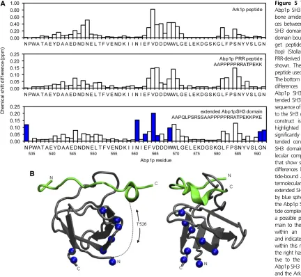
Since the Abp1pSH3 domain lies immediately adjacent to the PRR region, we hypothesized that an intramolecular in-teraction between these two regions could play a role in regulating Abp1p activity. To test this idea, we expressed and purified a version of the Abp1p SH3 domain that in-cluded 28 residues N-terminal to the start of the SH3 do-main. This region includes several PXXP motifs and the phosphorylation site at Thr526. To assess the ability of this region to interact with the SH3 domain, we collected an1H-15N HSQC NMR spectrum for this construct and
com-pared it to spectra previously collected for free and peptide-bound forms of theAbp1pSH3 domain (Stollaret al.2009). In this type of spectrum, a cross-peak can be observed cor-responding to each amide group in the protein. The posi-tions of these cross-peaks (i.e., their chemical shifts) are very sensitive to the chemical environment; thus, amide groups in residues close to the peptide-binding surface display sig-nificant chemical shift changes upon peptide binding. It can be seen in Figure 5A that similar changes in chemical shifts are seen when comparing the freeAbp1pSH3 domain to the Ark1p peptide-bound domain (top) or to the extended Abp1pSH3 domain construct (bottom). These data indicate that the PRR of the extended construct interacts with the
SH3 domain in a manner similar to a bound target peptide. We also found that a synthesized Pro-rich peptide derived from the Abp1p PRR was able to bind the Abp1p SH3 domain intermolecularly when added to the NMR sample (Figure 5A, middle). The dissociation constant of this inter-action as measured by isothermal titration calorimetry was 64.3 mM (E. J. Stollar, H. Lin, A. R. Davidson, and J. D. Forman-Kay, unpublished results). Comparison of the NMR spectrum of this intermolecular complex with the spectrum of the extended SH3 domain construct revealed additional residues in the extended construct with significant chemical shift changes (Figure 5A, bottom). A plot of these residues on the structure of theAbp1pSH3 domain:peptide complex shows that these residues lie on one surface of the domain (Figure 5B) and likely are positioned on the path that the PRR region would have to follow to engage in an intramo-lecular interaction with the peptide-binding surface of the domain.
Discussion
Members of theAbp1family of proteins have been found in diverse fungal and metazoan organisms ranging from yeast to humans. The high degree of conservation seen among these proteins indicates a crucial function in all of these organisms. It is surprising, therefore, that deletion of the ABP1gene in yeast causes no detectable phenotype and that measurable effects of ABP1 mutations are seen only when they are combined with other gene deletions (Holtzman
et al.1993; Lila and Drubin 1997; Copeet al.1999; Haynes
et al.2007). Thesefindings emphasize that deletion of
func-tionally important proteins or regions of proteins will not always cause a detectable phenotype when tested under laboratory conditions. Thus, uncovering the roles of these conserved regions may require phenotypic assays to be car-ried out under a wide variety of growth conditions and ge-netic backgrounds. The results presented in this work clearly demonstrate the difficulties associated with assigning func-tions to the conserved features of yeastAbp1p, yet also show that a thorough investigation can reveal requirements for regions that at first may appear to be dispensable.
association between CFM1 and actin would be weak because deletion of the ADF-H domain was observed to abrogate lo-calization of Abp1p to cortical actin patches as assessed by GFP localization experiments (Quintero-Monzonet al.2005). An important conclusion from our studies on CFM1 is that, even when a motif is highly conserved, its function may not become apparent unless the protein is sensitized by other perturbations, such as deletion of the ADF-H domain in this case. This type of“intragenic synthetic lethality”has been re-cognized in a handful of other studies (Teixeiraet al.1997; Kou and Pugh 2004; Murphy et al.2004; Dias et al.2008; Garrey and Mackie 2011), but systematic searches for intra-genic synthetic lethality have not been carried out. Such experiments could provide a generally applicable approach for uncovering the roles of conserved regions within multi-domain proteins.
Our work has also illuminated other potentially impor-tant features ofAbp1p. We have demonstrated thatAbp1p is phosphorylated at Thr526 by the Cdc28p and Pho85p kinases (Figure 4), which recognize the same consensus phosphorylation sites and are functionally interchangeable
results from the presence of PEST sequences in the PRR that are located near the site of phosphorylation (Figure 1). Thus, phosphorylation at Thr526 appears to provide a means of regulating proteolysis mediated by the nearby PEST se-quences. Since PEST sequences are a conserved feature of Abp1p homologs in fungal and mammalian species and phosphorylation within the PRR is seen in mouse (Larbolette
et al.1999; Hanet al.2003; Zanivanet al.2008; Lariveet al.
2009; Boateng et al. 2012) and human Abp1 (Olsenet al. 2006; Molinaet al.2007), we propose that this regulation of Abp1pstability may be a conserved feature of this family. It is possible that the putative intramolecular interaction be-tween the SH3 domain and the PRR, which is supported by our NMR data, is affected by phosphorylation at Thr526 since this site lies between the SH3 domain and the PRR (Figure 5B). This intramolecular interaction may play a role in regulating Abp1pstability through modulating the acces-sibility of the PRR to other binding partners. In mammalian cortactin, a protein with a similar domain layout and function as Abp1p (Olazabal and Machesky 2001), phosphorylation has also been suggested to modulate the intramolecular in-teraction between a C-terminal SH3 domain and an upstream PRR (Lua and Low 2005).
The investigation ofAbp1pprovides an instructive exam-ple of a protein that is highly conserved in evolution, yet deletion of the gene encoding it in yeast results in no de-tectable growth defects under laboratory conditions. The presence of Abp1p orthologs in divergent species implies that organisms lacking this protein would certainly display a decreased evolutionary fitness. Clearly, the requirements for survival in the natural environment over hundreds of millions of years are much more stringent than those found in the laboratory. For this reason, detection of phenotypes forABP1deletions in the laboratory requires additional ma-nipulation of genetic backgrounds and growth conditions (Lila and Drubin 1997; Faziet al.2002; Stefanet al.2005; Haynes et al. 2007; Stollar et al.2009). In this work, we show that deletions of conserved domains and motifs of Abp1p, which also must be functionally important, do not always lead to detectable growth phenotypes, yet further manipulation of conditions can reveal these phenotypes. In some cases, multiple conserved regions within a protein may have to be deleted before a phenotype emerges as we ob-served here in the case of the CFM1 motif. Detection of this type of intragenic synthetic lethality could be an important new addition to the repertoire of systematic approaches that are generally employed for uncovering protein functions. Due to the frequent difficulty in establishing conditions in the laboratory that can reveal phenotypes for highly conserved protein regions, we suggest that the detection of conserva-tion is a more reliable approach for identifying funcconserva-tionally important regions in a protein than is the identification of phenotypes resulting from mutations. The future challenge is to detect phenotypes associated with protein regions that are strongly indicated to be important through conservation analysis. High-throughput genetics studies on strains
con-taining deletions of one or more conserved regions of a pro-tein may provide the means to achieve this goal.
Acknowledgments
We thank Brenda Andrews and Charlie Boone for plasmids and yeast strains; Bruce Goode for anti-Abp1p antibody; and Karen Maxwell for critical reading of the manuscript. This work was supported by an operating grant from the Canadian Institutes of Health Research (CIHR ARD grant MOP-13609 to A.R.D.). E.J.S. was supported by grants from the National Center for Research Resources (5P20RR016480-12) and the National Institute of General Medical Sciences (8P20GM103451-12) of the National Institutes of Health.
Literature Cited
Bishop, A. C., J. A. Ubersax, D. T. Petsch, D. P. Matheos, N. S. Gray
et al., 2000 A chemical switch for inhibitor-sensitive alleles of
any protein kinase. Nature 407: 395–401.
Boateng, L. R., C. L. Cortesio, and A. Huttenlocher, 2012
Src-me-diated phosphorylation of mammalian Abp1 (DBNL) regulates
podosome rosette formation in transformedfibroblasts. J. Cell
Sci. 125: 1329–1341.
Brachmann, C. B., A. Davies, G. J. Cost, E. Caputo, J. Li et al.,
1998 Designer deletion strains derived from Saccharomyces
cerevisiae S288C: a useful set of strains and plasmids for PCR-mediated gene disruption and other applications. Yeast 14:
115–132.
Carroll, A. S., A. C. Bishop, J. L. DeRisi, K. M. Shokat, and E. K.
O’Shea, 2001 Chemical inhibition of the Pho85
cyclin-depen-dent kinase reveals a role in the environmental stress response.
Proc. Natl. Acad. Sci. USA 98: 12578–12583.
Chi, A., C. Huttenhower, L. Y. Geer, J. J. Coon, J. E. Syka et al.,
2007 Analysis of phosphorylation sites on proteins from
Sac-charomyces cerevisiae by electron transfer dissociation (ETD)
mass spectrometry. Proc. Natl. Acad. Sci. USA 104: 2193–2198.
Cole, C., J. D. Barber, and G. J. Barton, 2008 The Jpred 3
sec-ondary structure prediction server. Nucleic Acids Res. 36:
W197–W201.
Cope, M. J., S. Yang, C. Shang, and D. G. Drubin, 1999 Novel
protein kinases Ark1p and Prk1p associate with and regulate the cortical actin cytoskeleton in budding yeast. J. Cell Biol. 144:
1203–1218.
Cortesio, C. L., B. J. Perrin, D. A. Bennin, and A. Huttenlocher,
2010 Actin-binding protein-1 interacts with WASp-interacting
protein to regulate growth factor-induced dorsal ruffle
forma-tion. Mol. Biol. Cell 21: 186–197.
Delaglio, F., S. Grzesiek, G. W. Vuister, G. Zhu, J. Pfeifer et al.,
1995 NMRPipe: a multidimensional spectral processing
sys-tem based on UNIX pipes. J. Biomol. NMR 6: 277–293.
Dias, C. A., V. S. Cano, S. M. Rangel, L. H. Apponi, M. C. Frigieri
et al., 2008 Structural modeling and mutational analysis of yeast eukaryotic translation initiation factor 5A reveal new crit-ical residues and reinforce its involvement in protein synthesis.
FEBS J. 275: 1874–1888.
Drubin, D. G., K. G. Miller, and D. Botstein, 1988 Yeast
actin-binding proteins: evidence for a role in morphogenesis. J. Cell
Biol. 107: 2551–2561.
Enserink, J. M., and R. D. Kolodner, 2010 An overview of
Cdk1-controlled targets and processes. Cell Div. 5: 11.
Fazi, B., M. J. Cope, A. Douangamath, S. Ferracuti, K. Schirwitz
of the yeast actin-binding protein Abp1: structural and
func-tional analysis. J. Biol. Chem. 277: 5290–5298.
Fenster, S. D., M. M. Kessels, B. Qualmann, W. J. Chung, J. Nash
et al., 2003 Interactions between Piccolo and the actin/dyna-min-binding protein Abp1 link vesicle endocytosis to presynaptic
active zones. J. Biol. Chem. 278: 20268–20277.
Ficarro, S. B., M. L. McCleland, P. T. Stukenberg, D. J. Burke, M. M. Rosset al., 2002 Phosphoproteome analysis by mass spectrom-etry and its application to Saccharomyces cerevisiae. Nat.
Bio-technol. 20: 301–305.
Garrey, S. M., and G. A. Mackie, 2011 Roles of the 59-phosphate
sensor domain in RNase E. Mol. Microbiol. 80: 1613–1624.
Gietz, R. D., and R. A. Woods, 2002 Transformation of yeast by
lithium acetate/single-stranded carrier DNA/polyethylene
gly-col method. Methods Enzymol. 350: 87–96.
Goode, B. L., A. A. Rodal, G. Barnes, and D. G. Drubin, 2001
Acti-vation of the Arp2/3 complex by the actinfilament binding
pro-tein Abp1p. J. Cell Biol. 153: 627–634.
Han, J., R. Kori, J. W. Shui, Y. R. Chen, Z. Yaoet al., 2003 The SH3
domain-containing adaptor HIP-55 mediates c-Jun N-terminal ki-nase activation in T cell receptor signaling. J. Biol. Chem. 278:
52195–52202.
Haynes, J., B. Garcia, E. J. Stollar, A. Rath, B. J. Andrewset al.,
2007 The biologically relevant targets and binding affinity
re-quirements for the function of the yeast actin-binding protein 1 Src-homology 3 domain vary with genetic context. Genetics
176: 193–208.
Ho, Y., M. Costanzo, L. Moore, R. Kobayashi, and B. J. Andrews,
1999 Regulation of transcription at the Saccharomyces
cerevi-siae start transition by Stb1, a Swi6-binding protein. Mol. Cell.
Biol. 19: 5267–5278.
Holt, L. J., B. B. Tuch, J. Villen, A. D. Johnson, S. P. Gygi et al.,
2009 Global analysis of Cdk1 substrate phosphorylation sites
provides insights into evolution. Science 325: 1682–1686.
Holtzman, D. A., S. Yang, and D. G. Drubin, 1993 Synthetic-lethal
interactions identify two novel genes, SLA1 and SLA2, that con-trol membrane cytoskeleton assembly in Saccharomyces
cerevi-siae. J. Cell Biol. 122: 635–644.
Huang, D., H. Friesen, and B. Andrews, 2007 Pho85, a
multifunc-tional cyclin-dependent protein kinase in budding yeast. Mol.
Microbiol. 66: 303–314.
Huang, D., S. Kaluarachchi, D. van Dyk, H. Friesen, R. Sopkoet al.,
2009 Dual regulation by pairs of cyclin-dependent protein
kinases and histone deacetylases controls G1 transcription in budding yeast. PLoS Biol. 7: e1000188.
Johnson, B. A., and R. A. Blevins, 1994 NMRView: a computer
program for the visualization and analysis of NMR data. J.
Bio-mol. NMR 4: 603–614.
Kay, L. E., P. Keifer, and T. Saarinen, 1995 Pure absorption
gra-dient enhanced hetero-nuclear single quantum correlation spectroscopy with improved sensitivity. J. Am. Chem. Soc. 114:
10663–10665.
Kessels, M. M., A. E. Engqvist-Goldstein, and D. G. Drubin,
2000 Association of mouse actin-binding protein 1 (mAbp1/
SH3P7), an Src kinase target, with dynamic regions of the cor-tical actin cytoskeleton in response to Rac1 activation. Mol. Biol.
Cell 11: 393–412.
Kessels, M. M., A. E. Y. Engqvist-Goldstein, D. G. Drubin, and B.
Qualmann, 2001 Mammalian Abp1, a signal-responsive
F-ac-tin-binding protein, links the actin cytoskeleton to endocytosis
via the GTPase dynamin. J. Cell Biol. 153: 351–366.
Kou, H., and B. F. Pugh, 2004 Engineering dimer-stabilizing
mu-tations in the TATA-binding protein. J. Biol. Chem. 279: 20966–
20973.
Lappalainen, P., M. M. Kessels, M. J. Cope, and D. G. Drubin,
1998 The ADF homology (ADF-H) domain: a highly exploited
actin-binding module. Mol. Biol. Cell 9: 1951–1959.
Larbolette, O., B. Wollscheid, J. Schweikert, P. J. Nielsen, and J.
Wienands, 1999 SH3P7 is a cytoskeleton adapter protein and
is coupled to signal transduction from lymphocyte antigen
re-ceptors. Mol. Cell. Biol. 19: 1539–1546.
Larive, R. M., S. Urbach, J. Poncet, P. Jouin, G. Mascre et al.,
2009 Phosphoproteomic analysis of Syk kinase signaling in
human cancer cells reveals its role in cell-cell adhesion.
Onco-gene 28: 2337–2347.
Lee, J., K. Colwill, V. Aneliunas, C. Tennyson, L. Moore et al.,
1998 Interaction of yeast Rvs167 and Pho85 cyclin-dependent
kinase complexes may link the cell cycle to the actin
cytoskele-ton. Curr. Biol. 8: 1310–1321.
Lila, T., and D. G. Drubin, 1997 Evidence for physical and
func-tional interactions among two Saccharomyces cerevisiae SH3 domain proteins, an adenylyl cyclase-associated protein and
the actin cytoskeleton. Mol. Biol. Cell 8: 367–385.
Longtine, M. S., A. McKenzie III. D. J. Demarini, N. G. Shah, A. Wach et al., 1998 Additional modules for versatile and
eco-nomical PCR-based gene deletion and modification in
Saccha-romyces cerevisiae. Yeast 14: 953–961.
Lua, B. L., and B. C. Low, 2005 Cortactin phosphorylation as
a switch for actin cytoskeletal network and cell dynamics
con-trol. FEBS Lett. 579: 577–585.
Measday, V., L. Moore, J. Ogas, M. Tyers, and B. Andrews,
1994 The PCL2 (ORFD)-PHO85 cyclin-dependent kinase
com-plex: a cell cycle regulator in yeast. Science 266: 1391–1395.
Mise-Omata, S., B. Montagne, M. Deckert, J. Wienands, and O.
Acuto, 2003 Mammalian actin binding protein 1 is essential
for endocytosis but not lamellipodia formation: functional anal-ysis by RNA interference. Biochem. Biophys. Res. Commun. 301:
704–710.
Molina, H., D. M. Horn, N. Tang, S. Mathivanan, and A. Pandey,
2007 Global proteomic profiling of phosphopeptides using
electron transfer dissociation tandem mass spectrometry. Proc.
Natl. Acad. Sci. USA 104: 2199–2204.
Murphy, M. W., B. L. Olson, and P. G. Siliciano, 2004 The yeast
splicing factor Prp40p contains functional leucine-rich nuclear
export signals that are essential for splicing. Genetics 166: 53–65.
Nishizawa, M., M. Kawasumi, M. Fujino, and A. Toh-e,
1998 Phosphorylation of sic1, a cyclin-dependent kinase
(Cdk) inhibitor, by Cdk including Pho85 kinase is required for
its prompt degradation. Mol. Biol. Cell 9: 2393–2405.
Olazabal, I. M., and L. M. Machesky, 2001 Abp1p and cortactin,
new“hand-holds”for actin. J. Cell Biol. 154: 679–682.
Olsen, J. V., B. Blagoev, F. Gnad, B. Macek, C. Kumar et al.,
2006 Global, in vivo, and site-specific phosphorylation
dynam-ics in signaling networks. Cell 127: 635–648.
Ptacek, J., G. Devgan, G. Michaud, H. Zhu, X. Zhu et al.,
2005 Global analysis of protein phosphorylation in yeast.
Na-ture 438: 679–684.
Quintero-Monzon, O., A. A. Rodal, B. Strokopytov, S. C. Almo, and
B. L. Goode, 2005 Structural and functional dissection of the
Abp1 ADFH actin-binding domain reveals versatile in vivo
adapter functions. Mol. Biol. Cell 16: 3128–3139.
Rechsteiner, M., and S. W. Rogers, 1996 PEST sequences and
regulation by proteolysis. Trends Biochem. Sci. 21: 267–271.
Rogers, S., R. Wells, and M. Rechsteiner, 1986 Amino acid
se-quences common to rapidly degraded proteins: the PEST
hy-pothesis. Science 234: 364–368.
Saunders, N. F., and B. Kobe, 2008 The Predikin webserver:
im-proved prediction of protein kinase peptide specificity using
structural information. Nucleic Acids Res. 36: W286–W290.
Saunders, N. F., R. I. Brinkworth, T. Huber, B. E. Kemp, and B.
Kobe, 2008 Predikin and PredikinDB: a computational
frame-work for the prediction of protein kinase peptide specificity and
Sopko, R., D. Huang, J. C. Smith, D. Figeys, and B. J. Andrews,
2007 Activation of the Cdc42p GTPase by cyclin-dependent
protein kinases in budding yeast. EMBO J. 26: 4487–4500.
Stefan, C. J., S. M. Padilla, A. Audhya, and S. D. Emr, 2005 The
phosphoinositide phosphatase Sjl2 is recruited to cortical actin
patches in the control of vesicle formation and fission during
endocytosis. Mol. Cell. Biol. 25: 2910–2923.
Stollar, E. J., B. Garcia, P. A. Chong, A. Rath, H. Lin et al.,
2009 Structural, functional, and bioinformatic studies
demon-strate the crucial role of an extended peptide binding site for the
SH3 domain of yeast Abp1p. J. Biol. Chem. 284: 26918–26927.
Taylor, J. W., and M. L. Berbee, 2006 Dating divergences in the
Fungal Tree of Life: review and new analyses. Mycologia 98:
838–849.
Teixeira, M. T., S. Siniossoglou, S. Podtelejnikov, J. C. Benichou, M. Mann et al., 1997 Two functionally distinct domains gener-ated by in vivo cleavage of Nup145p: a novel biogenesis
path-way for nucleoporins. EMBO J. 16: 5086–5097.
Ubersax, J. A., E. L. Woodbury, P. N. Quang, M. Paraz, J. D. Bleth-rowet al., 2003 Targets of the cyclin-dependent kinase Cdk1.
Nature 425: 859–864.
Wesp, A., L. Hicke, J. Palecek, R. Lombardi, T. Aust et al.,
1997 End4p/Sla2p interacts with actin-associated proteins
for endocytosis in Saccharomyces cerevisiae. Mol. Biol. Cell 8:
2291–2306.
Zanivan, S., F. Gnad, S. A. Wickstrom, T. Geiger, B. Maceket al.,
2008 Solid tumor proteome and phosphoproteome analysis
by high resolution mass spectrometry. J. Proteome Res. 7:
5314–5326.
Zimmermann, C., P. Chymkowitch, V. Eldholm, C. D. Putnam, J. M.
Lindvallet al., 2011 A chemical-genetic screen to unravel the
genetic network of CDC28/CDK1 links ubiquitin and Rad6-Bre1
to cell cycle progression. Proc. Natl. Acad. Sci. USA 108: 18748–
18753.
GENETICS
Supporting Information
http://www.genetics.org/content/suppl/2012/06/01/genetics.112.141739.DC1
The Importance of Conserved Features of Yeast
Actin-Binding Protein 1 (Abp1p): The Conditional
Nature of Essentiality
Bianca Garcia, Elliott J. Stollar, and Alan R. Davidson
oh#
DMSO#
1NM*PP1#
phase*contrast#images## Pho4p*GFP#
Figure S1 The direct Pho85 target , Pho4, is desphosphorylated upon Pho85 inhibition with 1NM-‐PP1 and shuttled from the cytoplasm to the nucleus. pho85-‐as/cdc28as strain was transformed with an expression plasmid containing a Pho4p-‐GFP fusion construct under the control of the ADH1 promoter (BA1828, B. Andrews lab). Cells were grown to an OD600 of 0.5 (0h) and treated with DMSO or 25 μM 1NM-‐PP1. Cellular localization of Pho4p was analyzed using fluorescence microscopy at 15 min after treatment. Representative fields are shown.
1N# 2N# 180#
165#
150#
135#
120#
105#
90#
75#
60#
45#
30#
15#
0# t#(min)# A#
Abp1p#probe#
coronin#probe#
log
#
ar
re
st#
0# 15# 30# 45# 60# 75# 90# 105# 120#135# 150#165# 180# B#
unphosphorylated#Abp1# phosphorylated#Abp1#
Coronin# αF#release#(min)#
Figure S2 Abp1p expression is not cell cycle regulated. (A) Flow cytometry analysis of DNA content of wild type yeast cells synchronized by release from α–factor at the indicated times in the left. (B) Protein extracts were analyzed by Western blotting using polyclonal anti-‐Abp1p antibody to monitor Abp1p levels or anti-‐coronin antibody to control for cellular protein levels. Time is minutes after α–factor release. The positions of phosphorylated and
unphosphorylated Abp1p are indicated.
RIO2# G# #MPS1# #CAK1# #PKC1# #SGV1# RIO1#
###vector#
D# G# D# G# D# G# D# G# D# G# D# G# D# WT# KSS1# YPK2# KCC4# PKH1#GIN4#IME2#MKK2#BUD32#PKH2#BUB1#VHS1#CLA4# MKK1#
A# B#
Figure S3 Representative Western blots of Abp1p expression in strains bearing single deletions of non-‐essential kinases (A) or over-‐expression of essential kinases (B). (A) Western blot analysis, using polyclonal anti-‐Abp1p antibody, of yeast extracts from kinase deleted strains backgrounds. (B) Western blot analysis, using polyclonal anti-‐ Abp1p antibody, of yeast extracts from transformants expressing various essential kinases from the GAL1 promoter. Extracts were made from mid-‐log phase cells grown in presence of glusose (D) or galactose (G).