DIRECT AND INDIRECT EVIDENCE FOR THE
ENDOSOMOLYTIC PROPERTIES OF
POLY(AMIDOAMINE)S
W
m
b y
N. G. Pattrick B.Sc. (Bons)
A thesis submitted to the University of London in partial fulfillment of the requirements for the degree of Doctor of Philosophy
Center for Polymer Therapeutics School of Pharmacy Faculty of Medicine University of London
ProQuest Number: U585748
All rights reserved
INFORMATION TO ALL USERS
The quality of this reproduction is dependent upon the quality of the copy submitted.
In the unlikely event that the author did not send a complete manuscript and there are missing pages, these will be noted. Also, if material had to be removed,
a note will indicate the deletion.
uest.
ProQuest U585748
Published by ProQuest LLC(2016). Copyright of the Dissertation is held by the Author.
All rights reserved.
This work is protected against unauthorized copying under Title 17, United States Code. Microform Edition © ProQuest LLC.
ProQuest LLC
789 East Eisenhower Parkway P.O. Box 1346
A cknowledgements
Firstly I would like to thank Professor Ruth Duncan for her help, support and belief in me throughout my Ph.D. I would also like to acknowledge Dr Steve Brocchini for his patience, help and support, especially during the move to Cardiff. I would also like to thank the collaborator Professor Paolo Ferruti, for giving me the opportunity to study in Milan, and supplying me with large amounts of PAAs.
Many thanks are also due to my fellow work colleagues at CPT, who not only provided me with intellectual stimulation, but gave me emotional support, happiness and many laughs. I would especially like to thank Elinor, for having a positive attitude to life and for showing me the delights of Cardiff nightlife. I would also like to thank Ruthie, for being there with me through thick and thin. Thanks are also due to Richard, for his common sense, Ryan, for his cheeky sense of humour, and Myrto, for her enthusiasm and constant willingness to help others. I would also like to acknowledge Sam, for her continued friendship and endless supply of gossip, and Rhian for taking me in. I would also like to thank Michelle and Peli for helpful discussions on PAA work and Maria for helping me with chemistry.
Abstract
Many potential therapeutics (such as genes, antisense and protein drugs) have their targets in a variety of intracellular compartments and thus it is essential to develop intracytoplasmic delivery vectors to mediate this process, in particular, escape of the therapeutic from endosomal vesicles to the cytoplasm. It is in this context that amphoteric, biocompatible, biodegradable poly(amidoamine) (PAA) polymers were studied. These polymers are able to display pH-dependent conformational changes in response to pH, thus displaying membrane-permeabilising properties at endosomal but not physiological pH. In this thesis different methods were used to demonstrate their endosomolytic properties.
The ability of PAAs to mediate transfection of the plasmid pSV-p-galactosidase in HepG2 cells provided first direct evidence for their endosomolytic ability. For the first time it was shown that the PAAs termed ISA 1 and 4 were able to mediate delivery of the non-permeant toxins ricin A-chain (RTA) and gelonin to the cytoplasm of B16F10 cells. Toxin cytosolic delivery was indicated by a decrease in the IC50 value
from > 1.8mg/mL to < 0.65mg/mL. To investigate the intracellular trafficking of PAAs alone and in combination with gelonin, fluorescent-labelled PAAs and gelonin were prepared for fluorescence microscopy. Uptake studies showed that the intracellular fate of the PAA and toxin differed according to the PAA structure used.
Direct evidence for PAA-mediated intracellular membrane disrupting capability was investigated using isolated rat liver lysosomes as a model membrane system. After intravenous (i.v.) administration of ISA 1 (50mg/kg), nearly a 3-fold increase in the lysosomal enzyme N-acetyl-p-D-glucosaminidase (NAGase) was observed compared to the control. Morphologically ISA 1-containing lysosomes appeared less densely stained compared to the control.
Finally, an ISA 1-gelonin construct was examined for endosomolytic ability. However conjugation of gelonin alleviated the pH-dependent membrane rupturing ability of the parent PAA.
Contents
Page number
Title page i
Dedication ii
Acknowledgements iii
Abstract iv
Contents V
List of figures xiii
List of tables xvii
Abbreviations XX
Chapter 1 Introduction 1
1.1 Intracytoplasmic Delivery 2
1.2 Targeted Drug Delivery Vehicles 3
1.3 Polymer Therapeutics 5
1.3.1 Polymer-Protein Conjugates 6
1.3.2 Lysosomotropic vs endosomotropic delivery 8 1.4 The Enhanced Permeability and Retention (EPR) Effect as a means
of T umour T argeting 10
1.5 Endocytosis and Intracellular Trafficking in the Context of Drug Delivery 10 1.5.1 Intracellular Trafficking of Toxins 17 1.6 Challenges of Intracytoplasmic Delivery 18 1.7 Intracytoplasmic Delivery Systems 21 1.7.1 Non-Polymeric Intracytoplasmic Delivery Systems 21
1.7.1.1 Viral Vectors 21
1.7.1.2 Cationic lipids and liposomes 22 1.7.1.3 Thiol-activated cytolysins 24
1.7.1.4 Photosensitizers 25
1.7.2 Polymeric Intracytoplasmic Delivery Systems 25
1.7.2.1 Cationic polymers 25
1.7.2.2 Endosmolytic Polymers 28
1.7.2.2.1 Polyethylenimine (PEI) 28 1.7.2.2.2 Poly[2-(dimethylamino)ethyl methacrylate]
1.7.2.2.3 Poly(ethyl acrylic acid) (PEAAc)and related
polymers 33
1.7.2.2.4 Poly(amidoamine)s (PAA)s 34
1.8 Aims 42
CHAPTER 2. MATERIALS AND METHODS 44
2.1 Materials 45
2.1.1 General Chemicals 45
2.1.2 Polymers 45
2.1.3 Tissue culture 45
2.1.4 Cytotoxicity assays 45
2.1.5 Fluorescence microscopy 46
2.1.6 IPEC formation 46
2.1.7 Amplification and transfection using pSV-p-galactosidase 46 2.1.8 N-acetyl-p-D-glucosaminidase (NAGase) assays 46 2.1.9 Sodium dodecyl sulphate polyacrylamide gel electrophoresis
(SDS-PAGE) 46
2.1.10 Transmission electron microscopy (TEM) and scanning electron
microscopy (SEM) 47
2.2 Apparatus 47
2.2.1 General laboratory equipment 47
2.2.2 Tissue culture 47
2.2.3 Fluorescence microscopy 47
2.2.4 Agarose gel electrophoresis 48
2.2.5 IPEC formation 48
2.2.6 SDS-PAGE 48
2.2.7 TEM and SEM 48
2.3 Methods 48
2.3.1 Maintenance of cells 48
2.3.2 Passaging of cells 48
2.3.3 Evaluation of cell number and cell viability 49
2.3.4 Freezing down cells 49
2.3.5 Cell recovery from cell banks 50 2.3.6 Evaluation of cell viability using the colourimetric tetrazolium-
bromide (MTT) assay 50
2.3.7 Toxin cytotoxicity 53
2.3.8 Evaluation of cytotoxicity of polymer/toxin combinations 53 2.3.9 Synthesis, purification and characterisation of PAA-oregon
green and gelonin-texas red conjugates for fluorescence
microscopy 57
2.3.9.1 Construction o f calibration curve o f Oregon green-succinimidyl ester concentration against fluorescence
units 61
2.3.9.2 Uptake studies of fluorescently labelled PAAs and gelonin in B16F10 and HepG2 cells using fluorescence
microscopy 61
2.3.10 Techniques used to investigate PAA/IPEC formation 62 2.3.10.1 Evaluation of the stability of DNA to DNase II 62 2.3.10.2 Evaluation of IPEC formation using a gel retardation
assay 63
2.3.10.3 Morphological analysis of IPECs using TEM 64 2.3.10.4 Evaluation of IPEC formation using an EtBr
displacement assay 64
2.3.10.5 Evaluation of IPEC stability to anionic challenge 65 2.3.10.6 Zeta potential determination of IPECs 65 2.3.11 Transfection assays using the plasmid vector pSV-p-
galactosidase 65
2.3.11.1 Amplification, isolation and characterisation of
pSV-p-galactosidase 65
2.3.11.2 Transfection assays 68
2.3.11.3 p-galactosidase assay 68
2.3.12 Bicinchoninic (BCA) assay 69
2.3.13 N-acetyl-P-D-glucosaminidase (NAGase) assay 72 2.3.14 Use of the red blood cell (RBC) lysis assay to determine
the pH-dependent lytic ability of PAAs 72 2.3.15 Scanning electron microscopy (SEM) of RBC’s after
2.3.16 SDS-PAGE and visualisation of gelonin, RTA and RTA in
combination with ISA 4 75
2.3.17 Isolation of rat liver subcellular fractions 77 2.3.18 Preparation of isolated light mitochondrial fractions containing
ISA 1 for transmission electron microscopy (TEM) 80 2.3.19 Investigation of the ability of PAAs to destabilise isolated
rat liver lysosomes 81
2.3.19.1 Evaluation of lysosomal breakage during incubation of polymers with isolated rat liver lysosomes (polymer
outside the lysosome) 81
2.3.19.2 Investigation of the ability of ISA 1 to destabilise rat
liver lysosomes (polymer inside the lysosomes) 82
Chapter 3. Evaluation of the ability of PAAs to form
interpolyelectrolyte complexes and transfect cells in vitro 83
3.1 Introduction 84
3.2 Methods 88
3.3 Results 91
3.3.1 Characterisation of pSV-p-galactosidase by restriction analysis 91 3.3.2 Ability of PAAs to form IPECs assessed by electrophoresis 91 3.3.3 Ability of PAAs to form IPECs assessed by an EtBr displacement
assay 96
3.3.4 TEM analysis of IPEC morphology 96 3.3.5 Evaluation of IPEC stability to DNase II 100 3.3.6 Measurement of the zeta potential of IP E C s 100 3.3.7 Transfection of HepG2 cells by IPECs formed from pSV-p-
galactosidase and various vectors 100
3.4 Discussion 107
3.5 Conclusions 111
Chapter 4 evaluation of the ability of PAAs to mediate intracytoplasmic
delivery of RTA and gelonin toxins 112
4.1 Introduction 113
4.2 Methods 117
4.3.1 SDS-PAGE analysis of RTA, gelonin and RTA/ISA 4
combinations 119
4.3.2 Cytotoxicity of toxins, polymers and toxin/polymer combinations towards B 16F10 and HepG2 cells 119
4.4 Discussion 133
4.5 Conclusions 137
Chapter 5 Direct evidence for PAA-induced lysosomal rupture 138
5.1 Introduction 139
5.2 Methods 143
5.3 Results 143
5.3.1 Isolation of rat liver lysosomes 143 5.3.2 Evaluation of the pH-, time- and concentration dependence of
lysosomal rupture caused by polymers incubated with isolated rat liver lysosomes (polymer outside the lysosome) 148 5.3.3 Evaluation of lysosomal stability after internalisation of ISA 1 :
Time and dose dependancy (polymer inside the lysosomes) 148 5.3.4 TEM of lysosomes containing ISA 1 148
5.4 Discussion 154
5.5 Conclusions 157
Chapter 6 Synthesis and characterisation of fluorescent-labelled PAA and
gelonin conjugates and visualisation of their endocytic capture 159
6.1 Introduction 160
6.2 Methods 167
6.3 Results 167
6.3.1 Purification and characterisation of fluorescent-labelled conjugates 167
6.3.2 Fluorescence microscopy 175
6.3.2.1 Uptake of ISA 23-OG and ISA 1-OG with lysotracker
red in B16F10 cells 178
6.3.2.2 Uptake of gelonin-TR alone or in combination with ISA 1- OG and ISA 23-OG with B16F10 cells 178
6 .3 3 3 Uptake of ISA 23-OG and ISA 1-OG with lysotracker
6.3.3.4 Uptake of geloninTR alone or in combination with ISA 1 -OG and ISA 23--OG in HepG2 cells 182
6.4 Discussion 182
6.5 Conclusions 199
Chapter 7 Preliminary evaluation of PAA structures with a view to
improving endosomolytic ability 200
7.1 Introduction 201
7.2 Methods 204
7.3 Results 204
7.3.1 Investigation of the effect of ISA 23 Mw on pH-dependent
haemolysis 204
7.3.2 SEM of RBC’s after incubation of ISA 23 of different Mw 204 7.3.3 Investigation of the effect o f ISA 23 Mw on their cytotoxicity alone
and in combination with gelonin towards B16F10 cells 210 7.3.4 Investigation of the cytotoxicity of an ISA 1-gelonin conjugate
towards B16F10 cells 210
7.4 Discussion 210
7.5 Conclusions 215
Chapter 8 General discussion 216
References 225
List o f Figures
Figure 1.1 Levels of targeted delivery
Figure 1.2 The EPR effect
Figure 1.3 Diagram showing the three categories o f pinocytosis
Figure 1.4 The endocytic pathway in a non-polarised cell
Figure 1.5 Structures of lipidic vectors
Figure 1.6 Alteration of intracellular trafficking of macromolecules by endosomolytic polymers
Figure 1.7 Structures of endosomolytic polymers
Figure 1.8 Proposed proton-sponge mechanism as occurs in endocytic vesicles after uptake of PEI
Figure 1.9 Proposed colloid osmotic mechanism to explain PEAAc-mediated RBC rupture
Figure 1.10 Synthesis of PAAs
Figure 1.11 Characteristics of an MBI-triton X-100 conjugate
Figure 1.12 Haemolysis of rat RBC’s after incubation with polymers (Img/mL) for 24h.
Figure 2.1 Schematic showing the reduction of MTT to an MTT formazan salt
Figure 2.2 B16F10 growth curve of cells seeded to a density of 1 x 10"^ cells/mL
Figure 2.3 HepG2 growth curve of cells seeded to a density of 1 x 10"^ cells/mL
Figure 2.4 Elution profiles of fluorescent probes eluted on PDIO columns equilibrated in PBS
Figure 2.5 Reduction of Cu (II) to Cu (I) by proteins
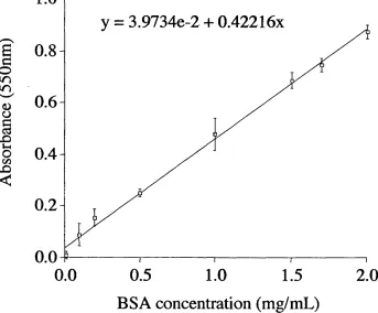
Figure 2.6 BSA calibration curve
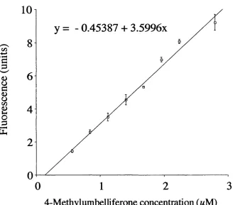
Figure 2.7 4-Methylumbelliferone calibration curve
Figure 2.8 Procedure for the differential centrifugation of rat liver
Figure 3.1 Schematic showing the multi-step process required for a non-viral delivery system
Figure 3.2 Restriction map of pSV-P-galactosidase from Promega
Figure 3.3 Effect of vehicle:DNA weight ratio on transfection of HepG2 cells
Figure 3.4 BCA assay using ISA 4 and ISA 23 as calibrants
Figure 3.5 Restriction analysis of recovered pSV-p-galactosidase
Figure 3.6 DNA retardation during agarose gel electrophoresis of polymer complexes
Figure 3.8 TEM’s showing morphology of IPECs
Figure 3.9 Protection of DNA from degradation by various polymers
Figure 3.10 Transfection of HepG2 cells by IPECs formed from psV-p-galactosidase and various vectors at a vehicle:DNA weight ratio of 5:1
Figure 4.1 Ribbon diagrams of toxins
Figure 4.2 Proposed intracellular trafficking o f toxins after endocytic uptake
Figure 4.3 SDS-PAGE of RTA and gelonin
Figure 4.4 Investigation of ISA 4/RTA interaction using SDS-PAGE
Figure 4.5 Cytotoxicity of RTA, ricin holotoxin and gelonin
Figure 4.6 Cytotoxicity of polymers and polymer/RTA combinations against B16F10 cells
Figure 4.7 Cytotoxicity of polymers and polymer/gelonin combinations against B16F10 cells
Figure 4.8 Cytotoxicity of polymers and polymer/gelonin combinations against HepG2 cells
Figure 4.9 Cytotoxicity of increasing concentrations of RTA when incubated with B16F10 cells in the presence of a fixed concentration of ISA 4
Figure 5.1 Enzyme and protein distributions of rat liver subcellular fractions
Figure 5.2 Subcellular distribution of NAGase in rat liver subcellular fractions
Figure 5.3 Release of NAGase from isolated rat liver lysosomes at 0.5 and Ih incubated in 0.25M sucrose with various polymers (0.5mg/ml)
Figure 5.4 Effect of ISA 1 concentration on the release of NAGase from isolated rat liver lysosomes at pH 5
Figure 5.5 Effect of endocytically internalised ISA 1 on the release of NAGase from isolated rat liver lysosomes
Figure 5.6 TEM of isolated lysosomes with and without ISA 1 in their interior
Figure 5.7 Morphology of lysosomes after incubation in the presence of the free radical generating system
Figure 6.1 Reaction of N-hydroxysuccinimide esters with nucleophiles
Figure 6.2 Typical reaction of OG-SE with PAAs containing a terminal NH] group
Figure 6.3 Structure of lysotracker red
Figure 6.4 Conjugation reaction of TR-SE with gelonin
Figure 6.5 Calibration curves showing the effect of OG-SE concentration on fluorescence intensity
Figure 6.7 Schematic showing TLC analysis of fluorescent probes after purification on a PDIO G-25 sephadex column using Alugram® SIL G/UV TLC plates and methanol solvent.
Figure 6.8 Emission and excitation spectra of fluorescent conjugates
Figure 6.9 Fluorescence micrographs of untreated cells
Figure 6.10 Cellular distribution of ISA 1-OG (Img/mL) and lysotracker (200nM) after incubation in B16F10 cells for 24h
Figure 6.11 Cellular distribution of ISA 23-OG (Img/mL) and lysotracker (200nM) after incubation in B16F10 cells for 24h
Figure 6.12 Cellular distribution of gelonin-TR (lOpg/mL) after incubation in B16F10 cells for 24h
Figure 6.13 Cellular distribution of ISA 1-OG (Img/mL) and gelonin-TR (lOpg/mL) after incubation in B16F10 cells for 24h
Figure 6.14 Cellular distribution of ISA 1-OG (Img/mL) and gelonin-TR (10|ig/mL) after incubation in B16F10 cells for 5h
Figure 6.15 Cellular distribution of ISA 23-OG (Img/mL) and gelonin-TR (lOpg/mL) after incubation in B16F10 cells for 24h
Figure 6.16 Cellular distribution of ISA 23-OG (Img/mL) and gelonin-TR (lOpg/mL) after incubation in B16F10 cells for 5h
Figure 6.17 Cellular distribution of ISA 1-OG (Img/mL) and lysotracker (200nM) after incubation in HepG2 cells for 24h
Figure 6.18 Cellular distribution of ISA 23-OG (Img/mL) and lysotracker (200nM) after incubation in HepG2 cells for 24h
Figure 6.19 Cellular distribution of gelonin-TR (5pg/mL) after incubation in HepG2 cells for 24h
Figure 6.20 Cellular distribution of ISA 1-OG (Img/mL) and gelonin-TR (5pg/mL) after incubation in HepG2 cells for 24h
Figure 6.21 Cellular distribution of ISA 1-OG (Img/mL) and gelonin-TR (5pg/mL) after incubation in HepG2 cells for 5h
Figure 6.22 Cellular distribution of ISA 23-OG (Img/mL) and gelonin-TR (5pg/mL) after incubation in HepG2 cells for 24h
Figure 6.23 Cellular distribution of ISA 1-OG (Img/mL) and gelonin-TR (5pg/mL) after incubation in HepG2 cells for 5h
Figure 7.1 Structure of ISA 23
Figure 7.3 Haemolysis of rat RBC’s after incubation with polymers (Img/mL)
Figure 7.4 Effect of pH on RBC morphology after incubation with PAAs (Img/mL) for Ih
Figure 7.5 Cytotoxicity of ISA 23 of different Mw towards B 16F10 cells
Figure 7.6 Cytotoxicity of ISA 1, ISA 1-gelonin and ISA 1 co-incubated with 1.4|ig/mL gelonin towards B16F10 cells
Figure 7.7 Structure of ISA 23-PEG
Figure 8.1 Gene therapy vectors used in clinical trials
List o f Tables
Table 1.1 Table 1.2
Table 1.3
Table 1.4 Table 1.5 Table 1.6 Table 1.7 Table 2.1. Table 2.2 Table 2.3 Table 3.1 Table 3.2 Table 3.3 Table 4.1 Table 4.2 Table 4.3 Table 5.1 Table 5.2 Table 5.3 Table 6.1
Polymer-protein conjugates on the market and in late phase development Requirements for lysosomotropic and endosomotropic drug delivery systems
Characteristics required for a non-viral intracytoplasmic gene delivery system
Cytotoxicity of polymeric gene delivery vehicles Protonation constants of amphoteric PAAs PAA-mediated cytotoxicity against B16F10 cells
Structure and chemical characteristics of the PAAs under evaluation Polymer concentrations used in the cytotoxicity assay
Polymer concentrations used in the cytotoxicity assay with a fixed concentration of toxin
RTA concentrations used in the cytotoxicity assay with a fixed concentration of polymer
Summary of the ability of polymers to exclude EtBr from DNA
Summary of the effect of IPEC formation on DNA degradation caused by DNase II
Zeta potential of IPECs formed at various weight ratios Intracytoplasmic delivery systems used to deliver toxins Cytotoxicity of polymers against B16F10 and HepG2 cells
Molar ratio of ISA 4:RTA responsible for the IC50 values seen in figure 4.9
Lysosomal membrane disrupting agents
Recovery of protein and NAGase activity in rat liver fractions The RSA of NAGase in rat liver fractions
Abbreviations ALL AIDS APS bp BDMA BCA BSA CCV CLSM CGC Dc-Chol DDSA DDW DMSO DMEAMA DNA DOGS DOPE DOTAP DOTMA EDTA EOF EM EPR EtBr PCS FDA FITC GPC h HPMA IPEC i.v. mRNA MTT Mw NAGase OG OG-SE OG-COOH PAAs PBS PCI PEAAc PEG PEI
acute lymphocytic leukaemia
acquired immune deficiency syndrome ammonium persulphate
base pairs
benzyldimethylamne bicinchoninic acid bovine serum albumin claithrin-coated vesicle
confocal laser scanning microscopy covalently closed circular
3P-(N-[N-(dimethylaminoethane)carbamoyl] cholesterol
dodecenyl succinic anhydride double distilled water
dimethyl sulphoxide
poly(2-dimethyl amino ethyl) methacrylate deoxyribonucleic acid
dioctadecylamidoglycylspermine dioleoyl phosphatidylethanolamine dioieoyI(trimethylammonio)propane (N [ 1 -(2,3 -dioleyloxy)propy 1] -N, N,
N-trimethyl ammonium)
ethylenediaminetetraacetic acid epidermal growth factor
electron microscopy
Enhanced Permeability and Retention Effect
ethidium bromide foetal calf serum
Food and Drug Adminstration fluorescein
gel permeation chromatography hour(s)
N-(2-hydroxypropyl)methacrylamide interpolyelectrolyte complex
intravenous
messenger ribonucleic acid 3-(4,5-dimethylthiazol-2-yl)-2,5- diphenyltetrazolium bromide molecular weight
N-acetyl-p-D-glucosaminidase Oregon green
Oregon green 488 succinimidyl ester Oregon green 488 carboxylic acid poly(amidoamine)s
PPAAc poly(propyl acrylic acid)
RBC red blood cell
RES reticulo-endothelial system
RNA ribonucleic acid
RSA relative specific activity
RTA ricin A-chain
RTB ricin B-chain
S.D. standard deviation
SDS sodium dodecyl sulphate
SDS-PAGE sodium dodecyl sulphate polyacrylamide gel electrophoresis
SEM scanning electron microscopy
SMANCS styrene-co-maleic
anhydride-neocarzinostatin
TEM transmission electron microscopy
TEMED N,N,N,N’-tetramethyethylenediamine
TLC thin layer chromatography
TR-COOH texas red carboxylic acid
TR-SE texas red succinimidyl ester
VLS vascular leak syndrome
(v/v) volume for volume
(w/v) weight for volume
Chapter I. Introduction
CHAPTER 1
C hapter t. Introduction
1.1 Intracytoplasmic Delivery
The emergence of molecular biology and the recent completion of the Human Genome Project have led to the identification of many new disease targets that have the possibility to be treated at the molecular level. Gene therapy, which acts to replace missing or defective genes, is one such therapeutic strategy, however, more recently, other methods have been examined (discussed in section 1.6). In practise, the efficacy of these macromolecular therapeutics is limited due to their inability to enter the intracellular compartments where their targets lie. For example genes have their target in the cell nucleus and anti sense oligonucleotides have their targets in the cell cytoplasm.
Thus, the main challenge at present is the design of a safe, efficient vector capable of large-scale manufacture, with the ability to deliver macromolecules not only to target cells but also to the appropriate intracellular location. Targeting, transport across biological membranes and trafficking of the vector to the designated organelle have all been shown to be rate-limiting steps for the cytosolic delivery of macromolecular therapeutics (Ferruti et al, 2002; Tan et al, 2000, Renes, et al, 2000). Therefore, a potential intracytoplasmic delivery system must be able to overcome all these obstacles if it is to be used successfully in the clinic. Although many different intracytoplasmic delivery systems are being explored (section 1.7), this study has focused on the use of PAA polymers as vectors to mediate intracytoplasmic delivery.
Systematic evaluation of PAAs over the last 20 years has shown that structures can be synthesised to include properties such as low toxicity, non-immunogenicity, ability to passively target solid tumours by the Enhanced Permeability and Retention Effect (EPR) effect (section 1.4), and membrane permeabilising properties in response to a decrease in pH (reviewed in Ferruti et al, 2002). This property in particular has conferred PAAs with potential endosomolytic properties and thus has made them attractive agents for the delivery of genes and proteins.
C hapter I. Introduction
In order to appreciate and understand this work, it is important to have background knowledge on the topics of drug delivery systems, endocytosis, polymer chemistry particularly in relation to PAAs, and endosomolytic polymers in general. Thus each of these topics will be reviewed below.
L2 Targeted Drug Delivery Vehicles
Targeting of a substance to specific regions in the body can be divided into three levels. First order targeting describes targeting to a specific organ; second order targeting is associated with cell/tissue specific targeting; and third order targeting describes targeting at the organelle level (figure 1.1). The idea of drug targeting to a specific region in the body was first proposed by Ehrlich who put forward the concept of the “magic bullet” in 1906. Since then a number of different vector targeting systems have been described, which can target either actively or passively.
Active targeting, which relies on ligand-receptor interactions, facilitates the selective localisation of a drug-construct carrying the ligand to a specific target, increasing its local concentration at that particular site. Recently, vectors for intracytoplasmic delivery that utilise active targeting are being developed. Antibody- drug conjugates (reviewed in Dubowchik and Walker, 1999), polymeric vectors such as N-(2-hydroxypropyl)methacrylamide (HPMA) copolymer doxorubicin-galactosamine (PK2) (Seymour et al, 2002) and polyethylenimine (PEI) which has been conjugated to various ligands, (reviewed in Kircheis et al, 2001a) are examples of such systems.
d io p te r I. Introduction
Order Targeting
(organ)
2nd Order Targeting
(cell)
3™ Order Targeting
(organelle)
#
C hapter 1. Introduction
Another example of passive targeting which is of relevance in this study is passive accumulation of macromolecules (which do not exceed 0.2 ^m in size) to regions of inflammation or tumour sites by the EPR effect. This is discussed in more detail in section 1.4.
As PAAs are part of a growing field known as polymer therapeutics, a brief introduction to this will be given including examples of polymers which exhibit both active and passive targeting.
1.3 Polymer Therapeutics
The use of polymeric drug delivery systems became widely established through the development of depot formulations. Depot formulations usually consist of an insoluble, biodegradable polymer which is implanted into the body and releases drug in a sustained manner over a period of time. Gliadel® (Gilford Pharmaceuticals), is a commercially available depot formulation which is used for the treatment of glioblastoma multiforme. As this implant provides local delivery of the anticancer agent, it can be implanted in the brain during surgery, post- removal of the primary tumour (Brem et al, 1995). The success of these types of systems led to the design and development of water-soluble polymers which are now the core element of an emerging field known as polymer therapeutics.
Polymer therapeutics (reviewed in Duncan et al, 1996) encompasses the field of water-soluble polymeric drugs (reviewed in Shaunak et al, 1998), polymer-drug conjugates (reviewed in Duncan et al, 2001), polymer-protein conjugates (reviewed in Nucci et al, 1991; reviewed in Maeda, 2001), polymeric intracytoplasmic delivery vehicles (Richardson et al, 1999a; Boussif et al, 1995) and polymeric micelles (reviewed in Jones and Leroux, 1999) that entrap drugs by covalent linkage.
C hapter I. Introduction
proteins, reduces immunogenicity (reviewed in Nucci et al, 1991). Importantly, circulating macromolecules such as polymers offer passive tumour targeting by the EPR effect (discussed in section 1.4).
Polymer-drug conjugates were first proposed by Helmut Ringsdorf and fellow colleagues 20-30 years ago who put forward the Ringsdorf model, in which a biostable or biodegradable polymeric backbone is used as a carrier for a drug, which is attached to the polymer via a biodegradable spacer. Additionally, a targeting moiety can be attached to the polymeric backbone to achieve specific localisation of the carrier in the body (Ringsdorf, 1975). Synthetic polymers typically exist as a range of molecular masses and this is characterised by polydispersity. Polydispersity is expressed as Mw/Mn. Weight-average molar mass (Mw) is defined as the sum of the products of the molar mass of each fraction multiplied by its weight fraction. Number-average molar mass (Mn) is defined as the sum of the products of the molar mass of each fraction multiplied by its mole fraction. Therefore the closer the polydispersity is to 1.0 the more uniform the polymer molecular weight (Young and Lovell, 1996).
1.3.1 Polymer-Protein Conjugates
One of the main aims of this thesis was to design PAA-protein conjugates with potential anti-cancer activity, whereby PAA intracytoplasmic ability would be utilised to deliver the conjugated cytotoxic protein to its destination in the cytosol. Therefore, it is important to understand the literature surrounding other polymer-proteins, in order to realise the advantages and limitations to this approach. Thus, an introduction to this topic is given below.
The study of the properties of polymer-protein conjugates such as antibodies and cytokines that occurred along with the development of polymer-drug conjugates, has led to several polymer-protein conjugates becoming commercially available or in late phase clinical testing (table 1.1).
C hapter 1. Introduction
Table 1.1 Polymer-protein conjugates on the market and in late phase development
Polymer-protein Company Use Status Reference PEG-adenosine
deaminase (ADAGEN®)
Enzon, Inc. Severe combined immunodeficiency
syndrome
Market Hershfield et al, 1987
PBG-L-asparaginase (ONCASPAR®)
Enzon, Inc. Acute lymphoblastoid
leukaemia
Market Ho and Frei, 1970 SMANCS Zinostatin stimulator Yamanouchi Hepatocellular carcinoma
Market Maeda, 2001
PEG-interferon 2 a (PEGASYS®)
Hoffman-La Roche
Hepatitis C Market Perry and Jarvis, 2001 PEG-filgrastim (PEGylated Neulasta) Amgen Febrile Neutropenia
Market Curran and Goa, 2002 PEG-interferon 2P
(PEG-INTROr^M)
Schering-Plough
C hapter I. Introduction
The first polymer-protein drugs for treating cancer became available in the early 1990’s. An example of this is PEG-L-asparaginase, which represents the class of PEGylated-protein conjugates. L-asparaginase is a protein used to treat acute lymphocytic leukaemia (ALL) in children, however, as with many proteins, itsuse is limited by its immunogenicity, short plasma half-life and a requirement for multiple daily injections. Poly(ethylene glycol) (PEG) has a number of characteristics that hasmade it attractive for conjugation to proteins. These include high water-solubility, itshigh degree of hydration resulting in a very large hydrodynamic volume, non toxicity, non-immunogencity and ability to be cleared rapidly from the body (reviewed in Zalipsky and Lee, 1992). In addition, it has been approved by the Food and Drug Administration (FDA) for human administration topically, by mouth, or injection. Conjugation of L-asparaginase to PEG increased plasma half-life, reduced protein immunogenicity and reduced the dosing schedule to weekly injections. PEG-L- asparaginase is now been approved for the treatment of pediatric ALL, adult ALL and Non Hodgkins Lymphoma (Harris et al, 2001).
Although polymer conjugation to proteins offers distinct advantages, there are some drawbacks to this approach. One main disadvantage is that the conjugate is often considered as a new chemical entity by regulatory authorities, which can hinder its progress through clinical trials. Additionally, conjugation of a polymer to a protein can block or sterically hinder the active site of the protein, or, if the polymer is bioactive, result in loss in activity of the polymer (Rungsardthong et al, 2001). Thus it is essential that these factors are considered carefully if this approach is to be used.
1.3.2 Lysosomotropic vs endosomotropic delivery
C hapter I. Introduction
Table 1.2 Requirements for lysosomotropic and endosomotropic drug delivery systems
Vector requirement for lysosomotropic delivery
Vector requirement for endosomotropic delivery
Biocompatible Biocompatible
Carry the payload Carry the payload
Protect the payload from degradation Protect the payload from degradation Avoid liver uptake Avoid liver uptake
Target appropriate cell type Target appropriate cell type Enter cell by endocytosis Enter cell by endocytosis
Trafficking to lysosome Exit the endosome
Chapter /. Introduction
1.4 The Enhanced Permeability and Retention (EPR) Effect as a means o f Tumour Targeting
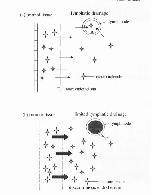
The EPR effect has been studied in depth by Maeda and co-workers, and is based on the fact that many different macromolecules passively accumulate in solid tumours (reviewed in Maeda and Matsumura, 1989). This phenomenon has been attributed to two factors; the discontinuous endothelium as often displayed by tumour vasculature which allows macromolecular extravasation to a greater extent than seen with most other endothelial barriers, and additionally lack of effective lymphatic drainage in tumours preventing clearance of extravasating macromolecules resulting in tumour accumulation by passive diffusion (reviewed in Maeda, 1994). This effect has been documented in many solid tumours, and it is now widely accepted that this phenomenon is crucial to the observed pharmacological effect of many macromolecular drug delivery systems (Duncan, 1992). A schematic of this concept can be seen in figure 1.2.
1.5 Endocytosis and Intracellular Trafficking in the Context o f Drug Delivery
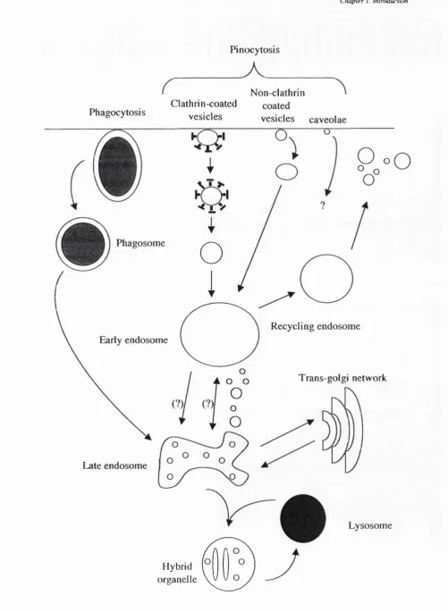
Understanding the endocytic pathway in cells and the complex mechanisms involved in the intracellular transport of internalised membrane components is paramount if efficient drug delivery systems utilising the endocytic pathway are to be successful. Although some vesicle trafficking events still remain controversial, there is fundamental agreement on the most important features of the pathway which will be described below.
All cells internalise extracellular material during the process of endocytosis. This can broadly be divided into two categories;
(1) Phagocytosis (cell eating), refers to the binding and internalisation of large (>0.75/im diameter) particles such as apoptotic cells. This mechanism was mainly established by the pioneering work of Elie Metchnikoff in the 1880’s. (A detailed review on phagocytic mechanisms is beyond the scope of this thesis).
C hapter /. Introduction
(a) normal tissue
lymphatic drainage
lymph node
macromolecule
— intact endothelium
(b) tumour tissue
limited lymphatic drainage
lymph node
i f
macromolecule
discontinuous endothelium
Chapter 1. Introduction
(a) Receptor-mediated pinocytosis
D
0
Clathrin-coated pit
(b) Non-specific adsorptive pinocytosis
@ 0
Negatively charged cell membrane
(c) Fluid-phase pinocytosis
Figure 1.3 Diagram showing the three categories of pinocytosis. Panel (a) shows receptor-mediated pinocytosis: receptor ligands bind to cell-surface receptors and are concentrated on the clathrin-coated pit; (b) Non-specific adsorptive pinocytosis:positively charged molecules bind electrostatically to the negatively charged cell membrane; (c) fluid-phase pinocytosis: non-diffusable uncharged molecules are intemaliscdAs a result of ongoing n i^ b r a n e invagination
Chapter 1. Introduction
(1) Fluid-phase pinocytosis is a constitutive phenomenon that is characterised by ongoing membrane invagination and capture of any non-diffusable molecule present in the extracellular medium.
(2) Non-specific adsorptive pinocytosis is mediated via the binding of a molecule to the cell surface non-specifically by ionic interactions between the molecule and theplasma membrane or through hydrophobic/hydrophilic interactions between non-polar moieties and the lipid bilayer. As a result of membrane binding, non specific absorptive pinocytosis is more efficient at substrate capture than fluid- phase.
(3) Receptor-mediated pinocytosis requires the presence in the plasma membrane of specific receptors that link the molecule destined to be endocytosed. As a large number of receptors can be concentrated in localised regions along the plasma membrane, this can be a very efficient method of ligand uptake.
Different types of endocytic pathways are known to exist within the cell to mediate the processes of non-specific absorptive, receptor-mediated and fluid-phase pinocytosis. These pathways will be discussed below.
Clathrin-dependent pinocytosis
The best-defined pathway of extracellular fluid and receptor-bound ligand uptake is the clathrin-coated vesicle (CCV) pathway. CCVs are found in virtually all cell types and form from domains at the plasma membrane known as clathrin-coated pits. CCVs comprise a protein clathrin coat, which forms lattices of hexagons and pentagons, forming a cage-like structure. These coats are linked to a heterotetrameric adaptin complex known as AP-2 (reviewed in Clague, 1998). AP-2 provides two functions; firstly it attaches clathrin to membranes via interaction of AP-2 with a high affinity receptor (Zhang et al, 1994), and secondly it recruits cargo-bearing receptors (bearing the appropriate signals) that are destined for incorporation into CCVs (Ohno et al,
1995). Invagination of CCVs occurs at the plasma membrane, under the control of a number of accessory/regu 1 atory proteins until a mature vesicle is ‘pinched o ff the
C h apter I. Introduction
Caveolae
Caveolae are small (~ 50nm-80nm diameter) specialised invaginations of the plasma membrane seen in many cell types, especially capillary endothelial cells, smooth muscle cells and adipocytes (Peters et al, 1985). The cholesterol-binding protein caveloin 1 (Vip21) is required for caveolae formation (Peters et al, 1985; Rothberg et al, 1992). In endothelium, the formation of caveolae is associated with the transcytosis of solutes across the cell, however, the contribution of caveolae to total endocytic activity is unclear (reviewed in Mellman, 1996).
Clathrin-independent pinocytosis
There is increasing evidence for pinocytosis that is independent of clathrin (Lamaze and Schmid, 1995). Although caveolae formation is a dynamin-dependent, clathrin-independent form of pinocytosis, studies have shown that another type of clathrin-independent pinocytosis can also occur, which is not dependent on dynamin, although the mechanisms concerning this pathway are not clear.
Vesicles formed at the cell surface undergo a series of fusion events with other organelles during the process of pinocytosis. The principal components of these intracellular sorting compartments are discussed below.
Endosomes
Early endosomes are often located on the cell periphery and receive most types of vesicles coming in from the cell surface. Although the initial sorting event occurs at the plasma membrane, many more sorting events occur in sorting endosomes, including the discharge of internalised ligands from their receptors, targeting of membrane proteins to their correct plasma membrane domains and transfer of ligands to late endosomes or back to the plasma membrane via recycling endosomes (reviewed in Mellman, 1996).
C hapter I. Introduction
higher pH values, in the range 6.4 6.5 depending on the cell type (reviewed in Mukheijee et al, 1997).
Late endosomes
Late endosomes are defined as vesicular structures that accumulate and concentrate internalised contents after passage through early endosomes, and are thought to mediate a final set of sorting events prior to delivery of material to lysosomes (reviewed in Mellman 1996; reviewed in Clague, 1998). These endosomes contain primary inward invaginations of the membrane (known as multi-vesicular bodies) and contain the highest concentration of the cation-independent mannose 6-phosphate
receptor, and also degradative enzymes. Late endosomes undergo extensive vesicular traffic with the Trans Golgi network to acquire these newly synthesised lysosomal enzymes and to return the mannose 6-phosphate receptor back to the Trans Golgi
network. These vesicles have a lower pH than early endosomes, characteristically in the range 5.0-6.0, as they contain degradative enzymes with pH optima within this range (reviewed in Mukheijee et al, 1997).
Lysosomes
Transfer of material between late endosomes and lysosomes appears to be a direct fusion event resulting in a hybrid organelle (Luzio et al, 2000). Lysosomes are distinguished from late endosomes by virtue of their higher density on Percoll gradients and their lack of the cation-independent mannose 6-phosphate receptor. A lower
lumenal pH of around 4.5-5.5 exists in these organelles (Asokan and Cho, 2002). Lysosomes represent the final step of traffic for macromolecules, as once transferred to this compartment, they are digested by a battery of lysosomal enzymes to small molecules (typically < IkDa). As a result of this digestion, the luminal concentration of nutrients rises above that of the cytoplasm. This leads to a concentration gradient across the vacuolar membrane which drives diffusion of these small molecules through specific channels or carriers in the lysosome membrane. These small molecules (amino acids, sugars) are then utilised in the cytoplasm for cell sustenance and growth (reviewed in Mukheijee et al, 1997).
C hapter /. Introduction
Pinocytosis
Phagocytosis
Non-clathrin Clathrin-coated coated
vesicles vesicles caveolae
Late endosome
Hybrid organelle
Recycling endosome Phagosome
Early endosome
P„»o
O
Trans-golgi network
Lysosome
CItapier I. Introduction
enzymes that may degrade therapeutic substances which are trafficked to the lysosome. In addition, the physical constraint of crossing the membranes of the endocytic pathway have also been reported to limit cytosolic access (Klemm et al, 1998; Lucas et al, 1999; Kircheis et al, 2001a).
1.5.1 Intracellular Trafficking o f Toxins
The mechanisms of intracellular trafficking of ricin and gelonin toxins will be discussed below and are of relevance to this thesis for a number of reasons: a) conjugates of these toxins with antibodies against tumour cell-surface antigens are currently being investigated as potential intracytoplasmic delivery systems (section
1.6); b) some plant and bacterial toxins are able to inherently enter the cytoplasm, and
understanding the mechanism of cytosolic access has implications for the future design of intracytoplasmic delivery systems; c) both RTA and gelonin have been delivered to the cytosol using endosomolytic systems.
The plant toxin ricin, has been particularly well documented as a marker for endocytosis and intracellular transport steps (reviewed in Sandvig and van Deurs, 1996). This protein (derived from Ricinus communis), is a highly cytotoxic, dimeric glycoprotein composed of RTA with molecular weight ~ 29,500 and a B-chain (RTB) of molecular weight 34,000, which are linked by a disulphide bridge (Lord et al, 1994).
RTB is the cell-binding moiety, which acts to promote binding and endocytosis of the protein by binding to glycolipids and glycoproteins with terminal galactose residues. The holotoxin is then taken up by both clathrin-dependent and independent endocytosis with about 95% of ricin subsequently transported to lysosomes. Of the remaining amount, about 75% is transported to the Golgi apparatus (reviewed in Sandvig and van Deurs, 1996). Once inside the cell, the toxin undergoes retrograde transport from the Golgi to the endoplasmic reticulum, where it is thought that the inter chain disulphide bond linking the A- and the B-chains is broken. Free RTA, the catalytic moiety, then interacts with Sec61p translocon and is perceived as a substrate for the ER-associated protein degradation pathway. It is then translocated to the cytosol in a partially unfolded state (Argent et al, 2000). Once in the cytosol, ribosomal inactivation occurs by cleavage of the N-glycosidic bond of adenosine"^^^"^ nucleoside in 28S RNA of the 60S ribosomal subunit, leading to inhibition of protein synthesis (Endo et al, 1987, Simpson et al, 1999). RTA alone displays a much higher IC50 value in vitro
C hapter I. Introduction
Gelonin is another ribosome inactivating toxin derived from the seeds of
Gelonium multiflorum. This toxin consists of a single polypeptide chain with 251 amino acid residues with molecular weight 30,500 (Stirpe et al, 1980). It has similar properties to RTA, by catalytically inactivating the 60S subunit of ribosomes in eukaryotic cells, however this only occurs in cell free systems as gelonin lacks the cell- binding domain equivalent to RTB. As a result of this, gelonin is non-toxic to intact cells (Stirpe, 1980; Selbo et al, 2000a), due to its poor uptake and subsequent trafficking to lysosomes.
As cytosolic access of macromolecules such as proteins (toxins) and genes is crucial for many therapeutic strategies (reviewed in Plank et al, 1998, Lackey et al, 1999a), there is a requirement for an intracytoplasmic delivery vehicle to mediate membrane translocation and avoid lysosomal degradation (Murthy et al, 1999, Mônkkônen and Urtti, 1998). The challenges faced by a potential intracytoplasmic delivery system in terms of use of these toxins and other therapeutic strategies will be discussed below.
1,6 Challenges o f Intracytoplasmic Delivery
The most widely researched therapeutic strategy which requires the intracytoplasmic delivery and nuclear trafficking of macromolecules is gene therapy. The classical definition of gene therapy is defined as the transfer of genetic material into cell nuclei to alter their function, usually for the benefit of the entire organism (Godbey and Mikos, 2001). This concept was first envisioned around 13 years ago and since then this field has expanded (encouraged by the recent completion of the Human Genome Project). Although gene therapy was initially envisioned for the treatment of monogenic disorders such as cystic fibrosis (Bellon et al, 1997), it is also currently being studied for a wide range of diseases including cancer, arthritis, neurodegenerative disorders and infectious diseases such as acquired immune deficiency syndrome (AIDS) (reviewed in Mhashilkar et al, 2001).
Cltapter I. Introduction
endocytosed and trafficked to lysosomes where again degradation will occur (reviewed in Wattiaux et al, 2000, Murthy et al, 1999).
Increasing stability of DNA to nucleases has been addressed by synthesising oligonucleotide analogues (phosphorothioate oligomers, methylphosphonate oligonucleotides, morpholino-oligonucleotides and peptide nucleic acids) (reviewed in Lebedeva et al, 2000; Hamilton et al, 1999), which are reported to resist rapid nuclease degradation. However, these analogues still require an intracytoplasmic delivery system, because when administered alone they display a similar pharmacokinetic profile as DNA (reviewed in Dokka et al, 2000, reviewed in Wattiaux et al, 2000).
At present, a safe, efficient vector capable of delivering DNA to the nucleus has yet to be synthesised (Brown et al, 2001). The main reason for this is because a non- viral intracytoplasmic gene delivery system requires many complex features and intracellular trafficking steps in order to deliver its payload to the nucleus. These requirements are set out in table 1.3. Although it is not unfeasible that a vector which possesses all these characteristics will eventually be developed, it is likely that it will involve a multi -component system to accommodate all these features.
Ribozymes are ribonucleic (RNA) structures that catalyse the cleavage of many forms of RNA (reviewed in Helene and Toulme, 1990). Their ability to cleave and deactivate messenger RNA (mRNA) means that they can be used to down-regulate gene expression. Ribozymes have advantages over conventional DNA delivery because they are smaller in size than plasmids and do not require delivery into the nucleus to exert biological activity (Merdan et al, 2002a). Thus, intracytoplasmic delivery vehicles are attractive agents for their delivery.
In addition to DNA and RNA, many therapeutic proteins require intracytoplasmic delivery (Lackey et al, 1999a). Research groups are currently examining using immunotoxins as a new approach to cancer chemotherapy. Immunotoxins are comprised of a protein toxin connected to a binding ligand such as an antibody or growth factor. These molecules then selectively bind to cells bearing the target antigen and antibody internalisation occurs.
C hapter I. Introduction
Table 1.3 Characteristics required for a non-viral intracytoplasmic gene delivery system
Property Reference
Protection of DNA from degradation by extracellular Godbey and Mikos, 2001; nucleases Zelphati et al, 1996; Wattiaux
et al, 2000 Appropriate organ and cell targeting/biodistribution Lui et al, 1997
Minimal toxicity to target cells Godbey and Mikos, 2001 Endocytic uptake/membrane fusion Zelphati et al, 1996
Exit from the endosome Kyriakides et al, 2002; Wattiaux et al, 2000 Release of DNA from complex Lucas et al, 1999
Nuclear trafficking/traversal through the nuclear pore Lucas et al, 1999; reviewed in Tachibana et al, 2001, Kamiya et al, 2001; reviewed in
Pouton, 1998; reviewed in Zuber et al, 2001
C h aplrr I. Introduction
displayed by certain toxins. Probably the most common limitation to immunotoxin therapy which confers dose-limiting toxicity is Vascular Leak Syndrome (VLS) (Baluna et al, 2000; reviewed in Kreitman, 1999) which is caused by toxin-mediated damage to endothelial cells. The problems associated with immunotoxin therapy leave a niche in the market which intracytoplasmic delivery vehicles could utilise. Degradation of the conjugate in lysosomes has been shown to be a rate-limiting step in immunotoxin therapy, which would not occur for intracytoplasmic delivery systems which lyse endomembranes before degradation occurs. Additionally, an intracytoplasmic delivery system alleviates the need for naturally cell permeant proteins, thus leaving a much wider choice of cytotoxic molecule to be delivered. Finally, chemical conjugation of a synthetic polymer to a protein such as a toxin can reduce its immunogenicity (Ulbrich et al, 2000; Seymour et al, 1991; Duncan, 2000).
Discussed below are the intracytoplasmic delivery systems currently being developed to utilise these therapeutic strategies. To date, the majority of these are being developed for gene delivery and therefore this will be the main focus of this section. It must be noted that as well as intracytoplasmic delivery vehicles, physical methods are also being developed for the delivery of genes into cells, which are not directly relevant to this thesis. These include electroporation, particle bombardment, microinjection and calcium phosphate precipitation (for reviews see Dokka and Rojanasakul, 2000; Chang,
1994).
1.7 Intracytoplasmic Delivery Systems
1.7.1 Nan-Polymeric Intracytoplasmic Delivery Systems 1.7.1.1 Viral Vectors
In order to take advantage of the natural processes of viral transfection, modified viral vectors are currently being developed as gene delivery vehicles because they display high levels of transgene expression (Robins et al, 1998). Among these, retrovirus and adenovirus are the two most commonly used viral vectors that have been tested in phases I/II clinical trials.
d t a p te r I. Introduction
associated with these vectors are addressed. Overlooking these problems led to the death of a research subject caused by a severe adverse reaction to a dose of over trillion modified adenovirus particles (Kielian and Hickey, 1999). For these reasons, an increasing amount of research has been focused towards developing non-viral systems for gene delivery.
7.7.2.2 Cationic lipids and liposomes
The potential of liposomes as drug carriers was first realised 25 years ago due to their ability to entrap solutes (reviewed in Gregoriadis et al, 1976). Hydrophilic drugs can be entrapped in the aqueous channels separating the hydrophobic lipid bilayers or in the liposomal lumen. Hydrophobic drugs can be sequestered in the acyl chains of phospholipid molecules within the bilayer (reviewed in Gregoriadis, 1990).
Oligonucleotides or plasmid DNA can be delivered with liposomes into cells either in encapsulated form or as a lipid-DNA complex (reviewed in Mônkkônen and Urtti, 1998). Of the non-viral gene delivery methods, liposomes have been the most widely tested vehicle to date (reviewed in Mhashilkar et al, 2001; Mônkkônen and Urtti, 1998). There are three common types of cationic lipids employed in lipid-based DNA delivery which are described below.
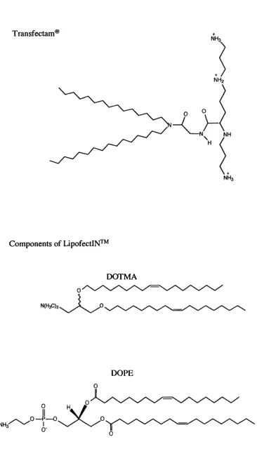
a) Lipids containing quaternary ammonium salts with long mono-unsaturated aliphatic chains, e.g. (N[l-(2,3-dioleyloxy)propyl]-7/,A,N-trimethyl ammonium) (DOTMA). A 1:1 mixture of DOTMA and the fusogenic lipid dioleoyl phosphatidylethanolamine (DOPE) make up the commercially available transfection agent LipofectIN™ (Feigner et al, 1987).
b) Cationic derivatives of cholesterol e.g. 3P-(N-(N-(dimethylaminoethane)carbamoyl) cholesterol (Dc-Chol).
c) Lipids with multi valent headgroups (lipospermines) e.g. dioctadecy 1 amidoglycylspermine (DOGS). This is available commercially as Transfectam® (Remy et al, 1998).
C hapter /. Introduction
Transfectam®
NH3
NH2
Components of LipofectlN^^
NH
NH3
DOTMA
NCHaOs
DOPE
NH3"
C hapter 1. Introduction
1996). Much work has been done to elucidate the mechanism of subsequent oligonucleotide release from cationic lipids once in the endosome. It has been proposed that cationic lipid fusion with the endosomal membrane occurs as a result of initial endosomal membrane destabilisation resulting in flip-flop of the anionic lipids located on the cytoplasmic face of the membrane. Membrane destabilisation could be initiated by close proximity of cationic and anionic lipids and the high radius of membrane curvature found in intracellular vesicles (Xu and Szoka, 1996). Anionic lipids can then diffuse laterally into the complex to form charge-neutralised ion-pairs with the cationic lipids. Neutralisation releases the oligonucleotide from its electrostatic interaction with lipids leading to its release into the cell cytoplasm (Zelphati et al, 1996).
Many cationic lipid formulations include the neutral co-lipid DOPE to promote membrane fusion (Mônkkônen and Urtti, 1998). This fusogenic lipid forms a stable lipid bilayer at pH 7, while in the acidic pH range of 5-6, a hexagonal-II structure is formed, which destabilises the membrane (reviewed in Kamiya et al, 2001).
In order to design more effective lipic vectors, lipid formulations have included cholesterol (Gao and Hui, 2001), PEG (reviewed in Kilbanov et al, 1990) and amphipathic peptides (Kichler et al, 1997). Additionally it was found that altering the liposome composition can result in pH-sensitive liposomes designed to undergo efficient fusion within the acidic environment of the endosome (Chu et al, 1990). Targeted liposomes have also been designed, which include immunoliposomes targeted to tumour cells over-expressing antigens (reviewed in Lebedeva et al, 2000).
A main disadvantage associated with DNA/lipid complexes includes instability in the extracellular environment due to interaction with serum proteins, leading to premature release of DNA (Zelphati et al, 1996; Moret et al, 2001). Serum protein interaction is most substantial with cationic liposomes, with opsonization of the liposome surface resulting in uptake of the complexes by the RES (Mônkkônen and Urtti, 1998; Finisinger et al, 2000; reviewed in Mahato et al, 1995).
1.7,1.3 Thiol-activated cytolysins
C hapter I. Introduction
oligomerisation of approximately 50 monomers to form pores in the lipid bilayer (reviewed in Provoda and Lee, 2000).
These transmembrane channels have been utilised for the delivery of antisense oligonucleotides to human leukaemia KYOl cells using the pore-forming toxin, Streptolysin-O (Giles et al, 1998). Although an antisense effect was seen, it is questionable as to whether the pore formation is reversible, as a non-reversible effect would ultimately result in cell death.
1.7,1.4 Photosensitizers
(AlPcS2a) is a photosensitive compound that is able to localise and rupture
endosomes and lysosomes upon light exposure. This novel method is denoted photochemical internalisation (PCI) and involves the release of highly reactive oxygen species upon light exposure, which are able to destroy membranes (Selbo et al, 2000a).
Photochemical treatment of human melanoma cells after incubation of (AlPcSza) substantially improved the efficiency of transfection mediated by poly-L-lysine and PEI (Prasmickaite et al, 2000). However, the inclusion of cationic polymers to complex and prevent premature degradation of the DNA has drawbacks in terms of vector toxicity and intravenous administration. In addition, the requirement of light exposure to induce membrane rupture limits this technique to in vitro mechanistic studies.
PCI has recently been utilised to demonstrate the translocation of gelonin toxin to the cytosol from endosomes and lysosomes. This study demonstrated the importance of endosomal escape, as in the absence of inhibition of endosome to lysosome transport, degradation of gelonin occurred prior to cytosolic translocation (Selbo et al, 2000a).
1.7.2 Polymeric Intracytoplasmic Delivery Systems 1.7.2.1 Cationic polymers
A variety of polymeric vectors are also currently being investigated as gene- delivery vectors. The advantages of using polymeric systems has already been discussed in section 1.3. Among these vectors, cationic polymers such as PEI, chitosans, poly-L-lysine, and PAMAM dendrimers have been the most widely documented (reviewed in Garnett, 1999). Although many of these polymers are not strictly endosomolytic, details of their mechanism of action will be discussed before the focus is moved to endosomolytic vectors.
C hapter !. Introduction
with cationic lipid-mediated gene delivery, condensation protects the DNA from degradation by nucleases and results in compact particles which can be taken up by cells via endocytosis. The positive surface charge of polycation/DNA complexes produced with high polycation/DNA ratios usually serves to bind cells via electrostatic interactions to sulphated proteoglycans on the cell membrane (Mislick and Baldeschwieler, 1996), which could be essential for complex internalisation.
In vitro cationic polymers are membranolytic (Nevo et al, 1955; reviewed in Garnett, 1999), and thus could act upon the endosomal membrane causing destabilisation leading to release of the polyplex into the cytosol. In practise, however, cell membrane destabilisation also occurs, resulting in concentration-dependent cytotoxicity for many cationic polymers (Godbey and Mikos, 2001). IC50 values for the
most widely used cationic gene delivery polymers are shown in table 1.4.
Efficiency of transfection is probably dependent on the rate of intracellular degradation of plasmid DNA (reviewed in Wattiiaux et al, 2000) and for many polyplexes, inability to promote endosomal membrane destabilisation results in degradation in the lysosome (Lucas et al, 1999). Many groups include membrane destabilising compounds such as fusogenic peptides in the transfection mixture which promote endosomal membrane destabilisation (Kircheis et al, 2001a). In addition weak bases such as chloroquine have been included during transfection which can increase transfection efficiency (Lucas et al, 1999).
C hapter 1. Introduction
Table 1.4 Cytotoxicity of polymeric gene delivery vehicles
Polymer Molecular weight
(kDa)
IC50
value (jiig/ml)
Cell lines Incubation time
(h)
Reference
PLL 56.5 50 L132 72 Richardson et al, 1999a, 56.5 50 CCRF 72 Richardson et al, 1999a 56.5 50 B16F10 72 Ferruti et al, 2000
25 40 HepG2 4 Choi et al, 1998
PEI 25
(branched)
6 293T 2h Lee et al, 2001 25
(branched)
40 NIH 3T3 48 Jeong et al, 2001
DMABMA 550 4.5 OVCAR 48 Van de Weltering et al, 1999
Chitosan 5 > 1,000 L132, 72 Richardson et al, 1999b 5 > 1,000 CCRF 72 Richardson et al, 1999b
p-cyclodextrin
6.1 380 BHK-21 24 Hwang et al, 2001
C hapter 1. Introduction
introduce cell-specific targeting and to improve efficiency of uptake into cells.
1.7,2.2 Endosomolytic Polymers
Endosomal escape has been documented to be a major barrier to non-viral mediated gene delivery. In the recent years, a number of polymeric systems have been developed with the ability to lyse endosomal membranes. A schematic demonstrating the advantages of endosomolytic delivery is shown in figure 1.6. A brief review of endosomolytic polymers is discussed below and their structures can be seen in figure
1.7.
1.7.2.2.1 Polyethylenimine (PEI)
PEI is a polymer that has been used for years in the paper industry and in water purification (reviewed in Godbey et al, 1999a, reviewed in Kircheis et al, 2001a). The polymer, made up of an ethylamine repeating unit, is available in two main forms, branched or linear. The most important feature of this polymer is its high cationic charge density, as every third atom is a potentially protonable amino nitrogen, and the overall protonation level increases from 20-45% between pH values of 7 and 5 (Remy et al, 1998). Endosomal membrane rupture has been proposed by Behr and colleagues to occur as a result of the proton sponge mechanism, although evidence and understanding of the release mechanism of PEI from endosomal/1 ysosomal compartments is limited (Godbey et al, 1999a; Godbey and Mikos, 2001; Godbey et al, 1999b). This mechanism is thought to occur as follows; within endosomes, the influx of protons by v-ATPases is coupled to an accumulation of chloride ions. In the presence of PEI, a net increase in the ionic concentration occurs within the endosome due to the buffering capacity of PEL In response to this, polymer swelling occurs due to internal charge repulsion. In addition, influx of water into the endosome occurs due to the increase in ionic concentration. It is proposed that these two mechanisms occurring simultaneously lead to endosomal rupture. A schematic of this mechanism is shown in figure 1.8.
Previously, PEI was shown to disrupt isolated rat liver lysosomes in an in vitro
C hapter I. Introduction
Intracellular trafficking of macromolecular drug
Intracellular trafficking of Drug-endosomolytic complex
endosome
/ o
\(b)
...
lysosome
(a) Uptake of macromolecular drug into endocytic vesicle
endosome
lysosome
(d) Uptake of macromolecular drug- endosomolytic polymer complex into endocytic vesicle
(b) Trafficking of drug to endosome (e) Trafficking of complex to
endosome; polymer-mediated membrane destabilisation occurs
(c ) Trafficking of drug to lysosome where degradation by lysosomal enzymes occurs
(f ) Drug and complex are delivered to cytoplasm, avoiding degradation
Cliapter I. Introduction
Poly(2-di methyl amino ethyl) methacrylate (DMAEMA)
(a) Poly(ethyl acrylic acid) (PEAAc) (b) Poly (propyl acrylic acid) (PPAAc)
a
-Poly(ethyleneimine) (PEI) (a) branched; (b) linear
W
h
/
OH
H ■N‘
N
(b)
N.
N ‘
N— C — C — h
H
Hg
Hg
H N.
-N
N ‘
C hapter 1. Introduction
Endosome
H,0
V-ATPase
PEI is endocytosedC l
pH < 7.0
PEI buffers vesicle acidification and PEI swells due to intra-chain repulsion.The influx of ions is coupled to an influx of Cl ions which results in a influx of HjO.
pH = 4.5-5.0
Vesicle bursts due to influx of HjO and ! polymer swelling