International Journal of Nanomedicine
Histologic and apoptotic changes induced by
titanium dioxide nanoparticles in the livers of rats
Saud Alarifi1 Daoud Ali1
Amin A Al-Doaiss1,2 Bahy A Ali1,3
Mukhtar Ahmed1
Abdulaziz A Al-Khedhairy1
1Department of Zoology, College of Science, King Saud University, Riyadh, Saudi Arabia; 2Anatomy and Histology Department, Faculty of Medicine, Sana’a University, Sana’a, Republic of Yemen; 3Genetic Engineering and Biotechnology Research Institute, City for Scientific Research and Technology Applications, Alexandria, Egypt
Correspondence: Daoud Ali Department of Zoology, College of Science, King Saud University, PO Box 2455, Riyadh 11451, Kingdom of Saudi Arabia Tel +966 5 5890 4621 Fax + 966 11 467 8514 Email [email protected]
Abstract: Titanium dioxide (TiO2) nanoparticles are among the top five nanoparticles used in consumer products, paints, and pharmaceutical preparations. Given that exposure to such nanopar-ticles is mainly via the skin and inhalation, the present study was conducted in male Wistar albino rats (Rattus norvegicus). Our aim was to investigate the effect of TiO2 nanoparticles on hepatic tissue in an attempt to understand their toxicity and the potential effect of their therapeutic and diagnostic use. To investigate the effects of TiO2 nanoparticles on liver tissue, 30 healthy male Wistar albino rats were exposed to TiO2 nanoparticles at doses of 63 mg, 126 mg, and 252 mg per animal for 24 and 48 hours. Serum glutamate oxaloacetate transaminase and alkaline phosphatase activity was altered. Changes in hepatocytes can be summarized as hydropic degeneration, cloudy swelling, fatty degeneration, portal and lobular infiltration by chronic inflammatory cells, and congested dilated central veins. The histologic alterations observed might be an indication of hepatocyte injury due to the toxicity of TiO2 nanoparticles, resulting in an inability to deal with accumulated residues from the metabolic and structural disturbances caused by these nanoparticles. The appearance of cytoplasmic degeneration and destruction of nuclei in hepatocytes suggests that TiO2 nanoparticles interact with proteins and enzymes in hepatic tissue, interfering with antioxidant defense mechanisms and leading to generation of reactive oxygen species which, in turn, may induce stress in hepatocytes, promoting atrophy, apoptosis, and necrosis. More immu-nohistochemical and ultrastructural investigations are needed in relation to TiO2 nanoparticles and their potential effects when used as therapeutic and diagnostic tools.
Keywords: TiO2 nanoparticles, rats, liver, histology, TUNEL assay, alkaline phosphatase, apoptosis
Introduction
Nanotechnology is a promising new field with potential applications in the domestic, industrial, and biomedical fields.1 Due to the growing number of applications, there
is an increasing risk to animals from environmental exposure to nanomaterials. Their potential toxicologic impact is still under investigation and our actual knowledge about the effects of nanosized contaminants on biological systems remains incomplete.2,3
These effects need to be assessed in order to provide a scientific basis for safe develop-ment of nanotechnologies. Use of nanotechnology has seen exponential growth in the areas of health care, consumer products, clothes, electronics, and sporting goods.2 This
is due to the unique chemical, mechanical, optical, magnetic, and biological properties of nanomaterials that make them desirable for commercial and medical applications.4
According to a recent survey, the number of nanotechnology-based consumer products available on the world market now exceeds 1000.5 TiO
2 nanoparticles have several
Dove
press
O R I G I N A L R E S E A R C H
open access to scientific and medical research
Open Access Full Text Article
International Journal of Nanomedicine downloaded from https://www.dovepress.com/ by 118.70.13.36 on 23-Aug-2020
For personal use only.
Number of times this article has been viewed
This article was published in the following Dove Press journal: International Journal of Nanomedicine
industrial applications, and as such, come in different sizes, shapes, chemical compositions, and crystalline structures.6
TiO2 occurs in four crystalline polymorphic forms, of which rutile and anatase are the most common.7 Rutile is considered
to be a more inert form whereas anatase is an active form of TiO2. Some studies indicate that anatase TiO2 nanoparticles are more cytotoxic than rutile TiO2 nanoparticles.8 Yeo et al9
have reported that different TiO2 nanoparticles induce differ-ent toxicities during embryogenesis of the zebra fish due to their different sizes and crystalline phases. The increased use of nanoparticles is a matter of great concern to health profes-sionals and environmental scientists because of the potential risks to humans and to the environment.10 Of the possible
exposure routes, inhalation and skin contact are considered to be the most important for nanoparticles. The toxic effects of nanoparticles can be attributed to their small size and hence large surface area, which increases their chemical reactivity and penetration into living cells.11 TiO
2 nanoparticles also
induce reactive oxygen species, leading to toxicity.12
The dimensions of TiO2 nanoparticles are critical from the toxicity point of view, given that TiO2 nanoparticles have more pronounced toxicity than conventional TiO2 particles.13
TiO2 nanoparticles have been shown to impair the function of macrophages, to cause persistent inflammatory reactions, and to increase pulmonary retention compared with fine TiO2 particles.14 TiO
2 nanoparticles can be absorbed into
the body by inhalation, ingestion, and dermal penetration, and are distributed to important organ systems, including lymph, brain, lung, liver, and kidney.15–17 Of note, it has been
observed in vivo that anatase TiO2 nanoparticles increased inflammatory indicators, cell proliferation, and histopathol-ogy in bronchoalveolar lavage fluid.18
Characteristics of TiO2 nanoparticles can be modified by several methods to improve their performance. Due to their small size, nanoparticles may cross biological barriers to reach a number of organs, and according to their size and surface properties, accumulation of metal nanoparticles has been observed previously in all organs in vivo.19 Generation
of free oxygen radicals and oxidative stress triggers a host of cellular events, including DNA damage and apoptosis.20
Therefore, in the present study, an attempt was made to assess the toxicity and apoptotic potential of TiO2 nanoparticles in the liver tissue of male rats.
Materials and methods
Chemicals and animals
Titanium (IV) oxide (TiO2) nanopowder (99.7% anatase, CAS 1317-70-0) was obtained from Sigma-Aldrich (St Louis,
MO, USA). A terminal transferase-mediated biotinylated 16-deoxy-uridine-triphosphate (dUTP) nick-end labeling (TUNEL) apoptosis detection kit (catalog number L00300) with anti-fluorescein antibody conjugated peroxidase (FITC-labeled POD) for paraffin-embedded tissue sections was purchased from GenScript Biology CRO (Piscataway, NJ, USA). All other chemicals were obtained locally in Saudi Arabia and were of analytical reagent grade. Forty healthy male Wistar albino rats (Rattus norvegicus) aged eight weeks with a mean weight of 126 g were obtained from the Animal Care Center, College of Pharmacy, King Saud University.
Preparation and characterization
of TiO
2nanoparticles
TiO2 NPs were suspended in Milli-Q water (Millipore Corporation, Billerica, MA, USA) at a concentration of 1 mg/mL. A stock suspension was probe-sonicated at 40 W for 15 minutes. Samples for analysis by transmission electron microscopy (TEM) were prepared by drop coating a TiO2 nanoparticle solution on carbon-coated copper TEM grids. The films formed on the TEM grids were allowed to dry prior to measurement. TEM measurements were performed using a JEOL instrument (model 1101F, JEOL Ltd, Tokyo, Japan) operated at an accelerating voltage at 200 kV.
Experimental design
The rats were housed in groups under standard lighting conditions with free access to water and food. Humidity and temperature (22°C ± 1°C) were controlled in ventilated cages on a 12-hour day/night cycle. Five animals from each group were anesthetized and euthanized by cervical dislocation after 24 and 48 hours of exposure to TiO2 nanoparticles. All experiments were conducted in accordance with the guide-lines approved by the local animal care and use committee at King Saud University.
TiO2 nanoparticle doses were calculated based on average body weight.21,22 The study was conducted to
compare the toxicity of the nanoparticles at three different doses. The animals were divided into four groups of ten rats each, which were injected intraperitoneally as follows for two days:
• group 1, normal animals, injected with Milli-Q water
• group 2, injected with 63 mg of TiO2 nanoparticles per animal
• group 3, injected with 126 mg of TiO2 nanoparticles per animal
• group 4, injected with 252 mg of TiO2 nanoparticles per animal.
Dovepress
Alarifi et al
International Journal of Nanomedicine downloaded from https://www.dovepress.com/ by 118.70.13.36 on 23-Aug-2020
Histopathology
The animals were exposed to different doses of TiO2 nanoparticles intraperitoneally for 24 and 48 hours. After euthanizing the animals, fresh portions of the lateral lobes of the liver from each rat were cut rapidly, fixed in neutral buffered formalin (10%), then dehydrated using grades of ethanol (70%, 80%, 90%, 95%, and 100%). Dehydration was followed by clearing the samples in two changes of xylene. The samples were then impregnated with two changes of molten paraffin wax, embedded, and blocked out. The tis-sue sections (4–5 µm) were stained according to the method described by Bancroft and Stevens23 using conventional
histologic stains. Stained sections from the control and treated rats were observed and photographs were taken using an optical microscope (Olympus, Tokyo, Japan) for alterations in architecture, hepatocytes, and sinusoids, and for the presence of degeneration, necrosis, fatty changes, and portal fibrosis.
Estimation of GOT and ALP
After exposure for 48 hours, blood samples were taken from five rats per group, and the serum was separated out for estimation of alkaline phosphatase (ALP) and glutamate oxaloacetate transaminase (GOT) activity using already reported methods.24,25
TUNEL assay
Formalin-fixed, paraffin-embedded tissue sections were dewaxed in xylene, rehydrated through graded ethanol, and pretreated with proteinase-K 20 µg/mL in phosphate-buffered saline for 15 minutes at 37°C in a humidified chamber. After washing twice in distilled water and rinsing in Tris-buffered saline (pH 7.6), the sections were incubated for 60 minutes at 37°C with a reaction mixture containing 0.3 U/µL terminal deoxynucleotidyl transferase (GenScript),
terminal transferase buffer (GenScript) 100 mM cacodylate buffer (pH 6.8), 1 mM cobalt chloride, and 0.5 mM DL-dithiothreitol with biotinylated 16-dUTP added (GenScript). The reaction was terminated by rinsing twice in Tris-buffered saline. Next, the sections were covered with 2% bovine serum albumin in Tris-buffered saline for 15 minutes and then incubated with avidin-biotin-conjugated ALP (Gen-Script) at 1:100 for 30 minutes. Staining was done using 5-bromo-chloro-indoxyl phosphate/nitro blue tetrazolium (GenScript) with 1 mM levamisole added to inhibit endog-enous ALP activity. The result imparted an orange/brown color to the nuclei of apoptotic cells.
Positive control slides was created by staining sec-tions of rat liver tissue with 50 µL of DNase I solution (GenScript). For the negative control, terminal deoxynucle-otidyl transferase was omitted from the reaction buffer. The slides for the control and treated rat hepatocytes were observed and the images captured using an optical Olympus microscope.
Statistical analysis
One independent experiment was carried out for evaluation. Data were expressed as the mean ± standard error and tested by one-way analysis of variance. A P-value less than 0.01 was considered to be statistically significant.
Results and discussion
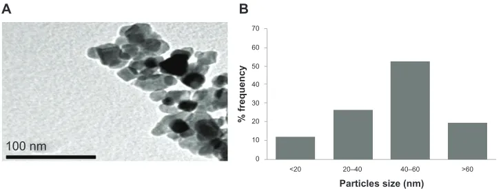
The TiO2 nanoparticles were characterized first for size and found to be in the nanoscale range, but formed small agglomerates in aqueous solution. TEM imaging revealed the morphology of the nanoparticles (Figure 1A). The average size measured by TEM was 50.40 ± 5.60 nm (Figure 1B). The typical TEM image shown in Figure 1A suggests that most of the TiO2 nanoparticles had a polyhedral morphology. Like other materials, when normal-scale TiO2 is converted
100 nm
A B
0
<20 20–40 >60
Particles size (nm)
40–60 10
20 30 40
% frequency
50 60 70
Figure 1 Characterizationof titanium oxide nanoparticles. (A) Transmission electron microscopic image and (B) size distribution histogram generated using transmission
electron micrography.
Dovepress Histologic and apoptotic changes induced by TiO2 nanoparticles
International Journal of Nanomedicine downloaded from https://www.dovepress.com/ by 118.70.13.36 on 23-Aug-2020
into nanoscale TiO2, the physicochemical properties change. The special physicochemical properties of nanoparticles come from their high surface-to-volume ratio. They also have a con-siderable higher percentage of atoms on their surface compared with bulk particles, which makes them more reactive.
We measured blood chemistry parameters, including enzymes, to evaluate organ function in our experimental animals. There was a significant (P , 0.05) increase in GOT and ALP levels (Figure 2). GOT and ALP levels are indicative of the functional efficiency of the liver, and are very sensitive to any disease process of the liver.26 The
his-tologic changes observed in the liver and the accompanying increase in ALP and GOT levels indicate compromised liver function. In comparison with the control group, histologic changes were detected in the liver tissue of rats treated with TiO2 nanoparticles (Figure 3). Apoptosis was also seen in the hepatocytes of rats treated with these nanoparticles. The appearance of inflammatory cells in hepatic tissue suggests that the TiO2 nanoparticles can interact with proteins and enzymes in the interstitial tissue of the liver, interfering with the antioxidant defense mechanism and leading to generation of reactive oxygen species, which in turn may imitate an inflammatory response.27 Distortion and swelling
of hepatocytes together with dilatation of the central vein
and blood sinusoids indicate that these nanoparticles may affect permeability of the cell membrane in hepatocytes and the endothelial lining of blood vessels. Swelling of hepatocytes on exposure to nanoparticles as seen in the present study might lead to adaptation of cell transporters.28
Binucleation is a consequence of cell injury and a type of chromosomal hyperplasia usually seen in regenerating cells.29 Cloudy swelling might be seen as a result of
dis-turbed membrane function, leading to a massive influx of water and sodium due to the effects of nanoparticles. Cell swelling might be accompanied by leakage of lysosomal hydrolytic enzymes, leading to degeneration of the cyto-plasm and macromolecular crowding.30 Hydropic
degenera-tion is a result of ion and fluid homeostasis, and leads to an increase in intracellular water.31 The vacuolated swelling
seen in the cytoplasm of hepatocytes from rats exposed to TiO2 nanoparticles indicates acute liver injury.
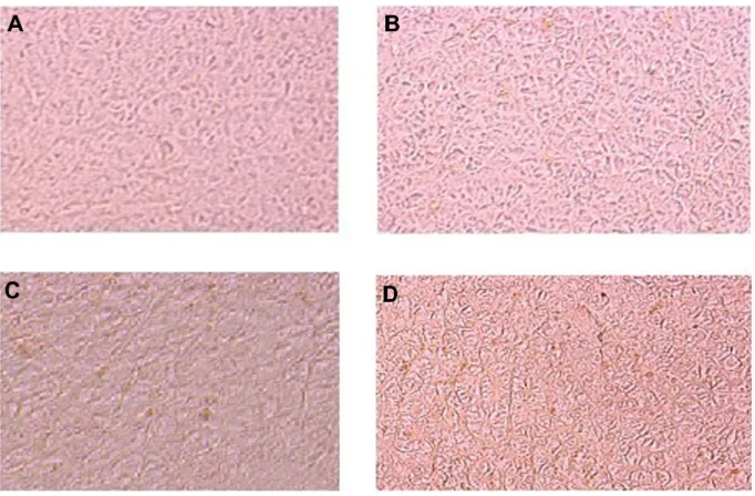
We also observed that apoptosis in hepatocytes exposed to TiO2 nanoparticles increased in a dose-dependent and time-dependent manner (Figure 4). Sporadic, spotty, and well defined necrosis was also noted in some hepatocytes from rats exposed to TiO2 nanoparticles, which might have been because of oxidative stress triggered by depletion of glutathione in these cells. Park et al32 reported that TiO
2
0
* *
* Concentrations (milligram/body weight)
A
Control 63 126
Concentrations (milligram/body weight)
B
252 0.1
0.2 0.3 0.4
ALP levels
0.5 0.6 0.7 0.8 0.9
0 1
0.2 0.4
GOT levels
0.6 1.2
0.8 1.4
Control 63
* *
*
126 252
Figure 2 Levelsof (A) alkaline phosphatase and (B) glutamate oxaloacetate transaminase after exposure of titanium oxide nanoparticles in the different experimental groups
at 48 hours. Each value represents the mean ± standard error of three experiments. *P, 0.05 versus control.
Dovepress
Alarifi et al
International Journal of Nanomedicine downloaded from https://www.dovepress.com/ by 118.70.13.36 on 23-Aug-2020
Figure 3 Lightmicrophotographs of liver tissue.
Notes: (A)Male rats injected with Milli-Q water for 48 hours demonstrating normal histologic architecture. Hematoxylin and eosin, 400×. (B) Male rats 24 hours
post-exposure to titanium oxide nanoparticles (252 mg per animal) by intraperitoneal administration demonstrating lymphocytic infiltration (*) in the hepatic portal space. Hematoxylin and eosin, 400×. (C) Male rats 24 hours post-exposure to titanium oxide nanoparticles (252 mg per animal) by a single oral administration demonstrating marked dilatation of central vein. Hematoxylin and eosin, 400×. (D) Male rats 24 hours post-exposure to titanium oxide nanoparticles (252 mg per animal) by intraperitoneal administration demonstrating marked necrosis (*) and scattered hemorrhages. Hematoxylin and eosin, 400×. (E) Male rats 24 hours post-exposure to titanium oxide nanoparticles (252 mg per animal) by intraperitoneal administration demonstrating dilatation of congested portal vein with edema (*) around the blood vessel in the portal triad. Hematoxylin and eosin, 400×. (F) Male rats 24 hours post-exposure to titanium oxide nanoparticles (252 mg per animal) by intraperitoneal administration demonstrating dilatation and congestion of blood sinusoids (arrows) and binucleation of hepatocytes (circles). Hematoxylin and eosin, 400×. (G) Male rats 48 hours post-exposure to titanium oxide nanoparticles (252 mg per animal) by intraperitoneal administration demonstrating swelling of hepatocytes (arrows) and presence of nanoparticle beneath the capsule (*). Hematoxylin and eosin, 400×. (H) Male rats 48 hours post-exposure to titanium oxide nanoparticles (252 mg per animal) by intraperitoneal administration demonstrating vacuolization of hepatocytes. Hematoxylin and eosin, 400×. (I) Male rats 48 hours post-exposure to titanium oxide nanoparticles (252 mg per animal) by intraperitoneal administration, hydropic degeneration (ballooning) of hepatocytes and presence of nanoparticle in blood sinusoids (arrows). Hematoxylin and eosin, 400×. (J) Male rats 24 hours post-exposure to titanium oxide nanoparticles (126 mg per animal) by intraperitoneal administration demonstrating dilatation of congested portal vein with hemorrhage and edema (*) around the blood vessel and lymphocytic infiltration (arrow) in the portal triad. Hematoxylin and eosin, 400×. (K) Male rats 24 hours post-exposure to titanium oxide nanoparticles (126 mg per animal) by intraperitoneal administration demonstrating focal necrosis (*) and hydropic degeneration of hepatocytes (arrows). Hematoxylin and eosin, 400×. (L) Male rats 48 hours post-exposure to titanium oxide nanoparticles (126 mg per animal) by intraperitoneal administration demonstrating marked dilatation of congested central vein. Hematoxylin and eosin, 400×. (M)
Male rats 48 hours post-exposure to titanium oxide nanoparticles (126 mg per animal) by intraperitoneal administration demonstrating dilatation of congested portal vein with edema (*) around the blood vessel in the portal triad. Hematoxylin and eosin, 400×. (N) Male rats 24 hours post-exposure to titanium oxide nanoparticles (63 mg per animal) by intraperitoneal administration demonstrating marked dilatation of congested central vein. Hematoxylin and eosin, 400×. (O) Male rats 48 hours post-exposure to titanium oxide nanoparticles (63 mg per animal) by intraperitoneal administration demonstrating focal necrosis (*) and hydropic degeneration of hepatocytes (arrows). Hematoxylin and eosin, 400×.
Figure 4 Photomicrographof apoptosis in liver tissue after exposure of titanium oxide nanoparticles. (A) Control, (B) 63 mg per animal, (C) 126 mg per animal, and
(D) 252 mg per animal.
Dovepress Histologic and apoptotic changes induced by TiO2 nanoparticles
International Journal of Nanomedicine downloaded from https://www.dovepress.com/ by 118.70.13.36 on 23-Aug-2020
nanoparticles induced oxidative stress and apoptosis in cultured BEAS-2B cells. In the present study, after intra-peritoneal administration of high-dose TiO2 nanoparticles, the difficulty encountered in clearance of these nanopar-ticles in vivo may have resulted in deposition of parnanopar-ticles in the liver and a hepatic lesion.33 The International
Pro-gramme on Chemical Safety34 shows that most ingested
titanium is excreted via the urine and is not absorbed by the organism. The liver, being the main detoxification organ in the body, is activated to eliminate the side effects induced by the ingested mass of TiO2 nanoparticles, and a proportion of these nanoparticles should be excreted by the kidneys. Because of their small size and difficult clearance, TiO2 nanoparticles were retained in vivo, and liver damage occurred after intraperitoneal exposure to a high dose.
Conclusion
In conclusion, our results indicate that TiO2 nanoparticles induce histologic changes in hepatocytes, which may be mediated by generation of reactive oxygen species and stress to induce atrophy and apoptosis. Long-term biological safety is another issue that will need clarification in future investigations.
Acknowledgment
The authors extend their appreciation to the Deanship of Scientific Research at King Saud University for funding this work through research group project RGP-VPP-180.
Disclosure
The authors report no conflicts of interests in this work.
References
1. Peralta-Videa JR, Zhao L, Lopez-Moreno ML, de la Rosa G, Hong J, Gardea-Torresdey JL. Nanomaterials and the environment: a review for the biennium 2008–2010. J Hazard Mater. 2011;186:1–15.
2. Singh N, Manshian B, Jenkins GJS, et al. NanoGenotoxicology: the DNA damaging potential of engineered nanomaterials. Biomaterials. 2009;30:3891–3914.
3. Skocaj M, Filipic M, Petkovic J, Novak S. Titanium dioxide in our everyday life: is it safe? Radiol Oncol. 2011;45:227–247.
4. Nel A, Xia T, Madler L, Li N. Toxic potential of materials at the nanolevel.
Science. 2006;311:622–627.
5. Project of the Emerging Nanotechnologies 2009. Available from: http:// www.nanotechproject.org. Accessed June 14, 2013.
6. Li JJ, Muralikrishnan S, Ng CT, Yung LY, Bay BH. Nanoparticle-induced pulmonary toxicity. Exp Biol Med (Maywood). 2012;235:1025–1033. 7. Madl AK, Pinkerton KE. Health effects of inhaled engineered and
inci-dental nanoparticles. Crit Rev Toxicol. 2009;39:629–658.
8. Hirakawa K, Mori M, Yoshida M, Oikawa S, Kawanishi S. Photo-irradi-ated titanium dioxide catalyzes site specific DNA damage via generation of hydrogen peroxide. Free Radic Res. 2004;38:439–447.
9. Yeo MK, Kang M. The biological toxicities of two crystalline phases and differential sizes of TiO2 nanoparticles during zebra fish embryogenesis development. Mol Cell Toxicol. 2012;8:317–326.
10. Oberdorster G, Oberdorster E, Oberdorster J. Nanotoxicology: an emerging discipline evolving from studies of ultrafine particles. Environ Health Perspect. 2005;113:823–839.
11. Pan Z, Lee W, Slutsky L, Clark RA, Pernodet N, Rafailovich MH. Adverse effects of titanium dioxide nanoparticles on human dermal fibroblasts and how to protect cells. Small. 2009;5:511–520. 12. Barnard AS. One-to-one comparison of sun screen efficacy, aesthetics
and potential nanotoxicity. Nat Nanotechnol. 2012;5:271–274. 13. Driscoll KE, Maurer JK. Cytokine and growth factor release by alveolar
macrophages: potential biomarkers of pulmonary toxicity. Toxicol Pathol. 1991;19:398–405.
14. Baggs RB, Fern J, Oberdorster G. Regression of pulmonary lesions pro-duced by inhaled titanium dioxide in rats. Vet Pathol. 1997;34: 592–597. 15. Bermudez E, Mangum JB, Wong BA, et al. Pulmonary responses of
mice, rats, and hamsters to subchronic inhalation of ultrafine titanium dioxide particles. Toxicol Sci. 2004;77:347–357.
16. Thomas T, Thomas K, Sadrieh N, Savage N, Adair P, Bronaugh R. Research strategies for safety evaluation of nanomaterials, part VII: evaluating consumer exposure to nanoscale materials. Toxicol Sci. 2006;91:14–19.
17. Wang J, Zhou G, Chen C, et al. Acute toxicity and bio distribution of dif-ferent sized titanium dioxide particles in mice after oral administration.
Toxicol Lett. 2007;168:176–185.
18. Warheit DB, Webb TR, Reed KL, Frerichs S, Sayes CM. Pulmonary toxicity study in rats with three forms of ultrafine TiO2 particles: dif-ferential responses related to surface properties. Toxicology. 2007;230: 90–104.
19. Li YF, Chen C. Fate and toxicity of metallic and metal-containing nanoparticles for biomedical applications. Small. 2011;7:2965–2980. 20. Ostrovsky S, Kazimirsky G, Gedanken A, Brodie C. Selective cyto-toxic effect of ZnO nanoparticles on glioma cells. Nano Res. 2009;2: 882–890.
21. Park EJ, Yoon J, Choi K, Yi J, Park K. Induction of chronic inflamma-tion in mice treated with titanium dioxide nanoparticles by intratracheal instillation. Toxicology. 2009;260(1–3):37–46.
22. Zhang XD, Wu HY, Wu D, et al. Toxicologic effects of gold nanopar-ticles in vivo by different administration routes. Int J Nanomedicine. 2010;5:771–781.
23. Bancroft JD, Stevens A. Theory and Practice of Histological Techniques, 4th ed. London, UK: Churchill-Livingstone; 1999.
24. King EJ, Armstrong AR. In vitro determination of alkaline phosphatase.
Can Med Assoc J. 1934;31:376–379.
25. Giray B, Gurbay A, Hineal F. Cypermethrin induced oxidative stress in rat brain and liver is prevented by vit-E or allopurinol. Toxicol Lett. 2011;118:139–146.
26. Tietz NW. Fundamentals of Clinical Chemistry, 4th ed. Philadelphia, PA: WB Saunders Company; 1996.
27. Johar D, Roth JC, Bay GH, Walker JN, Kroczak TJ, Los M. Inflammatory response, reactive oxygen species, programmed (necrotic-like and apop-totic) cell death and cancer. Rocz Akad Med Bialymst. 2004;49:31–39. 28. Johnson CE. Effects of fluid imbalances. In: Conn PM, editor.
Neurosciences in Medicine. New York, NY: JB Lippincott Company; 1995.
29. Gerlyng P, Åbyholm A, Grotmol T, et al. Binucleation and polyploidization patterns in developmental and regenerative rat liver growth. Cell Prolif. 2008;26:557–565.
30. Del Monte U. Swelling of hepatocytes injured by oxidative stress suggests pathological changes related to macromolecular crowding.
Med Hypotheses. 2005;64:818–825.
31. Schrand AM, Rahman MF, Hussain SM. Metal-based nanoparticles and their toxicity assessment. Nanomed Nanobiotechnol. 2010;2: 544–568. 32. Park EJ, Yi J, Chung KH, Ryu DY, Choi J, Park K. Oxidative stress
and apoptosis induced by titanium dioxide nanoparticles in cultured BEAS-2B cells. Toxicol Lett. 2008;180:222–229.
Dovepress
Alarifi et al
International Journal of Nanomedicine downloaded from https://www.dovepress.com/ by 118.70.13.36 on 23-Aug-2020
International Journal of Nanomedicine
Publish your work in this journal
Submit your manuscript here:http://www.dovepress.com/international-journal-of-nanomedicine-journal
The International Journal of Nanomedicine is an international, peer-reviewed journal focusing on the application of nanotechnology in diagnostics, therapeutics, and drug delivery systems throughout the biomedical field. This journal is indexed on PubMed Central, MedLine, CAS, SciSearch®, Current Contents®/Clinical Medicine,
Journal Citation Reports/Science Edition, EMBase, Scopus and the Elsevier Bibliographic databases. The manuscript management system is completely online and includes a very quick and fair peer-review system, which is all easy to use. Visit http://www.dovepress.com/ testimonials.php to read real quotes from published authors. 33. Johnston HJ, Hutchison GR, Christensen FM, Peters S, Hankin S,
Stone V. Identification of the mechanisms that drive the toxicity of TiO2 particulates: the contribution of physicochemical characteristics. Part Fibre Toxicol. 2009;6:33.
34. World Health Organization. International Programme on Chemical Safety. Environmental Health Criteria 24-Titanium. Geneva, Switzerland: World Health Organization; 1982. Available from: http://www.inchem. org/documents/ehc/ehc/ehc24.htm. Accessed June 13, 2013.
Dovepress
Dove
press
Histologic and apoptotic changes induced by TiO2 nanoparticlesInternational Journal of Nanomedicine downloaded from https://www.dovepress.com/ by 118.70.13.36 on 23-Aug-2020