Wai Khoon Ho
Deep vein
thrombosis
Risks and diagnosis
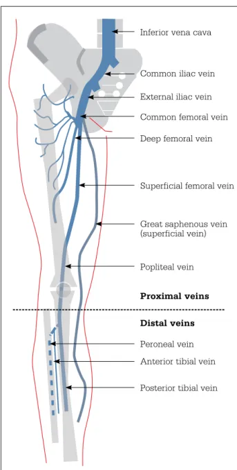
Venous thromboembolism (VTE), comprising deep vein thrombosis and pulmonary embolism (PE), is the third commonest vascular disorder in Caucasian populations.1 In Australia, DVT alone (without concomitant PE) affects 52 persons per 100 000 annually.2 Timely management of DVT is important as it is a common cause of morbidity. Thromboses of the deep veins in the upper limbs and ‘unusual sites’, such as mesenteric veins, constitute less than 10% of DVT cases.2 As they are uncommon, this article focuses only on the risks and diagnosis of lower limb DVT. Deep vein thrombosis usually starts in the calf area.3 most thrombi confined to the deep veins below the popliteal trifurcation (distal DVt, Figure 1) probably resolve spontaneously without causing any symptoms. Distal DVt can extend to the popliteal and femoral veins and other proximal veins. (note that in some imaging reports, the term ‘superficial femoral vein’ is applied to that part of the femoral vein between the popliteal vein and the confluence with the deep femoral vein.4 the superficial femoral vein is therefore a part of the proximal deep venous system.)
most patients present with symptoms when there is proximal vein involvement. About 50% of patients with untreated
symptomatic proximal DVt develop symptomatic PE within 3 months. Despite adequate treatment, DVt can recur. About 10% of patients with symptomatic DVt develop severe post-thrombotic syndrome within 5 years.3
Risk factors for VTE
Venous thromboembolism may be provoked by transient and reversible clinical risk factors such as surgery or oestrogen exposure, or long term and permanent factors, such as hemiparesis from stroke (Table 1).5 in 25% of cases, no clinical cause can be ascertained (idiopathic VtE).6
About 40–60% of VtE patients in caucasian cohorts have thrombophilia – a haemostatic disorder resulting in a thrombotic tendency.7 this may be heritable (eg. factor V leiden, prothrombin gene mutation and deficiencies of protein c, protein s and antithrombin),7 or acquired (eg. antiphospholipid antibodies).8
Background
Venous thromboembolism, comprising deep vein
thrombosis (DVT) and pulmonary embolism, is common in Australia and is associated with high morbidity.
Objective
This article provides a summary of the risk factors for DVT of the lower limb and discusses the diagnosis of the condition using a diagnostic algorithm incorporating clinical assessment, D-dimer testing and imaging studies. It also briefly reviews the clinical significance of isolated distal lower limb DVT and superficial vein thrombosis.
Discussion
Many conditions in the lower limb mimic DVT. Diagnosing DVT on clinical grounds without objective testing is unreliable. Patients incorrectly diagnosed as having DVT may be subjected to unnecessary anticoagulation and its associated risks of bleeding. In contrast, there is a risk of thrombus extension and embolisation when DVT is missed or inappropriately treated.
Keywords: venous thrombosis; thrombophlebitis; risk factors
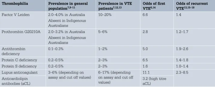
Reprinted from AustRAliAn FAmily PhysiciAn Vol. 39, no. 7, July 2010 469 the prevalence of thrombophilia varies greatly with ethnicity
(Table 2).7,9–13 in caucasian populations, the most common heritable thrombophilic defects are factor V leiden and prothrombin gene mutation but these are absent in indigenous Australians without caucasian admixture10 and in east Asians.9
heritable thrombophilia increases the risk of a first VtE14 but does not appear to be a major determinant of the risk of recurrence (Table 2).13,15–17 in contrast, the antiphospholipid syndrome is a strong risk factor for first and recurrent VtE and warrants consideration of long term anticoagulation.8,18–20
Assessment of the patient with
suspected DVT
the diagnosis without objective testing is unreliable as many common conditions, such as ruptured Baker cyst, haematoma and venous insufficiency, mimic DVt. in primary care among adults with suggestive symptoms and/or signs, only 29% have ultrasonographically proven DVt.21
to better evaluate the clinical probability of DVt before further testing, scoring systems based on symptoms, signs and risk factors have been developed. one such system is the modified Wells score (Table 3) which categorises patients as likely to have DVt (score of 2 or more points) or not (<2 points).22 this score is reproducible and is used together with laboratory and/or imaging studies in a diagnostic algorithm.
Laboratory testing
D-dimer is a degradation product of cross linked fibrin in thrombi. it is elevated in many conditions where fibrin is formed and then degraded, including acute VtE (Table 4).23,24 Various D-dimer assays are now available. they vary in turnaround times and sensitivity and specificity. Enzyme linked immunosorbent assays (ElisAs) are the most sensitive (sensitivity ≥90%) compared with the latex (sensitivity 80–85%) and whole blood (sensitivity ~87%) agglutination assays.25 the specificity of the various assays differ, but in general, the clinical utility of the D-dimer assays in diagnosing DVt is limited by their low specificity and low positive predictive value (Table 4). in contrast, a negative result (normal D-dimer) from a sensitive assay is useful to exclude DVt. D-dimer levels correlate with the size of the thrombus and clot activity. the sensitivity of an assay may be diminished in cases of
Table 1. Clinical risk factors for venous thromboembolism5
Strong clinical risk factors (odds ratio >10) • Fracture of the hip or lower limb
• Hip or knee replacement surgery • Major general surgery
• Major trauma • Spinal cord injury
Moderate clinical risk factors (odds ratio 2–9) • Arthroscopic knee surgery
• Hormonal therapy (eg. oral contraceptives, hormone replacement therapy)
• Pregnancy – postpartum • Paralytic stroke
• Previous venous thromboembolism Weak clinical risk factors (odds ratio <2)
• Immobilisation (eg. bed rest >3 days, air travel >8 hours)
• Pregnancy – antepartum • Obesity
• Advancing age Inferior vena cava
Common iliac vein External iliac vein Common femoral vein Deep femoral vein
Superficial femoral vein
Great saphenous vein (superficial vein)
Popliteal vein Proximal veins Distal veins Peroneal vein Anterior tibial vein Posterior tibial vein
Figure 1. Schematic diagram of the deep venous system of the lower limb
(Note that the superficial femoral vein – that portion of the femoral vein between the popliteal vein and the confluence with the deep femoral vein – is part of the proximal deep venous system4)
Deep vein thrombosis – risks and diagnosis FOCUS
small, isolated DVt compared with large proximal thrombosis. Further, the passage of time and/or the administration of anticoagulation may slow thrombus turnover thereby reducing assay sensitivity, hence, D-dimer testing may not be useful if a patient has already been effectively treated.23–26
Imaging studies
compression ultrasonography (cus) is the method of choice to evaluate the lower limbs for DVt.23,26,27 it is simple, noninvasive and its use has
been validated in management studies (Table 5). other methods such as computersied tomography (ct) and magnetic resonance imaging (mRi) are generally limited by availability.28,29
Diagnostic algorithm
A patient with symptoms and signs consistent with DVt should be assessed and the clinical (pretest) probability of acute DVt established by using a validated scoring system (Figure 2).22,26,30 thereafter, if the clinical probability of DVt is low, a D-dimer assay should be performed. in the modified Wells score, a pretest probability less than 2 (DVt unlikely) combined with a normal D-dimer assay result, reliably excludes DVt without the need for imaging studies. if D-dimer is raised, cus is indicated.
A high pretest probability (DVt likely) should be followed up by cus. With an abnormal cus, DVt is diagnosed. however, a normal cus does not exclude DVt and then a D-dimer assay should be performed. Anticoagulation can be withheld if the D-dimer result is normal. in the event of a raised D-dimer, imaging should be repeated within 1 week (or earlier if symptoms are worsening) as isolated distal DVt that have been missed initially on cus may extend into the proximal veins and be detected on repeated scanning.26,30 in patients with unexplained swelling of the entire leg but having negative cus, the possibility of pelvic vein thrombosis should be considered, in which case, ct, mRi or venography may be indicated.30
Figure 2 simplifies the diagnostic process and reduces cost by decreasing the number of patients undergoing both D-dimer testing and imaging studies while allowing room for the clinician to exercise clinical judgment: in the event that confirmatory testing is delayed and the clinical suspicion of DVt remains high, empirical anticoagulation (eg. low molecular weight heparin) should be started if there are no contraindications. note that this algorithm has been developed and validated for use predominantly in studies of outpatients. the D-dimer assay is frequently positive and has limited usefulness among inpatients (because of comorbidities) and pregnant women.26,30
Table 2. Thrombophilia: prevalence and risks of venous thromboembolism
Thrombophilia Prevalence in general
population7,9–11 Prevalence in VTE patients7,12,13 Odds of first VTE8,14 Odds of recurrent VTE13,15–18
Factor V Leiden 2.0–4.0% in Australia Absent in Indigenous Australians
10–20% 6.6 1.4
Prothrombin G20210A 2.0–3.2% in Australia Absent in Indigenous Australians 5–6% 2.8 1.2–1.7 Antithrombin deficiency 0.1–0.3% 1–2% 5.0 1.9–2.6 Protein C deficiency 0.2–0.5% 2–3% 6.5 1.4–1.8 Protein S deficiency 0.2–0.5% 2–3% 1.6 1.0–1.4
Lupus anticoagulant 3–6% (depending on assay and cut off values)
6–17% (depending on assay and cut off values) 11.1 2.3–8.5 Anticardiolipin antibodies (aCL) 3.2 (high titre aCL)
Table 3. Modified Wells score for predicting probability of deep vein thrombosis22
Clinical characteristic† Score*
Active cancer (treatment ongoing, administered within previous 6 months or palliative)
1 Paralysis, paresis or recent plaster
immobilisation of the lower extremities
1 Recently bedridden >3 days or major surgery within previous 12 weeks requiring general or regional anaesthesia
1
Localised tenderness along the distribution of the deep venous system
1
Swelling of entire leg 1
Calf swelling >3 cm larger than asymptomatic side (measured 10 cm below tibial tuberosity)
1 Pitting oedema confined to the symptomatic leg 1 Collateral superficial veins (nonvaricose) 1
Previously documented DVT 1
Alternative diagnosis at least as likely as DVT –2 * A score of ≥2 indicates that the probability of DVT is
likely; a score of <2 indicates that the probability of DVT is unlikely
† In patients who have symptoms in both legs, the more symptomatic leg is used
Deep vein thrombosis – risks and diagnosis FOCUS
472 Reprinted from AustRAliAn FAmily PhysiciAn Vol. 39, no. 7, July 2010
in pregnant women, if a DVt is suspected and initial cus is negative, repeat scanning in 5–7 days is indicated. if pelvic vein thrombosis is suspected, then mRi is the investigation of choice;31,32 alternatives are pulsed Doppler study of the iliac vein and ct venography.32
Further assessment
Evaluation of clinical risk factors for VtE helps determine the duration of anticoagulation.33 Venous thromboembolism provoked by a transient, reversible cause has a low risk of recurrence and short term anticoagulation is generally required. if the cause is permanent and irreversible (eg. hemiparesis from stroke), or if no provoking factor is identifiable (idiopathic VtE), the risk of recurrence is higher and longer term anticoagulation may be indicated.
laboratory testing for heritable thrombophilia among unselected patients is generally not indicated as results do not influence patient management.20,33–35 As long term anticoagulation may be warranted in the antiphospholipid syndrome, testing for lupus anticoagulant and anticardiolipin and anti-ß2 glycoprotein i antibodies should be considered in apparently idiopathic cases or among patients with concomitant connective tissue disorder, previous arterial thromboses (eg. stroke or myocardial infarction) particularly if premature, and/or a history of recurrent pregnancy failure or fetal death.19,20
Isolated distal DVT
the risk of symptomatic isolated distal DVt extending into the proximal veins if untreated or inadequately managed varies and ranges from
0–29%.30 one review estimated that 25% of untreated cases extended proximally within a week.3
At diagnosis, up to 40% of cases of symptomatic isolated distal DVt can have coexisting symptomatic PE.36 Among those with untreated distal DVt, 6.3% developed symptomatic PE within 7 days.37 Because of the variable risk of extension and/or embolisation, the optimal management of symptomatic distal DVt is uncertain38 but some authors suggest that anticoagulation for 6 weeks to 3 months is generally adequate.39–41
Table 4. Limitations of the D-dimer assay in the evaluation of suspected deep vein thrombosis23,24
Clinical situations other than acute VTE resulting in raised D-dimer (potentially diminishing the specificity and positive predictive value of the test)
• Surgery and/or trauma
• Haemorrhage and extensive bruises • Ischaemic heart disease
• Cerebrovascular accident (stroke) • Infections
• Malignancy
• Peripheral arterial disease and aneurysms • Pregnancy
• Advancing age of patients (ie. the very elderly) • Extensive burns
Clinical situations resulting in a false-negative D-dimer assay
• Small thrombus (eg. isolated distal deep vein thrombosis)
• Time lag between symptom onset and laboratory testing
• Concomitant anticoagulation (with heparin or warfarin)
Table 5. Imaging studies available for the diagnosis of deep vein thrombosis
Compression ultrasonography27
• Sensitivity and specificity exceed 95% and 98% respectively for symptomatic proximal DVT
• Sensitivity of 11–100% and specificity of 90–100% for symptomatic distal DVT
• Noninvasive: can be performed relatively rapidly and a portable technique allowing for the bedside assessment of critically ill patients
• Does not visualise the pelvic veins well and cannot be used in obese patients or in patients whose limbs are in plaster casts
Computerised tomography28
• Sensitivity and specificity of 96% and 95% respectively in a meta-analysis of studies
predominantly examining its use for the diagnosis of proximal DVT
• Can visualise the pelvic veins, define the upper limit of thrombus extension into the iliac veins and inferior vena cava
• Requires the injection of contrast medium, exposes the patient to radiation, may be difficult to interpret when artefact and insufficient venous filling is present, and is more expensive than ultrasonography • Limited by availability and technical expertise Magnetic resonance imaging29
• Sensitivity and specificity for the diagnosis of symptomatic DVT is 96% and 93% respectively • Sensitivity for distal DVT is much lower (about 62%) • Can be performed without the use of contrast
medium
• Can visualise the pelvic veins, define the upper limit of thrombus extension into the iliac veins and inferior vena cava
• Limited by availability and technical expertise Venography (phlebography)27
• Reference standard technique
• Reliably detects isolated distal DVT and thrombosis in the iliac veins and inferior vena cava
• Cumbersome to perform, requires the injection of contrast medium, exposes the patient to radiation, may be difficult to interpret when insufficient venous filling is present
FOCUS Deep vein thrombosis – risks and diagnosis
Summary of important points
• Diagnostic algorithms incorporating clinical assessment, D-dimer testing and imaging studies have been developed and validated for use in diagnosing DVt.
• Anticoagulation is generally indicated in symptomatic lower limb DVt. this includes thrombosis in the superficial femoral vein, which despite its name, is part of the proximal deep venous system. • The duration of anticoagulation is largely determined by the
site of thrombosis and clinical risk factors; testing for heritable thrombophilia does not generally influence management of the patient and is not warranted.
Author
Wai Khoon ho mmedsc, FRAcP, FRcPA, is consultant haematologist, Department of haematology, Austin health, melbourne, Victoria. [email protected].
conflict of interest: none declared.
Acknowledgment
i thank mr stephen Valentine for the illustration in Figure 1. Routine screening of the asymptomatic postoperative patient for
DVt is unwarranted.33 however, if isolated asymptomatic distal DVt is found incidentally, it is reasonable to withhold anticoagulation as many of these thrombi resolve spontaneously and do not cause clinically significant PE.3,42,43 if anticoagulation is withheld, repeat testing within the next 10 days should be considered to determine if the thrombus has extended.44 Repeat testing is also indicated if the patient becomes symptomatic.
Superficial vein thrombosis
concomitant DVt is observed in 2.6–65% of cases of superficial vein thrombosis (sVt) whereas symptomatic PE is present in 0.5–4.0%.45 in case series involving >15 patients with untreated sVt, DVt and/or PE occurred in 1.7% to >11% within 3 months.45,46
ultrasonography should be performed in cases of sVt to confirm the diagnosis and establish its extent, and to look for the presence of concomitant DVt. Where the proximal extent of sVt is close to the sapheno-femoral or sapheno-popliteal junction, some authors recommend the institution of anticoagulation due to the presumed high risk of DVt and/or PE.45,46
Figure 2. Diagnostic algorithm for clinically suspected deep vein thrombosis26,30
CUS = compression ultrasound scan; +ve = positive; –ve = negative
Swollen, painful and tender, erythematous lower limb
DVT unlikely
D–Dimer raised D–Dimer normal CUS –ve CUS +ve
CUS +ve
D–Dimer raised
CUS +ve No DVT
DVT Repeat CUS at 1 week
CUS –ve D–Dimer normal DVT No DVT No DVT No DVT DVT likely CUS –ve
Treat DVT. Evaluate clinical risk factors for DVT (including possible underlying malignancy). Clinical examination of patient and laboratory tests (including full blood examination) for possible myeloproliferative disorders (polycythaemia vera and essential thrombocythaemia). Consider laboratory testing for antiphospholipid antibody especially if DVT appears to be idiopathic or patient has connective
tissue disorders, or a history of arterial thromboses or recurrent pregnancy losses DVT
Deep vein thrombosis – risks and diagnosis FOCUS
474 Reprinted from AustRAliAn FAmily PhysiciAn Vol. 39, no. 7, July 2010
29. sampson Fc, Goodacre sW, thomas sm, van Beek EJR. the accuracy of mRi in diagnosis of suspected deep vein thrombosis: systematic review and meta-analysis. Eur Radiol 2007;17:175–81.
30. hirsh J, lee Ayy. how we diagnose and treat deep vein thrombosis. Blood 2002;99:3102–10.
31. scarsbrook AF, Evans Al, owen AR, Gleeson FV. Diagnosis of suspected venous thromboembolic disease in pregnancy. clin Radiol 2006;61:1–12. 32. marik PE, Plante lA. Venous thromboembolic disease and pregnancy. n Engl
J med 2008;359:2025–33.
33. Kearon c, Kahn sR, Agnelli G, Goldhaber s, Raskob GE, comerota AJ. Antithrombotic therapy for venous thromboembolic disease. American college of chest Physicians evidence-based clinical practice guidelines (8th edition). chest 2008;133:454–545s.
34. middeldorp s, van hylckama Vlieg A. Does thrombophilia testing help in the clinical management of patients? Br J haematol 2008;143:321–35. 35. Baglin t, Gray E, Greaves m, et al. clinical guidelines for testing for heritable
thrombophilia. Br J haematol 2010;149:209–20.
36. monreal m, Ruíz J, olazabal A, Arias A, Roca J. Deep venous thrombosis and the risk of pulmonary embolism. A systematic study. chest 1992;102:677–81. 37. Passman mA, moneta Gl, taylor lm, et al. Pulmonary embolism is
associ-ated with the combination of isolassoci-ated calf vein thrombosis and respiratory symptoms. J Vasc surg 1997;25:39–45.
38. Righini m, Paris s, le Gal G, laroche JP, Perrier A, Bounameaux h. clinical relevance of distal deep vein thrombosis. Review of literature data. thromb haemost 2006;95:56–64.
39. Gallus As, Baker Ri, chong Bh, ockelford PA, street Am. consensus guide-lines for warfarin therapy. Recommendations from the Australasian society of thrombosis and haemostasis. med J Aust 2000;172:600–5.
40. schulman s, Rhedin As, lindmarker P, et al. A comparison of six weeks with six months of oral anticoagulant therapy after a first episode of venous thromboembolism. n Engl J med 1995;332:1661–5.
41. Pinede l, Duhout P, chabaud s, et al. comparison of 3 and 6 months of oral anticoagulant therapy after a first episode of proximal deep vein thrombosis or pulmonary embolism and comparison of 6 and 12 weeks of therapy after isolated calf deep vein thrombosis. circulation 2001;103:2453–60. 42. Kim yh, Kim Js. incidence and natural history of deep-vein thrombosis after
total knee arthroplasty: a prospective, randomised study. J Bone Joint surg Br 2002;84–B:566–70.
43. Wang cJ, Wang JW, Weng lh, hsu cc, lo cF. outcome of calf deep-vein thrombosis after total knee arthroplasty. J Bone Joint surg Br 2003;85– B:841–4.
44. oishi cs, Grady-Benson Jc, otis sm, colwell cW, Walker Rh. the clinical course of distal deep venous thrombosis after total hip and total knee arthro-plasty, as determined with duplex ultrasonography. J Bone Joint surg Am 1994;76–A:1658–63.
45. marchiori A, mosena l, Prandoni P. superficial vein thrombosis: risk factors, diagnosis, and treatment. semin thromb hemost 2006;32:737–43. 46. leon l, Giannoukas AD, Dodd D, chan P, labropoulos n. clinical significance
of superficial vein thrombosis. Eur J Vasc Endovasc surg 2005;29:10–7.
References
1. naess iA, christiansen sc, Romundstad P, cannegieter sc, Rosendaal FR, hammerstrøm J. incidence and mortality of venous thrombosis: a population-based study. J thromb haemost 2007;5:692–9.
2. ho WK, hankey GJ, Eikelboom JW. the incidence of venous thromboembo-lism: a prospective, community-based study in Perth, Western Australia. med J Aust 2008;189:144–7.
3. Kearon c. natural history of venous thromboembolism. circulation 2003;107(23 suppl 1):i22–30.
4. Bundens WP, Bergan JJ, halasz nA, murray J, Drehobl m. the superficial femoral vein. A potentially lethal misnomer. JAmA 1995;274:1296–8. 5. Anderson FA, spencer FA. Risk factors for venous thromboembolism.
circulation 2003;107:19–16.
6. heit JA, o’Fallon Wm, Petterson tm, et al. Relative impact of risk factors for deep vein thrombosis and pulmonary embolism. A population-based study. Arch intern med 2002;162:1245–8.
7. lensing AWA, Prandoni P, Prins mh, Büller hR. Deep-vein thrombosis. lancet 1999;353:479–85.
8. Wahl DG, Guillemin F, de maistre E, Perret-Guillaume c, lecompte t, thibaut G. meta-analysis of the risk of venous thrombosis in individuals with antiphospholipid antibodies without underlying autoimmune disease or previ-ous thrombosis. lupus 1998;7:15–22.
9. Rees Dc, cox m, clegg JB. World distribution of factor V leiden. lancet 1995;346:1133–4.
10. Bennett JA, Palmer lJ, musk AW, Erber Wn. Prevalence of factor V leiden and prothrombin 20210A mutations in indigenous Australians. thromb haemost 2001;86:1592–3.
11. Khor B, Van cott Em. laboratory evaluation of hypercoagulability. clin lab med 2009;29:339–66.
12. Rance A, Emmerich J, Fiessinger Jn. Anticardiolipin antibodies and recurrent thromboembolism. thromb haemost 1997;77:212–4.
13. Baglin t, luddington R, Brown K, Baglin c. incidence of recurrent venous thromboembolism in relation to clinical and thrombophilic risk factors: pro-spective cohort study. lancet 2003;362:523–6.
14. Van der meer FJm, Koster t, Vandenbroucke JP, Briët E, Rosendaal FR. the leiden thrombophilia study (lEts). thromb haemost 1997;78:631–5. 15. ho WK, hankey GJ, Quinlan DJ, Eikelboom JW. Risk of recurrent venous
thromboembolism in patients with common thrombophilia. A systematic review. Arch intern med 2006;166:729–36.
16. marchiori A, mosena l, Prins mh, Prandoni P. the risk of recurrent venous thromboembolism among heterozygous carriers of factor V leiden or pro-thrombin G20210A mutation. A systematic review of prospective studies. haematologica 2007;92:1107–14.
17. De stefano V, simioni P, Rossi E, et al. the risk of recurrent venous throm-boembolism in patients with inherited deficiency of natural anticoagulants antithrombin, protein c and protein s. haematologica 2006;91:695–8. 18. Zhu t, martinez i, Emmerich J. Venous thromboembolism: risk factors for
recurrence. Arterioscler thromb Vasc Biol 2009;29:298–310. 19. Greaves m, cohen h, machin sJ, mackie i. Guidelines on the
investiga-tion and management of the antiphospholipid syndrome. Br J haematol 2000;109:704–15.
20. merriman l, Greaves m. testing for thrombophilia: an evidence-based approach. Postgrad med J 2006;82:699–704.
21. oudega R, moons KGm, hoes AW. limited value of patient history and physi-cal examination in diagnosing deep vein thrombosis in primary care. Fam Pract 2005;22:86–91.
22. Wells Ps, Anderson DR, Rodger m, et al. Evaluation of D-dimer in the diagno-sis of suspected deep-vein thrombodiagno-sis. n Engl J med 2003;349:1227–35. 23. Palareti G, cosmi B, legnani c. Diagnosis of deep vein thrombosis. semin
thromb hemost 2006;32:659–72.
24. Adam ss, Key ns, Greenberg cs. D-dimer antigen: current concepts and future prospects. Blood 2009;113:2878–87.
25. stein PD, hull RD, Patel Kc, et al. D-dimer for the exclusion of acute venous thrombosis and pulmonary embolism. A systematic review. Ann intern med 2004;140:589–602.
26. tan m, van Rooden cJ, Westerbeek RE, huisman mV. Diagnostic manage-ment of clinically suspected acute deep vein thrombosis. Br J haematol 2009;146:347–60.
27. Fraser JD, Anderson DR. Deep venous thrombosis: recent advances and optimal investigation with us. Radiology 1999;211:9–24.
28. thomas sm, Goodacre sW, sampson Fc, van Beek EJR. Diagnostic value of ct for deep vein thrombosis: results of a systematic review and meta-analysis. clin Radiol 2008;63:299–304.