PARTICULAR REFERENCE TO RISK FACTORS
A dissertation submitted in part fulfillment of the requirements for DM
(Branch IV, Gastroenterology) examination of the Tamil Nadu Dr.
This is to certify that that this dissertation entitled “A study of intestinal tuberculosis with particular reference to risk factors” is the bonafide work done by
Dr. Suresh Shenoy in partial fulfillment of rules and regulations for DM (Branch IV Gastroenterology) examination of the Tamil Nadu Dr. M.G.R. Medical University, Chennai to be held in February 2007.
B.S. Ramakrishna MD, DM, PhD, FAMS Professor of Gastroenterology
I wish to place my sincere gratitude to Dr. B.S. Ramakrishna, Professor and Head of Department of Clinical Gastroenterology and Hepatology, for guiding me to do this thesis as DM dissertation.
Special thanks to Mr. Pugazhendi S. and Mr. R. Balamurugan for meticulous analysis of study samples for interferon gamma polymorphisms.
I sicerely thank Mr.Thirumani for his excellent secretarial support in formatting this thesis.
I am grateful to the institution, which through the Fluid Research Grant has provided me the necessary financial aid required for the study.
PAGE NOS
HISTORY 1
INTRODUCTION 4
REVIEW OF LITERATURE 5
AIMS 38
PATIENTS AND METHODS 39
RESULTS 44
DISCUSSION 56
BIBLIOGRAPHY
HISTORY:
Tuberculosis has been present in humans since antiquity. The origins of the disease
are in the first domestication of cattle. Skeletal remains show prehistoric humans (4000 BC)
had TB, and tubercular decay has been found in the spines of Egyptian mummies from
3000-2400 BC. There were references to TB in India around 2000 BC.
Around 460 BC, Hippocrates called the disease phthisis (consumption), emphasizing
the dramatic aspect of general wasting associated with chronic untreated cases. He described
that diarrhea in a patient with phthisis is a mortal symptom.
Around 200 A.D., the Roman doctor Claudius Galen recognized the consumption as
incurable and recommended a treatment plan of fresh air, rest, and good food.
Results of postmortem examination on Louis XVIII evidenced an ulcerative lesion
with intestinal perforation associated with a cavitary lung disease.
In 1546, Girolamo Tracastoro explained the contagious nature of TB in his book “De
Morbis Contagiosis”. He wrote that bed sheets and clothing could contain contagious
particles.
In 1679, Franciscus de la Boe, more commonly known as Dr. Silvius, wrote the
“Opera Medica”. He described the tubercles and characterized the infection’s course
throughout the lungs and body of infected patients providing clear descriptions detailing the
In 1720, an English physician, Benjamin Marten, was responsible for the first theory
regarding Mycobacterium tuberculosis as “wonderfully minute living creatures”. He wrote
the book “A New Theory of Consumption” regarding his views.
In 1761, Austrian Leopold Avensbrugger wrote a book on tuberculosis about the
clinical symptoms and different pathologies of the disease.
In 1782, a physicist named Graumann proved conclusively that syphilis and
tuberculosis were not the same disease.
In1810, a London physician, Carmichael, wrote a dissertation demonstrating that
cattle tuberculosis is transmitted to humans through infected meat and milk.
In1854, Hermann Brehmer who suffered from TB himself theorized that TB was
treatable after his doctor recommended that he move to a more temperate climate, like the
Himalayas. After recuperating there, he returned home healthy and cured. He built the first
sanatorium where patients could recuperate under the influences of fresh air and healthy
eating habits.
In 1865, a French army physician, Jean-Antoine Villemin demonstrated the
transmission of TB from humans to cattle to rabbits. He theorized that the disease was caused
by a certain organism and did not arise from spontaneous generation as previously believed.
In March 24, 1882 Robert Koch identified and described the bacillus-causing
tuberculosis, Mycobacterium tuberculosis. He received the Nobel Prize in physiology or
tuberculosis were similar, which held back the recognition of infected milk as a source of
infection. Later, this source was eliminated by the pasteurization process. Koch announced a
glycerin extract of the tubercle bacilli as a "remedy" for tuberculosis in 1890, calling it
'tuberculin'. It was not effective, but was later adapted by von Pirquet in a test for
pre-symptomatic tuberculosis.
BCG (Bacillus of Calmette and Guerin) was developed from attenuated bovine-strain
tuberculosis by Albert Calmette and Camille Guerin in 1906. The BCG vaccine was first
used on humans on July 18, 1921 in France.
In 1890, an Italian doctor, Forlanini, created the first therapy for TB patients. He
found that collapsing the lungs had positive effects on recovery from tuberculosis.
In 1895, Wilhelm Konrad bon Rontgen used radiation to examine the progression and
assess the severity of the illness in TB patients.
In1920s, a treatise by Assmann cultivated the theory of reinfection.
Finally, in 1943, Selman A. Waksman developed the antibiotic Streptomycin. The
first time it was administered to a live, human patient was November 20, 1944. The
progression of the disease was halted, the bacteria were later absent from the sputum, and the
patient was fully healed. In the years following, more TB drugs were discovered.
Hopes that the disease could be completely eliminated have been dashed since the
rise of drug-resistant strains in the 1980s. The resurgence of tuberculosis resulted in the
INTRODUCTION:
Tuberculosis (TB) remains the single largest infectious disease causing high mortality
in humans, leading to 3 million deaths annually, about five deaths every minute.
Approximately 8-10 million people are infected with this pathogen every year12. In India,
there are about 500,000 deaths occurring annually due to TB13, with the incidence and
prevalence being 1.5 and 3.5 millions per year.
Approximately two billion individuals globally are infected with Mycobacterium
tuberculosis, yet only 10% develop clinical tuberculosis 1.There was a surge of tuberculosis
in mid-1980s in developed countries which may be due to multiple factors like infection with
human immunodeficiency virus (HIV), immunosuppression with prolonged steroid therapy,
immigration from countries with high prevalence of tuberculosis, and social problems such
as poverty, homelessness, and drug abuse 2, 3.
Tuberculosis continues to be a major health problem in India 4, 5. Deaths due to
tuberculosis accounts to around 50/100,000 population. Forty percent of the cases in India
contract tuberculosis by the age of 6 years and 80% by the age of 16 years 6. The incidence of
tuberculosis is also increasing in Western countries 7, 8, 9. Poor socio economic status, poor
sanitation and recent upsurgence of HIV infection enhance the susceptibility to tuberculosis
in India. The exact prevalence of intestinal tuberculosis in India is not known.
Several susceptibility-associated genetic polymorphisms have been proposed to
explain differential susceptibility to tuberculosis (TB) progression in different populations.
Though several gene polymorphisms have been associated with susceptibility or resistance to
TB in different ethnic populations, only few of these genetic associations have shown to have
REVIEW OF LITERATURE:
Intestinal tuberculosis (ITB) was common in the early 20th century. It was responsible
for most cases of small intestinal obstruction and stricture. There was a decline in ITB cases
in the middle of the century in developed countries which was caused by 1) an increased
standard of living 2) pasteurization of milk 3) control of bovine tuberculosis through
slaughter of reactive animals and 4) the introduction of antituberculous chemotherapy in the
1950s.
Gastrointestinal tract (GIT) is a common location for tuberculosis infection and is the
sixth most frequent site of extra pulmonary involvement. Intestinal tuberculosis still remains
the most common granulomatous disease of the bowel in India 10, 11. Intestinal tuberculosis is
an important cause of morbidity in the Indian population, especially because the diagnosis is
often delayed due to non-specific nature of its signs and symptoms.
The incidence in hospital admissions for ITB has been reported to be 0.8%4. In India,
TB is responsible fir 7% of hospital admissions for intestinal obstructions and 6% of
perforations21. Any segment of the GIT can be involved by tuberculosis, but the ileocecal
region is the most commonly involved part of the tract, noted in up to 90% of cases with
intestinal tuberculosis14, 15, 29, 30. In India, the organism isolated from all intestinal lesions has
been Mycobacterium tuberculosis and not Mycobacterium Bovis 16,17,18,19 .In Bhansali’s
series4, including 196 patients with gastrointestinal tuberculosis, ileum was involved in 52%
and cecum in 51% of cases. Of the 300 patients in a study, ileocecal involvement was
present in 54% of cases20. The frequency of bowel involvement declines as one proceeds
ITB is thought to result from swallowed organisms (infected sputum in active
pulmonary tuberculosis or ingestion of contaminated milk) that directly penetrate the
intestinal mucosa. Hematogenous spread (active pulmonary tuberculosis, miliary TB, or
silent bacteremia during primary phase of TB) and direct extension from adjacent organs can
also occur. Although any area of the GIT can be involved, the ileocaecal region is involved in
approximately 75% of cases and studies in India have suggested that approximately 20% of
the patients may have associated pulmonary involvement 22. It has been estimated that only
10% of persons infected with Mycobacterium tuberculosiswill ever develop clinical disease.
Of the total number of patients with TB, only about 1% will have intestinal disease.
Socioeconomic and environmental factors have long been known to influence the
occurrence of tuberculosis in a community. A survey carried out in Wardha district
(Maharashtra) is the only source of survey data (unpublished) linking tuberculosis in the
community to socio-economic criteria 23. The prevalence rates in the survey had depended on
literacy (lowest in the graduates and highest among the illiterates) and present employment
(highest among the professionals, followed by cultivators and agricultural labour). These had
also depended on income, living standard (those living in “Kutcha” houses had a higher
prevalence than “pucca” house dwellers). Of the total cases in women, 48 percent were
among those unemployed (include housewives). For all demographic variables, rates in
female were less than those in males.
As per Dholakia 24, evidence is lacking to assume a differential prevalence rate of
tuberculosis among workers than among non-workers. Of the ‘workers’ group, estimated to
be suffering from tuberculosis in India, about 52 percent were in the age group 15- 44 years.
tuberculosis in the urban and rural areas respectively. There was much lower proportion of
women among workers with tuberculosis in higher ages, especially in the urban areas. In the
Wardha survey 23, the urban professionals and rural service workers, who had a higher
prevalence, had a low proportion of the female population in them, and had consequently
accounted for a small proportion of the total cases among females. The extent of tuberculosis
morbidity in the males in the economically active age and in females in the reproductive age
marks it out as a priority among the public health problems in India.
The available literature strongly suggests the possible role of genetic factors in the
control of host responses to Mycobacterium tuberculosis (M.TB) 25, 26. Stead 27, has proposed
that susceptibility to infection with M. TB has changed from being the norm of all humans to
being an infection of certain population as a result of natural selection of resistance among
ancestors who came in contact with the bacterium and survived the illness during the
preantibiotic era. Distinct environmental and natural selective factors have likely resulted in
population – specific immunogenetic adaptations to clinical tuberculosis.
Convincing evidence exists from twin studies that host genetic factors are important
in determining susceptibility to the infection. Kallmann and Reisner28 found an appreciably
high concordance of pulmonary TB in monozygotic than dizygotic twins. Also the
aggregation of pulmonary tuberculosis in families emphasizes the importance of heredity.
The different manifestations of infection with M.TB reflect the balance between the
bacilli and hostdefense mechanisms. Traditionally, protective immunity to tuberculosishas
been ascribed to T-cell-mediated immunity, with CD4+ T cellsplaying a crucial role. Recent
immunological and genetic studiessupport the long-standing notion that innate immunity is
There are several evidences to suggest that the production of the human leukocyte
antigen (HLA) system, the major histocompatibility complex of humans, have an important
function in controlling the cellular immune response to infectious agents31. Though cellular
immunity in TB is regulated by HLA system, the exact role of HLA –DR genes on both the
development and course of TB, as well as cellular and humoral immune response remains
unknown. It is possible that the gene products are involved in the activation of T helper and
inducer cells rather than in subsequent T-cell activation of B cells. Singh et al 32 showed that
the DR2 antigen had a preferential tendency to be transferred from TB parents to their
affected children. In a group of pulmonary TB patients from North India, he found that TB
patients had an increased frequency of DR2 antigen and a marked decrease of DRW6 in
comparison to healthy subjects. In a group of pulmonary TB patients from South India,
Brahmajyothi V et al 33 noted that the frequencies of HLA-A10 and B8, but not DR2 were
greater in control subjects. Hence DR2 may be involved in the pathogenesis of advanced
pulmonary tuberculosis. The MHC genes may be physically close to the chromosome region
that carries a gene conferring susceptibility or resistance to a particular disease. This
association may explain the lack of complete association and geographic variation, due to
linkage disequilibrium. Though HLA –DR2, DQ1 and their subtypes are significantly
associated with the susceptibility to tuberculosis, and they may not be the sole genetic
markers predisposing to tuberculosis suggesting that non-HLA genes may have a role in
infection.
M.TB is an intracellular parasite and cellmediated immunity is crucial for
containment of infection34. After macrophage stimulation by mycobacterialinfection, the
interferon-γ secretionby natural killer cells, differentiation of antigen-driven CD4+T cells into interferon-γproducing Th1 cells, and activationof these Th1 cells to secrete interferon-γ
and possibly othermacrophage-activating factors35. Secretion of interferon-γ,in turn, results in macrophage secretion of TNF-α; enhanced antigen presentation, activationof macrophage
mycobactericidal mechanisms such as nitric oxideproduction; and impaired proliferation of
interleukin-4–secretingTh2 cells.
Interferon-γ induces cellular activation by binding to a receptorcomplex consisting of at least two subunits: the interferon-γ binding subunit (interferon-γ receptor 1) and a
chromosome21–encoded transmembrane accessory factor (interferon-γreceptor 2). Both components of the receptor arethought to be required for normal signal transduction. Binding
of interferon-γinduces dimerization of the interferon-γreceptor1, which subsequently associates with interferon-γ receptor 2.Interferon-γ interacts with both interferon-γreceptor 1 and interferon-γreceptor 2 during the process ofassociation of the two-receptor proteins36. The Janus proteinkinases Jak 1 and Jak 2 are associated with the intracellulardomains of
interferon-γ receptor 1 and interferon-γreceptor 2,respectively, and are brought together and activated by phosphorylationby the binding of interferon-γto the receptor complex. This results in the phosphorylation of tyrosine at position 457 ofthe interferon-γ receptor 1 chain and produces a binding sitefor Stat 1 α (signal transduction and activation of transcription
protein), leading to the phosphorylation, homodimerization,and subsequent dissociation of
Activation of infected macrophages by interferon-gamma (IFN-γ)derived from T cells and natural killer cells are the principalantimycobacterial effector mechanisms. The
importance of IFN-γin human mycobacterial immunity was established by the identification of mutations in the gene encoding the IFN-γ receptor ligand bindingchain as a cause of susceptibilityto mycobacterial infection 37, 38.
Recently several studies have shown that genes coding for different cytokines may
affect host susceptibility to tuberculosis. Interferon-gamma (IFN-γ) is a proinflammatory Th1-type cytokine produced by T cells that appears to be necessary for the containment of
mycobacterial infections. IFN-γ knockout mice were found to be highly susceptible to infection with M.TB. A recent study39demonstrated association between the (+874 A /T)
polymorphism in the IFN-γ gene and pulmonary TB, with subjects lacking the T allele was found to be at risk for pulmonary and meningeal tuberculosis. In a Spanish population,
patients who were homozygous for the (+874A) allele of IFN-γ had a 3.75 fold increased risk of developing compared to healthy controls64. Reports have shown that a complete deficiency
of interferon-γreceptor 1 may lead to BCG infection in vaccinated childrenor to atypical mycobacterial infection in unvaccinated persons38.
Although IFN-γ production may vary among subjects, some studies suggest that
IFN-γ levels are depressed in patients with active TB 40, 41. In a study comparing the immune response to pre and post- BCG vaccination, it was seen that BCG had little effect in driving
the immune response towards IFN-γ and a protective Th1 response42. In a study done to determine whether the effect of balance of T cell cytokines during initial stages of infection
most severely depressed in patients with moderately advanced and far advanced pulmonary
disease and in malnourished patients though production of IL-12, IL-4 and IL-10 was similar
in TB patients and healthy tuberculin reactors. Hence it is found that polymorphisms in the
genes of these cytokines have functional significance43.
Several polymorphic-derived deletions and point mutations of the mannose–binding
lectin (MBL) 44-48, human analogue of the murine natural resistance associated macrophage
protein 1 (NRAMP1) gene 49-53, the vitamin D receptor (VDR) gene 54-55, the interleukin -1
(IL -1 αand ß) 56-58, IL -1 receptor antagonist (IL-1RA) 58,59,60, IL-10 56,61, IL -12 receptor
antagonist (IL-12R) 62, tumor necrosis factor- alpha and beta (TNF-αand ß) 56,63, interferon-γ 39,64and interferon-γreceptor 138 genes have been associated with susceptibility or resistance
to TB in different ethnic groups. Only few of these genetic associations have been shown to
have relevant functional impact on the containment of the bacteria by the host immune
system.
Besides genetic differences in Th1/Th2 responses, conditioning of the mucosal
immune system in childhood may be important in determining susceptibility to ITB. It has
been postulated that helminthic infections (associated with less domestic hygiene) may
Association of important candidate gene variants of HLA and non-HLA genes with the
susceptibility or resistance to pulmonary tuberculosis in Indian population72
Candidate genes Effect Reference
HLA
HLA-DR2 Susceptibility 32,33,73
Sub-type
-DRB1 * 1501, * 1502 -DRB1 * 1501
Susceptibility Susceptibility
74 75.76 HLA-DQ1
- DQB1*0601 Susceptibility Susceptibility 73.75 75
HLA-DP
-DPB1*02 Susceptibility 75
Haplotype:
DRB1and1501- DQB1*0601 DRB1 * 11(5)
DRB1 * 10 DQB1*0501 Susceptibility Resistance Resistance Resistance 75 75 75 75 Non-classical HLA
Transporter Associated with Antigen Processing (TAP) gene TAP 2 and DR2.
Susceptibility 77
Non-HLA
Functional mutants Homozygotes to Mannose
Binding Lectin (MBL) gene (codon 52,54 and 57)
- Heterozygotes of MBL codon
57
Vitamin D receptor (VDR) gene variants (Bsml, Apal, TaqI and FokI) NRAMP1 [(CA) n 823C/T, TGTG+/del and D543N G/A]
Susceptibility
Resistance to bacteriological relapse
Differential susceptibility and resistance in males and females No association with
susceptibility or resistance
47
47
60,78
79
Cytokine gene TNF- α -238, -308
TNF- ß No association No association 63 63
Haplotypes
HLA-B17- TNF-α-238/A HLA-B17- TNF-α-308/2 HLA-B17- TNF-ß -2
Associated with bacteriological
Tuberculosis in HIV:
Extrapulmonary disease is more common in patients with AIDS; 50% of the AIDS
patients with tuberculosis have extrapulmonary involvement compared to only 10-15% of
non-HIV tuberculosis patients 67. While 10 percent of those infected with TB will progress to
active disease over their life times, those who are co-infected with both TB and HIV on the
other hand progress rapidly, at the rate of 10 percent annually and about 60 percent in their
life time. The pathogenesis of TB can be altered by HIV either through reactivation of latent
tuberculosis infection to active disease (more common) or by causing rapid progression from
recent infection with M. TBto tuberculosis disease. With progression of HIV infection,
CD4+ T-lymphocytes decline in number and function. The immune system is therefore, less
able to prevent the growth and local spread of M.TB. As a result, disseminated and
extra-pulmonary disease is more commonly seen. Nevertheless, extra-pulmonary TB is still the most
common form of TB even in HIV infected patient; pulmonary involvement can occur in
70-90 percent of all patients with TB 68.
Tuberculosis may precede the diagnosis of Acquired immunodeficiency syndrome
(AIDS) by few months and the disease may be severe and progress rapidly in AIDS and vice
versa 69, 70. Multidrug resistant TB is more common in patients with AIDS71.The risk of
tuberculosis infection progressing to active tuberculosis is estimated to be 8 percent per year
in an HIV positive person, as opposed to 10 percent life time risk in an immunocompetent
PATHOPHYSIOLOGY:
The pathophysiology of intestinal tuberculosis has been attributed to four
mechanisms80: 1) swallowing of infected sputum in patients with active pulmonary TB; 2)
hematogenous spread from active pulmonary or milary TB; 3) ingestion of contaminant milk
or food; 4) contiguous spread from adjacent organs. After the tubercle bacillus enters the
gastrointestinal tract, it traverses the mucosa to lodge in the submucosa. There, the presence
of the bacillus induces inflammatory changes, including serosal and submucosal edema,
cellular infiltration, and lymphatic hyperplasia. Eventually appearance of granulomas causes
small papillary mucosal elevations81. Lymphangitis, endarteritis, and fibrosis ensue which
lead to mucosal ulceration, caseation necrosis and narrowing of intestinal lumen82. Regional
lymph node involvement occurs by lymphatic spread.
In a review of 596 patients with abdominal TB3, the highest incidence of TB was
noted in the GIT and in the peritoneum, followed by mesenteric lymph nodes. Within the
GIT, the ileocecal area was the most common site of involvement. Infact, disease of the
jejunoileum and ileocecal areas together comprised > 75% of cases, with the disease in the
colon was found in 12% of the cases 83-86.The predilection of the bacillus for the ileo-cecum
is attributed to three factors: 1) relative physiologic stasis of the area 2) the high rate of
absorption with more complete digestion; and 3) the abundance of lymphoid tissue in
ileocecal region.
Pathology:
Hoon et al 87originally classified the gross morphological appearance of the
Prakash 88described the bowel lesions as ulcerative and ulcerohypertrophic types.
Ulcerative form has been found more often in malnourished adults, while
hypertrophic form is classically found in relatively well nourished adults. The bowel wall is
thickened and the serosal surface is studded with nodules of variable size. These ulcerative
and stricturous lesions are usually seen in the small intestine. In the less common
hypertrophic form, inflammatory response and reactive tissue produce a multinodular
mucosal pattern resembling neoplastic masses. The ulcero-hypertrophic pattern, most
commonly seen in ileo-cecal region may produce "cobblestone" appearance 14, 88, 89. The
patient often presents with a right iliac fossa lump constituted by the ileocecal region,
mesenteric fat and lymph nodes.
Adjacent tuberculous adenitis can cause colonic traction diverticula with narrowing,
local fixation, and sinus tract development 14. Other characteristics of tubercular intestinal
lesions include increased mesenteric fat and mesenteric adenopathy with caseation, which
grossly resemble Crohn’s disease. Rarely colonic TB can present as diffuse tuberculous
colitis which must be differentiated from inflammatory bowel disease (IBD), because steroid
treatment can be lifesaving in IBD and lethal in ITB 90.
Tuberculous granulomas are initially formed in the mucosa or the Peyer’s patches.
These granulomas are of variable size and characteristically tend to be confluent, in contrast
to those in Crohn’s disease. The presence of central caseation is the hallmark of granulomas
caused by tuberculosis 88. Granulomas are often seen just beneath the ulcer bed, mainly in the
submucosal layer. Submucosal oedema or widening is inconspicuous. Tubercular ulcers are
single or multiple, usually 3-6 mm in diameter, with an irregular margin and usually present
as transverse lesion parallel to each other. This orientation is related to the arrangement of the
submucosal lymphatic structures except when a Peyer’s patch alone is involved resulting in a
longitudinal orientation .The intervening mucosa is usually uninvolved. These ulcers are
usually transversely oriented in contrast to Crohn’s disease where the ulcers are longitudinal
or serpiginous 91. These circumferential 'girdle ulcers’, usually cicatrize during healing and
form strictures. Occlusive arterial changes may produce ischemia and contribute to the
development of strictures92. Endarteritis also accounts for the rarity of massive bleeding in
cases of intestinal tuberculosis. Shah et al 93 correlated findings on barium studies and
superior mesenteric angiography in 20 patients. Angiograms were abnormal in all and
showed arterial encasement, stretching and crowding of vessels, and hypervascularity.
Patients with strictures had occlusion of the vasa recta, while ulcerated lesions had
hypervascularity. In long-standing lesions there may be variable degree of fibrosis of the
bowel wall, which extends from submucosa into the muscularis. Many sections may show
only non-specific chronic inflammation and no granulomas. Pulimood 94et al., described
histological changes characteristic of TB and CD. They suggested that multiple (mean
number of granulomas per section: 5.35), large (mean widest diameter: 193µm), confluent
granulomas often with caseation necrosis are characteristic of TB. Other features were ulcers
lined by conglomerate epithelioid histiocytes and disproportionate submucosal inflammation.
The features characteristics of Crohn’s disease (CD) were infrequent (mean number of
granulomas per section: 0.75), small (mean widest diameter: 95µm) granulomas,
microgranulomas (defined as poorly organized collections of epithelioid histiocytes), focally
normal appearing areas. They also described that granulomas larger than 400µm in maximum
dimension, more than four sites of granulomatous inflammation per site, caseation, a band of
epithelioid histiocytes in ulcer bases and location of granulomas in the caecum favored a
diagnosis of TB compared to CD95.
Mesenteric lymph nodes may be enlarged matted and may caseate. Characteristic
granulomas may be seen only in the mesenteric lymph nodes. This is especially common in
patients who have taken antitubercular therapy for some time. The reverse, i.e., the presence
of granulomas in the intestine and no granulomas in the draining lymph nodes is rare87.
Clinical features:
ITB is predominantly a disease of young adults. Two-thirds of the patients are 21-40
years old 96, 97 and the mean age of patients is 30-40 years96. Although some studies mention
female preponderance, it seems that the disease affects both sexes equally. Intestinal lesions
are present in only 10% of cases of abdominal TB in children97. ITB is characterized by
different modes of presentation, viz acute (no previous history of obstruction), chronic or
acute on chronic (episode of acute obstruction with history of sub acute obstruction).
Most patients have constitutional symptoms of fever with evening rise (40-70%),
abdominal pain (80-95%), diarrhea (11-20%), constipation, alternating constipation and
diarrhea, weight loss (40-90%), anorexia, malaise and night sweats. Pain can be either
colicky due to luminal compromise, or dull and continuous when the mesenteric lymph nodes
are involved. Some patients, particularly those with miliary tuberculosis, may have features
of toxemia 20with high fever, tachycardia, anemia and leucocytosis. Patients in India
history of TB, which is reported in about one –third of patients in UK19, 84. Peripheral lymph
nodes (cervical or axillary) may be involved in 3-10% of the patients 17,22,97,98. Other clinical
features depend upon the site, nature and extent of involvement and are detailed below:
Esophageal tuberculosis
Tuberculosis of the esophagus is a rare and constitutes 0.2% of cases of abdominal
tuberculosis99. The relative rarity is thought to be due to rapid transit time and the vertical,
smooth stratified squamous epithelium. Esophageal involvement occurs with a secondary
spread from adjacent sites such as lymph nodes, pulmonary, and vertebral tuberculosis 100,101
or retrograde lymphatic spread 100. Primary esophageal tuberculosis is very unusual102.
Midoesophagus is the most common site of involvement. Rarely, upper one-third may be
involved by direct extension from pharynx and larynx. The commonest presenting symptoms
are dysphagia , weight loss , retrosternal pain and coughing on swallowing .The disease
usually mimics esophageal carcinoma and extra esophageal focus of tuberculosis may not be
evident 103.Rarely , there may be massive hematemesis from an aortoesophageal fistula due
to tuberculous aortitis 104.
Barium examination demonstrates extrinsic compression of the enlarged lymph nodes
to the esophagus, esophago-bronchial fistula, sinus tract formation, mucosal irregularity, and
ulceration 105. Computed tomography is a more reliable method to determine full extent of
Tuberculosis of stomach
Gastric involvement is very rare and constitutes around 0.2-1% of abdominal
tuberculosis. It occurs with a secondary spread from adjacent lymph nodes or hematogenous
route. The infrequent involvement of the stomach is due to the relative resistance of the
gastric mucosa, high acid content in stomach, lack of lymphoid tissue in the stomach and
rapid passage of gastric contents into the intestines. Any part of the stomach can be involved
but the usual sites are lesser curvature and antrum99. Lesions may be ulcerative or
hypertrophic infiltrative type.
Gastro duodenal tuberculosis may mimic peptic ulcer disease with a shorter duration
of history and non-response to anti -secretary therapy107. Constitutional symptoms may be
present in affected individuals. Hematemesis is a common presentation. It may also simulate
gastric carcinoma. Chowdhary et al 108 reported the rare concurrence of carcinoma and
tuberculosis of stomach in the same patient. Primary gastric tuberculosis has also been
reported 99.Patient may present with features of outlet obstruction due to inflammatory
changes, fibrosis, or enlarged lymph nodes 109,110. Caseating granulomas seen on microscopy
are usually located in mucosa and submucosa, but rarely extend to the muscularis layer. This
may the reason for rarity of free perforation in gastric tuberculosis99.
Radiological features are nonspecific, mimicking the signs of benign ulcer in the
ulcerative form of tuberculosis, and of malignancy in the hypertrophied form. Computed
Gastric tuberculosis should be differentiated radio graphically from gastric
carcinoma, non-Hodgkin's lymphoma, syphilis, and sarcoidosis. Endoscopic biopsy is
indicated to obtain the diagnosis. There is no specific picture of duodenal tuberculosis on
endoscopy, and demonstration of granulomas or acid fact bacilli on endoscopic biopsy
material is unusual. Because of the lack of accurate clinical diagnosis, most patients end up
with surgical intervention, and the diagnosis is made by surgery.
Tuberculosis of duodenum:
Duodenal involvement is extremely rare, occurring in up to 0.2- 2% of the patients
with ITB 111. It may present as duodenal obstruction with narrowing and sometimes fistula
due to adjacent lymph nodes. Duodenal narrowing due to enlarged lymph nodes is
recognized in the third or fourth part of duodenum.
The largest published series of duodenal tuberculosis reported 30 cases from India 112.
Most patients (73%) had symptoms of duodenal obstruction. In a majority of these cases
obstruction was due to extrinsic compression by tuberculous lymph nodes, rather than by
intrinsic duodenal lesion. The remainder (27%) had a history of dyspepsia and was suspected
of having duodenal ulcers. Patient can present with hematemesis 113,114.Other reported
complications by various authors are perforation 115, fistulae (pyeloduodenal,
duodenocutaneous, blind) 115, excavating ulcers extending into pancreas116 and obstructive
jaundice by compression of the common bile duct117.
Barium studies reveal evidence of segmental narrowing. Duodenal strictures are
enlarged lymph nodes and thickened mesenteric root is demonstrated by US or CT118. There
is no specific picture of duodenal tuberculosis on endoscopy, and demonstration of
granulomas or acid fact bacilli on endoscopic biopsy material is unusual. Surgical bypass has
been required in the majority of cases to relieve obstruction but successful endoscopic
balloon dilatation (TTS balloon, Microvasive) of duodenal strictures has been reported by Vij
et al119 in two cases.
Tuberculosis of jejunum and ileum
Jejunal or ileal involvement, except of the terminal ileum is rarely seen. Nonspecific
changes are demonstrated on barium studies and CT 89.
Tuberculosis of the appendix:
In India, tuberculosis is found in 2.3 % of appendicectomies 120. It is usually
secondary to tuberculosis elsewhere in the abdomen: local extension of the disease,
lymphatic spread, or peritoneal disease. The rare primary disease of the appendix may
present with perforation 121.
Ileocecal tuberculosis:
Colicky, midabdominal and/or right lower quadrant pain , distension, gurgling
,vomiting, feeling of a ball of wind moving in the abdomen , and visible loops and peristalsis
which are relieved spontaneously after passage of flatus reflects intermittent partial small
bowel obstruction, is the presenting complaint in 90-100% of patients 80,122. Abdominal
examination may reveal no abnormality or a doughy feel. A well-defined, firm, usually
cases. Associated lymphadenitis is responsible for the presence of one or more lumps which
are mobile if mesenteric nodes are involved and fixed if para-aortic or iliac group of nodes
are enlarged 4 .The most common complication is obstruction due to narrowing of the lumen
by hyperplasic caecal tuberculosis, by strictures of the small intestine, which are commonly
multiple, or by adhesions. Adjacent lymph nodal involvement can lead to traction, narrowing
and fixity of bowel loops. In India, around 3 to 20 percent of all cases of bowel obstructions
are due to tuberculosis 4,123,124. In a large series of 348 cases of intestinal obstruction,
Bhansali and Sethna 123 found tuberculosis to be responsible for 54 (15.5%) cases; 33 cases
were small bowel and 21 large bowel obstruction. Tandon et al 22 studied 186 patients over 5
yr and observed a change in clinical profile with an increase in patients with more protracted
course and subacute intestinal obstruction in recent years.
Tuberculosis accounts for 5 to 9 percent of all small intestinal perforations in India,
and is the second commonest cause after typhoid fever 125,126. Evidence of tuberculosis on
chest X -ray and a history of subacute intestinal obstruction are important clues.
Pneumoperitoneum may be detected on radiographs in only half of the cases. Tubercular
perforations are usually single and proximal to a stricture 92. Acute tubercular peritonitis
without intestinal perforation is usually an acute presentation of peritoneal disease but may
be due to ruptured caseating lymph nodes 4,126.
Malabsorption is a common complication. It is the most important cause of
malabsorption syndrome in India, next to tropical sprue 4. In a patient with malabsorption, a
history of abdominal pain usually suggests the diagnosis of tuberculosis 127. Pimparkar and
Donde 128 studied 40 patients with malabsorption and divided them into those with and
faecal fat and Schillings test for B12 malabsorption and found them to be abnormal in 28, 22,
57, 60 and 63 percent respectively in patients with stricture compared to 0, 0, 8, 25 and 30
percent respectively without strictures. Tandon et al 129 also reported biochemical evidence of
malabsorption in 75 percent of patients with intestinal obstruction and in 40 percent of those
without it. The cause of malabsorption in ITB is postulated to be bacterial overgrowth in a
stagnant loop, bile salt deconjugation, diminished absorptive surface due to ulceration, and
involvement of lymphatics and lymph nodes. Minor rectal bleeding in ITB has been
described frequently 130,131; however massive bleeding is a rare manifestation of the disease
132-135. It has been suggested that intestinal TB increases the capillary vascularity, and small
arteries undergo obliterative endarteritis that makes bleeding uncommon 136
Segmental colonic tuberculosis:
Segmental or isolated colonic tuberculosis refers to involvement of the colon without
ileocecal region, and constitutes 9.2 percent of all cases of abdominal tuberculosis and
around 15-20% of intestinal tuberculosis 137,138. It commonly involves the sigmoid, ascending
and transverse colon 139. Multifocal involvement is seen in one third (28 to 44%) of patients
with colonic tuberculosis 130,140.The median duration of symptoms at presentation is less than
1 yr 131. Abdominal pain is the predominant symptom in 78-90 percent of patients and
hematochezia occurs in less than one third 140,141. The bleeding is frequently minor and
massive bleeding is less common. Singh et al 131 reported rectal bleeding in 31 percent of
patients with colonic tuberculosis, and it was massive in 13 percent. Bhargava et al 142
reported bleeding in 70 percent cases. Overall, tuberculosis accounts for about 4 percent of
patients with lower gastrointestinal bleeding 92. The diagnosis is suggested by barium enema
diagnosis include Crohn’s disease, ulcerative colitis (in case of diffuse colitis), carcinoma of
colon, amoeboma, Yersinia infection, gastrointestinal histoplasmosis, ischemic colitis,
pseudomembranous colitis and periappendiceal abscess. The coexistence of colonic TB and
colonic cancer has been described143. Colonic tuberculosis might be complicated by
perforation causing peritonitis 144.
Rectal and anal tuberculosis
Rectal tuberculosis is rare 147 and may occur as isolated lesions. Clinical presentation
of rectal tuberculosis is different from more proximal disease. Hematochezia is the most
common symptom (88%) followed by constitutional symptoms (75 %) and constipation
(37%) 141. The high frequency of rectal bleeding may be because of mucosal trauma caused
by stool traversing the strictured segment. Digital examination usually reveals an annular
stricture. The stricture is usually tight and of variable length with focal areas of deep
ulceration. It is usually within 10 cm of the anal verge 131. Associated perianal disease is very
rare. Excessive fibrosis associated with the rectal inflammation results in an increase in
presacral space.
Anal tuberculosis is less uncommon and has a distinct clinical presentation. Most anal
and perianal lesions are ulcerative, although lupoid and verrucous forms have been described.
There is an association with anorectal fistula 145 and perianal tuberculous abscess 149.
Tubercular fistulae are usually multiple. Most ulcers are shallow with bluish undermined
edges. Progression of the ulcers is usually slow. There may be associated inguinal
lymphadenopathy 145.Biopsy should differentiate the lesions of Crohn’s disease, squamous
12 out of 15 multiple perianal fistulae were of tubercular origin, as compared to only 4 out of
61 solitary perianal fistulae. Shukla et al 145 reported that in India, tuberculosis accounted for
up to 14 percent of cases of fistula in ano. Anal discharge was present in all cases and
perianal swelling in one third. Constitutional symptoms were not present in any patient 145.
Anal tuberculosis is also seen in paediatric patients 148.
Diagnosis and investigations:
Despite a high index of suspicion, ITB can be difficult to diagnose. Symptoms are
vague, signs are nonspecific and the disease closely mimics many other diseases.
Paustian stated that atleat one of the following four criteria must be fulfilled to
diagnose abdominal tuberculosis: (i) A positive culture from enteric, mesenteric or lymphatic
tissue (ii) typical findings at surgery with histological evidence of tuberculosis in mesenteric
nodes (iii) histological demonstration of caseating granuloma ;(iv) histological demonstration
of acid fast bacilli in a lesion152. A fifth criterion defined by Logan 149 as indicating “probable
tuberculosis” is a favorable response to antituberculous treatment of concurrent tuberculosis.
These criteria are rarely present together153. These criteria must be considered and the
diagnosis substantiated by adequate radiological and histopathological studies.
Laboratory results can be nonspecific or normal. The most common abnormal
laboratory finding is an elevated erythrocyte sedimentation rate (50-80%) 20, 97, 98. Mild
anemia is also relatively frequent (50-80%) 20, 97, 98. Hypoalbuminaemia may suggest
malnutrition or malabsorption84.
Tuberculin skin test: A positive purified protein derivative (PPD) test is found in
patients. The test has a lower specificity for abdominal (77%) 153 than for pulmonary (84%)
tuberculosis154. It doesn’t differentiate between active disease and previous sensitization by
contact or vaccination.
Radiological studies: Chest X-ray: It is positive in only 25% of the patients 22.
Evidence of tuberculosis in a chest X-ray supports the diagnosis but a normal chest X-ray
does not rule it out. Chest X-ray may show evidence of active or healed tuberculosis. Sharma
et al 151 studied 70 cases of abdominal tuberculosis and found evidence of active or healed
lesions on chest X-ray in 32 (46%). X-rays were more likely to be positive in patients with
ulcerative intestinal types and with acute complications 151. In Prakash’s series of 300
patients, none had active pulmonary tuberculosis but 39 percent had evidence of healed
tuberculosis10.
Plain X-ray abdomen: Plain X -ray abdomen may show enteroliths, features of
intestinal obstruction i.e., dilated bowel loops with multiple air fluid levels, evidence of
ascites, perforation or intussusception. In addition, there may be calcified lymph nodes,
calcified granulomas and hepatosplenomegaly.
Small bowel barium meal: The following features may be seen: mucosal
irregularity and rapid emptying (in ulcerative type); hyper segmentation of the barium
column (“chicken intestine”), precipitation, flocculation and dilution of the barium (due to
malabsorption); stiffened and thickened folds; luminal stenosis with smooth but stiff
contours (“hour glass stenosis “), multiple strictures with segmental dilatation of bowel
Barium enema: The following features may be seen:
(i) Early involvement of the ileocecal region manifesting as spasm and edema of the
ileocecal valve. Thickening of the lips of the ileocecal valve and/or wide gaping of
the valve with narrowing of the terminal ileum (“Fleischner” or “inverted umbrella
sign”) are characteristic.
(ii) Fold thickening and contour irregularity of the terminal ileum, better appreciated
on double contrast study.
(iii) “Conical caecum”, shrunken in size and pulled out of the iliac fossa due to
contraction and fibrosis of the mesocolon. The hepatic flexure may also be pulled
down.
(iv) Loss of normal ileocecal angle and dilated terminal ileum, appearing suspended from
a retracted, fibrosed caecum (“goose neck deformity”).
(v) “Purse string stenosis” - localized stenosis opposite the ileocecal valve with a rounded
off smooth caecum and a dilated terminal ileum.
(vi) “Stierlin ’s sign ” is a manifestation of acute inflammation superimposed on a
chronically involved segment and is characterized by lack of barium retention in the
inflamed segments of the ileum, caecum and variable length of the ascending colon,
with a normal configured column of barium on either side. It appears as a narrowing
of the terminal ileum with rapid emptying into a shortened, rigid or obliterated
caecum.
Both Stierlin and String signs can also be seen in Crohn's disease and hence are not
specific for tuberculosis.
Barium studies are sensitive for ileocecal and colonic lesions 98. Tandon et al 22
reported false-negative barium studies in 25% of patients. Radiological studies may not
always differentiate ITB from Crohn’s disease and malignancy.
Ultrasonography:
Barium studies though accurate for intrinsic bowel abnormalities, do not detect
lesions in the peritoneum. Ultrasound is very useful for imaging peritoneal tuberculosis.
The following features may be seen, usually in combination 29.
(i) Intra -abdominal fluid, which may be free or loculated; and clear or complex (with
debris and septae). Fluid collections in the pelvis may have thick septa and can mimic
ovarian cyst.
(ii) “Club sandwich” or “sliced bread” sign is due to localized fluid between radially
oriented bowel loops, due to local exudation from the inflamed bowel (interloop
ascitis).
(iii) Lymphadenopathy may be discrete or conglomerated (matted). The echotexture is
mixed heterogenous, in contrast to the homogenously hypoechoic nodes of
lymphoma. Small discrete anechoic areas representing zones of caseation may be
seen within the nodes. With treatment the nodes show a transient increase in size for
3-4 weeks and then gradually reduce in size. Calcification in healing lesions is seen as
discrete reflective lines. Both caseation and calcification are highly suggestive of a
tubercular etiology, neither being common in malignancy related lymphadenopathy.
is uniform and concentric as opposed to the eccentric thickening at the mesenteric
border found in Crohn’s disease and the variegated appearance of malignancy.
(v) Pseudokidney sign - involvement of the ileocecal region, which is pulled up to a
subhepatic position.
Computed tomographic (CT) scan:
Ileocecal tuberculosis is usually hyperplasic and well evaluated on CT scan. In early
disease there is slight symmetric circumferential thickening of caecum and terminal ileum.
Later the ileocecal valve and adjacent medial wall of the caecum is asymmetrically
thickened. In more advanced disease gross wall thickening, adherent loops, large regional
nodes and mesenteric thickening can together form a soft tissue mass centered on the
ileocecal junction 155. Mural stratification does not occur in any of the cases. In comparison,
Crohn's disease has a uniform pattern of wall thickening, which is concentric or largely
symmetrical, and ranges from 0.6 to 1.7 cm. Some of patients show mural stratification
164.CT scan can also pick up ulceration or nodularity within the terminal ileum, along with
narrowing and proximal dilatation. Other areas of small and large bowel involvement
manifest as circumferential wall thickening, narrowing of the lumen and ulceration. In the
colon, involvement around the hepatic flexure is common. Complications of perforation,
abscess, fistula and obstruction are also seen. Although amebiasis may produce the typical
shrunken cecum seen in tuberculosis, small bowel association is very rare with amebiasis.
Cecal carcinoma is always limited by ileocecal valve162.
Tubercular ascitic fluid is of high attenuation value (25-45 HU) due to its high
protein content. Strands, fine septae and debris within the fluid are characteristic, but are
peritoneal nodules may be seen.
Mesenteric disease on CT scan is seen as a patchy or diffuse increase in density,
strands within the mesentery, and a stellate appearance. Lymph nodes may be interspersed.
Omental thickening is well seen often as an omental cake appearance. A fibrous wall can
cover the omentum, developing from long standing inflammation and is called omental line.
An omental line is less common in malignant infiltration 155.
Caseating lymph nodes are seen as having hypodense centers and peripheral rim
enhancement. Along with calcification, these findings are highly suggestive of tuberculosis.
In tuberculosis the mesentery, mesenteric root, celiac, porta hepatis and peripancreatic nodes
are characteristically involved, reflecting the lymphatic drainage of the small bowel. The
retroperitoneal nodes (i.e., the periaortic and pericaval) are relatively spared, and are almost
never seen in isolation, unlike lymphoma 155.
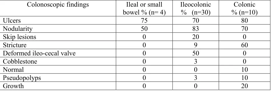
Colonoscopy:
Colonoscopy with ileoscopy is an excellent tool to diagnose colonic and terminal
ileal involvement, but is still often underutilized. Mucosal nodules of variable sizes (2 to 6
mm) and ulcers in a discrete segment of colon, 4 to 8 cm in length are common. The nodules
have a pink surface with no friability and are most often found in the caecum especially near
the ileocaecal valve. Large (10 to 20 mm) or small (3 to 5 mm) ulcers are commonly located
between the nodules. The intervening mucosa may be hyperemic or normal 130. Areas of
strictures with nodular and ulcerated mucosa may be seen. Other findings are
pseudopolypoid edematous folds, and a deformed and edematous ileocecal valve. Diffuse
ulcerative colitis. Lesions mimicking carcinoma have also been described130, 131,141. The
ileocecal angle is distorted and often obtuse. Both sides of the ileocecal valve are usually
involved leading to incompetence of the valve, another point of distinction from Crohn’s
disease.
For better yield, 8-10 colonoscopic biopsies are suggested for histopathology
(hematoxylin and eosin staining), staining for acid-fast bacteria (Ziehl-Neelsen stain) and
culture (Lowenstein-Jensen medium or BACTEC method). Biopsies should be taken from
the edge of the ulcers, some suggest even from the base of the ulcer. However, there is a low
yield on histopathology because of predominant submucosal involvement. Granulomas have
been reported in 8-48 percent of patients and caseation in a third (33-38%) of positive cases
131. The yield of acid-fast bacilli stains has been variable in studies. Culture positivity is not
related to the presence of granulomas. Endoscopic fine needle aspiration cytology may be
positive even when the biopsy has been negative156.
Microbiological diagnosis:
Microbiological diagnosis of ITB may be difficult; the yield of organisms may be low
because extrapulmonary disease is paucibacillary. Mycobacterial culture should be
performed in all cases (although results take 6 weeks) because it may be positive even in the
absence of a characteristic histological picture.
Radiactive Scintigraphy:
Gallium 67 citrate is superior to Indium 111-labelled leucocytes for detecting areas of
Immunological tests:
Chawla et al158 reported that an optical density (OD) of 0.81 on ELISA and
fluorescent coefficient of 2.56 on soluble antigen fluorescent antibody (SAFA) as cut-off
gave positivity of 92 and 83 percent, respectively, with 12 and 8 percent false positives
respectively. Bhargava et al 159 used competitive ELISA with monoclonal antibody against 38
kD proteins and found a sensitivity of 81 percent, specificity of 88 percent and diagnostic
accuracy of 84 percent. However, ELISA remains positive even after therapy, the response to
mycobacteria is variable and its reproducibility is poor. Hence the value of immunological
tests remains undefined in clinical practice92.
Polymerase Chain Reaction (PCR) analysis:
The PCR is a unique method of amplifying tiny quantities of DNA and RNA. It is
ideally suited for diagnosis of conditions in which infective organisms are present in very
minute quantities to be detected by conventional methods such as staining techniques and
culture. It has been that PCR can detect as few as 50 organisms per reaction, which is atleast
a five fold lower limit of detection compared to culture. PCR analysis of the involved
gastrointestinal mucosa is a useful tool for diagnosis of ITB and it assists in differentiation of
ITB from Crohn’s disease. Two studies found that the sensitivity, specificity, positive
predictive value and negative predictive value of PCR assay on tissue specimens in
differentiating ITB from Crohn’s disease were 21-64.1%, 95-100%, 92-100%, and 28-
68.2%, respectively160, 161. In a PCR study done on fecal samples of subjects including treated
positive predictive value and negative predictive value of fecal PCR were 88.8%, 100%,
100%, and 93.7%, respectively165.
Ascitic fluid examination:
The ascitic fluid in tuberculosis is straw coloured with protein >3g/dl, and total
cell count of 150-4000/ µl, consisting predominantly of lymphocytes (>70%). The ascites
to blood glucose ratio is less than 0.9650 and serum ascitic albumin gradient is less than
1.1 g/dl.
The yield of organisms on smear and culture is low. Staining for acid-fast bacilli is
positive in less than 3 percent of cases. A positive culture is obtained in less than 20
percent of cases, and it takes 6-8 weeks for the mycobacterial colonies to appear.
However Singh et al167 in an earlier study cultured 1 litre of ascitic fluid after
centrifugation and obtained 83 per cent culture positivity. Finding an ascitic fluid/blood
glucose ratio of less than 0.96 may be useful for distinguishing tuberculosis from other
causes of ascites 166.
Adenosine deaminase (ADA) is an aminohydrolase that converts adenosine to
inosine and is thus involved in the catabolism of purine bases. The enzyme activity is
more in T than in B-lymphocytes, and is proportional to the degree of T cell
differentiation. ADA is increased in tuberculous ascitic fluid due to the stimulation of
T-cells by mycobacterial antigens. ADA levels were determined in the ascitic fluid of 49
patients by Dwivedi et al168. The levels in tuberculous ascitis were significantly higher
than those in cirrhotic or malignant ascitis. Taking a cut off level of 33 U/L, the
sensitivity, specificity and diagnostic accuracy were 100, 97 and 98 percent
fluid ADA level above 36 U/L and a ascitic fluid to serum ADA ratio > 0. 985 were found
suggestive of tuberculosis170. In coinfection with HIV the ADA values can be normal or
low. Falsely high values can occur in malignant ascites. High interferon-γ levels in tubercular ascites have been reported to be useful diagnostically171. A cutoff level of 3.2
U/ml gave the assay a sensitivity of 93% and a specificity of 98%. Combining both ADA
and interferon estimations may further increase sensitivity and specificity.
Laparoscopic findings:
Bhargava et al172 studied 87 patients with high protein ascites, of which 38 were
diagnosed as having tuberculosis. They found visual appearances to be more helpful (95%
accurate) than histology, culture or guinea pig inoculation (82.3 and 37.5% sensitivity
respectively). Caseating granulomas may be found in 85-90 percent of the biopsies. The
laparoscopic findings in peritoneal tuberculosis can be grouped into 3 categories:
(i) Thickened peritoneum with tubercles: Multiple, yellowish white, uniform sized
(about 4 - 5 mm) tubercles diffusely distributed on the parietal peritoneum. The
peritoneum is thickened, hyperemic and lacks its usual shiny luster. The omentum,
liver and spleen can also be studded with tubercles.
(ii) Thickened peritoneum without tubercles.
(iii) Fibroadhesive peritonitis with markedly thickened peritoneum and multiple thick
adhesions fixing the viscera.
Pre-operative diagnosis is difficult even in areas where tuberculosis is common and
was obtained in only 40% 98 to 50 %173 of patients in India, 33% in Kuwait174 and 25% in
UK84. Reports of patients who were not diagnosed at life for tuberculosis but were revealed
strictures, which are not amenable to endoscopic or percutaneous biopsy or fine needle
aspiration cytology. With advances in endoscopic modalities (enteroscopy), small bowel
tuberculosis may be diagnosed at earlier stages at present.
Operative findings are infiltrated thickened and rolled up omentum, increased
mesenteric fat wrapping the bowel, short and fibrotic strictures and soft to firm hypertrophic
lesions. The opened specimen may show thickened mucosal fold with ulceration and fibrosis.
Besides these; ascites, yellow nodules over the visceral and parietal peritoneum (tubercles),
adhesions, enlarged and calcified mesenteric lymph nodes may be seen. A frozen section
may help to rule out malignancy. A mesenteric lymph node should always be removed
because granulomas and caseation are more likely to be found in the nodes than in intestinal
lesions4, 88.
Management:
All patients should receive conventional antitubercular therapy for at least 6 months
including initial 2 months of rifampicin, isoniazid, pyrazinamide and ethambutol, followed
by rifampicin and isoniazid for next 4 months. A randomized comparison of 6-month short
course chemotherapy with a 12 month course of ethambutol and isoniazid (supplemented
with streptomycin for the initial 2 wks) was conducted by Balasubramanium et al 175 in 193
adult patients. Cure rate was 99 and 94 percent in patients given short -course and the
12-month regimen respectively. Kim et al, in a randomized comparison in patients with ITB
found that 9 months of treatment is as effective as 15 months course of chemotherapy 176.
Acute-on-chronic intestinal obstruction usually responds to conservative
management; these patients can be electively investigated and treated accordingly4.
stenosed segment by entero-enterostomy or by ileo-transverse colostomy was practiced
when effective antitubercular drugs were unavailable, as any resectional surgery was
considered hazardous in the presence of active disease. This practice however, produced
blind loop syndrome, and fistulae and recurrent obstruction often occurred in the
remaining segments. With the advent of more effective antituberculous drugs, various
reports recommended the use of radical procedures in an attempt to eradicate the disease
locally. These included right hemicolectomy with or without extensive removal of the
draining lymph nodes and wide bowel resections. These procedures were often not
tolerated well by the malnourished patient. Moreover the lesions are often widely spaced
and not suitable for resection.
The recommended surgical procedures today are conservative; tuberculosis is a
systemic disease and cannot be eradicated by surgery alone. A period of pre operative
drug therapy is controversial .A segment of bowel bearing multiple strictures or a single
long tubular stricture or with almost complete obstruction may merit resection. Resection
is segmental with a 5 cm margin.
Tubercular perforations are usually ileal and are associated with distal strictures.
Resection and anastomosis is preferred as simple closure of the lesions is associated with
a high incidence of leak and fistula formation. Two reports suggest that obstructing
intestinal lesions may relieve with antitubercular drugs alone without surgery. Anand et al
179 reported clinical and radiological resolution of tuberculous strictures with drug therapy
even in patients with subacute intestinal obstruction. They treated 39 patients with
obstructive symptoms using medical therapy. At the end of one year 91 percent showed
needed in only 3 cases (8%). Predictors of need for surgery were long strictures (>12 cm)
and multiple areas of involvement 179. Balasubramaniam et al 170 made similar
observations. The mean time required for the relief of obstructive symptoms was 6
months, although systemic symptoms improved within 2 months.
Postoperative complications include anastomotic leak resulting in a fecal fistula,
peritonitis and intra-abdominal sepsis, persistent obstruction, wound infection and
dehiscence4. Re-operation may be required during the follow-up for recurrent obstruction
due to strictures or adhesions 84, 97.
Mortality rate due to ITB ranges from 4-12 % 20, 97, 98,181is partly due to the
associated malnutrition, anemia and hypoalbuminaemia. Mortality may be higher
(12-25%) 4, 97 in the presence of acute complications. Delayed diagnosis and injudicious
treatment due either to limited experience and poor understanding of the disease are
AIMS OF THE STUDY:
To perform a case control study in patients with intestinal tuberculosis and with matched
healthy controls in order:
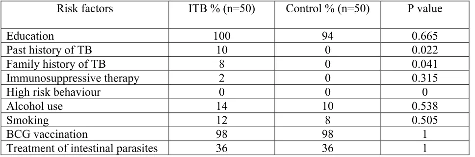
1. To identify the prevalence of specific risk factors such as diabetes mellitus, HIV
infection, family history of tuberculosis, history of BCG vaccination, in patients with
intestinal tuberculosis compared to age- and sex-matched control subjects.
2. To determine whether there are specific associations of intestinal tuberculosis with
factors connected with childhood hygiene, as well as a history of treatment for intestinal
parasitic infections.
3. To determine the frequency of interferon- gamma (IFN-γ) polymorphisms in patients with intestinal tuberculosis compared to appropriately matched healthy controls.
4. To study the clinical and investigation profile of intestinal tuberculosis in our institution
METHODOLOGY:
SUBJECTS:
Patients:
Confirmed intestinal tuberculosis patients included those who had their diagnosis
confirmed by colonoscopic biopsies demonstrating caseating granulomas, AFB on smear, or
AFB on culture, as well as surgical patients who had surgical resection with a histological
diagnosis of tuberculosis made on the resected specimen.
Presumptive intestinal tuberculosis included those patients whose colonoscopic
biopsies showed granulomatous or nongranulomatous chronic inflammation of colon or
ileum, with or without extra intestinal tuberculosis, and response to anti-tuberculous therapy.
Controls:
Controls included patients with irritable bowel syndrome, acid peptic disease and
non-ulcer dyspepsia who were matched for age, sex and geographical region with no history
of pulmonary or extra pulmonary TB in the past, in the family or even in the close contacts.
These subjects were used for risk factor analysis.
Healthy elderly controls matched for sex and region were used to analyze frequency
of interferon–gamma polymorphisms in the general population.
METHODS:
Informed written consent was obtained. A detailed questionnaire including risk
factors of intestinal tuberculosis were provided to the patient and controls and all the
responses were recorded as given by the patient, and these were maintained in a file.
Questionnaire for risk factors concerned with hygiene were demarcated as “during
household, and availability of toilets. Questionnaire also probed other risk factors like
diabetes mellitus, HIV infection, and family history of tuberculosis, history of BCG
vaccination, and history of treatment for intestinal parasitic infections. These responses were
later entered into a computer for analysis. Relative risks for each of these factors were
evaluated .All subjects were investigated as clinically necessary. Clinical and investigation
profile of intestinal tuberculosis (ITB) was also analyzed. The questionnaire is provided in
the Appendix to this thesis.
A 10 ml sample of blood was drawn by one of the laboratory co-investigators into
Vacutainers. DNA was immediately isolated from the whole blood and stored at -20°C
pending analysis. Analysis of IFN- γ polymorphisms was done using allele-specific PCR. The genotype was analyzed and comparisons made between patients and controls.
Laboratory analyses
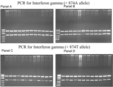
The IFN-γ (+874 A/T) polymorphism was genotyped using allele-specific PCR as described by Lopez-Maderuelo et al64. Genomic DNA was amplified in two different PCRs
for each polymorphism; each reaction has a generic antisense primer and one of the
allele-specific sense primers. One internal control, human growth hormone, was amplified in all the
reaction tubes to assess the success of PCR amplification. The amplified PCR products were
resolved on 2% agarose gel, stained with ethidium bromide, and documented using a gel
documentation system. PCR was performed under the following conditions: initial
denaturation at 94°C for 5 minutes; 40 cycles of 94°C for 30 seconds, 50°C for 30 seconds
DNA extraction and mutation analysis was as follows
For DNA extraction the materials required were:
EDTA coated vacutainer tube, centrifuge, plastic Pasteur pipette (sterile), 15ml of self
standing centrifuge tube (sterile), RBC lysis buffer, WBC lysis buffer, Proteinase K, 10%
Sodium Dodecyl Sulphate (SDS), saturated NaCl, absolute alcohol, Eppendorf tubes (sterile),
70 % ethanol, Tris-EDTA (TE) buffer.
Reagents were prepared in the following manner:
RBC lysis buffer: Dissolve 8.725g of ammonium chloride and 1 g of potassium
bicarbonate in 1 liter of water. Check the pH and that should be 7.4. Sterilize the solution by
autoclaving and store it at room temperature for a period of 1year.
WBC lysis buffer: Mix 25ml of 0.5M EDTA and 2.19 g of NaCl in 250ml of water
and adjust the pH to 8.0 and make the volume to 500ml with water. Sterilize by autoclaving
and store it at room temperature for 3 months.
TE buffer (10mM Tris & 0.1mM EDTA): Mix 1ml of 1M Tris (pH 8.0) and 400 l
of 0.25 M EDTA (pH 8.0) and make up the volume to 100ml with water. Sterilize by
autoclaving and store in at room temperature for 1 year.
10% SDS: Dissolve 10g of SDS in 100ml of water and heat the solution at 65ºC to
assist dissolution. Sterilization is not required and store in at room temperature for 6 months.
Proteinase K: Dissolve the lyophilized Proteinase K (Cat no. PK1, Bangalore Genie)
in 10ml of sterile water to obtain a concentration of 10mg/ml. Aliquot 500µl volume into