Functional
Genomics
Studies
of
Human
Brain
Development
and
Implications
for
Autism
Spectrum
Disorder
Mark
Nicholas
Ziats
Robinson
College
University
of
Cambridge
This dissertation is submitted for the degree of
Doctor
of
Philosophy
November 2013
Declaration
This dissertation is the result of my own work and contains nothing that is the outcome of work done in collaboration, except as specifically described in the Appendix.
The length of this thesis does not exceed the 60,000 word limit and it has been typeset using the specifications set by the Biology Degree Committee.
This dissertation is not similar to any other that I submitted for a degree, diploma, or other qualification at any other University. Furthermore, I state that no part of this dissertation has been, or is concurrently being, submitted for any degree, diploma, or other qualification.
Mark N. Ziats
Summary
Human neurodevelopment requires the coordinated expression of thousands of genes, exquisitely regulated in both spatial and temporal dimensions, to achieve the proper specialization and inter-connectivity of brain regions. Consequently, the dysregulation of complex gene networks in the developing brain is believed to underlie many neurodevelopmental disorders, such as autism spectrum disorders (ASD). Autism has a significant genetic etiology, but there are hundreds of genes implicated, and their functions are heterogeneous and complex. Therefore, an understanding of shared molecular and cellular pathways underlying the development ASD has remained elusive, hampering attempts to develop common diagnostic biomarkers or treatments for this disorder.
I hypothesized that analyzing functional genomics relationships among ASD candidate genes during normal human brain development would provide insight into common cellular and molecular pathways that are affected in autistic individuals, and may help elucidate how hundreds of diverse genes can all be linked to a single clinical phenotype. This thesis describes a coordinated set of bioinformatics experiments that first (i) assessed for gene expression and co-expression properties among ASD candidates and other non-coding RNAs during normal human brain development to discover potential shared mechanisms; and then (ii) directly assessed for changes in these pathways in autistic post-mortem brain tissue.
The results demonstrated that when examined in the context of normal human brain gene expression during early development, autism candidate genes appear to be strongly related to the neurodevelopmental pathways of synaptogenesis, mitochondrial function, glial cytokine signaling, and transcription/translation regulation. Furthermore, the known sex bias in ASD prevalence appeared to relate to differences in gene expression between the developing brains of males and females. Follow up studies in autistic brain tissue confirmed that changes in mitochondrial gene expression networks, glial pathways, and gene expression regulatory mechanisms are all altered in the brains of autistic individuals. Together, these results show that the heterogeneous set of autism candidate genes are related to each other through shared transcriptional networks that funnel into common molecular mechanisms, and that these mechanisms are aberrant in autistic brains.
Acknowledgements
This work would not have been possible without the tremendous amount of support I received from many individuals and organizations.
First, I would like to thank my graduate program and my two thesis advisors. The National Institutes of Health-University of Cambridge Biomedical Scholars Program has been a tremendous training opportunity for me. I have had great support from both NIH and the University of Cambridge throughout my studies, and I am incredibly grateful to have been given this opportunity. I am especially indebted to Dr. Azim Surani at the University of Cambridge for his guidance and flexibility with me as this work developed away from our original plans, and for his continued mentoring and guidance. To Dr. Owen Rennert at the National Institute of Child Health and Human Development, I will be forever grateful for all that he has done for me.
Throughout my training I have been funded by or received awards from a number of organizations that have allowed me to pursue this work. The National Institute of Child Health and Human Development at NIH, the NIH-Oxford/Cambridge Biomedical Scholars Program, the NIH MD/PhD Global Doctoral Partnership program, Baylor College of Medicine Medical Scientist Training Program, the Allen Institute for Brain Science, the American Academy of Neurology, Robinson College, and the University of Cambridge have all provided me with generous support.
I would like to thank others with whom I have worked, learned from, and have been given help during my studies, including members of Dr. Surani’s and Dr. Rennert’s laboratories, the administrators at NICHD, the NIH-Oxford/Cambridge Program, Robinson College, and Baylor College of Medicine, and our collaborators at TU Delft.
Most importantly, I would like to thank my parents, Nicholas and Lucille Ziats, for their unwavering support, guidance, and inspiration.
Table of Contents
Publications Resulting From This Work...7
List of Abbreviations ...8
List of Figures and Tables...9
1 Introduction ...12
1.1 Autism Spectrum Disorders ...13
1.2 Functional Genomics of Human Brain Development ...22
1.3 Previous Functional Genomics Studies of ASD ...42
1.4 Major Unanswered Questions and Motivations of this Work ...46
2 Characterizing ASD Candidate Genes During Normal Human Neurodevelopment ...47
2.1 Expression Profiling of Individual Autism Candidate Genes ...55
2.2 Co-expression Network Analysis of Autism Candidate Genes ...75
2.3 Global Sex Differences in Gene Expression ...95
2.4 Identification of Differentially Expressed MicroRNAs and their Relationship to ASD ...101
3 Functional Genomics Studies of Autistic Post-mortem Brain Tissue ...111
3.1 Long Non-coding RNAs are Dysregulated in Autistic Brain ...115
3.2 Altered Glial Marker Expression in Autistic Brains ...126
3.3 Altered Expression of the Mitochondrial Genome in Autism ...136
4 Conclusions 4.1 Summary ...146
4.2 Future Perspectives ...148
Appendix Theoretical Hypothesis on the Role of the Cerebellum in Autism ...149
Description of Work Performed in Collaboration ...152
Additional Tables and Figures ...153
References ...167
Publications Resulting From This Work
Mahfouz A,* Ziats MN,* Rennert OM, Lelieveldt BP, Reinders MJ. Co-expression Network
Analysis of the Developing Human Brain Transcriptome Reveals Shared Pathways among Autism Candidate Genes. Revision Submitted. *equal contribution
Edmonson C*, Ziats MN*, Rennert OM. Altered glial marker expression in autistic
post-mortem pre-frontal cortex and cerebellum Mol Autism. 2014;5(1):3. *equal contribution
Ziats MN, Rennert OM. The cerebellum in autism: pathogenic or an anatomical beacon?
Cerebellum. 2013 Oct;12(5):776-7.
Ziats MN, Rennert OM. Identification of differentially expressed microRNAs across the developing human brain. Mol Psychiatry. 2013 Aug 6. [Epub ahead of print]
Ziats MN, Rennert OM. Sex-biased gene expression in the developing brain: implications for autism spectrum disorders. Mol Autism. 2013 May 7;4(1):10.
Ziats MN, Rennert OM. Aberrant expression of long noncoding RNAs in autistic brain. J Mol Neurosci. 2013 Mar;49(3):589-93.
Ziats MN, Rennert OM. Expression profiling of autism candidate genes during human brain development implicates central immune signaling pathways. PLoS One. 2011;6(9):e24691.
List of Abbreviations
Amyg, Amy amygdala
ASD autism spectrum disorder
ATP adenosine triphosphate
BA Broadman’s area
CDC Center for Disease Control and Prevention
Cere cerebellum
CGH comparative genomic hybridization
CNV copy number variation
DSM Diagnostic and Statistical Manual of Mental Disorders
DLPC, DFC dorsolateral prefrontal cortex
ECM extra cellular matrix
endo-siRNA endogenous small interfering RNA
ETC electron transport chain
FAD falvin adenonucleotide
FC fold change
FDR false discovery rate
GABA gamma aminobutyric acid
GO gene ontology
Hipp, Hip hippocampus
IHC immunohistochemistry
ILTC inferior lateral temporal cortex
IPA ingeunuity pathway analysis
lncRNA long non-coding RNA
miRNA micro RNA
MPC, MFC medial prefrontal cortex
mRNA messenger RNA
mtDNA mitochondrial DNA
ncRNA non-coding RNA
NCX neocortex
OPC, OFC orbital prefrontal cortex
PCW post-conception weeks
PET positron emission tomography
piRNA Piwi-interacting RNA
PMC primary motor cortex
PMI post mortem interval
PSTC posterior superior temporal cortex
qRT-PCR quantitative, real time, polymerase chain reaction
RISC RNA-induced silencing complex
RNA-seq RNA sequencing
RNAi RNA interference
RPKM reads per kilobase of exon model per million mapped reads
rRNA ribosomal RNA
Stri, Stry striatum
tRNA transfer RNA
List of Figures and Tables
Figure 1.2.1. Trajectory of major brain developmental processes and their relationship to
work performed in this thesis ...23
Figure 1.2.2. Schematic of some experimentally-validated functions of ncRNAs ...32
Figure 1.2.3. Representation of precursor and mature miRNAs ...33
Figure 1.2.4. Examples of gene interaction networks ...39
Figure 2.0.1. Temporal description of the number and sex of the assessed donor brains ...51
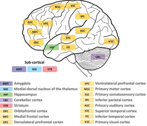
Figure 2.0.2. Graphical representation of brain regions assessed ...52
Figure 2.1.1. Summary of all genes analyzed from AutDB, CarpeDB, and SZgene ...61
Figure 2.1.2. Summary of the subset of highly expressed genes identified ...63
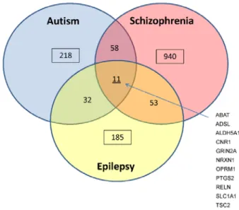
Figure 2.1.3. Overlapping gene-networks in ASD ...68
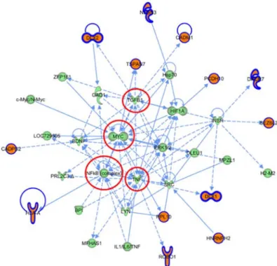
Figure 2.1.4. Network 1 derived from the ASD highly expressed gene set ...68
Figure 2.1.5. Network 2 derived from the ASD highly expressed gene set ...69
Table 2.1.1 GO enrichment analysis of the 11 genes shared by Autism, Schizophrenia and Epilepsy...61
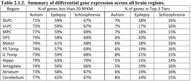
Table 2.1.2. Summary of differential gene expression across all brain regions ...62
Table 2.1.3. GO enrichment analysis of highly expressed autism genes ...64
Table 2.1.4. GO enrichment analysis of highly expressed schizophrenia genes ...65
Table 2.1.5. Canonical pathways implicated in ASD when considering all genes versus only highly expressed genes ...66
Table 2.1.6. Canonical pathways implicated in schizophrenia when considering all genes versus only highly expressed genes ...66
Table 2.1.7. Canonical pathways implicated in epilepsy when considering all genes versus only highly expressed genes ...67
Table 2.1.8. Cell-type specific protein expression of highly expressed ASD genes from the Human Protein Atlas database ...70
Table 2.1.9. Correlation of AutDB genes with published transcriptome studies in ASD brain ...71
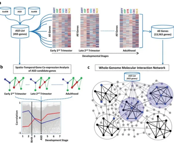
Figure 2.2.1. Graphical representation of methodologies used in this analysis ...79
Figure 2.2.2. Spatio-temporal gene co-expression analysis of ASD candidate genes ...81
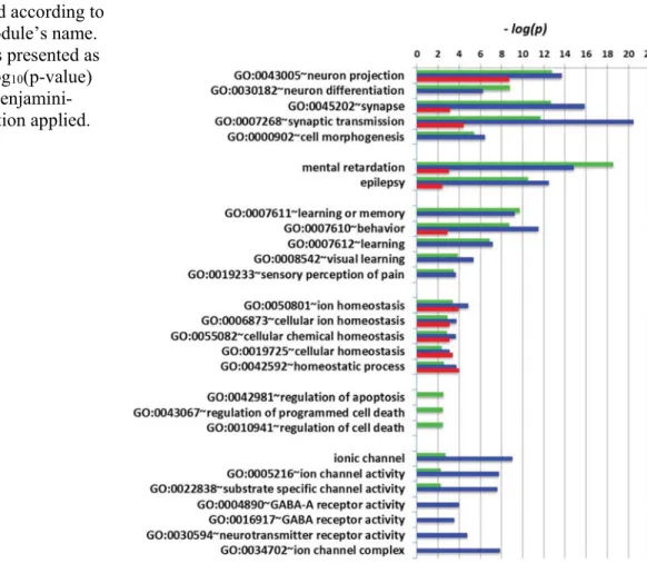
Figure 2.2.3. Gene ontology terms enriched in each of the three modules ...82
Figure 2.2.4. Enrichment scores for each of the ASD modules in neurons, astrocytes, and oligodendrocytes ...82
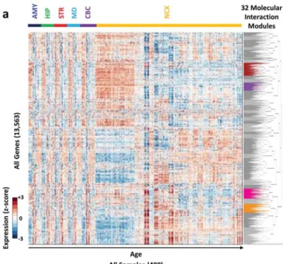
Figure 2.2.5. Transcriptome-wide Molecular Interaction Networks ...84
Figure 2.2.6. ASD modules ...85
Figure 2.2.7. Enrichment of the ASD modules in cell-type specific genes ...86
Figure 2.2.8. Hub genes of ASD modules ...87
Table 2.3.1. List of all significant gene ontology results from analysis of male genes ...99
Figure 2.4.1. Number of differentially expressed miRNAs within each brain region over development ...104
Figure 2.4.2. Number of differentially expressed miRNAs between brain regions over development ...105
Figure 2.4.3. Enrichment of differentially expressed miRNA target genes by brain region for
disease associated genes ...107
Figure 2.4.4. Enrichment of differentially expressed miRNA target genes among male versus female sets for disease associated genes ...107
Figure 2.4.5. Temporal, spatial, and isoform-specific miRNA regulation of three autism candidate genes ...108
Table 2.4.1. Developmental periods and average number of donor tissue samples assessed ...102
Table 2.4.2. Differentially expressed miRNAs between male and female prefrontal cortex over development ...105
Table 3.0.1. Clinical characteristics and RNA quality of autistic and control samples ...113
Figure 3.1.1. Summary of differentially expressed lncRNAs and mRNAs ...120
Figure 3.1.2. Distribution of differentially expressed lncRNAs by genomic origin ...120
Figure 3.1.3. qRT-PCR analysis of select lncRNAs ...121
Figure 3.1.4. Relative orientation and distance to the nearest transcriptional start site (TSS) of all differentially expressed lncRNAs ...122
Table 3.1.1. Characteristics of patients from whom brain samples were obtained ...117
Table 3.1.2. Source of lncRNAs contained on ArrayStar lncRNA microarray ...117
Table 3.1.3. Gene ontology analysis of the 381 mRNA loci nearby differentially expressed lncRNAs ...121
Table 3.1.4. Genes near differentially expressed lncRNAs that were previously implicated in ASD or shown to be differentially expressed in ASD brains ...122
Table 3.1.5 Gene ontology analysis for differentially expressed mRNAs between autism and control prefrontal cortex ...123
Table 3.1.6 Gene ontology analysis for differentially expressed mRNAs within control prefrontal cortex versus cerebellum ...124
Figure 3.2.1. Expression of cell-type specific markers in pre-frontal cortex samples of autistic cases relative to controls ...130
Figure 3.2.2. Expression of cell-type specific markers in cerebellum samples of autistic cases relative to controls...131
Table 3.2.1. Primers used for qRT-PCR ...123
Figure 3.3.1. Map of the human mitochondrial genome ...137
Figure 3.3.2. Schematic of mitochondrial ATP generation ...138
Table 3.3.1. Differentially expressed mtDNA genes in the prefrontal cortex of ASD ...142
Table 3.3.2. Differentially expressed mtDNA genes in the cerebellum of ASD ...142
Table 3.3.3. Significant gene ontologies of differentially expressed nuclear-encoded mitochondrial genes ...143
Table A1. Demographic information of donor brains in the BrainSpan Atlas used in this analysis ...153
Table A2. List of autism candidate genes used in Chapter 2 analyses...154
Supplementary Figure A2. Distribution plot of the number of strongly correlated gene-pairs per gene set from Chapter 2.2 ...122
Chapter 1. Introduction
The human brain is exceedingly complex, and the mechanisms underlying its development and functioning are only beginning to be understood. As a consequence, the etiology underlying disorders of neurodevelopment, such as autism spectrum disorder (ASD), remains unclear. However, recent efforts have demonstrated that a significant component of the etiology of ASD is genetic, but the exact genetic and molecular mechanisms underlying the disorder have proven exceedingly difficult to define. This is largely because the genetics guiding normal human brain development are still not clear, and studies of genes implicated in ASD mostly have not considered the unique function of these genes in the specific context of human neurodevelopment.
Therefore, in order to more comprehensively understand the genetic etiology of autism spectrum disorders, it is critical to understand the function and regulation of autism candidate genes during normal human brain development. To do so requires integrating what has previously been discovered about the genetics of autism with what is known about normal human neurodevelopmental genomics, as is reviewed in this chapter. This information can then be applied to analyze new datasets of human gene expression during neurodevelopment. Furthermore, it is critical that parallel efforts are made to determine what molecular genetic mechanisms are aberrant in autistic brain tissue directly, as the overlap of these two lines of evidence may help focus on the main inherited genetic etiologies of autism.
Finally, as is shown in this chapter, there is clearly a need to more comprehensively understand the expression and regulation of ASD candidate genes during normal human brain development, and to determine if the molecular mechanisms they implicate are abnormal in autistic brains. This chapter concludes by describing these major unanswered questions in the field of ASD genomics, which are then addressed in the studies described in Chapter 2 and Chapter 3 of this thesis.
1.1
Autism Spectrum Disorders
The autism spectrum disorders are a heterogeneous set of neurodevelopmental syndromes defined by impairments in verbal and non-verbal communication, restricted social interaction, and the presence of stereotyped patterns of behavior. The prevalence of ASD is rising, and the diagnostic criteria and clinical perspectives on the disorder continue to evolve in parallel. Although the majority of individuals with ASD will not have an identifiable cause, almost 25% of cases have genetic lesions (Huguet et al. 2013). The rapidly improving ability to identify genetic mutations because of advances in next generation DNA sequencing, coupled with previous epidemiological studies demonstrating high heritability of ASD, have led to many recent attempts to identify causative genetic mutations underlying the ASD phenotype. However, although hundreds of mutations have been identified to date, they either are rare variants affecting only a handful of ASD patients, or are common variants in the general population with only a small effect size on the risk for ASD (Devlin and Scherer 2012). Furthermore, the genes implicated thus far are heterogeneous in their structure and function, hampering attempts to understand shared molecular mechanisms among all ASD patients; an understanding that is crucial for the development of targeted diagnostics and therapies. Therefore, a major unmet need in the field of ASD research—and the main goal of this work—is to integrate the heterogeneous genetic findings in ASD in order to begin to understand common molecular and cellular pathways that are perturbed in patients with the disorder.
Clinical
Phenotype
and
Incidence
Autism was first described seventy years ago by the American child psychiatrist Leo Kanner (Kanner 1943). While originally reported by Kanner as an isolated syndrome with the core components being ‘obsessive insistence on the preservation of sameness’ and ‘autistic aloneness,’ autism was considered mainly as a childhood form of schizophrenia for more than thirty years (Eisenberg and Kanner 1955). Autism was first formally recognized as its own clinical diagnostic entity in 1980 (American Psychiatric Association, DSM-III, 1980), defined as encompassing three essential features: impairment in communication, lack of interest in other people, and ‘bizarre’ behaviors. Since that time, the criteria required to obtain a diagnosis of ASD, and its relation to other similar disorders such as Asperger’s and Rett syndrome, have changed multiple times—reflecting both the clinical heterogeneity of the disorder and the poor understanding of its underlying pathophysiology.
The most recent definition of ASD recognizes abnormalities in two clinical domains: ‘social and communication defects’ and ‘fixed interests and repetitive behaviors’ (American Psychiatric Association, DSM-V, 2013). All of the following three symptoms describing persistent deficits in social interaction and communication must be present for a diagnosis of ASD to be made: (i) problems reciprocating social or emotional interaction, inability to initiate an interaction, and problems with shared attention or sharing of emotions and interests with others; (ii) problems maintaining relationships and problems adjusting to different social expectations; and (iii) nonverbal communication problems such as abnormal eye contact, posture, facial expressions, tone of voice and gestures, as well as an inability to understand these. Additionally, these interaction/ communication deficits cannot be better accounted for by general developmental delay. Two of the four following symptoms related to restricted and repetitive behavior must also be present: (i) stereotyped or repetitive speech, motor movements, or use of objects; (ii) excessive adherence to routines, ritualized patters of verbal or nonverbal behavior, or excessive resistance to change; (iii) highly restricted interests that are abnormal in intensity or focus; and (iv) hyper- or hypo-reactivity to sensory input or unusual interest in sensory aspects of the environment.
Furthermore, the severity of each symptom must be defined based on the level of support required for that symptom, in an attempt to more thoroughly capture the ‘spectrum’ nature of the disease. In all cases, symptoms must have been present in early childhood (even if initially unrecognized); although they may not become fully manifest until later in life when social demands exceed capacities. The symptoms must impair everyday functioning, and cannot be better described by another Diagnostic and Statistical Manual of Mental
Disorders-5th Edition (DSM-5) diagnosis.
Autism spectrum disorders are one of the most common neurodevelopmental problems affecting children in the Western world. The most recent estimates have shown that ASD affects between 1 in 88 children (Centers for Disease Control and Prevention (CDC) 2012) and perhaps as many as 1 in 50 (Blumberg et al. 2013) depending on the methodology employed. This represents a staggering 1.17%-2% of all children. Boys are at least four times more likely to receive a diagnosis of ASD as compared to girls (CDC 2012), and this ratio
increases significantly when only mildly affected children are considered (Gilberg et al.
years—form 1 in 150 children in the year 2000—although it is unclear to what extent this represents a true biological increase or is a result of expanding diagnostic criteria and better clinical recognition of the disorder (Fombonne 2009).
The costs associated with autism are similarly great. Economically, direct and indirect medical costs are estimated to be nearly £2 million pounds person over his or her lifetime, or more than £21 billion pounds per year for all people with ASD (Moldin and Rubenstein 2006). Perhaps more importantly, the emotional toll placed on parents and caregivers of children with autism is immense, unrelenting, and has a serious impact on family relationships (Rao and Beidel 2009), marriages (Benson and Kersh 2011), and couples’ future reproductive decisions (Selkirk et al. 2009).
Consequently, it is of upmost urgency to patients with ASD, their caregivers, and society at large that the underlying cause(s) of the disorder are understood. Doing so will enable the development of better, more specific diagnostic tests that can recognize ASD earlier in life, which has been shown to be important to improve long-term outcomes (Howlin et al. 2009), provide parents with an explanation for their child’s symptoms, and may eventually enable the development of targeted therapeutics. Moreover, by understanding the mechanisms that lead to the altered higher cognitive functioning seen in patients with ASD, the field of human neuroscience as a whole can be advanced, as it will provide insights into the genetic and molecular basis of higher cognition.
However, the underlying pathophysiology of autism spectrum disorders has long been a mystery. Various hypothesis ranging from psychosocial abnormalities to environmental insults have been purported, yet it was not until twin and sibling epidemiological studies were undertaken in the 1980s that the strong heritability of ASD began to be realized. Subsequently, a large amount of work has firmly established a significant genetic component to ASD’s etiology.
Genetic
Etiology
Evidence for a strong heritable risk of ASD was initially described in twin and sibling epidemiological studies of autism (Folstein and Rutter 1977), and has since been firmly established through multiple genetic approaches (Berg and Geschwind 2012; Geschwind
2011). It was first recognized that the risk of having a second child with autism was higher in families that already had one child with ASD than was the risk of having a child with ASD in the general population. Originally this recurrence risk was estimated to be 5% (compared with approximately 1% in the general population), although more recent estimates suggest it may be as high as 20% (Ozonoff et al. 2011). Following these initial observations, the first twin studies in ASD demonstrated a concordance rate approaching 90% in monozygotic twins and 10% in dizygotic twins (Bailey et al. 1995; Steffenburg et al. 1989; Smalley et al. 1988; Ritvo et al. 1989). Subsequently, larger studies have shown the dizygotic concordance rate to be greater than 20% (Hallmayer et al. 2011).
These observations, coupled with the identification of causative genetic mutations in monogenic disorders with autism as a component, such as Fragile X and Rett syndromes (Amir et al. 1999; Pieretti et al. 1991), led to an ongoing effort to identify genetic causes of ‘idiopathic’ ASD using a number of genomic approaches. As the technology behind these approaches has improved, the ability to identify mutations with incredibly sensitivity and genomic resolution has resulted in over 200 genetic loci implicated in ASD to date (Freitag 2007; Anney et al. 2010; Holt et al. 2010). However, as more genes and loci are identified, it is becoming increasingly clear that the genomic architecture of ASD is incredibly heterogeneous and complex, necessitating a functional integration in order to decipher common molecular mechanisms underlying ASD.
Genomic
Architecture
of
ASD
The identification of genomic loci and individual genes disrupted in patients with ASD has progressed in tandem with the rapid development of sensitive genomic tools. Initially, microscopically-visible chromosomal aberrations were observed in patients with ASD who received karyotyping analysis. These case reports were variable, but a number of loci were repeatedly implicated, including 7q11, 15q11-13, and 22q11.2 (Vorstman et al. 2006)— regions already associated with syndromes that had autistic symptoms as a component, and known to contain a number of critical neurodevelopmental genes and some of the first identified functional non-coding RNAs (Mabb et al. 2011; Szafranski et al. 2010).
Subsequently, the development of microarray technology such as comparative genomic hybridization (CGH, Alkan et al. 2011), allowed the unbiased assessment of copy-number
variation (CNV) across the whole genome at a resolution of as low as 100 kilobases. The first of these analysis indicated that individuals with ASD had 10-20 times the number of CNVs as controls (Jacquemont et al. 2006; Sebat et al. 2007). Numerous studies have since used CGH or similar approaches to follow up and improve upon these initial reports with larger and more homogenous patient populations, with thousands of individuals with ASD having been analyzed to date (Christian et al. 2008; Cooper et al. 2011; Gilman et al. 2011; Glessner et al. 2009; Itsara et al. 2010; Marshall et al. 2008; Pinto et al. 2010; Sanders et al. 2011; Szatmari et al. 2007; Huguet et al. 2013). These studies have consistently demonstrated that individuals with ASD have more CNVs than non-related controls. Furthermore, studies employing a family cohort model have been able to compare individuals with ASD to their parents and unaffected siblings, which has revealed that de novo mutations in particular are more frequent in children with ASD.
Functionally, it was also shown that larger CNVs (i.e. affecting more genes) are associated with decreased cognition (Girirajan et al. 2012), and that females with ASD tend to have larger CNVs than males with ASD (Itsara et al. 2010; Sanders et al. 2011), suggesting they are somehow more ‘genetically tolerant’ of these disruptions. Moreover, some of the identified loci result in nearly opposite phenotypes depending on whether they are duplicated or deleted (Jacquemont et al. 2011). Taken together, these functional CNV findings suggest that identification of the genes in these regions is not sufficient to understand the mechanisms underlying autism, as it appears that a finely-regulated dosage of each gene is necessary to avoid neurodevelopmental problems such as ASD.
Despite the progress made with CGH arrays, the findings from these studies only identified CNVs in 5-15% of individuals with ASD, suggesting that other types of mutations must be operant in ASD as well. However, investigations at higher genomic resolution were traditionally limited to specific candidate genes until the recent advent of next-generation sequencing technologies. Since then, seven exome sequencing studies have been completed in ASD, encompassing more than 1,000 affected individuals (Klei et al. 2012; Kong et al. 2012; Neale et al. 2012; O’Roak et al. 2011; O’Roak et al. 2012a; O’Roak et al. 2012b; Sanders et al. 2012). In addition to identifying a number of high-confidence ASD candidate genes (likely representing 5-10% of ASD cases), these studies provided two other more broad insights into the functional genomics of ASD that are particularly motivational to the work
describe in this thesis. First, with the exception of a few identified genes, there was very little replication of ASD candidates among the studies. This suggest that common variants (i.e. those accounting for greater than 1% of cases) are unlikely to play a major role in ASD pathogenesis, confirming similar prior findings from genome-wide association studies and linkage analysis that failed to identified many replicable loci (Szatmari et al. 2007; Wang et al. 2009; Weiss et al. 2009). Consequently, it has been predicted that up to 1,000 genes may be found to be associated with ASD based on statistical modeling (Sanders et al. 2012; Iossifov et al. 2011). Therefore, understanding how such a large and varied number of genes can all be associated with one common clinical phenotype is a major challenge in the field, and one of the focuses of this work. Secondly, a meta-analysis of these studies at the group level showed that the average rate of mutations in individuals with ASD was not significantly different than controls—or even unaffected siblings—unless the analysis was restricted to genes that are known to be expressed during human brain development (Sanders et al. 2012). This highlights the tissue- and human-specific nature of gene function, which underscores the importance of understanding the function of ASD candidate genes in the context of human brain development specifically.
Lastly, there is a growing appreciation that the presence of multiple mutations and/or inherited protective or risk alleles—each at different loci within one individual—may interact with each other to result in the emergent ASD phenotype, and that this may help explain the complex and heterogeneous nature of ASD genomics. For instance, a number of studies have described individuals with ASD who have more than one deleterious mutation (Girirajan et al. 2010; Girirajan et al. 2012; Leblond et al. 2012), and the presence of more than one mutation correlates with an increased risk of developmental delay (Girirajan et al. 2012). Other studies have suggested certain inherited variants may be protective against other ASD-causing mutations, especially in females (Robinson et al. 2013). While the identification of multiple mutations within individuals is becoming a relatively straightforward task, the challenge of understanding how combinations of susceptibility genes interact during human brain development to cause disease (epistasis) has only begun to be explored, and is a major theme of this thesis.
In summary, much work has attempted to elucidate the molecular genetics underlying autism, with many linkage, genome-wide association, copy number variation, and whole-exome
sequencing projects having implicated hundreds of genes in ASD. Yet understanding how this diverse set of genes relates to the underlying molecular mechanisms and subsequent neuropathology of ASD is still unclear. The genetic etiology of ASD is variable, complex, and likely involves gene-gene, gene-environment, and epigenetic interactions, as is evidenced by the incomplete concordance among monozygotic twins, and the considerable variability within pedigrees (Piven et al. 1997; Ronald et al. 2006). This genetic heterogeneity reflects the overlying broad clinical presentation of ASD, and is captured by the ‘spectrum’ designation of the disorder. Furthermore, ASD shares considerable clinical and genetic overlap with other neuropsychiatric disorders such as schizophrenia and mental retardation (Mitchell 2011), and ASD patients have significantly increased neurologic co-morbidities like hypotonia, tics, and epilepsy (Levy 2009). In fact, many of the same gene mutations have been found to predispose to more than one of these neurodevelopmental disorders (Ching et al. 2010; Guilmatre et al. 2009). This body of evidence suggests that while identification of candidate genes in ASD is a critical first step toward understanding the genetic etiology of this disorder, a comprehensive, disorder-specific understanding of the molecular mechanisms cannot be realized until the functional genomics of ASD candidate genes are properly understood in the context of human brain development.
Cellular
Etiology
of
ASD
Although autism currently lacks any unifying principles at the genetic and molecular levels, both human and animal studies have begun to demonstrate that disruption of synaptogenesis and improper connectivity of local and distant brain networks likely underlie the cellular pathophysiology responsible for the broad ASD phenotype (Geschwind and Levitt 2007; Zoghbi 2003). Multiple different brain regions have been implicated in both post-mortem and neuroimaging studies, notably the prefrontal and temporal cortices, and the cerebellum (Abrahams and Geschwind 2010). Histological analysis has revealed increased cell densities, changes in synaptic spine morphology, mini-columnar disorganization, and glial activation (Pickett and London 2005). Intriguingly, many of the genes known to be integral to these processes have been independently linked to autism in genetics studies. For instance, the Shank family of proteins, which interact with themselves and other transmembrane proteins at the post-synaptic density, are one of the main regulators of synaptic spine morphology (Sala et al. 2001). Multiple Shank family genes, notably Shank 1 and Shank 3, have been repeatedly implicated in ASD (Bourgeron 2009). Similarly, genes involved both in formation
and maintained of cortical mini-columns, such as the cadhedrin family of proteins (Redeis et al. 2012), and genes involved in glial activation, such as members of the Wnt/B-catenin pathway like DOCK1 and WNT2 (Yang 2012), have been independently implicated in ASD through genetic studies (Wang et al. 2010; Kalkman 2012).
Despite these observations, the underlying mechanism(s) responsible for this “disconnection” phenotype remains obscure, as a complex interplay between diverse cell types and functions modulate the developing network architecture in both a temporally and spatially regulated manner (Levitt 2003; Vogel et al. 2010; Bolton et al. 2009).
In particular, studies have shown that long-distance communication between disparate neocortical areas may be disrupted in ASD, causing delays in information processing within the brain that manifest as the communication, language, and social development problems seen in children with autism (Just et al. 2007). Additionally, parallel research has shown that neuronal micro-circuitry within brain areas may also be disrupted in ASD, and that this may result in local processing deficits within brain regions related to higher functioning, such as the prefrontal cortex (Rubenstein and Merzenich 2003). Underlying these circuit disruptions is a large body of evidence that has demonstrated decreased numbers of neurons (and their various subtypes) throughout the autistic brain by early childhood in post-mortem studies (Courchesne et al. 2007).
In addition to the body of evidence implicating aberrant local and long-distance synaptic dysfunction in ASD, many studies have demonstrated microglial and astrocyte dysfunction in ASD brains. For instance, post-mortem pathological studies of autistic brain using immunocytochemistry (IHC) and/or stereology have identified microglial activation patterns (Vargas et al. 2005; Morgan et al. 2010; Morgan et al. 2012), and have demonstrated increased microglial cell density in multiple brain regions (Morgan et al. 2010; Tetreault et al. 2012). Furthermore, positron emission tomography (PET) using a microglial-specific radiotracer also demonstrated microglial activation in multiple brain regions of autistic cases (Suzuki et al. 2013). Additionally, studies in a Rett syndrome mouse model, a single-gene deletion disorder with autism as a component, have also demonstrated cellular microglial abnormalities (Maezawa and Jin 2010), and a remarkable study demonstrated that autistic-like phenotypes can be partially reversed by replacing mutant microglia with their respective wild-type cells (Derecki et al. 2012).
Increased numbers of astrocytes, with altered cell size and branching patterns, have also been demonstrated in post-mortem autistic brains (Cao et al. 2012). Additionally, astrocyte-specific cell marker proteins are increased in multiple autistic brain regions (Laurence and Fatemi 2005; Fatemi et al. 2008). Similar to microglial studies in ASD mouse models, astrocytes have been shown to be abnormal in number of single-gene ASD models, including Rett (Maezawa et al. 2009; Yasui et al. 2013), Fragile X (Yaskaitis et al. 2010), and Tuberous Sclerosis (Uhlmann et al. 2002). In parallel to the aforementioned microglial study, it was also shown that replacing mutant astrocytes in Rett syndrome mice could correct some aspects of the phenotype (Lioy et al. 2011).
Overall, the cellular pathology in the brains of individuals with ASD is equally as complicated as the underlying genetics. While there is strong evidence to suggest that the autistic phenotype ultimately results from aberrant local and long-distance synaptic wiring, it remains unclear if the repeated observation of altered microglia and astrocytes are contributory to the phenotype or represent a reaction to synaptic pathology. However, previous functional genomics studies of ASD brain tissue (discussed in Chapter 1.3) have demonstrated altered immune and glial gene expression in autistic brains, suggesting that glial cell abnormalities may contribute to defects in synaptic wiring. The complex interplay between ASD genetics and glial cell abnormalities is explored further throughout this work.
Conclusion
In summary, autism spectrum disorders are common, and have considerable consequences for individuals with ASD, their families, and society at large. Because the underlying causes of ASD are not understood, specific diagnostic tests and therapeutic strategies are unavailable. The evolution of ASD’s clinical definition is indicative of the heterogeneous and complex nature of the disorder. While ASD has recently been shown to have a significant genetic etiological component, the genes implicated are equally heterogeneous, hampering attempts to define common molecular mechanisms. In parallel, cellular studies have revealed that ASD likely ultimately results from disrupted synaptic function, but a large body of evidence has also implicated immune and glial abnormalities in autistic individuals. Therefore, studies that attempt to reconcile the heterogeneous and varied nature of ASD genomics, and the interplay between neurons and glial, are necessary to move the field forward toward a common understanding of the mechanisms underlying the development of ASD.
1.2
Functional Genomics of Human Brain Development
Transcription of the inherited DNA sequence into copies of messenger RNA (mRNA) is the most fundamental process by which the genome functions to guide development. Furthermore, encoded sequence information, inherited epigenetic marks, and environmental influences all converge at the level of mRNA gene expression to allow for cell type-specific, tissue-specific, spatial, and temporal patterns of expression. Thus, the transcriptome represents a complex interplay between inherited genomic structure, dynamic experiential demands, and external signals. This property makes transcriptome studies uniquely positioned to provide insight into complex genetic-epigenetic-environmental processes such as human brain development, and disorders with non-Mendelian genetic etiologies such as autism spectrum disorders.
As humans develop, an individual gene can be expressed in multiple ways depending on the particular developmental context; that is, the tissue, stage of development, and local or long-distance signaling mechanisms being received. Therefore, in order to understand how a gene may contribute to a developmental disorder, it is critical to assess its expression and function in the appropriate tissue and developmental time window. Human brain gene expression has been demonstrated to be particularly unique evolutionarily, compared to other human tissues, and in its complex regulatory processes, underscoring the need to understanding the functional genomics of genes implicated in autism spectrum disorders during human brain development.
A
Brief
Overview
of
Human
Brain
Development
at
the
Cellular
Level
The complex processes that lead to the fully formed human brain encompass a spectrum of mechanisms spanning genetic determinates to environmental and experimental influences. While the functional genomic mechanisms underlying human brain development remain poorly understood—motivating much of the work in this thesis—over the past several decades significant advances have been made to document the cellular and anatomical events that occur as the human brain develops and matures. It is therefore important to consider studies of gene expression in this context of cellular/anatomic brain developmental patterns.
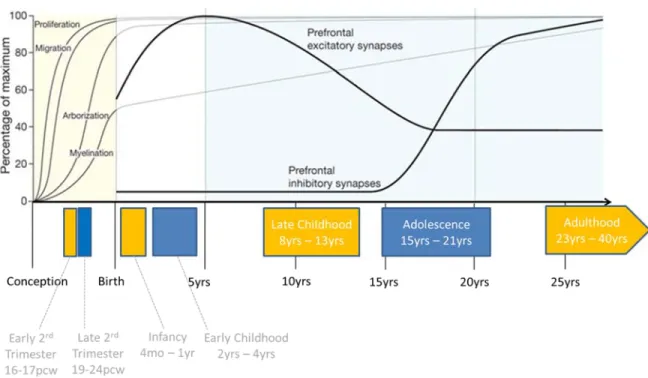
Cellular human brain development is a protracted process that begins around the third post-conception week (pcw) and arguably extends nearly into adulthood (Stiles and Jernigan, 2010). Conventionally, human brain development is considered in gross stages within which major cellular and anatomic transitions occur (Figure 1.2.1, Insel 2010); namely the embryonic, fetal, early and late postnatal, adolescent, and adult periods.
Figure 1.2.1. Trajectory of major brain developmental processes and their relationship to work performed in this thesis. Top: the trajectory of the major cellular phases of brain development are depicted as a percentage of their maximum abundance across human development. Bottom: Stages of brain development that are assessed in Chapter 2 of this thesis are depicted in colored blocks. Names and age ranges of the stages are given. Pcw; post-conceptional weeks. Figure adapted from: Insel TR.
Nature. 2010;468(7321):187-93.
Beginning early in the embryonic period (defined as conception to eight pcw), the basic structures of the brain, spinal cord, and peripheral nervous system are established. The first major differentiating event of the embryonic period is gastrulation, during which the single-layered blastula forms a trilaminar structure containing the ectoderm, mesoderm, and endoderm. Gastrulation is completed by the third pcw, at which time some cells of the ectodermal layer differentiate into neural progenitors (Ozair et al. 2013). The first well-defined neural structure, the neural tube, begins forming during the third pcw and serves as the basis of the early developing central nervous system, within which reside populations of neural stem cells. From this basic tubular structure, more specific neural patterning of what
will become the major brain structures and compartments occurs through the creation and migration of neural cells from the stem cell proliferative zones. Through graded patterns of molecular signaling, neural progenitors migrate outward from proliferative zones and begin differentiation such that a primitive map of the brain is established by the end of the embryonic period. For instance, through comparative studies of other mammals it has been projected that the sensimotor regions of the neocortex (Bishop et al. 2002), the major compartments of the diencephalon and midbrain (Nakamura et al. 2005; Kiecker and Lumsden 2004), and the organization of the hindbrain and spinal column are all well established by the end of the embryonic period in humans (Lumsden and Keynes 1989; Gavalas et al. 2003).
Around the ninth pcw, the fetal period of development ensues and extends until birth, during which time there is rapid growth of the structures established during the embryonic period. Grossly, the brain develops its characteristic gyri and sulci during the fetal period (Chi et al. 1977), reflecting the underlying dramatic cellular changes occurring during this period. The majority of neuronal and glial proliferation occurs between the 9th and 16th pcw, with the
peak period of migration of these cells to their appropriate region following closely thereafter (Volpe 2000). In fact, production of new neurons is largely finished by midgestation, except for the ongoing production of neurons in a few specialized areas (Bystron et al. 2008).
After their production in the proliferative regions, neurons migrate in an orderly manner to their final position in the developing brain. In the neocortex, the arriving cells establish a 6-layered structure, with the earlier migrating neurons forming the deeper layers and the later migrating neurons forming the more superficial layers (Cooper 2008). Their migration from the proliferative zone to their final position in the neocortex is helped by the guidance of radial glial cells, a population of stem cells that serve as a scaffold in the developing brain of all vertebrates (Borrell and Götz, 2014). Different layers of the neocortex contain different types of neurons as a result of both cell-intrinsic mechanisms operant in the progenitor cells from which they derive (Leone et al. 2008), and through soluble signaling cascades that direct progenitors toward a restricted mature neuronal type (Desai and McConnell 2000).
Of particular note in this migration process are a set of structures that appear only transiently during the fetal period to help guide the migration of progenitors to the developing
neocortical layers. The very first neurons to populate the developing neocortex form a primitive and transient structure termed the preplate, which is then split into two separate structures by arriving neurons—the marginal zone and the subplate (Molnár et al. 2006). The region between the marginal zone and subplate serves as a hub for new arriving neurons, and will eventually become layer 6 (the deepest) of the developing neocortex. Subsequently, all newly arriving cells will form progressively more superficial layers of the neocortex from this base structure. Intriguingly, both the marginal zone and subplate disappear by the end of the fetal period, yet they have been shown to highly express some of the genes most significantly linked to neurodevelopmental disorders such as autism and schizophrenia, such as Reelin (Bielle et al. 2005; Hoerder-Suabedissen et al. 2013). Consequently, an important caveat to post-mortem tissue research, both cellular and genetic, is the possibility of omitting the contribution of these transient structures to the proper formation and potential abnormalities in neocortical patterning.
Once the migrating neural cells have reached their destination, they begin to be incorporated into newly developing neural networks through a dynamic process of synaptogenesis and pruning that continues late into adolescence. Young neurons initially develop processes (dendrites and axons) that allow them to form synapses with other neurons both locally and long-distance. The growth cone of an axon is able to sample the neuron’s environment for both chemical and electrical signals that guide its wiring to other neurons to create a new synapse (Brown et al. 2001). Initial patterns of connectivity in the fetal and early postnatal brain are characterized by exuberant synaptic connections that will later be pruned away to leave only the connections indicated through postnatal experience (Stiles and Jernigan, 2010). This process of network refinement occurs through both synaptic rewiring and neuronal apoptosis, with rates of apoptosis as high as 70% of cells in some regions of the cortex (Rabinoqicz et al. 1996). Physiological neuronal apoptosis in development occurs both as the result of intrinsic neuronal cell death mechanisms mainly responding to the absence of local neurotrophic factors (Huang and Reichardt, 2001), and also through glial-initiated mechanisms which have recently become more widely recognized (Kettenmann et al. 2013), which are of particular relevance to much of the work presented in Chapters 2 and 3 of this thesis. This synaptic and network refinement continues through early adulthood, largely in response to interaction with one’s environment (Huttenlocher, 1987, Paus et al. 2001).
By the end of fetal development, all major adult brain structures are present, major connections between them are established, and the brain is poised for the rapid and dynamic growth that occurs in the first few years of life. The brain develops rapidly in the first few years after birth, reaching almost adult volume by age six (Lenroot and Giedd, 2006). While the production and migration of neurons are mainly prenatal events (with the notable exception of subventricular zone), glial progenitors have been shown to proliferate and differentiate throughout childhood (Cayre et al. 2009), like helping to sculpt the developing synaptic networks.
One main function of these proliferating glial cells during the early and late postnatal periods is to accomplish the extensive amount of axon myelination that occurs during this time. Increased myelination of axons allows for increased growth of axon diameter, and ultimately enables faster and long-distance neuronal connections (Zalc et al. 2008). Robust increases in myelination have been reported across the brain from ages 5 – 12 years, with a varying rate of fiber tract myelination in various brain regions (Lebel et al. 2008; Lebel and Beaulieu, 2009).
While the early postnatal period is characterized anatomically by an over-abundance of synaptic connections (“overconnectivity”) between neurons, these connections are gradually pruned back over the course of development by competitive experiential processes. In fact, this particular pruning mechanism is hypothesized to be one of the main altered mechanisms in neurodevelopmental disorders like autism (Just et al. 2004). Modern neuroimaging techniques such as diffusor tensor imaging (DTI) and functional magnetic resonance imaging (fMRI) have made significant recent advances in linking brain structural changes to functional and behavioral development, with longitudinal neuroimaging studies having demonstrated changes in grey matter density throughout the neocortex into the mid-twenties, with the prefrontal cortex being the last to mature (Paus et al. 2008). While much of the organization of the postnatal brain is genetically determined (Stiles and Jernigan, 2010), it has been clearly demonstrated that this intrinsic development remains extremely malleable to experience-dependent processes (Hubel and Wiesel, 1977; Markham and Greenough, 2004).
Moreover, epigenetic mechanisms that ultimately converge to influence gene expression have been shown to be one of the main mediators between environmental experiences and developmental synaptic plasticity (Fagiolini et al. 2009). For instance, studies in mice have
shown that environmental enrichment results in increased chromatin remodeling that modifies gene expression patterns in the hippocampus, resulting in improved spatial memory (Fischer 2007). Alternatively, an increase in methylation of the BDNF promoter and consequent decrease in BDNF mRNA in the prefrontal cortex was found in association with exposure to periods of abusive maternal care, and these effects are perpetuated to the F1 generation suggesting a role for transgenerational effects (Champagne 2008). Yet while studies of model organisms are beginning to demonstrate that gene expression represents a critical nexus of experience dependent plasticity, human studies of neurodevelopmental disorders in which this process may go awry are limited, and the general landscape of gene expression in the developing human brain as relates to neurodevelopmental disorders like autism is largely unexplored.
In summary, great progress in understanding the anatomical and cellular trends underlying human brain development have been made over the past few decades. We have come to appreciate though various approaches that human neurodevelopment is a dynamic and protracted process, characterized by an initial period of neurogenesis leading to the formation of the basic CNS framework in early embryonic development. This is following by substantial cellular proliferation, migration, and differentiation in the fetal period that establishes the main areas and pathways of the brain by birth. The early postnatal period is a time of rapid growth through glial proliferation, myelination, and organization of developing neural networks. Importantly, this process is very malleable particularly with regard to environmental and experiential events. Precise refinement of these developing neural networks occurs throughout adolescence and into early adulthood.
While the cellular events that lead to the initial formation and subsequent refinement of human neuroanatomy are fairly well defined, the underlying molecular and genetic determinates that in part encode for these events are much well-less understood. Recent efforts to profile genome-wide expression patterns in post mortem human brains across development have begun to expose the uniqueness of human brain functional genomics, as is discussed in the next section.
Human
Brain
Gene
Expression
Compared to other species, human brains express mRNA transcripts at much higher levels and with much greater complexity. For instance, comparisons of human brain gene expression with both mouse (Enard et al. 2002; Lockhart and Barlow 2001) and primates (Caceres et al. 2003; Khaitovich et al. 2004) has demonstrated that most of the differentially expressed genes between the species are up-regulated in humans, but this phenomena is not apparent in other tissues. Additionally, the human brain expresses ~86% of all genes encoded in the human genome at some point during development (Kang et al. 2011), which is greater than any other individual tissue type. It is hypothesized that this increased level of gene expression in the human brain is at least partially responsible for the higher level of neuronal activity and overall cognitive function in humans.
Within humans specifically, the brain also displays a distinct gene expression profile from other tissues. Using both array (de la Grange et al. 2010) and sequencing-based techniques (Ramskold et al. 2009), the brain has been shown to have higher expression levels and greater transcriptome complexity than other human tissue and cell types. In particular, human brain gene expression displays a high level of alternative splicing and a unique diversity of non-coding RNA types expressed. For example, studies have demonstrated that the human brain transcriptome has an unusually high level of alternatively spliced transcripts compared to other tissues (Yeo et al. 2004; Wang et al. 2008; Mortazavi et al. 2008), and the set of isoforms produced in brain differs considerably from other tissue types (Yeo et al. 2004; de la Grange et al 2010). In addition to increased numbers and types of spliced mRNAs, the human brain transcriptome also displays a uniquely high abundance of transcribed non-coding RNAs (ncRNAs). In fact, the brain displays the greatest abundance of transcribed ncRNAs among all tissues studied thus far (Qureshi and Mehler 2012). Both short ncRNAs, such as microRNAs (miRNAs) and piwi-interacting RNAs (piRNA), and long non-coding RNAs (lncRNAs) are highly enriched in the brain (Chodroff et al. 2010; Kuss and Chen 2008; Ponjavic et al. 2009; Schonrock et al 2010; St. Laurent et al. 2009). As ncRNAs are becoming increasingly recognized as important regulatory elements in genome processing during neurodevelopment and in the pathogenesis of neurodevelopmental disorders (Qureshi and Mehler 2011), their abundance in the brain further highlights the uniqueness of neurodevelopmental functional genomics (discussed further below).
While within a given brain region the human transcriptome has been shown to be incredibly complex, it is also of importance to consider the relationship among different anatomical regions of the brain, as ‘disconnectivity’ between disparate brain regions is thought to underlie a number of neurodevelopmental syndromes including ASD (Geschwind and Levitt 2007). Perhaps unsurprisingly, there is strong evidence that distinct regions of the human brain have distinct gene expression profiles, and animal studies have suggested that this variation is related to both structural and functional differences (Nadler et al. 2006). For instance, a microarray study of twenty distinct brain and spinal cord sites showed that expression profiles can cluster samples from different donors by anatomical origin, and that some anatomical regions have up to 2,000 region-specific genes (Roth et al. 2006). Multiple studies have shown that the cerebellum contains the most unique gene expression pattern compared to other brain structures (Lockhart an Barlow 2001; Roth et al. 2006; Strand et al. 2007), which is of consequence to autism in particular, as this region has been consistently implicated in the pathogenesis of the disorder (Fatemi et al. 2012). Even just within the neocortex, different cortical layers each express a detectably distinct profile of mRNA transcripts (Molveneaux et al. 2007). Underscoring the importance of region-specific expression are results that have shown gene expression differences between any two brain areas with one individual are more pronounced than are gene expression differences between two different individuals within the same brain region (Strand et al. 2007; Khaitovich et al. 2004; Naumova et al. 2008).
In summary, the human brain has been demonstrated to have a unique pattern and complexity of gene expression both compared to other species and compared to other human tissues, including region specific gene expression patterns, and pervasive transcription of ncRNAs. This highlights the importance of understanding human neuropsychiatric disorders, such as ASD, in the context of human brain gene expression specifically, as it is likely that animal, cellular, and other models do not recapitulate the uniqueness of human brain functional genomics with the appropriate level of fidelity. Moreover, recent evidence is accumulating that suggests gene expression patterns within the human brain vary considerably across developmental time, and therefore temporal patterns of gene expression are also an important consideration.
Changes
in
Gene
Expression
During
Human
Neurodevelopment
The developing human brain grows remarkably fast—the weight of a newborn’s brain is approximately 25% of its adult weight, but within two years, it nearly reaches its adult size (Dekaban and Sadowsky 1978). During this time, the brain grows mainly through glial multiplication, myelination, formation of new synaptic connections, and pruning of unused synaptic connections. While the human brain continues to mature up to the age of 25 years (Sowell et al. 2004), the greatest changes occur in the periods of infancy and early childhood. Coincidentally, most neurodevelopmental disorders, including autism spectrum disorders, become clinically recognizable around this age.
Underlying these dramatic early changes in brain development are complex and dynamic broad patterns of gene expression, which have only recently begun to be understood. The most comprehensive study to date of the developing human brain transcriptome (published after the onset of this work; Kang et al. 2011) documented that genome-wide patterns of gene expression correspond closely to the major stages of clinical development (namely prenatal, early infancy, childhood, adolescence, and adulthood), and that the molecular profile of these stages are distinct from each other. The most striking observation was that the greatest shifts in gene expression occur around the period of birth, where the authors found almost 60% of genes change their expression patterns in the neocortex (Kang et al. 2011). Other studies have demonstrated similar changes, and have showed that many of the genes identified during this shift are known to be involved in cortical development and higher order cognitive functioning (Johnson et al. 2009; Lambert et al. 2011).
The Kang et al. study, which profiled RNA expression using whole-genome microarrays on tissue derived from neurologically normal donor brains spanning the 2nd trimester through
adulthood, also demonstrated that after infancy the number of genes whose expression profile changes in the neocortex decreases dramatically to approximately 9% of expressed genes between infancy and adolescence, and less than 1% of genes between adolescence and adulthood. Functional annotation of these gene sets further revealed that genes expressed very early in prenatal development are highly related to the process of cell differentiation, proliferation, and migration, while genes expressed later in gestation are more related to synaptogenesis, suggesting that time-period specific gene expression patterns drive cell-level developmental programs. Again, these findings highlight the importance of assessing autism
candidate gene expression and function during the appropriate developmental time window, in order to gain the most relevant insight into this disorder.
In addition to the greatest number of genes shifting their expression trajectory shortly after birth, the changes in gene expression in early post-natal life have also been shown to have greater amplitude of change (Colantuoni et al. 2011; Somel et al. 2009; Somel et al. 2010). In fact, it was shown that many genes actually reverse their expression trajectory in early life (Calantuoni et al. 2011), mostly shifting from a pattern of increasing expression in fetal life and infancy to a decrease in expression beginning in childhood. Moreover, as the brain begins to mature, the gene expression profile within each anatomical region becomes more similar to other regions, with the exception of the cerebellum, suggesting that most of the region-specific development is completed early in life. Interestingly, these broad gene expression patterns appear to reverse themselves in older age, at least in the prefrontal cortex (Somel et al. 2010).
Gene expression dynamics in early human brain development are clearly both spatially and temporally specific. This suggests not only that they are highly regulated, but that different genes and gene networks will have dynamic expression throughout space and time. Despite this increasingly recognized property of human neurodevelopmental genomics, few studies of autism candidate genes have considered their expression and function in early human brain development. Furthermore, the molecular regulators of brain mRNA expression, such as non-coding RNAs, have not been extensively characterized in the developing human brain, and their potential involvement in ASD has hardly been studied. Accordingly, an understanding of the functional genomics of autism, the genetic interaction networks that ASD candidate genes participate in, and the potential ncRNA regulators of these genes, represent important unresolved avenues of research, and are addressed by work in this thesis.
Non
‐
coding
RNAs
in
Human
Brain
Development
Since the advent of high-throughput, unbiased, genome expression arrays and sequencing platforms, the recognition that the genome is pervasively transcribed at loci that do not encode for protein products is becoming well recognized. The term ‘non-coding RNA’ (ncRNA) is commonly employed for RNA that is transcribed in the cell but does not encode for a corresponding protein product (Mattick and Makunin, 2006). While originally consider
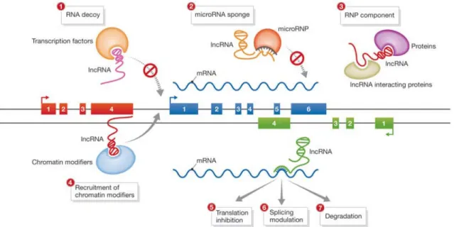
to represent transcriptional ‘noise’ (Ponting and Belgard 2010), this non-coding RNA component of the transcriptome is increasingly implicated in regulating the genomic landscape though a myriad of mechanisms (Figure 1.2.2), and as such are increasingly being recognized as important modulators of gene expression. Moreover, they are also beginning to be implicated in disease.
These ncRNAs can interact with DNA to induce methylation or histone modifications, recruit transcription factors, and modulate the three-dimensional architecture of chromosomes in the nucleus (Ponting et al. 2009). They can bind to other RNA molecules, especially mRNA with complementary sequences, to inhibit translation through RNA degradation, or they can act as ‘sponges’ and thereby dilute the effect of mRNAs or other ncRNAs (Hansen et al. 2013). Conversely, they can increase the rate of translation by acting as molecular stabilizing scaffolds. They can also interact with proteins and protein complexes to catalyze reactions by acting as linkers among otherwise scarce proteins in the cytosol, and participate in cellular trafficking of RNA binding proteins (Mansfield and Keene 2009). Additionally, ncRNAs have been shown to participate in intracellular communication by helping transport cargo between adjacent cells (Skog et al. 2008; Balaj et al. 2011).
Figure 1.2.2. Schematic of some experimentally validated functions of ncRNAs. Long non-coding RNAs, one class of ncRNAs, have been demonstrated to act through a variety of mechanisms