Bioactive Glasses at the Atomic Scale: the Complementary use of Advanced Probe and Computer Modelling Methods.
Jamieson K. Christiea, Alastair N. Cormackb, John V. Hannac, Richard A. Martind, Robert J. Newportd,1, David M. Pickupd and Mark E. Smithc
a) Department of Chemistry, University College London, WC1H 0AJ, UK.2
b) Inamori School of Engineering, Alfred University, NY 14802, USA.
c) Department of Physics, University of Warwick, Coventry, CV4 7AL, UK.3
d) School of Physical Sciences, University of Kent, Canterbury, CT2 7NH, UK.4
Abstract
Sol-gel synthesised bioactive glasses may be formed via a hydrolysis condensation reaction,
silica being introduced in the form of tetraethyl orthosilicate (TEOS) and calcium is typically
added in the form of calcium nitrate. The synthesis reaction proceeds in an aqueous
environment; the resultant gel is dried, before stabilisation by heat treatment. These
materials, being amorphous, are complex at the level of their atomic-scale structure, but
their bulk properties may only be properly understood on the basis of that structural insight.
Thus, a full understanding of their structure : property relationship may only be achieved
through the application of a coherent suite of leading-edge experimental probes, coupled
with the cogent use of advanced computer simulation methods. Using as an exemplar a
calcia-silica sol-gel glass of the kind developed by Larry Hench, to whose memory this paper
is dedicated, we illustrate the successful use of high-energy x-ray and neutron scattering
1 Corresponding author: [email protected]
2 Present address: Department of Materials, Loughborough University, LE11 3TU, UK. 3 Mark Smith’s present address: Vice-Chancellor’s Office, Lancaster University, LA1 4YW, UK.
(diffraction) methods, magic-angle spinning solid state NMR, and molecular dynamics
simulation as components to a powerful methodology for the study of amorphous materials.
Introduction
Neutron and x-ray diffraction
Synchrotron-based high-energy x-ray diffraction (HEXRD) and neutron diffraction (ND) are
powerful techniques that can be used to probe the structure of amorphous materials such as
sol-gel glasses. This may include key stages of materials processing as well as providing
insights into the glass’ final state [1, 2]. Conventional HEXRD and ND experiments on
amorphous materials yield a real-space pair-distribution function (PDF) which contains a
series of peaks that correspond to the correlations between pairs of atoms. The PDF can be
simulated to obtain structural parameters such as interatomic distances, coordination
numbers and disorder parameters. The major limitation of this method is the difficulty of
obtaining information on individual correlations from a single PDF where the correlations
overlap. For example, in the PDF from bioactive (CaO)0.3(SiO2)0.7 sol-gel glass the Ca–O
correlation appears as a broad feature at around 2.35 Å that overlaps with the strong O–
(Si)–O correlation at 2.64 Å; this makes a quantitative determination of the Ca environment
impossible [3, 4]. The case of (CaO)0.3(SiO2)0.7 sol-gel glass is further complicated by the
fact that calcium often adopts complex local environments in silicates, as evidenced by the
diverse calcium environment in crystalline calcium silicate minerals [5].
The approach adopted by Skipper et al. to circumvent the problems described above was to
use neutron diffraction with isotopic substitution (NDIS) in order to extract the atomic
correlations involving calcium [4]. This technique makes use of the fact there are stable
isotopes of calcium with different neutron scattering lengths. By preparing two samples that
are identical except for the isotope of calcium they contain, and taking a difference between
the measured ND datasets, a PDF can be obtained that contains only the correlations that
involve calcium (all other pairwise correlations being identical in the datasets collected from
Two (CaO)0.3(SiO2)0.7 samples were prepared by the sol-gel method [6]: one containing
natural calcium (natCa) which has an average coherent scattering length of 4.70 fm and one
containing calcium enriched with 44Ca, scattering length 1.42 fm. HEXRD data confirmed the
samples to be structurally equivalent. The ND data were collected on the GEM
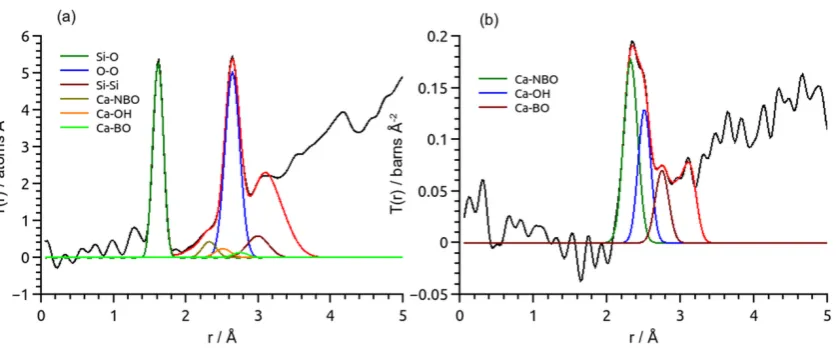
[image:4.612.100.519.266.442.2]diffractometer on the ISIS Spallation Neutron Source, UK. The resultant PDFs are shown in
Figure 1.
Figure 1. Neutron PDFs from (CaO)0.3(SiO2)0.7 (black lines) and their simulations (red lines)
showing key correlations: (a) PDF from (44CaO)
0.3(SiO2)0.7 and (b) natCa-44Ca difference PDF
with non-Ca correlations eliminated [nx4]. The fits and partial correlation functions were
generated using NXFit [nx7].
Analysis of the neutron PDFs from the two samples revealed that sol-gel (CaO)0.3(SiO2)0.7
has a structure based on an incomplete network of SiO4 tetrahedra with non-bridging
oxygen atoms (NBOs, i.e. those not providing a bonded link between network Si atoms)
terminated by protons and Ca2+ ions. The results of fitting the NDIS PDF showed that the
resolvable distances of 2.3, 2.5 and 2.75 Å. On the basis of molecular dynamics simulations,
Mountjoy and Mead assigned these correlations to Ca–NBO, Ca–OH and Ca–BO (BO =
bridging oxygen), respectively [8]. The Ca–NBO and Ca–BO correlations have since been
confirmed in the melt quench 45S5 analogue at ~2.33 and 2.75 Å using diffraction and
computer modelling; as anticipated, the Ca–OH is absent in the melt quench derived sample
[9]. The complex Ca–O environment provided the first clue towards explaining why calcium
loss from sol-gel (CaO)0.3(SiO2)0.7 occurs readily by simple ion exchange with body fluid.
Skipper et al. have shown that calcium is not incorporated into the silica network until the
glassy material is heated to ~400 °C, at which point the nitrate breaks down and Ca enters
the network [1]. Additionally, the NDIS study described above was extended by soaking the
heat-treated final materials in simulated body fluid (SBF) for 30 minutes and repeating the
NDIS experiments [10]. The results revealed that calcium associated with non-bridging
oxygens was preferentially leached into the SBF. The reduction in intensity of the peak at
2.3 Å in the first order difference function PDF was accompanied by a domination of the
feature at 2.5 Å by the peak at 2.7 Å. These changes were interpreted as the formation of
calcium phosphates after immersion in SBF, since the Ca–O distances are longer in calcium
phosphates [11]. FitzGerald et al. undertook a complementary in situ and time-resolved
HEXRD study of bioactive sol-gel (CaO)0.3(SiO2)0.7 foam immersed in SBF on beamline ID15
at the ESRF, France [3]. The results showed that after ~1 hour of exposure to SBF, weak
Bragg peaks could be observed, after ~3 hours a layer of tricalcium phosphate and
hydroxyapatite was evident and after ~5 hours the formation of hydroxycarbonate apatite
was observed. Again, evidence of preferential dissolution of calcium from the Ca–NBO
environment was observed. Furthermore, changes to the O–(Si)–O correlation associated
with SiO4 groups provided direct evidence of disruption to the underlying glass network as
Solid State NMR
Solid state NMR provides information about the details of local atomic scale structure of a
material, extending up to a few atomic neighbours away from the target atom’s position. A
fully multinuclear approach means that a perspective is provided from each different
nucleus [12]. In recent years, the increasing availability of first principles quantum
calculations of the NMR parameters strongly complements the NMR data by revealing the
intricacies of the atomic scale structure [13, 14], which for bioactive glasses directly
influences their osteogenic properties.
For the (CaO)0.3(SiO2)0.7 sol-gel glass considered here the three nuclei (17O, 29Si, 43Ca) can
all be observed by NMR with varying degrees of difficulty. 1H NMR has provided direct
evidence about the proton content in the porous structure, and on reaction with SBF 31P
NMR indicates how the phosphate phases develop over time. The differently connected SiO4
species – Q0 to Q4 – can usually be readily distinguished from their differing chemical shifts
within the resolution of the magic angle spinning (MAS) 29Si spectra, even for amorphous
solids [12]. The network’s average connectivity (Dc) is summarised as the sum of (n ×
(fraction Qn)). Ca(NO
3)2 is a common Ca source in sol-gel formed bioactive (CaO)0.3(SiO2)0.7
and Dc initially increases and was much higher than the composition predicted. Extensive
study and cross referencing the different characterisation techniques showed very clearly
that the 5-8 nm secondary silicate particles have Ca2+ and NO
3- interacting with the surface,
such that the Ca is not yet playing a direct role in the silicate network [15]. On stabilisation
at 600°C the nitrate has thermally decomposed and the Ca directly interacts with the silicate
to satisfy its charge-balance needs by creating non-bridging oxygens and decreasing the
Although Ca(NO3)2 has many advantages, the high stabilisation temperature required to
decompose the nitrate makes it unsuitable for producing hybrids as these temperatures
destroy the polymer component. In making a detailed comparison of Ca(NO3)2, CaCl2 and
Ca(OCH2CH2OCH3)2 as calcium sources 29Si NMR gave Dc for initially aged samples of 3.65,
3.55, 2.17 and those stabilised at 700°C of 3.28, 3.75 and 3.14 for the three calcium
sources respectively [16]. The data clearly show that CaCl2 is completely unsuitable as the
Ca appears to interact very little with the silicate network at any temperature. However, the
low Dc in the initial gel for Ca(OCH2CH2OCH3)2 indicates that Ca is already strongly
interacting with the network, making it highly suitable for low temperature processing
schemes. In gels, network connectivity can be reduced via both conventional
charge-balancing oxygens (i.e. Si‒O−) and Si‒OH. As protons are relatively dilute in these systems
modest MAS can produce 1H NMR spectra from which both the identity and amount of
proton species can be determined [17]. For monoliths produced from Ca(NO3)2 optical
microscopy clearly revealed two distinct macroscopic regions [18]. A combination of 1H and
29Si NMR was able to show that both the proton content (by a factor 3) and the connectivity
of the silicate framework (by 3%) were higher in the inner region, consistent with lower Ca
content.
17O has much potential as a direct observer of the framework species, with a relatively large
chemical shift range and usually modest quadrupolar interaction. The low natural abundance
(0.017%) necessitates isotopic enrichment, but this is relatively straightforward in a sol-gel
produced sample. The distinction between bridging O (BO) and non-bridging O (NBO) for
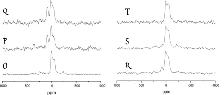
calcium silicates is straightforward even in direct MAS spectra. It is clear from Fig. 2(a) that
in a sample heated to 120°C there is no NBO peak at ~100 ppm. However, on increasing
the heat treatment temperature to 500 then 700°C the NBO peak can be seen progressively
Figure 2. 17O MAS NMR data collected at 14.1 T of (CaO)
0.3(SiO2)0.7 heated to (a) 120, (b)
500 and (c) 700°C, and of (CaO)0.2(SiO2)0.8 (d) before and after (e) 1 hour and 24 hours
reaction with SBF (adapted from [NMR8]).
43Ca is a more difficult nucleus for NMR due to its small magnetic moment, low natural
abundance and quadrupolar nature [12]. However natural abundance studies at high field
are often sufficient to produce interesting information. 43Ca NMR directly confirmed that at
temperatures ≤ 350°C, although no x-ray diffraction peaks of Ca(NO3)2 could be detected,
the 43Ca resonance was very similar to bulk Ca(NO
3)2 indicating that very highly dispersed
calcium nitrate is present. After higher temperature heat treatment the 43Ca NMR signal is
lost due to the chemical shift dispersion present as calcium has a wide range of local
environments in the stabilised gel. This makes an interesting comparison with the
melt-quench analogue where a strong signal is observed in the glass [9]. Inversion of 43Ca
3QMAS NMR data [9] could be rationalised with two signals: a Ca largely coordinated with
BO and the other largely associated with NBO [20]. This data provided good corroboration
of the ND data by cross-referencing the various Ca correlations.
The ultimate utility of these calcium silicates depends on their bioactivity, which may be
studied via subsequent reaction with SBF. The release of Ca2+ (and silicate) ions into the
c
b
a
f
e
solution is an important trigger event for subsequent cell development. 17O MAS NMR shows
that, on contact with SBF, the NBOs associated with Ca are rapidly lost (Fig. 2). It appears
that there is a rapid exchange between Ca2+ ions and H+ such that calcium is released and
the surface rapidly becomes hydroxylated [19]. During this process Dc drops slightly. This
then creates favourable conditions for the surface deposition of calcium phosphates on the
way to hydroxycarbonate apatite (HCA). The spin-½ 31P is an ideal probe nucleus with high
intrinsic signal sensitivity and a highly dispersed chemical shift range for detection of
different phosphate environments. The 31P NMR data shows that a phosphate phase forms
very rapidly (within a few minutes) and that, from a relatively early stage, its chemical shift
closely resembles quite well ordered HCA [19].
Molecular Dynamics simulation
Molecular dynamics (MD) simulation is a powerful computational method for probing a
material’s structure and properties. It is conceptually simple: for each timestep, starting
from the atomic positions, the interatomic forces are modelled via Newton’s Law F = ma,
and the atoms are then moved to new positions under those accelerations; these positions
are used in the next timestep. The interatomic forces can be modelled either
quantum-mechanically or with an empirical classical approximation. Because MD provides the atomic
positions throughout the trajectory, it can be used to extract information about structure
and properties at local and larger length scales, providing an effective complement to
experimental techniques.
The standard method for preparing a glass in the computer is to create an equilibrated melt
(or liquid) whose temperature is then reduced to body or room temperature [21], forming a
disordered solid. Bioactive glasses such as 45S5 Bioglass® and related phosphosilicate
basic composition space by substituting Sr for Ca [24] and introducing fluorine [25, 26].
Characterisation of these simulated structures show that the network connectivity
parameter introduced by Hill [27], whilst being a useful guideline, does not reflect the full
range of topological structure in these glasses. This is embodied in the Qn distribution, for
example, or in the greater extent of network fragmentation displayed by the more bioactive
glasses and the concomitant spatial distribution of the modifiers in these glasses (which, of
course, determines the Qn distribution).
For applications in tissue engineering, low-temperature sol-gel preparative routes are often
used. Glasses formed in this way still retain a number of –OH groups which leads to more
open, lower-density glasses. The most common composition of sol-gel derived bioactive
glasses is (CaO)0.3(SiO2)0.7, as investigated by Mead and Mountjoy [8]. Their structures
contained hydroxyl species not bonded to SiO4 tetrahedra, but which were preferentially
coordinated to the network modifying Ca ions. These ‘free‘–OH come from the dissociation
of added H2O molecules. Likewise, a quantum-mechanical study of water in Bioglass [28]
also contained free ‒OH, which arose from the dissociation of water molecules. A similar
observation was made by Tilocca [29] on a 45S5 glass in which a quantity of Na were
replaced by protons.
The presence of modifiers, such as calcium and protons (as hydroxyl ions), in the glass
structure should, in principle, reduce its network connectivity and enhance bioactivity.
However, as just noted, a significant fraction of the hydroxyl oxygen atoms bond directly to
the (sodium and) calcium, and not to silicon. In this case, the effect on the glass structure is
to increase the silicate network connectivity above that which might be expected [8, 28], as
also found in more complicated hydrated yttrium aluminosilicate glasses [3]. Clustering of
modifier cations is also potentially important because it increases bioactivity [31], and these
(CaO)0.3(SiO2)0.7, some slight clustering is observed: the Ca–Ca coordination number is
slightly larger than would be expected if Ca were randomly distributed. The amount of Ca
clustering appears to increase with decreasing Ca content, as has been found for modifier
ions in melt-quenched yttrium aluminosilicate glasses [31].
Although there has been no simulation of (CaO)0.3(SiO2)0.7 interacting with the body,
simulations of other bioactive glass compositions provide some insight. These bioactive
glass compositions contain both sodium and calcium as network modifiers, so the structures
are not exactly comparable, although there are many similarities. The glass surface is first
enriched with sodium, which interacts with water outside the glass, allowing water to enter
the glass itself, which begins the dissolution [8]. For these glasses, Ca–water interactions
occur after Na has been leached into the solution; for the Na–free (CaO)0.3(SiO2)0.7
composition, the Ca–water interactions will presumably occur first, although more slowly as
Ca is less mobile through the glass structure than Na [28]. Ca also binds to hydroxyl groups
created by water dissociation, stabilising this process [28]. One might speculate that the
migration of the Ca cation in such cases (e.g. surface gel layers) is a co-operative process
involving the hydrating ‒OH species as well as the Ca cation itself. Amongst the most
reactive sites on the glass surface are non-bridging oxygen atoms (often associated with
modifier cations) [28, 33, 34]; these can promote water dissociation and the formation of
silanol Si–O–H groups on the surface of the glass, which is one of the early steps in the
bioactive glass dissolution process. In addition, a large number of the free hydroxyl groups
introduced into the glass structure are bound to the modifier cations [29], as also observed
in (CaO)0.3(SiO2)0.7.
Conclusions
We have demonstrated the efficacy of using complementary experimental structural probes
suitable methodological approach to the problem of determining the details of the
atomic-scale structure of amorphous materials. Moreover, we have also shown that MD simulations,
whether classical or quantum mechanical, provide information about the structure and
structure-related properties, such as reactivity, which both complement and supplement
experimental data. Because they offer “direct” atomic scale pictures of the structure, the
simulations are valuable aids to the interpretation of that experimental data.
Acknowledgements
We thank our respective institutions for their support; the work undertaken in the UK was
funded via EPSRC and STFC awards.
References
[1] L.J. Skipper, F.E. Sowrey and R.J. Newport, Z. Lin and M.E. Smith; “x-ray and neutron
diffraction and solid state NMR studies of the growth of hydroxyapatite on bioactive
calcia-silica sol-gel glasses.” Phys & Chem Glasses 46, 372 (2005).
[2] R.J. Newport, L.J. Skipper, D. Carta, D.M. Pickup, F.E. Sowrey, M.E. Smith, P.
Saravanapavan and L.L. Hench; “The use of advanced diffraction methods in the study of
the structure of a bioactive calcia:silica sol-gel glass.” J Mater. Sci.-Mater. M. 17, 1003
(2006).
[3] V. FitzGerald, K.O. Drake, J.R. Jones, M.E. Smith, V. Honkimäki, T. Buslaps, M.
Kretzschmer, R.J. Newport; “In situ high-energy X-ray diffraction study of a bioactive
calcium silicate foam immersed in simulated body fluid.” J. Synchrotron Radiat. 14, 492-499
(2007).
[4] L.J. Skipper , F.E. Sowrey, D.M. Pickup, K.O. Drake, M.E. Smith, P. Saravanapavan, L.L.
Hench and R.J. Newport; “The structure of a bioactive calcia–silica sol–gel glass.” J. Mater.
[5] F.E. Sowrey, L.J. Skipper, D.M. Pickup, K.O. Drake, Z. Lin, M.E. Smith and R.J. Newport;
“Systematic empirical analysis of calcium-oxygen coordination environment by calcium
K-edge XANES.” Phys. Chem. Chem. Phys. 6, 188 (2004).
[6] P. Saravanapavan and L. L. Hench; “Mesoporous calcium silicate glasses: I. Synthesis.”
J. Non-Cryst. Solids 318, 1-13 (2003).
[7] D.M. Pickup, R.M. Moss and R.J. Newport; “NXFit: a program for simultaneously fitting
x-ray and neutron diffraction pair-distribution functions to provide optimized structural
parameters.” J. Appl. Cryst. 47, 1790-1796 (2014).
[8] R.N. Mead and G. Mountjoy; “Modeling the local atomic structure of bioactive
sol-gel-derived calcium silicates.” Chem. Mater. 18(17), 3956–3964 (2006).
[9] R.A. Martin, H.L. Twyman, G.J. Rees, E.R. Barney, R.M. Moss, J.M. Smith, R.G. Hill, G.
Cibin, T. Charpentier, M.E. Smith, J.V. Hanna, R.J. Newport; “An examination of the calcium
and strontium site distribution in bioactive glasses through isomorphic neutron diffraction,
X-ray diffraction, EXAFS and multinuclear solid state NMR.” J. Mater. Chem. 22, 22212
(2012).
[10] R.J. Newport, L.J. Skipper, V. FitzGerald, D.M. Pickup, M.E. Smith, J.R. Jones; “In vitro
changes in the structure of a bioactive calcia–silica sol–gel glass explored using isotopic
substitution in neutron diffraction.” J. Non-Cryst. Solids 353, 1854-1859 (2007).
[11] D.A. Fletcher, R.F. McMeeking and D. Parkin; The United Kingdom Chemical Database
Service; J. Chem. Inf. Comp. Sci. 13, 746-749 (1996).
[12] K.J.D. MacKenzie and M.E. Smith; Multinuclear Solid State NMR of Inorganic Materials,
Pergamon Press, UK, (2002).
[13] T. Charpentier; “The PAW/GIPAW approach for computing NMR parameters: A new
dimension added to NMR study of solids”, Solid State Nucl. Magn. Reson., 40, 1-20, (2011).
[14] C. Bonhomme, C. Gervais, F. Babonneau, C. Coelho, F. Pourpoint, T. Azais, S.E.
NMR parameters using the gauge including projector augmented wave method: A chemist's
point of view”, Chem Rev., 112, 5733-5779, (2012).
[15] S. Lin, C. Ionescu, K.J. Pike, M.E. Smith and J.R. Jones; “Nanostructure evolution and
calcium distribution in sol-gel derived bioactive glass”, J. Mater. Chem., 19, 1276-1282,
(2009).
[16] B. Yu, C.A. Turdean-Ionescu, R.A. Martin, R.J. Newport, J.V. Hanna, M.E. Smith and
J.R. Jones; “Effect of calcium source on structure and properties of sol-gel derived bioactive
glasses”, Langmuir, 28, 17465-11476, (2012).
[17] J.R. Jones, T.F. Kemp and M.E. Smith; “Effect of OH content on the bioactivity of
sol-gel derived glass foam scaffolds”, Key Engin. Mater., 309-311, 1031-1034, (2006).
[18] S. Lin, C. Ionescu, S. Baker, M.E. Smith and J.R. Jones; “Characterisation of the
inhomogeneity of sol-gel-derived SiO2–CaO bioactive glass and a strategy for its
improvement”, J. Sol-Gel Sci. Technol., 53, 255-262, (2010).
[19] Z. Lin, J.R. Jones, J.V. Hanna and M.E. Smith; “A multinuclear solid state NMR
spectroscopic study of the structural evolution of disordered calcium silicate sol-gel
biomaterials”, Phys. Chem. Chem. Phys., 17, 2540-2549, (2015).
[20] Z. Lin, M.E. Smith, F.E. Sowrey and R.J. Newport; “Probing the local structural
environment of calcium by natural-abundance solid-state 43Ca NMR”, Phys. Rev. B, 69,
224107(7), (2004).
[21] K. Vollmayr, W. Kob and K. Binder; “Cooling rate effects in amorphous silica: a
computer simulation”, Phys. Rev. B 54, 15808-27 (1996).
[22] A. Tilocca, N. H. de Leeuw and A. N. Cormack; “Shell-model molecular dynamics
calculations of modified silicate glasses”, Phys. Rev. B 73, 104209 (2006).
[23] A. Tilocca, A. N. Cormack and N. H. de Leeuw; “The structure of bioactive silicate
glasses: new insight from molecular dynamics simulations”, Chem. Mater. 19, 95-103
[24] Y. Xiang and J. Du; “Effect of strontium substitution on the structure of 45S5
bioglasses”, Chem. Mater. 23, 2703-17 (2011).
[25] G. Lusvardi, G. Malavasi, M. Cortada, L. Menabue, M. C. Menziani, A. Pedone and U.
Segre; “Elucidation of the structural role of fluorine in potentially bioactive glasses by
experimental and computational investigation”, J. Phys. Chem. B 112, 12730-39 (2008)
[26] J. K. Christie, A. Pedone, M. C. Menziani and A. Tilocca; “Fluorine environment in
bioactive glasses: ab initio molecular dynamics simulations”, J. Phys. Chem. B 115,
2038-45 (2011).
[27] R.G. Hill, “An alternative view of the degradation of Bioglass”; J. Mater. Sci. Lett. 15,
1122-25 (1996).
[28] E. Berardo, M. Corno, A. N. Cormack, P. Ugliengo and A. Tilocca; “Probing the fate of
interstitial water in bulk bioactive glass by ab initio simulations”, RSC Adv. 4, 36425-36
(2014).
[29] A. Tilocca; “Atomic-scale models of early-stage alkali depletion and SiO2-rich gel
formation in bioactive glasses”, Phys. Chem. Chem. Phys. 17, 2696-2702 (2015).
[30] J. Malik and A. Tilocca; “Hydration effects on the structural and vibrational properties
of yttrium aluminosilicate glasses for in situ radiotherapy”, J. Phys. Chem. B 117, 14518-28
(2013).
[31] J. K. Christie and A. Tilocca; “Aluminosilicate glasses as yttrium vectors for in situ
radiotherapy: understanding composition-durability effects through molecular dynamics
simulations”, Chem. Mater. 22, 3725-34 (2010).
[32] A. Tilocca and A. N. Cormack; ”Structural effects of phosphorus inclusion in bioactive
silicate glasses”, J. Phys. Chem. B, 111, 14256-14264 (2007).
[33] J. Du and A. N. Cormack; “Molecular dynamics simulations of the structure and
hydroxylation of silica glass surfaces”, J. Amer. Ceram. Soc. 88, 2352 (2005).
[34] A. Tilocca and A. N. Cormack; “Modelling the water-Bioglass Interface by ab initio
Figure captions
Figure 1. Neutron PDFs from (CaO)0.3(SiO2)0.7 (black lines) and their simulations (red lines)
showing key correlations: (a) PDF from (44CaO)
0.3(SiO2)0.7 and (b) natCa-44Ca difference PDF
with non-Ca correlations eliminated [4]. The fits and partial correlation functions were
generated using NXFit [7].
Figure 2. 17O MAS NMR data collected at 14.1 T of (CaO)
0.3(SiO2)0.7 heated to (a) 120, (b)
500 and (c) 700°C, and of (CaO)0.2(SiO2)0.8 (d) before and after (e) 1 hour and 24 hours