Printed in Great Britain © The Company of Biologists Limited 1984
MECHANICAL SYNCHRONIZATION OF CILIARY
BEATING WITHIN COMB PLATES OF CTENOPHORES
BY SIDNEY L. TAMM
Boston University Marine Program, Marine Biological Laboratory, Woods Hole, MA 02543 U.SA.
Accepted 9 May 1984
SUMMARY
The mechanism which synchronizes the beating of the hundreds of thousands of long cilia making up a ctenophore comb plate was investigated by microsurgical experiments on single comb plates of Mnemiopsis and
Pleurobrachia. Comb plates of lobate ctenophores (e.g. Mnemiopsis) are
triggered to beat by ciliated grooves which run between the centres of adja-cent plates. By creating gaps or introducing mechanical barriers between two parts of a plate, or by severing the cells at the base of a plate, it was shown that physical proximity of cilia, not tissue continuity, is required for synchronization of beating. In Pleurobrachia only the first comb plate of each row is activated by a ciliated groove, and similar experiments to those done on Mnemiopsis gave identical results. Although adjacent comb plates in Pleurobrachia are triggered mechanically by movements of the preceding plates without the need for an intraplate synchronizing mechanism, unilateral amputation of a plate showed that cilia within these plates may also be synchronized by mechanical coupling. Therefore, in cases where the beating of a comb plate is triggered by a ciliated groove - either at the head of a comb row (in all ctenophores) or along the row (lobates only) - the cilia within the plate are synchronized by hydrodynamic coupling forces between them, not by electrical coupling between their cells as assumed previously.
INTRODUCTION
The mechanism of metachronal coordination of ciliary activity has long been studied, and has been shown in all cases to depend on hydrodynamic interaction (viscous-mechanical coupling) between neighbouring cilia (Machemer, 1974; Murakami, 1963; Sleigh, 1974; Tamm, 1973, 1982, 1983). Little is known, however, about the mechan-ism which synchronizes the beating of component cilia within a compound ciliary or-ganelle, so that all the cilia beat together as a single unit. Examples of compound cilia in-clude the cirri and membranelles of protozoa, the large abf rontal cilium and laterof rontal cilium of mussel gills, maeroeilia of the ctenophore Beroe, and the giant comb plates of ctenophores. Microsurgical experiments on certain compound cilia indicate that syn-chronization of beating is achieved by mechanical coupling between the constituent cilia (Carter, 1924; Chambers & Dawson, 1925; Tsuchiya, 1969). For example, when the abfrontal cilium or laterofrontal cilium of Mytilus is split into component cilia by a
402 S. L. TAMM
microneedle, the cilia beat out of unison (Carter, 1924; Tsuchiya, 1969); upon remov ing the needle the individual cilia reunite and beat together as a single unit again. Alter-natively, membrane excitation and cell-to-cell electrical coupling have been proposed to account for the synchronous beating of the hundreds of thousands of long cilia, borne on a ridge of thousands of cells, which make up a ctenophore comb plate (Horridge & Mackay, 1964; Satterlie & Case, 1978). To date, no experimental studies have been made on the mechanism of ciliary synchrony in ctenophores. In this report I have inves-tigated this problem by performing microsurgical experiments on single comb plates. I show that the cilia within a comb plate are synchronized by mechanical coupling forces between them, not by electrical signals between their cells as assumed previously.
MATERIALS AND METHODS
Mnemiopsis leidyi and Pleurobrachia pileus were dipped carefully from the surface
of Great Harbor or Vineyard Sound near Woods Hole, Mass. Ctenophores were maintained in excellent condition in perforated buckets submerged in running sea water, and were fed daily with freshly caught plankton.
Long pieces of comb rows were cut out of the animal and held stationary in a 'micro-vice' apparatus mounted in a Lucite chamber as described previously (Tamm, 1973). A mixture of sea water and 7-0% MgCb (1:1) was commonly used in place of sea water for Mnemiopsis to prevent muscular retraction of the comb row; initiation and transmission of metachronal ciliary waves are not affected by excess Mg2+ (Horridge, 1965; Tamm, 1982). The preparation was viewed under a dissecting microscope by dark-field or bright-field transmitted light.
Glass microneedles were made from capillary tubing drawn out in an electrode puller, or pulled by hand in a small flame. Needles and other micro-tools were operated by a Jena micromanipulator. Amputation of comb plates (Fig. 3B,C) was done with iridectomy scissors (Weiss, London).
Cine films of certain experiments were taken through the dissecting microscope with a 16 mm cine camera at 25 or 50 frames s"1. Higher resolution images of microsurgical results were obtained by transferring the preparation to a microscope slide and viewing with Zeiss Nomarski optics using a 16X or 40X objective. Photographs (Fig. 2) were taken on Kodak High Contrast Copy film.
Synchronization of ctenophore cilia
o—A
403
D
404 S. L. TAMM
RESULTS
In all ctenophores, the first comb plate at the head of each comb row is triggered to beat by a narrow tract of short cilia, the ciliated groove, which runs from the abora: statocyst to the centre of the base of the plate (Tamm, 1973, 1982). In lobate ctenophores (e.g. Mnemiopsis) the ciliated grooves continue from plate to plate along the comb rows, and trigger the beat of adjacent plates during metachronal coordina-tion (Tamm, 1973, 1982). A signal must therefore be transmitted outward from the ciliated groove junction to either side of a comb plate to activate beating of all the cilia within the plate.
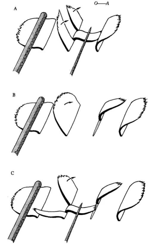
To investigate the nature of this synchronal coordination, microsurgical experi-ments were performed on single comb plates along comb rows of Mnemiopsis (Fig. 1). Cine films of some of these experiments have been presented previously (Tamm, 1980). First, a physical gap between two parts of a plate was made by slitting a plate longitudinally into three components with a microneedle, and bending back the middle part close to the body surface (Fig. 1A,B). If one of the side pieces was wider and connected to the ciliated groove, only this part beat during passage of a meta-chronal wave down the row; the other free piece, separated by a gap, did not beat (Fig. 1A). This effect was reversible: upon releasing the intermediate sliver the parts reunited and the whole plate beat as a unit again. At high beat frequencies, however, the separated parts of the plate usually beat together regardless of the presence of a gap between them. If the restrained part of the plate was medial and connected to the ciliated groove, metachronal waves caused twitches of this part, but no movements of the two free end pieces (Fig. IB). Release of the middle part resulted in syn-chronous beating of the entire plate.
A mechanical discontinuity between two parts of a plate, without preventing the movement of an intermediate pie.ce, was made by slitting a plate (but not the underly-ing tissue) with a chip of razor blade held just above the tissue surface (Fig. 1C). Only the larger part of the plate connected to the ciliated groove was stimulated to beat by metachronal waves; the narrower part on the other side of the barrier did not beat. Upon removing the razor blade, both parts reunited and beat synchronously again. Lastly, a deep cut was made with a glass needle across the ridge of polster cells at the base of a comb plate, producing a narrow cleft in the plate as well (Figs 1D,E, 2). Nevertheless, the two severed parts of the plate beat together. However, if the smaller part was moved a short distance away from the main part with a needle, this piece no longer beat with the main part of the plate (Fig. IE). Upon pushing the end part back against the main part to close the gap, the entire plate resumed synchronal beating. Physical proximity, not tissue continuity, is thus required for synchronization of beating between parts of a comb plate.
Similar microsurgical experiments on the first comb plate of a row in Pleurobrachia gave identical results.
Journal of Experimental Biology, Vol. 113
Fig. 2
Fig. 2. Comb plate (cp) of Mnemiopsis after cutting through its base with a glass needle, as shown in Fig. 1D,E. The ridge of polster cells is severed as well as the plate itself (arrow). Note the widening of the ciliated groove (eg) at its junction with the aboral side of the plate. Zeiss Nomarski optics, electronic flash. Scale bar, 50 ^m.
Synchronization ofctenophore cilia 405
fctroke of the preceding plate, without the need for an intraplate synchronizing mechanism. That this is the case was shown by slitting a plate along a comb row ofPleurobrachia into three parts, and then pressing down the middle piece against the
[image:7.451.100.349.116.528.2]O A
406
S. L. TAMMbody surface to create a gap between the two end parts (Fig. 3A). In contrast to tha results with Mnemiopsis (Fig. IB), both of the separated parts of the Pleurobrachick plate beat together during passage of metachronal waves down the row. Each end piece was thus stimulated directly by the movement of the preceding plate.
To test whether intraplate coordination can occur in Pleurobrachia, direct mechanical stimulation of one side of a plate by the preceding plate was avoided by amputating the corresponding side of the neighbouring plate (Fig. 3B). Nevertheless, the entire plate beat as a unit, demonstrating the existence of synchronal coordination between the side which was triggered by the active stroke of the neighbouring half-plate, and the other unstimulated side. To determine the nature of this coordination, a physical gap was made between the stimulated side and the unstimulated part of this plate by holding back an intermediate sliver with a needle. Activity of the neighbour-ing half-plate triggered beatneighbour-ing of the correspondneighbour-ing side of the experimental plate, but not of its separated end piece (Fig. 3C). Upon release of the intermediate piece, the entire plate beat together when stimulated by the preceding half-plate.
DISCUSSION
The present results show that in cases where the beating of a plate is triggered by a ciliated groove - either at the head of a comb row (in all ctenophores) or along the row (in lobates) - the cilia within the plate are synchronized by hydrodynamic coupl-ing forces between them. Such mechanical interaction is sufficient to ensure ciliary synchrony in naturally frayed plates, where the separation between adjacent parts is very narrow. As shown above, mechanical coupling is even possible across wider gaps if the cilia beat at high frequencies and create stronger water currents.
Synchronization ofctenophore cilia 407
The microsurgical experiments also show that electrical coupling between comb f late cells, suggested by the presence of gap junctions (Satterlie & Case, 1978), is not responsible for synchronizing the beating of cilia within a plate as proposed previously (Horridge & Mackay, 1964; Satterlie & Case, 1978). Gap junctions may serve to spread neurally-mediated signals for various motor responses of comb plates, such as ciliary arrest and reversal (Tamm, 1982; Tamm & Moss, 1985). This possibility is currently being investigated by intracellular electrophysiological recording and dye injection (A. G. Moss & S. L. Tamm, in preparation).How the short cilia (10—15 (*m) of the ciliated groove excite beating of the long cilia (> 1000 jUm) in the centre of a comb plate is not known. Since the cilia along the groove are coordinated by mechanical interaction (Tamm, 1982), they may stimulate the plate cilia directly ahead of them by a similar mechanism, providing this region of the plate is sufficiently mechanosensitive. The pronounced widening of the ciliated groove at its junction with the base of a comb plate (Fig. 2) suggests a device for mechanical amplification.
Once triggered by the ciliated groove, the beat is instantaneously transmitted out-ward to both sides of the comb plate by the high degree of mechanical coupling between neighbouring cilia (Fig. 4). The flange-like compartmenting lamellae which extend between adjacent cilia in the line of synchrony (Afzelius, 1961) undoubtedly contribute to this mechanical interaction.
The results onPleurobrachia comb rows (Fig. 3) show that under conditions where intraplate coordination occurs, the cilia within the comb plates of cydippid ctenophores are also synchronized by mechanical coupling. In this regard, compart-menting lamellae are present between adjacent cilia in the comb plates of all ctenophores, regardless of the mechanism of metachronal coordination (Tamm, 1982). Although an intraplate synchronizing mechanism is normally not needed in
Pleurobrachia, it may be advantageous in ensuring metachronal wave transmission in
cases where comb plates have become partially damaged.
In conclusion, these findings emphasize the mechanosensitive nature of motile cilia (see Tamm, 1982, 1983; Wiederhold, 1976), and show that mechanical activation of the motile elements (Machin, 1958; Brokaw, 1966) plays a key role in the initiation and phasing of ciliary beating during synchronal as well as metachronal coordination.
Supported by NIH Grant GM 27903.
R E F E R E N C E S
AFZELIUS, B. (1961). The fine structure of the cilia from ctenophore swimming-plates. J. biophys. biochem.
Cytol. 9, 383-394.
BROKAW, C. J. (1966). Bend propagation along flagella. Nature, Land. 209, 161-163.
CARTER, G. S. (1924). On the structure and movements of the latero-frontal cilia of the gills of Mytilus. Proc.
R.Soc.B 96, 115-122.
CHAMBERS, R. & DAWSON, J. A. (1925). The structure of the undulating membrane in the ciliateBlepharisma.
Biol. Bull. mar. biol. Lab., Woods Hole 48, 240-242.
HORRIDGE, G. A. (1965). Relations between nerves and cilia in ctenophores. Am. Zool. 5, 357—375. HORRIDGE, G. A. & MACKAY, B. (1964). Neurociliary synapses in Pleurobrachia (Ctenophora). Q.Jlmicrosc.
Sci. 105, 163-174.
408 S. L. TAMM
MACHIN, K. E. (1958). Wave propagation along flagella. J. exp. Biol. 35, 796-806.
MURAKAMI, A. (1963). Analysis of metachronal co-ordination in ciliary pads of Mytilus gill. J . Fac. Sri.
Univ. 10, 23-35.
SATTERLIE, R. A. & CASE, J. F. (1978). Gap junctions suggest epithelial conduction within the comb plates of the ctenophore Pleurobrachia bachei. Cell Tissue Res. 193, 87-91.
SLEIGH, M. A. (1974). Metachronism of cilia of metazoa. In Cilia and Flagella, (ed. M. A. Sleigh), pp. 287-304. London: Academic Press.
TAMM, S. L. (1973). Mechanisms of ciliary co-ordination in ctenophores. J. exp, Biol. 59, 231—245. TAMM, S. L. (1980). The mechanism of intra-plate synchrony in ctenophores. Biol. Bull. mar. biol. Lab., Woods
Hole 159, 446.
TAMM, S. L. (1982). Ctenophora. In Electrical Conduction and Behaviour in 'Simple' Invertebrates, (ed. G. A. B. Shelton), pp. 266-358. Oxford: Oxford University Press.
TAMM, S. L. (1983). Motility and mechanosensitivity of macrocilia in the ctenophore Beroe. Nature, Land. 305, 430-433.
TAMM, S. L. & Moss, A. G. (1985). Unilateral ciliary reversal and motor responses during prey capture by the ctenophore Pleurobrachia. J. exp. Biol. (in press).
TSUCHIYA, T . (1969). Synchronal coordination within a compound cilium of Mytilus gill. Annotnes zool.jap. 42, 113-125.
WIEDERHOLD, M. L. (1976). Mechanosensory transduction in "sensory" and "motile" cilia. A. Rev. Biophys.