REGIONS IN KENYA
SHARON CHEROTICH CHEPKWONY (B. Ed. Se.)
I56/CE/220OS/20 10
A THESIS SUBMIlTED IN PARTIAL FULFILLMENT OF THE REQUIREMENTS FOR THE AWARD OF THE DEGREE OF MASTER OF
SCIENCE (IMMUNOLOGY) IN THE SCHOOL OF PURE AND APPLIED
r.
SCIENCES OF KENYA1TA UNIVERSITY
KENY ~TT~
I,INI\!ERSn,
PO BOX 438.114 00100NAIROBI KENyA N~~E1t)"15
RECE1VE.D
QRAOUATeSCHOOL
DECLARATION
I Sharon Cherotich Chepkwony hereby declare that this thesis is my original work and has not been presented for degree or other awards in any other university.
Signature ~ ..~ Date ..
~o\~~\\?"
.
Sbaron CberoticbChepkweny Department of Zoological Sciences,
SUPERVISORS
Thisthesis has been submitted with our approval asUniversity supervisors.
Prof. Micbael M. Gicberu
Department of Zoological Sciences, Kenyatta University ~
--KW-a~'
2Y{1l1
Zo\~~
Signature
7J.\-:
-.
G
Date .Dr. Atunga Nyacbieo
DepartmentofReproductive Health and Biology,
Institute ofPrimate Research
DEDICATION
This thesis is dedicated to my dad Julius Chepkwonyand my mum Christine
Chepkwony who laid foundation for·my education, and to my siblings, Joselyne Chepkwemoi, Linet Chepkwony, Dorine Chepkorir, Paul Koech, Mercy Chepkwony,
David Kiprop, Emma Chepchumba, Ruth Chebet and EIsie Cherono for their support
ACKNOWLEDGE~NTS
I wish to thank my supervisors, Prof Michael Gicheru of Kenyatta University for the advice and guidance he provided throughout this study and Dr.Atunga Nyachieo of the Institute of Primate Research who funded my project and gave me invaluable
intellectual advice. I am grateful to the Institute of Primate Research for the support accorded tome in the Animal Science Department, Molecular and Female Reproductive
health laboratories during my studies.
I am also grateful to Kenyatta University, Department of Zoological Sciences for the
advice they gaveme during myproposal defence.
I wish to acknowledge Mr. Eric Omolo and Mary Galo (Female reproduct~vehealth
laboratory), Mr. Samson Mutura ~(Molecular laboratory) for continued technical
assistance, Mr. Nicholas Kiulia who helped me with bioinformatics analysis and Ruth
Nyakundi forteaching me how to do ELISAs.
I also acknowledge Dr. Daniel Chai, Dr.George Omondi, Mr. Habib Saibulu,Mr. Jason Alembi, and Ms. Priscillah Kimiti all of Animal Science Department, for their support during the entire period of sample collection. I recognize the support of my fellow student colleagues at the Institute of Primate Research.
I would finally like to thank Prof Richard Eberle of the Department of Veterinary Pathobiology, Oklahoma State University, USA, for providing me with HVP 2 antigens,
ELISA positive and negative controls, TK primer set and HVP 2 DNA as a positive control and for sponsoring my trip to the USA where I acquired invaluable laboratory
TABLE OF CONTENTS
DECLARATION II
DEDICATION _ III
ACKNOWLEDGEMENTS : IV
LIST OF TABLES : ; IX
ACRONYMS AND ABBREVIATIONS ; XI
ABSTRACT : : XIII
CHAPTER ONE: INTRODUCTION 1
1.IBackground Information 1
1.2Problem statement : :..3·
l.3Study Justification ; : 4
1.4 Research questions : 5
1.5 Null hypotheses ...•... 5
1.6 Objectives
,.
5.
1.6.1General Objective 5
1.6.2 Specific objectives : 6
1.7Significance ofthe study ; 6
~
CHAPTER TWO: LITERATURE REVIEW 7
2.1 Herpes Simplex Virus : 7
2.1.1 Viremia in Herpes Simplex Virus Infection 10
2.1.2 Role of thymidine kinase in the virulence and latency of HSV 11
2.1.3 Transmission of Herpes Simplex Virus 13
2.2 Herpes virus in Non-Human Primates 15
2.2.1 Herpesvirus papio 2 - 17
2.2.2 Herpesvirus papio 2 in non primates 22
2.2.3 Transmission of human herpes to Monkeys 22
2.3Role ofinterferons in herpes virus infections: : 24
2.3.1Role of interferons in Replication of HSV 25
2.3.2Role of type 1 interferon on HVP-2 infection 25
2.4 Importance of using a baboon as a model to studyHSV 27
CHAPTER mREE: MATERIALS AND l\IETHODS .••••..••••_••_••••• •.••..•.••••••.•••.•32..• >
3.1Studysite c.: : : 32
3.2 StudyDesign 32
3.3 Animal capture 32
3.4 Sample size determination r...•...33
3.5 Sample collection 34
3.5.1 Blood sample 35
3.5.2 Swabs 35
~.
3.5.3 Trigeminal ganglia 35
3.6Viral antigenpreparation 36
3.6.1 Lyophilized HVP-2 antigen resuspension 36
3.7 Screening forHVP-2 antibodies using Enzyme Linked Immunosorbent Assay ....37
3.8Extractionof Deoxyribonucleic Acid (DNA) 38
3.8.1 Oral and genital swabs 38
3.8.3 Confirmation of the extracted Deoxyribonucleic Acid _ _._ 40
3.9 Amplification of deoxyribonucleic Acid ; 40
3.10 Polymerase Chain Reaction conditions ~ 40
3.11 Gel electrophoresis :
41
3.12 Purification of PeR products : : 41
3.13 Clean up of DNA before sequencing ~ 42
3.14 Sequence analysis : 42
3.15 Phylogenetic analysis ;.:: 43 .
3.16 Data Analysis : : : ,.., 43
3.17 Ethical Considerations ; : 43
CHAPl'ER FOUR.: RESUL TS _ _.._ ....•... _ _.. .._ _..4S
.
.
4.1 Anti- HVP-2 antibodies : 45
4.1.1 Seroprevalence ofHVP-2 inbaboons by region __ _.._.45
4.1.2 Seroprevalence ofHVP-2 in baboons by sex ._ _ 47
4.1.3 Seroprevalence ofHVP-2 in baboons by Age ..__..•... _ , 47 4.1.4 Exposure rates in freshly-caught and wild captive colony baboons 48
"
4.2 Confirmation of the presence of extracted DNA _ _ 49
4.3Polymerase Chain Reaction : 50
4.3 Sequencing analysis 51
4.4 Phylogenetic analysis 56
-CHAPTER FIVE: DISCUSSION, CONCLUSIONS AND
RECOMMENDA TIONS •••••_•..•.•.•...••.•...•.••.••._•••.•.•..•.•.•••..•••.•.•.•.••.•••••••••....•••.•••58•.•.
5.2SeroprevalenceofHVP-2 58 5.2.1 Seroprevalence ofHVP-2 in baboons from different regions in Kenya 58
5.2.2 Sero-prevalence ofHVP-2 by age 59
5.2.3 Seroprevalence ofHVP-2 by sex 59
5.2.4 Sero-prevalence in colony baboons with freshly caught wild baboons 60 5.3 Amplification ofthe thymidine kinase coding region ofHVP-2 60
5.4 HVP 2 strain ~ 61
5.5 Studylimitations : ; 63
5.6Conclusion ., e•••••••••• 64
. .
.
5.7 Recommendations : 65
REFERENCES 66
APPENDICES ._.._ _ "" _ _ _ _ 75
~ Appendix I:Ethical clearance ; ! 75
Appendix II:Reagents for IgG antibody ELISA 76
Appendix Ill: ELISA plates , 77
Appendix IV: Images ~ 78
~.
LIST OF TABLES
Table 2.1:Summary of herpes viruses andtheir hosts 16
Table4.1: Seroprevalence ofHVP 2 comparing gender exposure rates 47
Table 4.2: Sero-prevalence ofHVP-Z comparing age exposure rates 48
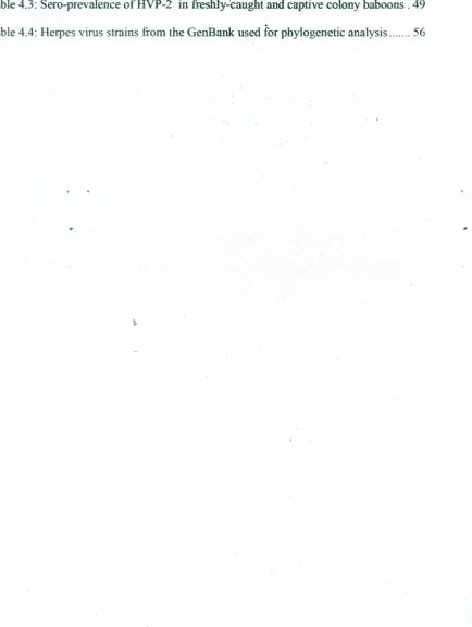
Table 4.3: Sero-prevalence ofHVP-2 infreshly-caught and captive colonybaboons. 49
LIST OFFIGURES
Figure 2.1: Lesions caused by HSV ~ 9
Figure4.2: Structure ofthymide kinase from herpesvirus 13
~igure 2.3:Phylogenetic tree of HSV and simian herpes viruses 17
Figure 2.4: Oral lesion in the baboon mouth caused by HVP 2 18
Figure 2.5: Ulcers appearing on the baboon penis following infection by HVP-2 20
Figure 2.6: A troop of baboons in the wild 21 .
Figure 2.7: Wild caught baboons in the animal facility ofIPR 28
Figure 4.1: A graph showing regional prevalence ofHVP 2 in baboons , 46
Figure4:2: Image of gel electrophoresis run to confirm thepresence ofDNA 50
Figure 4.3: Gel image for PCR amplicons : 51
Figure4.4: A chromatogram for sample Sll. 53
Figure4.5: Pair wise Alignments of the DNA sequences of sample OPC 54
Figure4.6: Pair wise alignments of the DNA sequences of sample S55 55
AAALAC BSA
BV
CDC
ChHV
CNS
CPE
DNA
dTdTMl)
DPI
EBSCO
EDTA
ELISA
HIV
HSV
HVAHVP2
HVS
IFN
IPR.ACRONYMS AND ABBREVIATIONS
Association for Assessment and Accreditation of Laboratory Animal
Care
Bovine Serum Albumin
Monkey B Virus
Center for Disease Control
Chimpanzee Herpes Virus
Central Nervous System
Complete cytopathologic effect
Deoxyribonucleic Acid
Deoxythymidine
Deoxythymidine- 5'-monophosphate
Days Post Infection
Elton B Stephens Company
Ethylene DiamineTetraacetate
Enzyme Linked ImmunosorbentAssay
Human Immunodeficiency Virus
Herpes Simplex Virus
Herpesvirus ateIes
Herpesvirusl'apio 2
Herpesvirus saimiri
Interferon
IRe
Institutional Review CommitteeKAIS
Kenya AIDS Indicator SurveyNHPs Non-Human Primates
NMP Nucleoside monophosphate
ope
Oklahoma positive controlPBS
Phosphate Butler SalinePBST
Phosphate Buffer Saline+
TweenpeR
Polymerase Chain ReactionSA8
Simian Agent 8SDS
Sodium Dodecyl SulphateTAE
Tris Acetate EDT A'butlerTG
Trigeminal GangliaTK
Thymidine KinaseTMB Tetra Methyl Benzidine
UL
Unique Long regionUS
Unique Short regionUSA
United States of AmericaABSTRACT
CHAPTER Ol\1J::INTRODUCTION
1.1 Background Information
Herpessimplex virus (HSV) is an alphaherpesvirus that infects humans (Finnen and
Banfield, 2010). There are two serotypes; HSV-l andHSV-2. HSV-l causes oral ulcers
while HSV-2isthe leading cause of genital ulcers, a co-factor inHIV-1 acquisition and
transmission as well as a cause for neonatal herpes infection (Joyce et al., 2011).
Reactivated HSV-2 infections are estimated to cause 45% of neonatal herpes
transmission (Adeset al., 1989;Brown et al., 1997). Neonatal herpes infection can also
be caused by HSV-l infection in about 20010 of the instances (Ades et al., 1.989;Brown
et al., 1997).The acquisition of genital herpes during pregnancy has been associated
with spontaneous abortion, prematurity, and congenital and neonatal herpes (Zane et al.,
1997). Infection at the time of delivery may result in infection of the newborn infant
during passage through the infected birth canal. or by ascending infection after rupture
of the membranes. Neonatal herpes is a serious condition with about 60% mortality
(Adeset al., 1989). Studies in the African continent have shown variation in HSV-2
prevalence across diverse populations- of women, ranging from 22% among adults in
Tanzania and 68% among adults in Kenya to 90% among sexual workers in Zaire
(Weisset al.. 2001). In Western Kenya, a HIV survey among women aged 13-34 years
conducted in2003-4 revealed a HSV-2 prevalence of 53% (Amornkul et al., 2009). A
more recent National AIDS Indicator Survey among general women aged 16-64 years
A large proportion of viral pathogens that have emerged in humans have originated
from various non-human primates including chimpanzees, gorilla orangutans, baboons,
cynomogus marmosets and rhesus monkeys. Non- human primates are thus the best
models for investigating the biological roots of human diseases (Gessain and Calattin,
2008). After initial interspecies .transmission, these viruses have evolved and
disseminated in the human population through various distinct mechanism. However,
understandingof the initial step of emergency of some viruses and associated diseases remainspOOL Baboons shares 91% ofhuman genes (Karin, 2004). For this reason, they
playa critical role to biomedical research in the advancement of human m;tdanimal
.
healthbytargeting the cause,progression, prevention and treatment ofawide variety of
diseases.
Herpes viruses that infect Non human primates (NHPs) include; Cercopithecine
herpesvirus 16(Herpesvirus papio 2, HVP-2) of baboons, Cercopithecine herpesvirus 1
(monkey B virus, BY) of macaques, Cercopithecine herpesvirus 2 (Simian Agent 8.
SA8)of African green monkeys, andSaimirine herpesvirus 1(Herpesvirus saimiri 1)of
~
squirrelmonkeys.Of great concern in this study is HVP 2 since the baboon is closer in
terms of phylogenetic relationship to man than the other mentioned NHPs (Rogers and
Hixson, 1997). Herpesvirus papio 2 (HVP-2) formally, is a simian alphaherpesvirus belongingto the family Herpesviridae and subfamily Herpesvirinae.
Baboons have been identified as animalmodels to study HSV. This study therefore, sort
•
the exposurerates in baboons from different geographical regions, as well as identify
the circulatingHVP 2 strains,
1.2 Problem statement
Herpes simplex virus infection in pregnant women has been associated with risk of
miscarriage, premature labor, low fetal growth rate, or transmission of the herpes
infection to the infant ..during vaginal delivery leading to meningitis, chronic skin
infection and sometimes physical disability. It has also been associated with RN-I
infection.No cure for HSV has been identified and once one is infected, the virus
remains in the body system. Viral reactivation always occur following sunburn,
upper-respiratory infections, gastrointestinal tract infections, fevers, emotional stress, or
anxiety (Whitley and Gnann, 2002). Available antiviral medic~ help shorten the
length of a herpes outbreak and cut down on recurring outbreaks, but do not cure the
disease.Baboons are phylogenetically related to man and HVP-2 that affects baboons
producesa disease that is clinically similar to HSV (Bigger and Martin, 2003),yet much
~.
consideration has not been given to studying HVP-2 infection in baboons in Kenya.
Hence,serocreening and molecular characterization ofHVP-2 is required to give some
light in fullyunderstanding the pathogenecity of HVP-2 in baboons which could serve
as a model for studying HSV in humans .
Survey,KAIS, 2007), more than a third of Kenyan men and women are infected with
herpes simplex virustype 2(HSV-2). Women have a greater risk of being infected than men (Christie et al., 1997). Preventing and treating herpes simplex viruses will
significantly reduce the cases of miscarriages and fetaldisability inaffected women as well asreducing the cases ofHN-1 acquisition and transmission. Baboons are infected with HVP-2 which produces a disease that is clinically similar to HSV. There is however no scientific data on seroprevalence and circulating strains of HVP-2 in
Kenyanbaboons. In order to come up with the best preventive and curative methods,
molecular biology of HSV must be well understood. One way to achieve this is by
studying the molecular characterization of HVP-2, which produces a disease that is
clinically similar to HSV. The results obtained from this study will help in identifying
andtargeting specific genes for future study and also provide additional insight into the
evolutionary biology of herpes viruses. Establishing levels of HVP-2 infection of
baboonsin Kenya therefore, willprovide a model to study thepathogenesis of simplex
virusin the context of a natural, primate host andpossibly help scientists come up with
ways of managing HSV infections. This baboon model would alsopermit studies of the
different geographical regions of Kenya?
11. What is the difference in HVP-2 sero-prevalence between male and female
baboons from selected regions of Kenya?
Ill. What are the circulating strains ofHVP-2 in baboons?
1.5Null hypotheses
1. There is no difference in HVP-2 prevalence m baboons from' different
geographical regions in Kenya.
n. There is no difference in HVP-2 infection prevalence between female and male
baboons.
1.6 Objectives
1.6.1 General Objedive ~
To determine the sero-prevalence and identify HVP-2 strains in wild caught olive
geographical regions in Kenya.
ll. To determine the difference in HVP-2 infection rates between male and
female baboons.
111. To identify HVP-2 strains in baboons trapped from different geographical regions in Kenya.
1.7 Significance
of
the studySince there is no available scientific data on HVP 2 surveillance in baboons in Kenya,
this studywill shade some light on whether baboons in Kenya get infected by HVP 2or
not. It will provide information on HVP 2 exposure rates in baboons from different
.•
geographical regions. Also establishing the infection rates of HVP 2 between male and
female baboons is important given that the rate of HSV infection is higher in women
than in men. The development of a model requires that there is a high degree of
similarity in pathogenesis
between
the host in question and the animal model to be. Thestudy will also provide. information on the circulating strains of HVP 2 which will be
important in characterization of a baboon as an infection model for HSV. Information
•
on the circulating strains ofHVP-2 and its prevalence will immensely influence the use
of baboons as models to study the pathogenesis of HSV and in designing vaccines and
CHAPTER TWO: LITERATURE REVIEW
2.1 Herpes Simplex Virus
Herpes simplex virus is a member of the herpes virus family, Herpesviridae, which
infects humans (Ryan and Ray, 2004). Herpes simplex virus (HSV) exists as two types,
I and 2 (HSV-l and HSV-2), and causes a lasting infection with recurrent lesions.
Herpessimplex virus-I has been associated with oro-labial disease, with most infections
occurring during childhood, and HSV -2 with genital disease with infection following
sexual debut (Nahmias et al., 1990). However, it is possible for HSV -2 to cause or
o-labial herpes and HSV-1 to cause genital herpes (Lafferty et ai., 1987). Genital infection
with HSV -I is associated with less severe disease and fewer recurrences than genital
infection with HSV-2 (Wald etal.,2002), although it is more likely to result in neonatal
infection (Brown et al., 2003). In addition, the severity of clinically apparent first
..
episodes and reactivation with HSV -2 infection are lower in those with prior HSV-l
(Corey et al., 2004).
During an infection, the virua passes through broken skin or the Iining of the mouth, vagina, or anus and goes to the nuclei of the cells and tries to reproduce itself (CDC,
2006). The virus's replication process destroys the cells it has invaded causing blisters
or ulcers toform on the skin. The blisters or ulcers crust over and heal without scarring.
The virus is then transported back through the nerve to important nerve branching
•
points called ganglia deep in the body. The virus is mainly found in the trigeminal
ganglia (TG). It stays in the ganglia in an inactive, or latent, form. During this time, the
(Heather,2014). Certain triggers may cause the virus to travel back down the nerve to
the skin and cause symptoms again. The first episode of genital herpes .includes
symptomssuch as;fever, headache, muscle aches, urinary pain or difficulty and swollen
glandsinthegroin area (Beauman, 2005). A few days following the initial symptoms,
soresorlesionserupt at the site of the infection. Tbese sores can occur inside the vagina
or on thecervix in women, as well as in the urinary passage in both men and women.
Genitalherpes lesions may frrstappear as small red bumps that develop into blisters
which become painful, open sores (Figure 2.1, 2.2 and 2.3) (Whitley and Roizman,
2001).Afterseveral days these sores become crusted and then heal without scarring, .
.
(AmericanAcademy ofPediatrics, 2003).
.
.
Infectionwith HSV -1 occurs worldwide, equally between the sexes. There are uo
seasonalvariations for the infee ion with this partieular virus. In the United States, it is
estimatedthat there are approximately 500,000 primary infections annually (Scott et al.,
1997).Herpes simplex virus type 2 is a common infection in many countries, with
prevalencein some regions, such as sub-Saharan Africa, higberthan in the USA (Weiss,
~
2001).According to the U.S. Centers for Disease Control and Prevention, 45 million
.peoplein theUnited States aged 12years and older, or lout of 5 of the total adolescent
and adult population, are infected with HSV -2. Most data from Central and South
Americaarefromwomen, in whom HSV-2 prevalence ranges from about 20% to40010.
Prevalence in the general population in developing Asian countries appears to be lower
(10-30%). Prevalence in adult general populations in sub-Saharan Africa ranges from
developedworld,HSV -2 seropositivity is uniformly higher in women than in men and
increaseswith age (Weiss, 2004). In Kenya, since the late 1970s,the number of people
withgenital herpes infection has increased to 30 percent (KAIS, 2(07).
2.1.1 Viremia in Herpes Simplex Virus Infedion
Herpes simplex virus was isolated from the blood of an immunocompetent child with
herpeticrhinitis in the mid 1900 (Ruchman et al., 1950), signaling the possibility of
more widespread infection in clinically benign disease. Sensitive HSV culture
techniques were however unable to identify viremia in immunocompetent pediatric
patients with clinical HSV infection (Halperin et al., 1983). More recently, highly
sensitive polymerase chain reaction (PCR) assays have altered the understanding of the
natural history of HSV infection. Herpes simplex virus DNA has been found in the
blood in 34% of children with primary gingivostomatitis (Harel etal., 2004), in 24% of
adultswith primary genital herpes (Johnston et al., 2008), and in 20% of persons with
reactivation herpes labial is (Brice et al., 1992). Patients with PCR evidence ofviremia
have been shown to have ahigher rate of systemic symptoms and meningeal signs than
those without such evidence (Johnston et al., 2008). Herpes simplex virus viremia has
been reported most often in patients with some identifiable form of
immunocompromised conditions (eg, transplant recipients, neonates, and pregnant
women). It is therefore evident that primary HSV infection and reactivation may
frequently expand beyond the mucocutaneous surface in immunocompetent and
immunocompromised individuals.
Astudy by Richard (2009), indicated that HSV viremia can occur with primary disease
andwith reactivation, as suggested by serologic testing that was performed for a subset
of these patients. The study also found high mortality rates in patients with detectable
2.1.2 Role of thymidine kinase in the virulence and latency of HSV
Thymidine kinase is an enzyme, a phosphotransferase, present in two forms in
mammalian cells, TKI and TK2 (Kit, 1985; Wintersberger, 1997). Certain viruses
includingthe alphaherpesviruses, also have genetic information for expression of viral
thymidine kinases. The enzyme plays a role in DNA synthesis and inhibition of this
enzyme by antiviral drugs (acyclovir, IDU) inhibits virus multiplication. The drugs are
effective onlyagainst viruses from the herpes group with their specific thymidine kinase
(Maret al., 1985).
In a studyto investigate the role of thymidine kinase in promoting virulence and latency
in mice, itwas shown that there was a correlation between TK expression and virulence
andlatency ofHSV-I in mice (Gordon etal., 1983). Thymidine Kinase-positive (KT) strain of HSV 1proved tobe the most neurovirulent as compared toThymidine
Kinase-positive/negative (TK"/") and the Thymidine Kinase-negative (TIC) strains used in the
study.Theresults of the study thus indicated that TK gene activity is essential for
HSV-I murine neurovirulence that is, efficient CNS invasion, replication and establishment
of latency. However, another study conducted using thymidine kinase deficient HSV 2
10induce genital infection in guinea pigs suggested that virus coded thymidine kinase
mayinfluence virulence but not latency (Stanberry, 1985). Thymidine kinase negative
mutants established latent infections thus implying that thymidine kinase activity isnot
necessary for latency, but in reactivation from latency. This is according to a study by
used to activate a range of antivirals against herpes infection, and thus specifically
target the therapy towards infected cells only.
The structure ofTK is complexed with ADP at the ATP-site and
deoxythymidine-5'-monophosphate (dTMP), deoxythymidine (dT), or idoxuridine-5' -phosphate
(5-iodo-dUMP) at the substrate-site and exhibits important structural similarities to the
nucleoside monophosphate (NMP) kinase family, which are known to show large
conformational changes upon binding of substrates. TK catalyzes the phosphorylation
of dT resulting in an ester, and the phosphorylation of dTMP giving rise to an
anhydride.Numerous anti-Herpes drugs have been developed; they have been,reviewed
.
by De Clercq (J 993). The initial target of these drugs is nearly in all cases the viral .
thymidine kinase (TK). This enzyme is part of the thymine salvage pathway, catalyzing
.
.
the transfer of the y-phosphate from A TP t the 5' -hydroxyl of deoxythymidine (dT) to
form deoxythymidine-St-monophosphate (dlMP). In contrast to the corresponding host
cell enzyme, TK from HSV -1 is not very specinc and phosphorylates its primary
product, dTMP, as well as a variety of substrate analogues such as the uridine
derivatives idoxuridine and brivudin or guanosine derivatives aciclovir and ganciclovir. ~
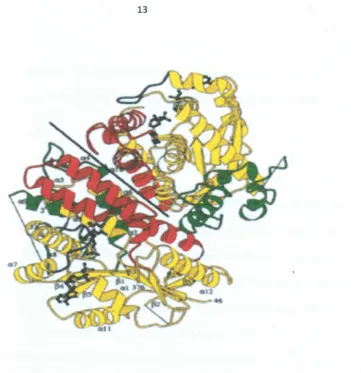
Figure 2.2: Structure of thymide kinase from herpesvirus. Stereoview of tbe
symmetric TK dimer with bound ADP and dTMP. Tbe domains are defined
according to the NMP-kinases (residues 46-81, 143-218, 227-250, 323-376, yellow),
NMP biRd (82-142, red). 72 additional residues around position 290 are green
(KIemenset01., 1997)
2.1.3Transmission of Herpes Simplex Virus
Transmissionof HSV is through direct contact with a lesion or body fluid of an infected
individual (David, 2012). People often get genital herpes by having sexual contact with
others who don't know they are infected or who are having outbreaks of herpes without
any sores. A person with genital herpes also can infect a sexual partner during oral sex
(Wald,2006). The virus is spread only rarely, if at all, by touching objects such as a
(1999). Infection of the mother may lead to severe illness in pregnancy and may be
associated with virus transmission from mother to foetus/newborn (Anzivino et al.,
2009).
2.1.4 Treatment
Antiviralmedications such as acyclovir, famciclovir, and valacyclovir are among the
drugs used to treat the symptoms of herpes. Valacyclovir is available as a generic as
well as acyclovir (Agrawal et al., 1990). Evidence supports the use of acycIovir and
valacyclovirin the treatment of herpes labialis (Chon et al., 2007) as well as herpes
infectionsin people who have cancer (Glenny etal.,2009). The evidence to support the
use of acyclovir in primary herpetic gingivostomatitis is less strong (Nasser et al.,
2008). Asmuch as these antiviral medicines are used, they do not cure the disease but
only help shorten the length of a herpes outbreak. Certain dietary supplements and
alternative remedies are also claimed to be beneficial in the treatment of herpes
(EBSCO, 2012). There is, however, insufficient evidence to support use of many of
these compounds including euchinecea, eleuthero, L-Iysine, zinc, monolaurin bee
productsandAloe vera (Perfect et al., 2005). While there are anumber ofsmall studies
showingpossible benefits from monolaurin, L-Iysine, aspirin, lemon balm, topical zinc
or licorice root cream in treatment, these are preliminary studies that have not been
confirmed by higher quality randomized controlled studies (Beauman, 2005). It has
beendifficult to develop effective vaccines against herpes viruses, many of which have
complexlife cycles and can lie dormant in the body for longperiods of time. The only
isthechickenpoxvaccine, which uses alive, weakened virus. (Ledford, 2012). There is
need therefore, to study effectively the biology of HSV in order to come up effective
drugsandvaccines.
2.2Herpes virus in Non-Human Primates
Thereare several neurotropic alpha-herpesviruses that are related to human herpes
simplex viruses (HSV-l and HSV-2), that infect non-human primates (Eberle and
Hilliard,1995: Wertheim et al., 2014). All these viruses produce a disease that is
similarto HSV in their hosts. Cercopithecine herpesvirus J (Monkey B virus [BY]) is
common in macaques tMacaca spp.). B Virus is of major zoonotic concern. When
transmittedto humans via bites or scratches, BV replicates in the peripheral epithelial
tissue where it invades the sensory ganglia. The virus then progresses into the central
nervoussystem (CNS) where itcontinues to replicate and spread resulting in extensive
tissue damageleading to death in most cases (Huff and Barry, 2003). Cercopithecine
herpesvirus 2 or Simian Agent 8 (SA8) infects the African Green Monkeys
(Cercopithecus aethiops). Simian Agent 8 was originally isolated from the African
Green Monkeys but it has also been found to be common in both wild and captive
baboons(Hull, 1973;Eberle et al., 1995).Herpesvirus saimiri and Herpesvirus ateles
arecommonin squirrel monkeys andspider monkeys respectively.
The baboons' Cercopithecine herpesvirus 16 or Herpesvirus Papio 2 (HVP-2) had
earlierbeen misidentified as SA8, but recent molecular analyses has shown it to be
humaninfectionhave been reported in the other simian viruses (Rogers et al., 2003). A
recentstudy by Wertheim et af. (2014) has linked HSV 2 infection to cross-species
transmission.Herpes simplex virus 2 is thought to have originated from chimpanzee;
theclosestrelative of man. Table 2.1 gives a summary of herpes viruses and their hosts.
Table2.1: Summary ofberpes viruses and their hosts (Wertbeim et al.,2014)
Virus
Virus Abbreviation Host Latin Name Host Common Name. . .
HerpesvirusPapio 2 HVP-2 Papio spp Baboon
SimianAgent 8 SA8 Cercopithecus aethiops African green monkey
Chimpanzeeherpes virus ChHV Pan troglodytes Chimpanzee
Herpessimplex virus 1 HSV-l Homo sapiens Human
Herpessimplexvirus 2 HSV-2 Homo sapiens Human t.
MonkeyB Virus BV Macacaspp. Macaques Saimiriineherpes virus HVS-J Saimiri sciureus Squirrel monkey
Spidermonkeyherpes virus RV A-1 Ateles geoffroyi Spider monkey
HSV-2
ChHV
HVP-2
HSV-l
SA8
BV
HVS-l
HVA-l
Figure 2.3: Pbylogenetic tree sbowing the relationship between human and simian
herpes viruses (~V-2-Herpes Simplex Virus type 2; ChDV-Chimpanzee Herpes
Virus; HVP-2-Herpesvirus Papio 2; SAS-Simian Agent 8; DV-Monkey D Virus;
HVS-I-Saimiriine herpes virus; DVA-I-Spider Monkey Herpes Virus)(Wertheim
et al: 2014)
2.2.1 Herpesvirus papio 2
Herpesviruspapio2(HVP-2) isan indigenous B-Iymphotropic virus of baboons (Papio
sp.) present in latent form in baboon lymphoblastoid cell lines (Lawrence, 1976).
Herpesviruspapio 2 exists as two major serotypes, HVP apathogenic (HVPap) and
HVP neurovirulent (HVP nv) (Ritchey et al., 2002). This difference in pathogenesis
however,is only seen in the mouse model. For instance, HVP 2 strain A951 has been
baboons were infected with different strains of HVP-2 (both apathogenic and
neurovirulent in a mice model) showed no distinction in terms of pathogenesis in
baboonsas they all caused the development of lesions. Juveniles were used in the study,
wherethe virus was inoculated intradermally in the oral mucosa of the bottom lip.
Lesionsdeveloped as seen in figure 2.4.
Figure 2.4: Increasing size of oral lesion in the baboon mouth following inoculation
with HVP 2 (Rogers et at.,2005)
The lesions continued increasing up to 21 days post infection (DPD, and then they
disappeared. The study was conducted by Rogers et al. (2005). The study proves that
mice are good models for studying cross-species transmission of herpes viruses. The
lesionsdevelopment regardless of the strain shows that baboon is a good model for
HSVstudy.
Infectionof baboons (Papio species) with HVP-2 produces a disease that is clinically
similar to herpes simplex virus (HSV-l and HSV-2) that affects human (Bigger and
Martin,2003). Infected adult baboons develop genital lesions whereas oral lesions
representthe predominant form of disease in younger animals (Levin et al., 1988). The
lesionsrange from vesicles to ulcers (Figure 2.5). Spontaneous reactivation from
latency has been demonstrated, a pattern consistent with HSV infection of humans
(Martinoet al., 1998). Acquisition of genital infection is primarily associated with the
onsetofsexual activity in baboons. Secondary bacterial infection is common, epidermal
necrosiswithcentral erosions and peripheral parakeratosis is also observed (Eberle et
aI., 1998). Like as with other members of the genus Simplex virus, HVP-2 also causes
Urogenital and neuronal infections that closely resemble those caused by HSV in
humans(Martinoet al., 1998).
HVP-2 outbreaks in baboon breeding colonies have been associated with genital
infectionsof adults and an increased incidence of stillbirths and hydrocephaly in
Figure 2.5: Ulcers appearing on the baboon penis following infection by HVP-2 (Borrowed from Dr. Richard Eberle from Department of veterinary pathobiology, Oklahoma State University, USA)
The prevalence of HVP-2 in several groups of captive and wild-caught baboons was
determined by detection of anti-HVP-2 antibodies in 133 sera of adult baboons. Over
90%of newly imported (wild-caught) adult olive baboons (Papio anubis) from Kenya
and chacma baboons (P. ursinus) from South Africa were found to have anti-HVP-2
antibodies. Similarly, approximately 85% of captive breeding colony baboons (P.
anubis and P. cynocephalus) were seropositive for HVP-2 (Eberle et al., 1997).
Baboons are social animals and move in large groups called troops (Figure 2.6) and this
could be the cause of high rates of infection by HVP-2, especially for the sexually
TheHVP-2 genome contains unique long (UL) and unique short (US) regions separated
by regions of inverted repeat DNA. To date some 27 partial and four complete open
readingframes have been analyzed, all of which demonstrate high degrees of identity
between the proteins of HVP 2 and their HSV homologues (Eberle and Black et al.,
1995;1997).The similarity between HVP 2 and HSV at both the clinical and genetic
levels indicates that the HVP 2 system has the potential to be a powerful surrogate
modelto study the biology of herpes simplex virus replication in the context of a natural
primatehost.
Becauseof lack of any reported human infections by HVP2, the virus has largely been
considered unimportant from a zoonotic standpoint. Studies have however shown that
intraperitoneal inoculation of mice with HVP 2 causes rapid death (Eberle et al., 1995),
suggestingthat HVP 2could affectother mammalian species.
2, HVP2 apathogenic (HVPap) and HVP 2 neurovirulent, (HVP2nv) were found to
readilyinvade the central nervous system (CNS) following peripheral intramuscular
inoculation(which most closely resembles a bite or scratch), producing paralysis and
death(Ritcheyet aI.,2002). Herpesvirus papio 2ap is not competent for spread within
thebrain. Herpesvirus papio 2 nv on the other hand readily invades the CNS and
produces a fulminant ascending encephalomyelitis which proves fatal once the virus
reachesthe brainstem (Rogers et al., 2003). Previous studies implicated the type I
interferon (IFN) response as being a major factor in controlling infection by
apathogenicisolates.
2.2.3Transmission of human herpes to Monkeys
Infectionby herpes 1 from human to monkeys can cause fatal herpes I disease in
monkeys(Herpes simplex, cold sores, fever and blisters). Just as the human immune
systemis not equipped to deal with the Monkey B virus, the immune system of
monkeysis not equipped to deal with herpes 1 virus from humans. If the monkey does
not die immediately, the same treatment used in humans can be given. A case of
transmissionof herpes from human to monkeys was observed by Matz-Rensing et al.
Anoutbreak occurred in a group of Callithrix jacchus housed as a family group after
contact with a person who had recrudescent HSV-I disease. On the first day of the
outbreak,a male marmoset showed apathy, anorexia, weakness, and marked salivation and diedthe same afternoon. At that time, no other animals inthe group showed clinical
signs(Mat-Rensing etal.. 2003). The following morning, one young adult and a female
developed excessive salivation and apathy. Closer examination showed ulceration of the
lipsand the oral cavity. In total, eight animals were infected and all had a 1-2 day
history of vesicular to ulcerative gingivitis and stomatitis accompanied by severe
salivation, serous nasal discharges, anorexia, and depression. Three of the animals
developed lesions covered with fibronecrotic exudate at the mucocutaneous junctions.
At necropsy, all animals showed an acute gingivitis and stomatitis. The tongues had
multifocal single to coalescing lingual erosions with rough borders up to 0.5-1.0 cm in
diameter. Some lesions appeared to be lingual or gingival ulcers covered by a
fibrinopurulent exudates. Mild splenomegaly and severe lymphadenopathy of the
regional lymph nodes was also observed. These animals had had contact with a
caretaker suffering from a herpes labialis-Iike illness a few days before the outbreak
occurred (Mat-Reusing et al.,2003).
Naturally occurring herpes simplex was also reported to be fatal in a family of white
faced saki monkeys. From the study, a family of three white-faced saki monkeys
tPithecia pithecia pitheciay died 48-96 hours after the onset of anorexia, nasal
discharge, pyrexia and oral ulceration. One animal also had clonic seizures. Lesions
necrosis,meningoencephalitis and sporadic neuronal necrosis. Intranuclear inclusion bodies and syncytial cells were present in oral lesions and affected areas of liver. Herpes simplex virus I (HSV-l) was identified as the etiology of disease by virus isolation,polymerase chain reaction, or in situ hybridization in all three animals. Immunohistochemistryfor detection of apoptotic DNA and activated caspase-3 showed significantlevels of apoptosis inoral andliver lesions and occasional apoptotic neurons in the brain. The finding of the study demonstrated the vulnerability of white-faced saki monkeys to HSV-l (Schrenzel et al., 2003). In most of the reported infections in variousnonhuman primates species, itwas indicated that contact with humans was the sourceof herpes virus infection. Visitors, students, or caretakers might be able to infect theanimals, forexample, by passing partly eaten food intothe cage.
2.3Role of interferons in herpes virus infections
2.3.1Role of interferons in Replication of HSV
A study that was performed to analyze the effect of IFNson HSV replication in human
primary cells, showed that Human macrophages were sensitive to the antiviral activity
of exogenous IFN a and IFN B, whereas IFN y exhibited only very little antiviral
activity (Ingrid etal., 1986). The effect was macrophage specific, because inhibition of
HSV replication on HEp-2 cells by IFN-y was comparable to virus inhibition by other
IFN types. The results of the study also indicated that IFN blocks a step before HSV
DNA synthesis. The main block during the viral replication cycle occurs either during
or before the synthesis of immediate early a-proteins (Ingrid etal., 1986).
Another study describes differences in biological activities of IFN-y on human
monocytes and WISH cells; a comparison of the IFN-y receptor on both cell types
revealed different binding characteristics (Orchansky et al., 1986). Thus, the lack of
antiviral activity of IFN- Y against HSV on macrophages could be related to a different
receptor on these cells. This is in agreement with the finding that IFN- yis ineffective in
potentiating the antiviral activities of IFN-a or IFN-B (Ingrid etal., 1986).
2.3.2Roleoftype 1 interferon on HVP-2 infection
A critical component of the innate anti-viral response is production of the type I
interferons a and B (IFN-a, IFN-B). IFN-a is rapidly produced by infected plasmacytoid
viatoll-likereceptors (TLRs) either through endocytosis of viral particles or autophagy
of infectedcells(Leeet al., 2007).
IFN-~israpidly induced in many cell types (including those infected byherpesviruses
such as fibroblasts and epithelial cells) due to their constitutive expression of IRF-3.
IFN-a and
-
1
3
produced by infected cells binds to the IFN-alf3 receptor (IFNAR)initiating a signal transduction cascade via the Janus associated kinase - signal
transducer and activator of transcription (Jak-Stat) pathway (Darnell et al., 1994;
Goodbournet al., 2000).
Followinginoculation of mice with HVP 2, two subtypes (HVPnv and HVPap) of the
viruswere identified. These subtypes vary in their pathogenesis in mice. Since the
pathogenesis of HVP2nv and HVP2ap infection in mice diverges well before an
adaptive immune response develops, innate immunity likely plays a major role in
effectingthe divergent pathogenicity of the two HVP2 subtypes. 1FN-f3appears to be
involvedin controlling HVP2ap infections sinceHVP2nv isolates do not induce and/or
overcome an IFN-f3 response, and are thus not as susceptible to IFN-f3 in vitro as
HVP2ap isolates are (Rogers et al., 2007). The experiments presented by Rogers et al.
(2009),extend the above observations and confirm a critical role for the type I IFN
2.4Importance of using a baboon as a model to study HSV
There are several advantages of using captive baboons for studies on herpesvirus
associated diseases. First, the animals are subjected to the same environmental
conditions,which naturally trigger reactivation of the endogenous herpesviruses; the
animals are genetically diverse (Sidebottom et al., 2001), yet most are related, and
familialassociations within the breeding cohort are easily established. The animals are
alsoavailablefor intervention as well as extensive observational studies ona long-term
basis.Femalebaboons have aprominent sex skin, which swells visibly and can beused
to follow the menstrual cycle. Since herpesviruses are transmitted in utero or during
.
.
birth,and since in particular the beta herpesviruses are a leading cause of congenital
infectionsandassociated diseases, this unique feature in the natural history of baboons
lends itself for detailed studies of theses pathologies (Sidebottom et al., 2001). At the
Institute of Primate .Research, the baboons (Papio anubis) are housed in special
facilities (Figure 2.7).· The animals are housed according 10 the Association for
Figure 2.7: Wild caught baboons in the animal facility of the Institute of Primate Research
Vaccine research is one of the applications for baboon models of infectious disease.
Since baboons carry a full complement of the gamma isotype of immunoglubulins,
humanreagents can be used to measure immune response and the animals can be
challenged to assess vaccine efficacy. Therefore, baboons are an outstanding model for
vaccine studies. There is already an existing extensive history of testing vaccines in
baboons(Jeong et al., .2004). The vaccines are for various infectious agents including
that Hepatitis C virus-like particles, produced usmg a recombinant baculovirus
containingthe cDNA of HCV structural proteins, can induce both cellular and humoral
immunityin baboons (Jeong et al., 2004). Other viral vaccine. studies have
demonstratedthe ability of baboons to produce an antibody response to a commercially
availableencephalomyocarditis virus vaccine, and baboons were the chosen model for
analyzingthe safety of using Escherichia coli heat-labile toxin
a
s
an adjuvant for thenasalinfluenzavaccine (Huneke et al., 1998;Zurbriggen et al., 2(03). Other studies
haveproven the baboon to be an effective model for elucidating the necessity of an
antibodyresponse when vaccinating against the parasite, Schistosoma mansoni. In the
.
studies,IgGlevels directly correlated with the level ofprotection (Soisson et al., 1993;
Kariukiet al., 2004). Capitalizing upon the high similarity between baboon and human
reproductivebiology, studies have demonstrated the ability of the fetus to mount a fetal
specificantibody response.and the neonates go on to develop immunological memory
againsthepatitisB surface antigen (Watts etal., 1999; Bot etal., 2001).
A surveyon theprevalence of HSV -2 in the world was done by Katherine etal. (2008). ~
According to the survey, the estimated total number of people aged 15-49 years who
wereliving with HSV-2 worldwide in 2003 was 536 minion. More women than men
were infected,with an estimated 315 million infected women compared to 221 million
infectedmen. The number infected increased with age, most markedly in the younger
ages,until it peaked in the age stratum 35-39 years of age, after which it declined
slightly.According to the same survey, analysis by region reveals similar trends. The
prevalence,with more women than men infected, and higher number of cases among
thoseat older age. Many studies that have been conducted on prevalence of HSV-2 in
Kenyahave shown similar results. According to Mugo etal.(2011), the total prevalence
ofHSV-2 in Kenya is 35%, of which 42% were women and 2~% were men.
Theprevalence of HVP-2 in baboons is 90%. This prevalence was obtained after
samplingbaboons from Southwest Foundation for Research in San Antonio in Texas,
NewEngland Regional Primate Center and also baboons that had been captured in
Kenyaand South Africa (Eberle etal., 1997).There is however, no data showing either
thecomparative prevalence of HVP-2 in baboons of different sexes or age groups. The
prevalence of HVP-2 in Kenyan baboons alone is also not known. Several strains of
HVP-2 in baboons have been identified. These strains have been classified as the
neurovirulentsubtype, and the apathogenic subtype. The neurovirulent strains include;
AI89164, OUI-76, OU3-i, OU3-I8, X313 and 960. The apathogenic strains on the
otherhand include; A951, OU2-5, OU2-9, OU2-12, OU4-2, OU4-8 and OU4-15. This
classificationhowever was determined based on HVP-2 infection in mice (Rogers et01.,
2003). In baboons, there is no distinction in terms of pathogenecity of the strains as
theyall causedevelopment of lesions (Rogers etal., 2005)
Currentlyin Kenya, there isno available scientific data on circulating strains of HVP-2
in non human primates which include the olive baboons (Papio anubis) and theAfrican
green monkeys (vervets). Such data would provide an insight into the evolutionary
The similarity between HVP-2 and HSV at both the clinical and genetic levels indicates
thatthe HVP 2 system has the potential to be a powerful surrogate model to study the
CHAPTER THREE: MATERIALS AND METHODS
3.1Studysite
Thisstudy was conducted at the Reproductive health and Biology Laboratories of the
Instituteof Primate Research (IPR) in Karen, Nairobi County, Kenya. The Institute of
PrimateResearchis ~ biomedical research facility mandated to undertake research using
non-humanprimates (NHPs) as models with a mission to improvehumanhealth. Inthis
study,sampleswere collected from the captive baboons at the !PR animal facility and
alsofromIPR blood bank. The baboons were captured fromsix geographical regions of
Kenya.These regions include; Laikipia, Nyandarua, Kirinyaga, Machakos, Kajiado and
Nairobi.
3.2Study Design
Thiswasa cross sectional study where 5 ml of blood was drawn from wild caught olive
baboons. These baboons were captured from the following regions; Laikipia,
Nyandarua,Kirinyaga, Machakos, Kajiado and Nairobi,
to
3.3Animal capture
The baboons were trapped using rectangular traps (1.5m high and O.9mwide) made of
iron bars, wire mesh and weighted trap doors designed by Institute of Primate research
(IPR)staff. The NHPs were baited for a week using maize cobs, carrots and bananas.
The monkeys then became habituated. The traps were set in the evening after the
day(Jenebyet al., 2002; Eley et al., 2005). This technique gave an equal chance for all
the animals regardless of their gender or age to have an equal opportunity to feed and
thus trapped as they came for the bait. The animals were trapped then transferred to
their holding cages and transported to Institute of Primate Research quarantine area,
3.4 Sample size determination
This study involved serum samples, trigeminal ganglia and swabs collected from
baboons. The sample size was determined using the following formula-according to
Delletal. (2002).
er
dWhere;
n =Minimumsample sizerequired
pc=Observedprevalence (90%), (Eberle et al., 1997)
ps= Postulated prevalence (85%), (Eberleet al., 1997)
qc= i-pc
qe=1-pe
C=Constant that depend on chosen valuesof a and
P;
10.519n
=
10.519(0.9x
0.1+
0.85x
0.4)+
L
+
2=
59Thus, aminimum of 59 baboon sampleswas required in this study. However, a totalof
189baboons were sampled in thestudy.
3.5Sample collection
Five milliliters of blood was collected from the baboons. Genital and oral swabs were
collected from 100out of the 189 baboons. Trigeminal Ganglia (TG) were collected
from 9 asymptomatic baboons at autopsy. Blood wasused to screen for anti-HVP2 IgG.
Deoxyribonucleic Acid (DNA) extraction was done for swabs and TG from
asymptomatic animals. Thymidine kinasecoding region of HVP-2 was amplified using
TKIITK2 primer set. Positive sample amplicons from peR were purified before
sequencing. During sampling, baboons quarantined and those housed atthe Institute of
Primate Research cages were injected with anaesthesia; a mixture ofxylazine (2%) and
Ketaminehydrochloride (10010) in the ratio of 3:i respectively. The dosage given was
3.5.1 Blood sample
Bloodsamples were collected from 105 freshly captured baboons from the wild. Five
millilitersof venous blood were collected in sterile 9 m1 tubes (Greiner bione, France)
for serum separation. Serum was stored at-20°C before ELISA was done. Eighty four
serumsamples were acquired from the IPR serum bank. These sera were from baboons
thathad been at IPR formore thanthreemonths.
3.5.2 Swabs
Swabswere collected from asymptomatic baboons. Since baboons acquire virus as an
oral infection while young or as a genital infection after reaching sexual maturity, Oral
swabswere collected from 31 juveniles while genital swabs were collected from 69
adult baboons. For the juveniles, swabbing was done in the chicks and the tongue. As
for the adults, swabbing was done on the vaginal lining and the cervix for the females,
andin the urethra for the males. Cotton-tipped applicators were used as swabs and
placedin l-ml transport medium (digene® Female Swab Specimen Collection Kit,
QIAGEN). Atotal of 100 genital and oralswabs from the baboons were collected.
3.5.3 Trigeminal ganglia
Nine TG tissues were collected. At the time of autopsy, all TG were aseptically
removedandtrimmed of dura and fat. All TGwere aseptically removed after opening
theskull, and trimmed of dura and fat. The three branches of the TG were dissected
Clinicalswabs and the brain tissues were then submitted to the Reproductive Health
laboratory at IPR Nairobi, where analysiswas done.
3.6Viral antigen preparation
The HVP 2 antigens, positive and negative controls were generously provided by
Professor Richard Eberle of Veterinary Pathology Department of Oklahoma States
University,USA. Herpesvirus Papio 2 antigens were prepared by Or. Richard Eberle in
hislaboratory (Eberle et al., 1997). During the preparation, HVP2 was isolated from an
adult baboon. Theviruswaspropagated in African green monkey (vero) cells. Snbstrate
antigenfor enzyme-linked immunosorbent assays (ELISA) was propagated by inducing
an infection to a 150 cm2 flask of vero cells with HVP2. When complete cytopathologic
effect (CPE) had taken place, cells were scraped into a medium and pelleted by
centrifugation at400xg for 5 min and then, the medium removed. The infected cells
werefrozen for 30 mins in ice,thawed and then resuspended in 0.5% Triton x-lOO
Tx-100(in water) at a concentration of 2 x 10⁷ cells/ml. Two microfuge tubes containing
500 ~lof infected cells in suspension were prepared. The negative control constituted
uninfected vero cells. The cells, positive and negative controls were then lyophilized
beforebeing shipped to IPR from Oklahoma State University.
3.6.1 Lyophilized HVP-2 antigen resuspension
Thelyophilized antigens were resuspended in 500 Ill/tube of sterile water containing
pipeting was done to resuspend the cells. Vortexing was done every few minutes for a
period of 10 min. Centrifugation was done at full speed (14,000 rpm) for 30 seconds.
The supematant (antigen) was aliquoted and stored at -80°C while the pellet was
discarded.
3.7 Screening Cor HVP-2 antibodies using Enzyme Linked Immonosorbent Assay
(ELISA)
For ELISA, 96 well flat-bottomed plates were coated with HVP 2 antigen (1OOJ1JJwell)
diluted at 1:600 in lx Phosphate Buffered Saline (Ix PBS), and incubated overnight at
4°C. The plate was washed 3 times in the ELISA washer. Excess binding site was
blocked by addition of 100 J11of PBS containing 3% Bovine serum albumin BSA and 5
Jll Tween 20 (PBSrrwIBSA) for 1hour at 37°C. The excess blocking buffer was then
aspirated and the plate washed 5 times. Fifty microlitres of sample sera diluted in
PBS/TW containing 0.3% of BSA at 1:200 was added to each well and incubated at
room temperature for 2 hours. Wells were washed 5 times with PBSITW. Fifty
microlitres per well of goat anti-human IgG peroxidase conjugate diluted in 1:2000 was
added and incubated for lhour at room temperature. Wells were washed again in
PBS/TW and 50 J11of substrate solution (TMB) per well was added, then incubated in
dark for 10min (when +control was blue and -control clear). Optical density OD at a
wavelength of 630 nm was determined using a microplate reader (DYNA TECH
3.8Extraction of Deoxyribonucleic Acid (DNA)
Deoxyribonucleic acid was extracted from 76 swabs (both oral and genital) from
animalsthat were seropositive by ELISA A total of 24 swabs were left out since the
animals fromwhich the swabs were collected tested negative for HVP 2 when ELISA
wasdone. DNA was also extracted from the9TG samples that were collected.
3.8.1 Oral and genital swabs
Viral DNA was extracted from swabs of seropositive animals, usmg the ZYMO
RESEARCH ZR -DNA extraction Minikit (Inqaba, South Africa) according to the
manufacturer's protocol. These swabs had been stored at -80°e. Swabs were picked
fromthe transport media. The stick part of the swab was cut off and the cotton swab
insertedinto an eppendorf tube. Five hundred microlitres of genomic lysis buffer was
added to each eppendorf tube. Vortexing was done for 10sec and the latter was left to
stand for IO min at room temperature. The mixture was transferred to a zymospin
column in a collection tube. Centrifugation was done at 11,000 rpm for 1 min. The
zymospin column wastransferred to a new collection tube and 200 J.lI ofDNA pre-wash
buffer added. Centrifugation was done at 11,000 rpm for 1 min. Five hundred
microlitres of g-DNA wash buffer was added to the spin column. Centrifugation was
done at 11,000 rpm for 1 min. The spin column was then transferred to a clean
eppendorftube. Thirty microlitres of DNA elution buffer was added to the spin column.
(14,000 rpm) wasdone for 30 sec. The eluted DNA was stored at -20°C before being usedforPCR.
3.8.2 Trigeminal ganglia
Deoxyribonucleic acid extraction was done using the Qiagen kit according to the manufacturer's protocol. Twenty five milligrams of the trigeminal ganglia tissue was cut up into small pieces and put in a 1.5 microfuge tube. A hundred microlitres of the
ATL buffer was added. Twenty microlitres of proteinase K was added and mixed by vortexing thereafter incubation at 56°C overnight followed. Occasional vortexing
duringincubation was done to disperse the sample. Centrifugation was done to remove
dropsfrom theinside of the lid. Two hundred micro litres of ALbuffer was added tothe
sampleand pulse-vortexing done for 15 sec. Incubation followed at
ire
for 10 min.Twohundred microlitres of ethanol (99%) was added tothe sample and mixed bypul
se-vortexingdone for 15min. Themixture was carefully applied to aQIAmp spin column in acollection tube and centrifugation done for 1 min at 8,000 rpm. The QIAmp spin
column was carefully opened and 500 J.LI of AWl buffer added. Centrifugation was
done at 8,000 rpm for 1 min. The QIAmp spin column was transferred to a new collection tube. Five hundred microlitres of AW2 buffer was added and centrifugation done at full speed (14,000 rpm) for 3min. The QIAmp spin column was transferred to
another collection tube. Two hundred microlitres of buffer (AE) was added to the spin column. Incubation at room temperature for was done for 5 min. The eluted DNA was
3.8.3 Confirmation of the extracted Deoxyribonucleic Acid
Gel electrophoresis was done to confirm whether the DNA had been extracted. 1%
agarosegel was prepared. Tenmicrolitres per well of the extracted DNA was added in
each well. It was then allowed to run for 40min. The presence of DNA was confirmed
by viewingthe gel ina UV trans-illuminator.
3.9 Amplification of deoxyribonucleic Acid
ForPCR assay, the unique longregion (UL 23) of herpes virus that code forthymidine
kinase was amplified using primer set TKl (5'TTCACCGTGGGCTGGACTGG 3') and
TK2 (5'GCGGTTCTGGAGCTCGGACCA 3'). For virus characterization, PCR
products were purified before being sent for direct sequencing,
3.10 Polymerase Chain Reaction conditions
A ready mix PCRmaster mix from zymogen was used. The PCR components consisted
of95 ulofPCR master mix, 1 uloftheforward primer, 1 ul of thereverse primer, I J.lI
of the DNA template and 9.5 J.lIof nuclease free water to bring the final Volume to 25
Ill.PCR was run using 30cyclesof initial denaturation at 94°C for 5 min, denaturation
at 94°C for 60sec, annealing at 60°C for 60 sec, andelongation at
n
O
c
for 120secin a3.11Gel electropboresis
Agarose gel electrophoresis was used to determine and visualize the presence of PCR products.-Specific-DNA amplification "was confirmed by electrophoresis using 1.5% agarose gels prepared by dissolving 1.5 gm of agarose into 100 ml of Tris acetate EDTA (TAE) buffer and then adding 5 IIIof ethydium bromide. The gel wells were
loaded with 9III .of the amplicons. The gel was then visualized using a UV
transiluminator.
3.12 Purification ofPCR products.
Polymerase chain reaction products were purified prior to sequencing using Gene JETlMPCR Purification kit. Binding buffer was added to the PCR mixture at a ratio of
1:1 and mixing done vigorously. The solution was transferred to the Gene JETlM
.
purification column. Centrifugation was then done for 1 min at 14,000rpm. The flow-through was then discarded. Seven hundred microlitres of the wash buffer was added to
the Gene JET purification column. Centrifugation was done at 14,000 rpm for 1 min.
The flow-through was discarded and the purification column put back into the collection tube. The Gene JET purification column was centrifuged for an additional 3 min tocompletely remove any residual wash. The Gene JET purification column was
~ .
transferred to aclean microfuge tube.Twenty five micro litres of the DNA elution buffer was added to the centre of the column. Incubation was done for 1 min at room
3.13 Clean upof DNA before sequencing
Deoxyribonucleic Acid clean up was done at the Inqaba biotech laboratories in South
Africa where samples were sent for sequencing. The clean up was done using the ZR-96
DNA sequencing clean up kit TM. During clean up, 240 f.11of sequencing binding buffer
was added to 5 f.11of the sequencing reaction. The mixture was transferred to a z
ymo-spinTMIB - 96 plate mounted onto a collection plate. Centrifugation was done at 5000
xgfor 2min. Three hundred micro litres of sequencing wash was added to each well of
the plate. Centrifugation was done at 3000 xg for 5 min. Twenty micro litres of water
was added directly to the matrix of the filter plate. The Zymo - Spin 1MIB .:....96 was
placed on top of the supplied 96 - well PCR plate and the assembly mounted on the
collection plate. Centrifugation was done at 3000 xg for 2 min to elude the DNA. The
ultra-pure DNA was ready to be loaded onto a sequencer. The primers that were used
during PCR amplification, that is, TKl and TK2 were used for sequencing.
3.14 Sequence analysis
AB! V3.1 Big Dye kit was used for sequencing. The samples were sequenced on an
AB! Prism 3130 automated sequencer (Applied Biosystems, Foster City, CA, USA)
according to the manufacturer's instructions. Analysis was done on the ABl 3500 XL
genetic analyzer using a 50 cm array and POP7. Sequences were generated with the
MEGA version 5 program package and blasted at the NeBI database using a blast
algorithm. The purpose of blast analysis is to find regions of sequence similarity, which
will yield functional and evolutionary clues about the structure and function of the
3.15 Phylogenetic analysis
Nucleotide sequences for HVP 2(samples S55 and OPC) were edited and analysed
using BioEdit Sequence Alignment Editor (V.7.0.9.0) [Hall, 1999]. A Basic Local
Alignment Search Tool (BLAST) search (http://blast.ncbi.nlm.nih.gov) was performed
to compare the identity of the sequenced with the reference strains from GenBank by
pairwise comparison. The nucleotide sequences were aligned with human herpes virus
and other HVP 2 virus strains reference sequences from GenBank using MAFFT
version 7 (http://mafft.cbrcjp/alignment/server/). After manual adjustment of the
alignment, phylogenetic analysis was performed with MEGA 6 using the
neighbour-joining methods (Tamura et al., 2013). The HSV strains were assigned based on the
clustering in the phylogenetic tree (>70010 bootstrap support).
3.16 Data Analysis
Pearson Chi- X2 square (two bytwo table) was used to compare the number of positive
samples to negative samples (proportions) for both female and male baboons in both
groups that is, captive and wild caught. Graph pad prism was used for data analysis
(Motulsky, 1999).Percentages of exposed animals were determined where Age,sexand
locationwere compared. AP value of lessthan 0.05wasconsidered significant.
3.17 Ethical Considerations
Ethical approval before conducting the study was obtained from theInstitute of Primate
maintenance were carried out as according to the guidelines of Institutional Review
CHAPTER
FOUR: RESULTS
4.1 Anti- HVP-2 antibodies
The prevalence of HVP-2 was compared among baboons from different regions, sexes
and ages by detection of anti-HVP 2 antibodies. Some of the baboons had been in the
IPR colony for more than 3 months therefore the study also compared the prevalence of
HVP-2 in freshly captured baboons from the wild with those that had been in the colony
forover 3 months.
4.1.1 Seroprevalence of HVP-2 in baboons by region
A total of 189 baboons from six counties in Kenya were sampled. The counties include
Laikipia (65), Nyandarua (33), Kirinyaga (31), Machakos (24), Kajiado (16) and
Nairobi (20) Counties. These counties experience differen climatic conditions, with
Nyandarua County being the coldest of the six counties and Machakos and Kajiado
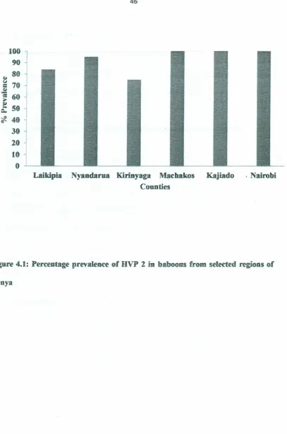
counties, the hottest. Findings from this study showed a high prevalence of HVP-2 .
infection in baboons from all the regions ranging from 71% in Kirinyaga county to
~,
Laikipia Nyandarua Kirinyaga Machakos Kajiado . Nairobi Counties
Figure 4.1: Percentage prevalence of HVP 2 in baboons from selected regions of
4.1.2 Seroprevalence of HVP-2 in baboons by sex
Inthis study more females compaired tomales sampled were found to have anti-HVP-2
IgG antibodies. The prevalence of HVP-2 in females was 90% (94 out of 105) while in
males it was 83% (70out of 84)(Table 4.2; X2=1.6; df=l; P>0.05 ).
Table 4.1: Seroprevalence of HVP 2 antibodies comparing gender exposure rates
Gender Total Animals Positive Negative % Prevalence
Males
Females
84
105
70
94
14
11
,83
90
Total 189 164 25 87
••
There was no statistical significance in gender prevalence.
4.1.3 Seroprevalence of HVP-2 in baboons byAge
In relation to the three age groups sampled in the study (Adults, Sub-adults and
Juveniles), more adults (94%) and sub-adults (90%) were found to be sero-positive as
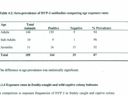
Table 4.2: Sero-prevalence of HVP-2 antibodies comparing ageexposure rates
Age Total
Animals Positive Negative %Prevalence
Adults 148 139 9 94
Sub-Adults 10 9 1 90
Juveniles 31 16 15 52
Total
189
164
2587
The difference in age prevalence was statistically significant.
4.1.4 Exposure rates in freshly-caught and wild captive colony baboons
A comparison in exposure frequencies of HVP 2 in freshly caught and captive colony
baboons was made. The prevalence of HVP 2 in captive colony baboons (89%) was
found to be slightly higher than in wild-caught baboons (85%) (Table 4.3; X2=O.83;