ELECTRICAL RESPONSES TO CHEMICAL STIMULATION OF
SQUID OLFACTORY RECEPTOR CELLS
BY MARY T. LUCERO, FRANK T. HORRIGAN AND WM F. GILLY Hopkins Marine Station, Stanford University, Pacific Grove, CA 93950, USA
Accepted 19 August 1991
Summary
Electrical properties of isolated olfactory receptor cells were studied using voltage- and current-clamp techniques based on whole-cell patch-clamp methods. Squid olfactory receptor cells contain voltage-gated Na+ and K+ channels and are capable of generating action potentials. Chemicals that elicit escape-jetting responses in behavioral experiments affect the excitability of isolated receptor cells. One set of such chemicals, including quaternary ammonium ions and aminopyridines, blocks K+ channels and increases excitability. Squid ink and L-Dopa also elicit escape jetting, but these substances increase membrane conductance, hyperpolarize the receptor cell and decrease excitability. These experiments indicate that sensory neurons of the olfactory organ are capable of detecting chemical signals and that at least two different transduction mechanisms can lead to similar behavioral responses.
Introduction
Anatomical studies have shown that squids (and other cephalopods) have what is termed an 'olfactory organ' generally located posterior and ventral to each eye (Watkinson, 1909; cited by Emery, 1975; Wildenburg and Fioroni, 1990). This olfactory organ is the site of a sensory epithelium composed of ciliated support cells and several types of putative receptor cells. The receptor cells are bipolar neurons that send an apical dendritic stalk to the surface of the olfactory organ where the specialized sensory cilia are exposed to the ambient sea water. A small unmyelinated axon leads from the basal surface of each receptor neuron to the 'olfactory lobe' and other areas of the brain (Messenger, 1979). Within the brain, afferent tracts from the olfactory lobe impinge on motor centers that control swimming and escape jetting and on the optic gland, a structure thought to affect gonadal maturation (Wells and Wells, 1959). The anatomy thus suggests links between chemoreception and escape jetting as well as sexual or reproductive activity.
It is surprising, given these anatomical studies describing structures and pathways so similar to their likely counterparts in vertebrate olfactory systems,
232 M. T. LUCERO, F. T. HORRIGAN AND W. F. GILLY
that behavioral or physiological studies on the squid olfactory system have been so limited. Our behavioral study (Gilly and Lucero, 1992) provides the first evidence for chemoreception of water-borne chemical signals by the olfactory organ in squid.
In the present study, we extend the behavioral work (Gilly and Lucero, 1992) to the receptor cell level. Primary receptor neurons isolated from the squid olfactory organ contain voltage-gated Na+ and K+ channels and are capable of generating repetitive action potentials. When tested on isolated receptor cells, chemicals that cause escape jetting fall into two categories. Chemicals in one category block K+ channels, depolarize the cell and increase excitability. Such blockers include quaternary ammonium ions and 4-aminopyridine (4-AP). The second category includes squid ink and L-Dopa, both of which act in the opposite way to increase membrane conductance, hyperpolarize the cell and decrease excitability. Thus, it appears that squid olfactory receptor cells convey sensory information about certain chemicals in their environment by at least two mechanisms that change the firing pattern of the receptor cells. A preliminary report of this work has appeared in abstract form (Lucero et al. 1991).
Materials and methods
Dissection of olfactory organ and culture conditions
The olfactory organ of adult Loligo opalescens Berry was excised under a dissecting microscope and treated with non-specific protease (lOmgmP1 Sigma type XIV) in sterile filtered sea water at 18-20°C for 50 min. Following a 3-5 min rinse in fresh filtered sea water, the isolated olfactory organ was placed on a nylon screen (100 ^m mesh size) in a tissue culture medium consisting of Leibovitz's L-15 (Gibco, Inc) supplemented with 263mmoll~1 NaCl, 4.64mmoll~1 KC1, g.OSmmoir1 CaCl2, 49.54mmoir1 MgCl2, 2 m m o i r1 Hepes (pH7.8),
2mmoll~1 L-glutamine, SOi.u.mP1 penicillin G and O i m g m l "1 streptomycin. The olfactory organ remained relatively intact with this treatment and was incubated at 16-17°C for periods ranging from l h to 2 days before use in electrophysiological experiments.
Whole-cell and nystatin patch recordings
Isolated olfactory cells were obtained for electrophysiological study by remov-ing the nylon screen and adherent olfactory organ from the culture dish with sterile forceps and dipping the screen into the bath solution in the glass-bottomed experimental chamber (100jd volume). Cells were allowed to settle for about 5 min before the chamber was thoroughly flushed to remove debris and non-adherent cells. Experiments were conducted at 10°C.
test solution and 1 mmol P1 blue no. 1 (Brilliant Blue) dye. It took the pipette 0.5 s to travel from the surface of the bath to the bottom of the chamber. The pipette was positioned approximately 50 pan from the apical portion of the cell. To ensure that bath solution was not unintentionally drawn up into the puff pipette, 10-20kPa of pressure was applied immediately before entering the bath. Thus, test solution was ejected during the entire time (8-15 s) that the pipette was in the bath. Control and test substances were applied either through the same pipette, by pulling bath solution into the pipette after a test run, or by using a multibarrelled pipette. Complete removal of the pipette from the bath also took 0.5 s.
For whole-cell recordings (Hamill etal. 1981), electrodes were pulled from either KG12 or 0010 glass (Garner Glass, Claremont, CA), coated with Sylgard (Dow Corning, Midland, MI), and had resistances of 1-2 MQ when filled with an internal solution. To establish the whole-cell recording configuration, the elec-trode was pressed against the cell while tip resistance was being monitored. Once a high-resistance seal had formed (>109Q), the holding potential was set to —70 mV, and gentle suction was applied to rupture the membrane under the pipette. An advantage of this conventional whole-cell technique is that macro-scopic currents can be studied while controlling the cell's internal solution. A disadvantage is that second messenger systems are washed out of the cell.
The nystatin 'perforated' patch technique (Horn and Marty, 1988) was also used to examine cells under voltage- and current-clamp, presumably with second messenger systems more intact. In these experiments, a thick-walled (0.64mm) Boro-glass pipette with a capillary filament (Sutter Instrument Co., Novato, CA) was back-filled with 50/zgmP1 nystatin internal solution. Positive pressure was applied to the patch electrode during the descent through the bath. Just before touching the cell, the pressure was released and the pipette current was balanced to zero. Upon touching the cell, gentle suction was applied to the patch electrode only until a high-resistance seal formed between glass and cell. The holding potential was set to — 70 mV. Within lOmin the electrical transient associated with the cell's input capacitance grew larger and faster, indicating that nystatin pores had formed to allow electrical access to the cell interior.
25-65 % of the resistance in series with the cell membrane was compensated for by positive feedback. Linear leak currents and the small residual, uncompensated capacity currents were subtracted on-line using a 'P/4 procedure' (Armstrong and Bezanilla, 1974). All voltage-clamp records presented in this paper have been leak subtracted. Averages are presented as the mean±l standard error. Holding potential was always - 7 0 mV.
Data acquisition
234 M. T. LUCERO, F. T. HORRIGAN AND W. F. GILLY
converter at a sample rate of 1.3-100 kHz and filtered at 3-10 kHz. Continuous recordings in current-clamp mode were recorded on a Toshiba digital hi-fi VHS DX900 using PCM (Unitrade, New York) with a sample rate of 44.1kHz and played back through the Labmaster/TL-1-125 for further analysis.
Solutions and channel blockers
Compositions of external bath and internal pipette solutions for the different experiments are given in each figure legend. Internal solutions were buffered to pH7.2, and bath solutions were buffered to pH7.4 with Hepes. For nystatin patch experiments, a nystatin stock solution of 25mgml~1 in dimethylsulfoxide was made daily and added to the filtered internal solution. All chemicals were obtained from Sigma Chemical Co. (St Louis, MO).
Tetramethylammonium (TMA+), tetraethylammonium (TEA+), tetrabutyl-ammonium (TBA+) and 4-aminopyridine (4-AP) were added in powdered form to the appropriate external artificial sea water (ASW). Methadone was made up as a lOOmmoll"1 stock solution in ASW and diluted to SOOjimoll"1 just prior to use. L-Dopa was added in powdered form to ASW, sonicated for 2min, and used within 2 h. Squid ink extract was prepared by placing a cut open ink sack in 1 ml of ASW, vortexing it for 2min, centrifuging at 14 000 g for 2min and filtering the supernatant through a 0.2/an filter. This extract, hereafter referred to simply as 'ink', was freshly prepared on the day of use.
Results
Receptor cell morphologies
Fig. 1A-C shows examples of living olfactory organ cells that have been freshly dissociated. Several kinds of putative chemoreceptor cells are recognizable, and the morphologies to be described persist for 1-2 days in vitro. The most common type (Fig. 1A) is termed pyriform and has a long axonal process arising from the basal pole containing the nucleus and a spike-like process that projects from the apical pole. Most of the cell body appears to be filled with granular material or cristae, and the bulk of the cell's 'interior' is actually an invaginating extracellular cavity filled with cilia (Emery, 1975). These cells appear equivalent to the 'Type 4' receptors described on ultrastructural grounds in Lolliguncula brevis (Emery,
1975). A low-power scanning electron micrograph of an aldehyde-fixed pyriform receptor (see Materials and methods in Gilly and Lucero, 1992) is shown in Fig. IE; the spike-like process is evident.
236 M. T. LUCERO, F. T. HORRIGAN AND W. F. GILLY
A second distinctly different receptor cell type is termed floriform (Fig. IB). This type also displays a prominent axonal process, but in this case a long, thin neck extends from the cell body and is crowned by a swelling covered with non-motile cilia arranged in a petaloid fashion. Such floriform cells are probably those Emery labeled Type 2 or Type 5 receptors. Fig. 1D,F shows scanning electron micrographs of a fixed floriform receptor. Pyriform and floriform receptor cells were the most distinctly identifiable and most common types of cells. Electro-physiological experiments were conducted on both.
A third, less common receptor may be related to the pyriform-type cell on the basis of the presence of a 'spike-like process' on the apical pole (Fig. 1C). In this type of cell, however, the portion of the cell body containing the nucleus is separated by a short neck from the apical swelling filled with cilia and granular material. This type of receptor appears to be equivalent to Emery's Type 3.
Passive properties of floriform and pyriform receptor cells
Resting potentials were determined in receptor cells immediately after obtain-ing electrical access to the cell's interior, with the whole-cell electrode measurobtain-ing voltage (current-clamp mode). Pyriform receptors have a mean resting potential of — 58.2±1.2mV (7V=91). Floriform cells have less negative resting potentials that are not stable and generally fluctuate rapidly from —50 mV to —25 mV (N=5). Thus, we were not able to obtain a meaningful average value of the floriform cell resting potential. Resting potential data are also displayed in Table 1 (measured with 400-450 m m o i r1 K+ in the pipette).
Passive electrical properties of voltage-clamped floriform and pyriform receptor cells were examined by applying voltage steps from the holding potential of - 7 0 mV to -120mV. No voltage- or time-dependent currents were activated in this voltage range. The mean values for input resistances, input capacitances and
Table 1. Passive membrane properties of single receptor cells of the olfactory organ
Cell type Pyriform (N) Floriform (N) Resting potential (mV) -58.2+1.2 (91)
-25 to -50 (5) Input resistance (MO) 406+77 (30) 1700+400 (14) Input capacitance (PF) 11.9+1.0 (40) 5.7+0.5 (11) Capacity current time constant Fast 0*0 78+6 (16) 88+8 (14) Slow (ms) 1.7+0.2 (16)
Means±ls.E. are given; the number of cells (TV) is also indicated.
Input capacitance refers to the fast component only for pyriform cells; it was determined after the slow component had vanished (see text for additional details).
time constants for the capacity current transients measured from both pyriform and floriform cells are listed in Table 1. The highest input resistance measured for a floriform receptor of 5.4GQ was considerably higher than the maximum of 1.8 Gfi in a pyriform receptor cell.
Input capacitance of pyriform cells was larger than that in floriform cells, and pyriform cell capacity current transients showed both fast and slow components with time constants of 7 8 ± 6 J I S and 1.73±0.17ms, respectively (N=16). The slow component sometimes suddenly disappeared during an experiment with no change in the amplitude of the fast component. This phenomenon often occurred after 15 min or so and is almost certainly associated with loss of electrical access to the large invaginating cavity in the interior of the cell. Voltage-dependent currents under study at the time were not obviously altered by this event. The average value of input capacitance after loss of the slow component was 11.9+1.0 pF
Sodium currents
Both pyriform and floriform receptor cells are excitable and contain a complement of ion channels similar to those found in other squid neurons (Brismar and Gilly, 1987; Gilly and Brismar, 1989). Na+ currents sensitive to tetrodotoxin (TTX) (obtained by subtracting voltage-clamp records obtained in the presence of 100 nmol P1 TTX from those recorded in the absence of drug from the same cell) are illustrated in Fig. 2A. These currents were elicited by voltage steps to - 4 0 mV and beyond (10 mV increments to +70 mV) from a pyriform cell bathed in a zero-potassium artificial sea water (ASW) and internally dialyzed with a Na+-containing internal solution. The conductance-voltage relationship calcu-lated by dividing the peak current at each potential by the driving force shows that the channels first activate near - 3 0 mV and reach a half-maximal conductance at —5mV (Fig. 2B). Inactivation of Na+ current is steeply voltage-dependent between —50 and OmV and is half-maximal at approximately — 25 mV (data not illustrated).
Na+ currents recorded from a floriform cell under the same conditions are shown in Fig. 2C. The properties of the floriform cell Na+ conductance (Fig. 2D) are similar to those observed in the pyriform cells. A major difference between cell types lies in the amplitude of the currents. The voltage-gated Na+ currents in pyriform cells are about five times larger than those in floriform cells. Based on capacitance measurements (Table 1), a pyriform cell is only about twice as large as an average floriform cell.
Potassium currents
238
A
M. T . LUCERO, F . T. HORRIGAN AND W . F . GlLLY
l n A
5 ms
D
-20 0 20 40
Voltage (mV)
1.0 T
60
0.2 nA
-20 0 20 40 Voltage (mV)
60
Fig. 2. Voltage-gated Na+ currents are found in both pyriform and floriform olfactory receptor cells. (A) Superimposed TTX-subtracted Na+ currents from a pyriform cell rapidly activate and inactivate during 5 ms voltage steps. (B) Relative Na+ conduc-tance is plotted from the data in A. (C) Na+ currents from a floriform cell recorded using the same voltage protocol as in A are similar to, but smaller than, Na+ currents in pyriform cells. (D) The conductance-voltage relationship for the floriform Na+ currents in C reveals similar voltage dependence of Na+ channel activation in the pyriform cell. Zero-K+ bath solution: 480mmoirl NaCl, 50mmoir' MgCl2,
lOmmoll"1 CaCl2, lOmmoir1 Hepes. Internal solution: SOmmolF1 NaCl,
lOOmmoll"1 sodium glutamate, 50mmoll~L NaF, SOOmmolF1 tetramethylammo-nium glutamate, 25mmoir1 TEAC1, lOmmolP1 EGTA, lOmmolT1 Hepes.
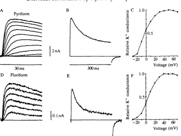
concentrations indicates that these currents are carried by K+-selective channels (not illustrated). The kinetic properties evident in Fig. 3A and the voltage-dependence of the peak K+ conductance (Fig. 3C) are very similar to analogous data obtained in squid giant axon or giant fiber lobe (GFL) neurons of the stellate ganglion (Llano and Bookman, 1986). As in GFL cells, the K+ currents in pyriform receptors partially inactivate during a long depolarization (Fig. 3B).
Pyriform C 1.0
2nA
30 ms D Floriform
300 ms
0.1 nA
- 2 0 0 20 40 60
Voltage (mV)
8 F
3
8 1.0
0.5
[image:9.451.49.406.61.328.2]- 2 0 0 20 40 60 Voltage (mV)
Fig. 3. Delayed rectifier K+ currents from pyriform and floriform cells have similar kinetics but differ in amplitude. (A,D) Outward K+ currents activate with a delay in response to 30 ms voltage steps to levels between — 30 mV and +70 mV (10 mV increments) in a pyriform cell (A) and a floriform cell (D). Note the difference in scale bars between the two cell types. (B,E) K+ current inactivates during a long (300ms) pulse to +70 mV. lnactivation is slightly greater and more rapid in the floriform cell (E) than in the pyriform cell (B). (C,F) Relative K+ conductance is plotted from the data in A and D, respectively. K+ currents from both cell types activate between -30 and -20 mV and reach a half-maximal conductance between - 5 and +10 mV. Artificial sea water bath solution: 470mmoir1 NaCl, lOmmoll"1 KC1, SOmmolT1 MgCl2,
lOmmoll"1 CaCl2, lOmmolP Hepes, (plus lOOnmoll L
KF, SSOmmoir1 solution: 50mmoir' KC1,
glutamate, lOmmoll"1 EGTA, lOmmolF1 Hepes.
TTX). 100K internal tetramethylammonium
qualitatively similar to, but again consistently smaller than, those in pyriform cells (Fig. 3D-F). In this case, however, the kinetics of inactivation are slightly more rapid than those in pyriform cells (Fig. 3E).
Calcium currents
240 M . T. LUCERO, F . T. HORRIGAN AND W. F. GlLLY
voltage-dependent inward current (maximum peak current of 180 pA) was observed that was blocked by 2mmoll Cd . This presumptive Ca current was labile and disappeared after approximately 5min; thus, there was little recovery of inward current after washout of Cd2+ (data not shown).
Action potentials in current-clamped receptor cells
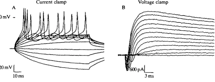
Isolated olfactory receptor cells are capable of generating action potentials (APs) when current-clamped using either conventional whole-cell or nystatin-patch techniques. In Fig. 4A, current steps to subthreshold voltages elicit passive membrane voltage responses in a pyriform cell. Larger current pulses bring the membrane voltage to threshold and elicit trains of action potentials. Measure-ments made from the data illustrated in Fig. 4A reveal an AP threshold voltage of - 3 1 mV, a peak amplitude of +55 mV (measured from threshold) and a duration at half-peak amplitude of 1.9 ms. These APs are generated by voltage-dependent Na+ channels and are blocked by lOOnmolF1 TTX (not illustrated). There is no obvious plateau phase, and it appears that Ca2+ channels probably play a minor role in shaping the AP. This is consistent with the very small Ca2+ currents seen under voltage-clamp conditions. Repolarization of the AP involves the delayed rectifier K+ channels (see also below).
Repetitive firing was regularly observed in all pyriform cells with high-input resistance values, and some cells fired action potentials spontaneously without any injected current. Rates of spontaneous and induced firing varied in cells from 0.08 to 17.00 Hz. Some pyriform cells fired bursts of APs; others fired continuous
0mY
" 11
)M\>
R
20mVl
10 ms
Current clamp
__ — " ^
Voltage clamp B
^ f//C- ^ =
V S O OPA | _
[image:10.451.49.407.398.534.2]3 ms
Fig. 4. Isolated olfactory receptor cells are capable of generating action potentials. (A) Voltage traces obtained from a pyriform cell (resting membrane potential of -64mV) show repetitive firing upon reaching a threshold near — 30 mV. Current pulses were applied for 100 ms in 50 pA increments from — 200 pA to +400 pA. (B) Voltage-clamp measurements on the same cell reveal both inward Na+ and outward K+ currents during voltage steps between —50 and +60 mV. Bath solution: Fig. 3). Potassium-nystatin internal solution: 20mmoir' KC1, ASW (see
380mmoll~' potassium aspartate, lOOmmolF1 glucose, 50 jig ml"1
lOmmoir nystatin.
trains. Only a few presumably damaged cells, with input resistances of 100 MQ or less, showed a single action potential regardless of the amplitude or duration of the current pulse stimulus. Data from such cells were not analyzed further.
Although floriform cells have voltage-gated Na+ channels and produce a single action potential in response to electrical stimulation, the membrane potential fluctuates dramatically in current-clamp and trains of action potentials were not observed. Thus, the majority of work on floriform cells was carried out in voltage-clamp.
Voltage- and current-clamp experiments can be performed on the same cell. Fig. 4B shows a family of current traces (from the cell used in Fig. 4A) for voltage steps to levels between - 5 0 and +60mV. Both inward Na+ and outward K+ currents flow with the high-K+-nystatin internal solution. There are no obvious differences between Na+ and K+ currents obtained with the nystatin method and those recorded with conventional whole-cell dialysis. The ability to examine both membrane currents and action potentials in the same cell is very useful when studying the mechanisms of chemosensory transduction.
Effects of channel-blocking compounds on isolated receptor cells
As the next step in studying the mechanisms involved in transducing chemical signals by squid olfactory cells, the effects of agents that elicited strong behavioral responses in vivo (Gilly and Lucero, 1992) were examined on isolated receptor cells. The first chemical tested was propyl paraben, a preservative in blue food coloring that caused escape jetting in behavioral tests (Gilly and Lucero, 1992). When bath-applied to isolated floriform or pyriform receptor cells lOO^moll"1 propyl paraben reversibly blocked about 50 % of the peak K+ current and 20 % of the peak Na+ current (data not shown). This finding led us to test the effects of better-known K+ and Na+ channel blockers on squid escape jetting and on the electrical properties of receptor cells.
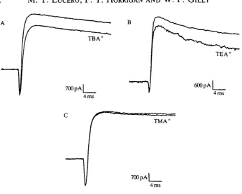
In an examination of three related quaternary ammonium compounds: tetra-methyl-, tetraethyl- and tetrabutylammonium (TMA+, TEA+ and TBA+, re-spectively), our behavioral studies showed that, although TMA+ produced little behavioral effect, both TBA+ and TEA+ reliably elicited escape responses. In the present electrophysiological studies, we found that TBA+ and TEA+ blocked K+ currents in isolated pyriform receptor cells, whereas TMA+ did not.
Fig. 5 shows results from a voltage-clamp experiment in which 'puff application (see Materials and methods) of 20 mmol P1 TBA+ blocked approximately 20 % of the K+ current in a pyriform cell during a pulse to +40 mV (Fig. 5A), without affecting inward Na+ current. The average block of peak K+ current at +60 mV by bath-applied TBA+ (20mmoir1) was 52±9% (N=5).
242 M . T. LUCERO, F . T. HORRIGAN AND W . F . GlLLY
TEA4
600 pA
4 ms 4 ms
[image:12.451.55.402.55.332.2]4 ms
Fig. 5. Quaternary ammonium compounds reduce K+ current without affecting Na+ current in isolated pyriform cells. (A) 20 mmol I"1 T B A+ blocks approximately 20 % of the outward K+ current at +40 mV but has no effect on inward Na+ current. (B) In a
different cell, 20mmoH~1 T E A+ blocks approximately 1 5 % of the outward K+ current at +40 mV but has no effect on inward Na+ current. (C) 20 mmol I"1 T M A+ has very little effect on either the K+ current or the Na+ current. Test substances were applied via a perfusion pipette for a few seconds before, during and after each voltage pulse. Bath solution: ASW (see Fig. 3; drugs were added to this solution). 400K internal solution: 20 mmol I"1 KC1, 355 mmol I"1 potassium glutamate, 25mmoll~l KF, 10 mmol r1 EGTA, 15 mmol I"1 Hepes.
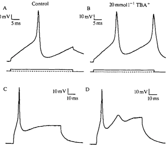
TBA+ also affected the shape of action potentials and induced hyper-excitability in current-clamped pyriform receptor cells. Fig. 6A,B compares membrane voltage responses to a 30 ms current pulse of +200 pA before (control) and during the puff application of 20mmoll~1 TBA+. The blocking of K+ channels by TBA+, as discussed above, increased the cell's excitability and facilitated a second AP during the current pulse. In addition, the AP was broadened in the presence of TBA+ from a half-amplitude duration of 2.3 ms to 2.9 ms. A similar example of the effects of TBA+ on another pyriform cell is shown in Fig. 6C,D. Such changes in the cell's excitability would presumably alter information encoded by the pattern of impulses passing from an olfactory receptor cell to the brain of a living squid.
Control 20mmoir'TBA+
1 0 m V | _ D
10 ms
10 mV 10 ms
Fig. 6. 20 mmol 1 ' TBA+ affects excitability of current-clamped olfactory cells. (A,C) Control action potentials from two different pyriform receptor cells were elicited during a 30ms (A) or60ms(C) current injection of200pA. (B,D) 20 mmol 1~' TBA+ was applied from a perfusion pipette to each cell for several seconds before and during the matching current pulse. Bath solution: ASW (see Fig. 3). 400K internal solution: see Fig. 5.
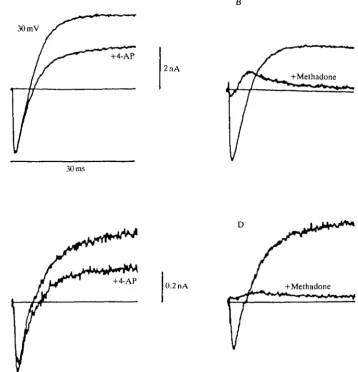
pyriform receptor cell, approximately 50% of the outward K+ current during a voltage step to + 30 mV is blocked by bath application of 5 mmol PJ 4-AP. Inward Na+ currents are not affected. A similar K+-selective block by 4-AP is shown in Fig. 7C for a floriform cell.
A third type of channel blocker that caused escape behavior in squid is methadone. This tertiary amine was chosen because its effects on the ionic currents in squid GFL neurons had already been characterized (Horrigan, 1990). As was found in giant fiber lobe neurons, 500//moll"1 methadone almost completely blocks both Na+ and K+ currents in pyriform and floriform cells (Fig. 7B,D). These effects are readily reversible (not illustrated) and do not involve opiate receptors.
[image:13.451.96.364.75.313.2]244 M. T. LUCERO, F. T. HORRIGAN AND W. F. GILLY
A B
30 mV
2nA
30 ms
D
[image:14.451.53.412.77.450.2]0.2 nA
Fig. 7. 4-AP and methadone block K+ currents in isolated olfactory receptor cells. (A,C) Superimposed current traces are illustrated for 30ms pulses to +30 mV in the presence (+4-AP) and absence of 5mmoll~' 4-AP for a pyriform cell (A) and a floriform cell (C). (B,D) Currents recorded at +30mV are shown in the presence (+Methadone) and absence of 500/imolP1 methadone for a pyriform cell (B) and a floriform cell (D). Bath solution: ASW (see Fig. 3; drugs added to this solution). 100K internal solution: see Fig. 3.
Na+ channels, but FIX, which selectively blocks Na+ currents, does not produce behavioral effects.
ASW r ASW
^ t *t ii. , . '
Squid ink D L-Dopa
4s Dopamine
[image:15.451.31.425.74.295.2]10 mV
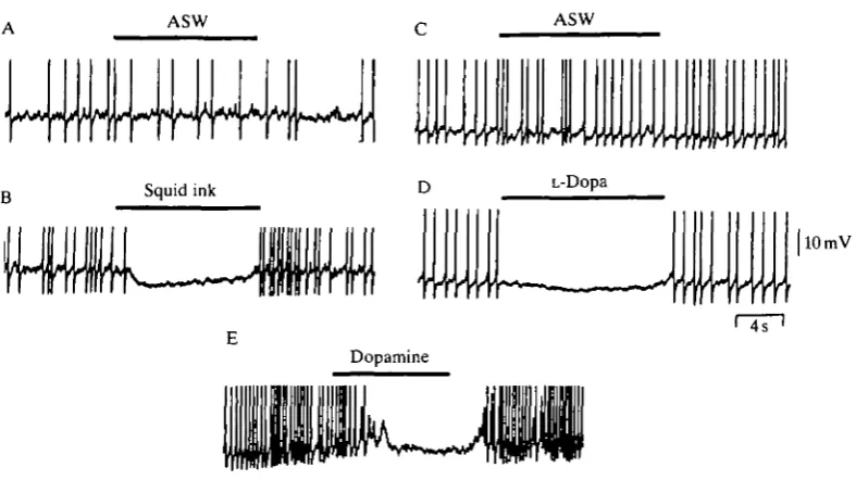
Fig. 8. Squid ink, L-Dopa and dopamine hyperpolarize the membrane and block action potentials in isolated pyriform receptor cells. (A) ASW was applied via a small perfusion pipette for the duration of the solid bar. The pipette was then rapidly removed from the bath. The peaks of the action potentials have been truncated. (B) Squid ink extract was applied in the same manner to the same cell. (C) A control puff application of ASW in a different cell. (D) 10 mmol P1 L-Dopa was applied to the same cell as in C. (E) 5 0 / i m o l P1 dopamine was applied to a different cell. Bath solution: ASW (see Fig. 3). 400K-nystatin internal solution: 20 mmol I"1 KC1, 355 mmol 1~' potassium glutamate, 25 mmol I"1 KF, lOmmoll"1 EGTA, 15mmoll~L Hepes, 50 jig m l " ' nystatin.
caused escape jetting in behavioral experiments (Gilly and Lucero, 1992). In the following experiments, the nystatin patch recording method was used in an attempt to keep any second messenger systems as intact as possible (see Materials and methods). In these experiments, only current-clamp measurements were made, and the electrical resistance through the pipette and patch of membrane containing the nystatin pores was considerably higher than that required for good voltage-clamp control. Attempts to obtain the results to be described using conventional whole-cell recording techniques were unsuccessful.
246 M . T. L U C E R O , F . T. HORRIGAN AND W . F . G I L L Y
hyperpolarized the membrane potential by approximately 5 mV and inhibited AP firing (Fig. 8B).
Not all pyriform cells were responsive to ink application. For a cell to respond by inhibiting action potentials, it had to be capable of sustaining prolonged (1-2 min) tonic firing. Although 17 of the 25 cells tested were capable of tonic firing, only 11 of the 17 tonically active cells responded to ink. Many of the sensitive cells responded to repeated applications, but most became electrically leaky after the third or fourth trial.
Membrane resistance was monitored before, during and after application of ink (in experiments like that in Fig. 8B) by periodically delivering brief hyperpolariz-ing current pulses (-50 pA for 200 ms) through the patch pipette (data not shown). In such experiments, the response to ink was associated with a 28±4% (N-3) decrease in membrane resistance. This decrease in membrane resistance was significantly different from the 1.5±0.6 % (N=3) decrease in membrane resistance associated with control application of l m m o l P1 blue no. 1 dye (independent t-test, P<0.05). Thus, it appears that K+, or possibly C\~, channels are opening in response to squid ink and this serves to hyperpolarize the cell.
We have not been successful at detecting a membrane current associated with the application of squid ink under voltage-clamp conditions. However, in a pyriform cell with an average input resistance of 406 MQ, an ink-induced hyperpolarization of 5mV would correspond to a current as small as 12.5 pA. A current of this size was beyond the limit of resolution in our whole-cell recordings.
The pigment in squid ink is melanin, the precursor of which is L-Dopa (Fox and Crane, 1942). l m m o l P1 L-Dopa, like squid ink, elicited escape jetting in behavioral experiments (Gilly and Lucero, 1992). Similarly, 1-lOmmolP1 L-Dopa mimicked the effect of squid ink in pyriform cells when puff-applied (Fig. 8D). This result was obtained in six of eight cells tested. As with squid ink, membrane resistance transiently decreased during the L-Dopa-induced hyper-polarization. There was no dramatic change in firing rate or membrane conduc-tance during control application of ASW, as shown in Fig. 8C.
Preliminary studies show that 50/xmolP1 dopamine also hyperpolarizes the membrane potential and inhibits action potential firing (Fig. 8E). The dopamine-induced hyperpolarization appears to develop more slowly and to persist for a longer time than the effect produced by ink or L-Dopa, but detailed comparisons are not yet possible. It seems likely, however, that L-Dopa or a compound derived from it plays a role in the hyperpolarization induced by squid ink.
Discussion
less negative, varying from - 5 0 to - 2 5 mV. Floriform cells were difficult to record from in current clamp because the resting membrane potential tended to fluctuate sporadically.
Voltage-clamp studies on isolated receptor cells showed that channel-blocking chemicals used in the behavioral studies could be divided into three classes: (1) those that block only K+ channels, such as tetrabutylammonium (TBA+), tetraethylammonium (TEA+) and 4-aminopyridine (4-AP), (2) those that block K+ and Na+ channels, including methadone and propyl paraben, and (3) a strictly Na+ channel blocker, TTX. It appears that the common underlying mechanism of transduction for the first two classes of compounds involves K+ channel block and an increase in excitability of the receptor cell. Detection of TTX is not evident from our behavioral studies (Gilly and Lucero, 1992).
Block of K+ currents changes the shape and frequency of action potentials in isolated olfactory receptor neurons and presumably these changes, upon reaching the brain of a living squid, initiate the series of events culminating in a powerful escape jet. At present we do not know the precise spatial distribution of K+ channels in squid chemoreceptors, but presumably they exist in both the exposed apical region as well as the basolateral region, which gives rise to the axon. The exposure of the entire cell to K+ channel blockers poses the potential problem that K+ channel block is primarily affecting non-sensory pathways. However, the finding that externally acting chemicals, such as the membrane-impermeant quaternary ammonium ions, elicit escape responses, strongly suggests that at least some of a receptor cell's K+ channels are located in the apical region that lies in direct contact with chemical stimuli in sea water (Gilly and Lucero, 1992). In this respect, squid olfactory receptors appear to be similar to vertebrate taste receptors in which signal transduction pathways include blockage of K+ conductances to produce depolarizing receptor potentials (Kinnamon and Roper, 1988; Bigiani and Roper, 1991).
Some indirect evidence for localization of Na+ channels to discrete regions on the squid receptor cell comes from experiments with TTX, a chemical that blocks Na+ currents and action potentials in isolated squid olfactory cells. Inhibition of firing by TTX applied to a living animal presumably does not occur, because TTX does not induce escape responses. Squid ink and L-Dopa (which also block action potentials), in contrast, are potent stimulants that reliably induce escape responses in living squid (Gilly and Lucero, 1992). The lack of behavioral sensitivity to externally applied TTX suggests that most Na+ channels are located in an area that does not come in direct contact with the external milieu. Thus, the bulk of the Na+ channels appear to be localized to the basolateral, axonal pole of the neurons.
248 M. T. LUCERO, F. T. HORRIGAN AND W. F. GILLY
experiments on individual receptor cells would be useful in determining whether, as behavioral experiments suggest, Na+ channels are absent from dendritic regions of squid olfactory receptors.
K+ channel block as a mechanism of chemosensory transduction is undoubtedly only one of several mechanisms involved in squid olfaction. Squid ink appears to elicit escape jetting by a mechanism opposite to K+ channel block. Squid ink suppresses activity in the isolated receptor cells by increasing membrane conduc-tance to presumably K+ or Cl~ and hyperpolarizing the cell's membrane potential. Although the only evidence for hyperpolarizing responses to odorants by vertebrate receptor cells was obtained in the mudpuppy (Dionne, 1990), recent work on lobster olfactory receptor cells shows an odor-activated K+ conductance that reduces both the rate and the magnitude of an odor-activated depolarization and, thus, potentially modulates stimulus coding (Michel et al. 1991). Further studies with squid receptor cells will be necessary to determine both the selectivity of the ink-induced conductance increase and the extent to which it might modulate responses to depolarizing stimuli.
The major component of squid ink is melanin (Fox and Crane, 1942). L-Dopa, the precursor for melanin, is also found in squid ink (Jimbow et al. 1984) and elicits escape jetting in squid (Gilly and Lucero, 1992). In isolated olfactory receptor cells, L-Dopa and dopamine mimic the squid ink-induced suppression of action potentials. D2 dopamine receptors may be localized in olfactory nerve terminals in
the rat olfactory bulb (Boyson et al. 1986), and in situ hybridization studies suggest that olfactory receptor cells are capable of synthesizing dopamine receptors (Shipley et al. 1991). Although it is known that certain molluscan neurons express dopamine receptors and hyperpolarize when stimulated with dopamine (Walker, 1986), it remains to be seen whether the hyperpolarizing responses to squid ink and L-Dopa of olfactory neurons are the result of activation of a similar or novel dopaminergic transduction pathway.
We wish to thank Jimmy Lucero for performing the microdissection and tissue culture of the olfactory organ and Bruce Hopkins for providing the squid and scanning electron micrographs. This work was supported by the Office of Naval Research grant N0014-89-J-1744.
References
ARMSTRONG, C. M. AND BEZANILLA, F. (1974). Charge movement associated with the opening and closing of the activation gates of Na channels. /. gen. Physiol. 63, 533-552.
BIGIANI, A. R. AND ROPER, S. D. (1991). Mediation of responses to calcium in taste cells by modulation of a potassium conductance. Science 252, 126-128.
BOYSON, S. J. MCGONIGLE, P. AND MOLINOFF, P. B. (1986). Quantitative autoradiographic localization of the Dj and D2 subtypes of dopamine receptors in rat brain. J. Neurosci. 6,
3177-3188.
BRISMAR, T. AND GILLY, W. F. (1987). Synthesis of sodium channels in the cell bodies of squid giant axons. Proc. natn. Acad. Sci. U.S.A. 84, 1459-1463.
EMERY, D. G. (1975). The histology and fine structure of the olfactory organ of the squid Lolliguncula brevis Blainville. Tissue & Cell 2, 357-367.
FIRESTEIN S. AND WERBLIN, F. S. (1987). Gated currents in isolated olfactory receptor neurons of the larval tiger salamander. Proc. natn. Acad. Sci. U.S.A. 84, 6292-6296.
Fox, D. L. AND CRANE, S. C. (1942). Concerning the pigments of the two-spotted octopus and the opalescent squid. Biol Bull. mar. biol. Lab., Woods Hole 82, 284-291.
GILLY, W. F. AND BRISMAR, T. (1989). Properties of appropriately and inappropriately expressed sodium channels in squid giant axon and its somata. J. Neurosci. 9, 1362-1374. GILLY, W. F. AND LUCERO, M. T. (1992). Behavioral responses to chemical stimulation of the
olfactory organ in the squid Loligo opalescens. J. exp. Biol. 162, 209-229.
HAMILL, O. P., MARTY, A., NEHER, E . , SAKMAN, B . AND SIGWORTH, F. J. (1981). Improved
patch-clamp techniques for high-resolution current recording from cells and cell-free membrane patches. Pfliigers Arch. 391, 85-100.
HORN, R. AND MARTY, A. (1988). Muscarinic activation of ionic currents by a new whole cell recording method. J. gen. Physiol. 92, 145-159.
HORRIGAN, F. T. (1990). Methadone block of neuronal K current. Biophys. J. 57, 515a.
JIMBOW, K., MIYAKE, Y., HOMMA, K., YASUDA, K., IZUMI, Y., TSUTSUMI, A. AND ITO, S. (1984).
Characterization of melanogenesis and morphogenesis of melanosomes by physicochemical properties of melanin and melanosomes in malignant melanoma. Cancer Res. 44,1128-1134. KINNAMON, S. C. AND ROPER, S. D. (1988). Membrane properties of isolated mudpuppy taste
cells. / . gen. Physiol. 91, 351-371.
LLANO, I. AND BOOKMAN, R. J. (1986). Ionic conductances of squid giant fiber lobe neurons. /. gen. Physiol. 88, 543-569.
LUCERO, M. T., HORRIGAN, F. T., LEVITT, J. L. AND GILLY, W. F. (1991). Chemoreceptive
capabilities of the squid olfactory organ. Biophys. J. 59,184a.
MCCLINTOCK, T. S. AND ACHE, B. W. (1989). Ionic currents and ion channels of lobster olfactory receptor neurons. J. gen. Physiol. 94, 1085-1099.
MESSENGER, J. B. (1979). The nervous system of Loligo. IV. The peduncle and olfactory lobes. Phil. Trans. R. Soc. Lond. B 285, 275-309.
MICHEL, W. C , MCCLINTOCK, T. S. AND ACHE, B. W. (1991). Inhibition of lobster olfactory
receptor cells by an odor-activated potassium conductance. J. Neurophysiol. 65, 446-453. RUDY, B. (1988). Diversity and ubiquity of K channels. Neuroscience 25, 729-750.
SCHMIEDEL-JAKOB, I., ANDERSON, P. A. V. AND ACHE, B. W. (1989). Whole cell recording from lobster olfactory receptor cells: responses to current and odor stimulation. / . Neurophysiol. 61, 994-1000.
SHIPLEY, M. T., NICKELL, W. T., NORMAN, A. B . AND GERFEN, C. (1991). Localization of D2
dopamine receptor messenger RNA in primary olfactory neurons. ACHEMSAbstr. 13,108a. THOMPSON, S. (1982). Aminopyridine block of transient potassium current. / . gen. Physiol. 80,
1-18.
WALKER, R. J. (1986). Transmitters and modulators. In The Mollusca, vol. 9 (ed. A. O. D. Willows), pp. 314-347. New York, London: Academic Press, Inc.
WATKINSON, G. (1909). Untersuchungen iiber die sogenannten Geruchsorgane der cephaloden. Jena Z. Naturw. 44, 353-414.
WELLS, M. J. AND WELLS, J. (1959). Hormonal control of sexual maturity in Octopus. J. exp. Biol. 36, 1-33.