PREDICTION OF GESTATIONAL AGE BY ULTRASONOGRAM USING LINEAR REGRESSION MODEL
1
Rajathi, G.,
2,*2
Director, Research Centre for Cellular Genomics, Sree Balaji Medical College
3
Assistant Professor, Dept of Anatomy, Sree Balaji Medical College
ARTICLE INFO ABSTRACT
Introduction:
providers are dependent on gestational age to schedule maternal and fetal investigations, to gauge parameters of fetal growth and apply interventions timely for the manage
complications. This study was done to develop a linear regression model using ultrasound measurement of fetal parameters to predict the gestational age in pregnancy.
Methodology:
and one private tertiary care hospitals in Chennai among 145 antenatal women. Gestational age was determined by measuring fetal parameters like Mean sac diameter, Biparietal diameter, Crown rump length, Abdominal circumference, Head
Results:
30 years and nearly 23 % of the subjects were in the age group 20
participants were in 3rd trimester. There was a statistically significant correlation between the fetal parameters and gestational age in the second trimester (p<0.001).
Conclusion:
abnormalities. It is essential to assess the gestational age using multiple parameter. It is likely that the technological development of USG will continue and increases in ultrasound frequency will further improve image resolution of early
assess early pregnancy viability and multiple gestations.
Copyright © 2017, Rajathi et al. This is an open access distribution, and reproduction in any medium, provided
INTRODUCTION
Ian Donald, a Scottish physician in 1958 published the first scientific report on medical use of ultrasound entitled "Investigation of abdominal masses by pulsed ultrasound D o na l d , 1 9 5 8 ). Initially ultrasound was done in antenatal women only for those with any medical problem or at high risk pregnancy. It was introduced in routine medical practic 1970's and in antenatal women to confirm the fetal viability, to assess gestational age and to detect whether single or multiple pregnancies. Radiographic techniques were used to measure fetal dimensions prior to ultrasound, which had the drawback of exposing radiation to the fetus. Currently with increasing use of ultrasound, a non invasive diagnostic procedure, there is a decrease in maternal morbidity and mortality.
an accurate due date is of both social and medical significance. Providers of obstetric care are dependent on gestational age to
*Corresponding author: Johnson, W.M.S.,
Director, Research Centre for Cellular Genomics, Sree Balaji Medical College and Hospital.
ISSN: 0975-833X
Vol.
Article History:
Received 17th September, 2017
Received in revised form 10th October, 2017
Accepted 28th November, 2017
Published online 27th December, 2017
Citation: Rajathi, G., Johnson, W.M.S., Archana, R. and Rahe, R. International Journal of Current Research, 9, (12), 62
Key words: Fetal parameters, Gestational age, Pregnancy, Ultrasound,
RESEARCH ARTICLE
PREDICTION OF GESTATIONAL AGE BY ULTRASONOGRAM USING LINEAR REGRESSION MODEL
,*
Johnson, W.M.S.,
3Archana, R. and
3Rahe, R.
1
Assistant Professor, PRIMST,
Director, Research Centre for Cellular Genomics, Sree Balaji Medical College
Assistant Professor, Dept of Anatomy, Sree Balaji Medical College and
ABSTRACT
Introduction: Estimation of an accurate due date is of both social and medical significance. Care providers are dependent on gestational age to schedule maternal and fetal investigations, to gauge parameters of fetal growth and apply interventions timely for the manage
complications. This study was done to develop a linear regression model using ultrasound measurement of fetal parameters to predict the gestational age in pregnancy.
Methodology: This study was carried out as a multicentre cross sectional
and one private tertiary care hospitals in Chennai among 145 antenatal women. Gestational age was determined by measuring fetal parameters like Mean sac diameter, Biparietal diameter, Crown rump length, Abdominal circumference, Head circumference, Femur length and Effective fetal weight. Results: The mean age of the participants was 27.5 years. About 57 % of subjects were between 25 30 years and nearly 23 % of the subjects were in the age group
20-participants were in 3rd trimester. There was a statistically significant correlation between the fetal parameters and gestational age in the second trimester (p<0.001).
Conclusion: Fetal parameters are key predictors of gestational age. They are useful t
abnormalities. It is essential to assess the gestational age using multiple parameter. It is likely that the technological development of USG will continue and increases in ultrasound frequency will further improve image resolution of early pregnancies. A 3D and 4D USG will also improve our ability to assess early pregnancy viability and multiple gestations.
access article distributed under the Creative Commons Attribution License, the original work is properly cited.
Ian Donald, a Scottish physician in 1958 published the first ultrasound entitled al masses by pulsed ultrasound (I a n Initially ultrasound was done in antenatal women only for those with any medical problem or at high risk pregnancy. It was introduced in routine medical practice in 1970's and in antenatal women to confirm the fetal viability, to assess gestational age and to detect whether single or multiple pregnancies. Radiographic techniques were used to measure fetal dimensions prior to ultrasound, which had the drawback exposing radiation to the fetus. Currently with increasing use of ultrasound, a non invasive diagnostic procedure, there is a decrease in maternal morbidity and mortality. Estimation of an accurate due date is of both social and medical significance. ders of obstetric care are dependent on gestational age to
Director, Research Centre for Cellular Genomics, Sree Balaji Medical
schedule maternal and fetal investigations, to gauge
of fetal growth and apply interventions timely for the management of prenatal complications. Proper timing of a repeat caesarean section also needs accurate dates. When an anomaly is detected, interventional modality is influenced by gestational age. For this, ultrasonography is one of the methods commonly used. Accuracy depends on the quality of images obtained. Gestational age is approximately 280 days calculated from first day of last menstrual period and so dating of pregnancy starts even before fertilisation. Ultrasonography is commonly used to estimate gestational age by measuring fetal dimensions like gestational sac diameter, crown rump length,
biparietal diameter, abdominal circumference, head
circumference and femur length. When ultrasou performed with quality and precision, it is far superior and reliable compared to clinical and other methods of dating the pregnancy (Butt, KimberlyLim
International Journal of Current Research Vol. 9, Issue, 12, pp.62426-62432, December, 2017
Johnson, W.M.S., Archana, R. and Rahe, R. 2017. “Prediction of gestational age by ultrasonogram using linear
62426-62432.
Available online at http://www.journalcra.com
z
PREDICTION OF GESTATIONAL AGE BY ULTRASONOGRAM USING LINEAR REGRESSION MODEL
Rahe, R.
Director, Research Centre for Cellular Genomics, Sree Balaji Medical College and Hospital
and Hospital
Estimation of an accurate due date is of both social and medical significance. Care providers are dependent on gestational age to schedule maternal and fetal investigations, to gauge parameters of fetal growth and apply interventions timely for the management of prenatal complications. This study was done to develop a linear regression model using ultrasound measurement of fetal parameters to predict the gestational age in pregnancy.
This study was carried out as a multicentre cross sectional study in one government and one private tertiary care hospitals in Chennai among 145 antenatal women. Gestational age was determined by measuring fetal parameters like Mean sac diameter, Biparietal diameter, Crown rump
circumference, Femur length and Effective fetal weight. The mean age of the participants was 27.5 years. About 57 % of subjects were between
25--25 years. Overall, 55(37.9%) of the participants were in 3rd trimester. There was a statistically significant correlation between the fetal
Fetal parameters are key predictors of gestational age. They are useful to detect fetal abnormalities. It is essential to assess the gestational age using multiple parameter. It is likely that the technological development of USG will continue and increases in ultrasound frequency will further pregnancies. A 3D and 4D USG will also improve our ability to
License, which permits unrestricted use,
schedule maternal and fetal investigations, to gauge parameters of fetal growth and apply interventions timely for the management of prenatal complications. Proper timing of a repeat caesarean section also needs accurate dates. When an anomaly is detected, interventional modality is influenced by age. For this, ultrasonography is one of the methods commonly used. Accuracy depends on the quality of images Gestational age is approximately 280 days calculated from first day of last menstrual period and so dating of ore fertilisation. Ultrasonography is commonly used to estimate gestational age by measuring fetal dimensions like gestational sac diameter, crown rump length,
biparietal diameter, abdominal circumference, head
femur length. When ultrasound is performed with quality and precision, it is far superior and reliable compared to clinical and other methods of dating the
Butt, KimberlyLim et al.)
INTERNATIONAL JOURNAL OF CURRENT RESEARCH
Objectives
To develop a linear regression model using ultrasound measurement of fetal parameters to predict the gestational age in pregnancy. Materials and Methods
Study Settings
This study was carried out as a multicentre cross sectional study between February and April 2015. All the tertiary care hospitals were approached for permission. The study was carried out in one government and one private tertiary care hospitals in Chennai.
Study Population
All pregnant women who attended the study facility during the study period and fulfilled the study criteria were selected for the study. The participants were selected consecutively. A total of 145 antenatal women participated in the study.
Inclusion Criteria
Singleton pregnancy (5 - 40 weeks)
Known last menstrual periodul
History of regular menstrual cycles
Exclusion Criteria
Gestational Diabetes
Hypertension (Systemic and pregnancy induced )
Anaemia
Maternal Disease
Fetal anomalies
Multiple Pregnancy
Placental anomalies
Unknown last menstrual period / irregular menstrual
cycles
Ethical approval and informed consent
Approval from Institutional Ethics committee was obtained prior to data collection. The study participants were explained in detail about the study and informed consent was obtained prior to data collection.
Data collection
Subjects were made to lie down in a supine position with full bladder while doing ultrasonogram. Ultrasonography machine used was Siemens Acuson X 300,3-5 MHz transducer. Each fetus was measured only once during the whole study. Gestational age was determined by measuring fetal parameters like Mean sac diameter, Biparietal diameter, Crown rump length, Abdominal circumference, Head circumference, Femur length and Effective fetal weight.
Operational Definitions
Gestational sac diameter: Gestational sac is an echo free
space containing the fluid, embryo and extra embryonic structures. It is measured inside the hyperechoic rim, including only the echo free space (MacGregor, 2008)
Biparietal diameter: The biparietal diameter is imaged in
the transaxial plane of the fetal head at a level depicting thalami in the midline, equidistant from the temporal bone and usually the cavum septum pellucidum anteriorly (MacGregor, 2008)
Crown-Rump length: Crown rump length is a
measurement of embryo along its longest axis. The technique involves measurement of the fetal length from the tip of cephalic pole to the tip of caudal pole. (MacGregor, 2008)
Head circumference: Head circumference is measured by
tracing the outer perimeter of the head.(MacGregor, 2008)
Abdominal circumference: Abdominal circumference is
measured in the transaxial view at the level of fetal liver using the umbilical portion of the left portal vein as the landmark.(MacGregor, 2008)
Femur length: Femur length is measured along the long
axis of the bone, disregarding the curvature.(MacGregor, 2008).
Data analysis
Data was computed in Microsoft Excel 2010 and statistical analysis was performed using SPSS ver. 21 software. Mean and standard deviation was calculated for all the fetal parameters. Correlation co-efficient was calculated and linear regression was used for developing a predictor model.
RESULTS
This study was carried out among 145 pregnant mothers visiting one government and one private health facility. The mean age of the participants was 27.5 years. About 57 % of subjects were between 25-30 years and nearly 23 % of the subjects were in the age group 20-25 years. Nearly 1 1 % of the subjects belonged to the age group of 30 -35 years and nearly 7 % of the subjects belonged to the age group of 35 -40 years. 1 % belonged to 1 5 -20 age group and 0. 7 % to 40- 45 years. The age distribution of the study participants is given in Figure 1.
Figure1. Age distribution of the study participants
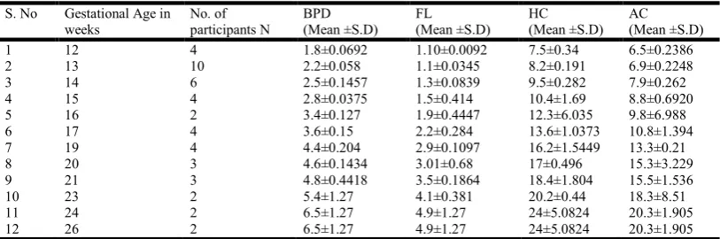
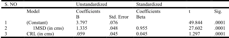
The distribution of the participants based on the trimesters is given in figure-2. Overall, 55(37.9%) of the participants were in 3rd trimester. About 44(30.3%) and 46(31.7%) were in 2nd trimester. The mean and standard deviation of the fetal parameters for first trimester are given in Table-1. During the first trimester Mean Sac Diameter (MSD) and Crown Rump Length (CRL) were measured. The mean and standard deviation of the fetal parameters for second trimester are given in Table-2. During the first trimester Mean Sac Diameter (MSD) and Crown Rump Length (CRL) were measured. The mean and standard deviation of the fetal parameters for third trimester are given in Table-3. During the first trimester Mean Sac Diameter (MSD) and Crown Rump Length (CRL) were measured. To prove a correlation between BPD ,FL,HC,AC and gestational age in the second trimester correlation coefficient was calculated and found to be statistically significant 0. 997, 0. 991 , 0.994, 0.992 and the values were less than 0.001, there by showing positive correlation between the variables.(Table-4 ,5). The linear regression was computed for the 1st trimester parameters with the dependent variable being gestational age. Statistically significant results were obtained (Table-6). From Table 7, As per Anova, Regression model was accepted and the equation was given as follows GA (USG) = 3.797 + 1.335(MSD) +0.59(CRL). Both CRL and MSD were contributing towards gestational age assessment and highly significant. Linear regression and coefficients were computed for 2nd trimester, with Gestational age as dependent variable and BPD, FL and AC as predictor variables.
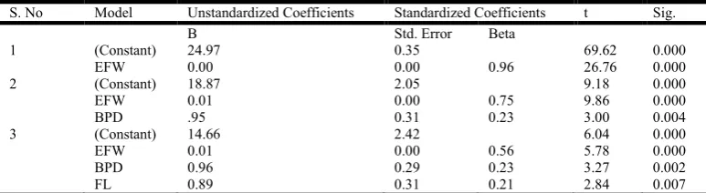
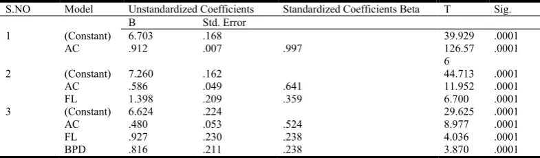
The results were statistically significant (Table-8, 9). From Table 10 and 11, As per Anova, Regression model was accepted and the equation was given as follows: GA (USG) = 14.657+0.965 (BPD) +0.894 (FL) +0.002 (EFW). EFW was highly significant than BPD and FL. HC and AC were excluded in this model since they are not contributing. From Table 12 and 13, As per Anova, Regression model was accepted and the equation was given as follows GA (USG)=6.624+0.816(BPD) +0.927(FL)+ 0.480(AC). BPD, FL, AC were highly significant. HC was excluded in this model since it was not contributing.
DISCUSSION
This study was carried out to predict the gestational age based on ultrasound measurements of fetal parameters using linear
regression model. Patricia M .Dietz et al ., (2007 in their study
provided evidence that substantial amount of misclassification results on using LMP based gestational age estimates and this can lead to preterm delivery rates. Estimation of gestational age by USG is of high importance for diagnosis, investigation and treatment of fetus. The importance and use of ultrasound in estimating the gestational age has been well emphasized in
studies done by Nielson (1998) Caroline A Crowther et al.,
(2005) Daniel Salpou, Torvid Kiserud (2008), Verburg Bo et
al., (2008) and George M Graham (2010). According to Laing
[image:3.595.93.487.83.154.2]et al. (2000) during the first 3 to 5 menstrual weeks an intrauterine pregnancy is first signalled by the presence of gestational sac.
Table 1. First trimester fetal parameters among the study participants
S. No Gestational age in weeks No. of participants N MSD (Mean ±S.D) CRL (Mean ±S.D)
1 6 7 1.8± 0.045128 0.49±0.078372
2 7 7 2.45±0.21923 1.03±0.195784
3 8 13 3.22±0.110752 1.73±0.08136
4 9 11 3.84±0.168257 2.34±0.149644
5 10 3 4.23±0.143422 2.83±0.108281
[image:3.595.90.497.188.323.2]6 12 3 5.76±0.286844 5.00±0.416664
Table 2. Second trimester fetal parameters among the study participants
S. No Gestational Age in weeks
No. of participants N
BPD (Mean ±S.D)
FL (Mean ±S.D)
HC (Mean ±S.D)
AC (Mean ±S.D)
1 12 4 1.8±0.0692 1.10±0.0092 7.5±0.34 6.5±0.2386
2 13 10 2.2±0.058 1.1±0.0345 8.2±0.191 6.9±0.2248
3 14 6 2.5±0.1457 1.3±0.0839 9.5±0.282 7.9±0.262
4 15 4 2.8±0.0375 1.5±0.414 10.4±1.69 8.8±0.6920
5 16 2 3.4±0.127 1.9±0.4447 12.3±6.035 9.8±6.988
6 17 4 3.6±0.15 2.2±0.284 13.6±1.0373 10.8±1.394
7 19 4 4.4±0.204 2.9±0.1097 16.2±1.5449 13.3±0.21
8 20 3 4.6±0.1434 3.01±0.68 17±0.496 15.3±3.229
9 21 3 4.8±0.4418 3.5±0.1864 18.4±1.804 15.5±1.536
10 23 2 5.4±1.27 4.1±0.381 20.2±0.44 18.3±8.51
11 24 2 6.5±1.27 4.9±1.27 24±5.0824 20.3±1.905
[image:3.595.92.495.358.466.2]12 26 2 6.5±1.27 4.9±1.27 24±5.0824 20.3±1.905
Table 3. Third trimester fetal parameters among the study participants
S. No Gestational Age in weeks
No. of participants N
BPD (Mean ±S.D)
FL (Mean ±S.D)
HC (Mean ±S.D)
AC (Mean ±S.D)
1 31 4 7.79 ± 0.10 5.94 ± 0.67 28.89 ± 0.85 27.09 ± 2.53
2 32 6 7.93 ± 0.22 6.17 ± 0.16 28.75 ± 0.74 28.02 ± 0.64
3 33 11 8.39± 0.16 6.38 ± 0.16 30.04 ± 0.55 28.95 ± 0.57
4 34 10 8.48 ± 0.24 6.60 ± 0.15 31.47 ± 1.25 30.23 ± 0.38
5 35 4 8.69± 0.23 6.73 ± 0.35 31.98 ± 1.14 30.07 ± 1.34
6 36 3 8.85 ± 0.52 6.93 ± 0.55 32.68 ± 0.55 31.4 ± 3.11
7 37 6 9.04 ± 0.22 7.1 ± 0.16 32.77± 0.78 32.78 ± 1.40
8 38 3 9.42 ± 0.30 7.51 ± 0.22 33.89 ± 1.33 34.07 ± 3.25
9 39 2 9.36 ± 3.05 7.95 ± 3.18 34.54 ± 2.03 34.13 ± 15.57
Timor-Tritsh IE et al. (1988) stated that the gestational sac represents the chorionic cavity, and its echogenic rim represents the implanting chorionic villi and associated decidual tissue. The smallest gestational sac size that can be clearly distinguished by high frequency transvaginal transducers is 2 to 3 mm, which corresponds to a gestational age of about 32 to
33 days as per Rowling et al. (1999).
The MSD is a commonly used, standardized, way to estimate gestational age during early pregnancy. It is less reliable when the MSD exceeds 1 4 mm or when the embryo can be
identified according to Nyberg DA et al. (1987).MacKenzie AP et al., (2008) stated that
[image:4.842.128.720.86.157.2]the growth of theMS D is approximately 1 mm per day. CRL is measured transabdominally from 6th - 7th week.
Table 4. Correlation between the GA and first trimester measurements
S . N o Pearson Correlation Coefficients Lower Limit of Correlation Coefficients Upper Limit of Correlation Coefficients
G A / U S G B P D F L H C A C G A / U S G B P D F L H C A C G A / U S G B P D F L H C A C
G A / U S G 1 0.99 0.99 0.99 0.99 0.99 0.98 0.98 0.98 0 . 9 9 0 . 9 9 0 . 9 9 0 . 9 9
B P D 0.99 1 0.98 0.99 0.98 0.97 0.99466 0.97 0 . 9 9 0 . 9 9 0 . 9 9
F L 0.99 0.98 1 0.98 0.98 .968 0.97 0 . 9 9 0 . 9 9
H C 0.99 0.99 0.98 1 0.98 0.97 0 . 9 9
A C 0.99 0.98 0.98 0.98 1
Table 5. Correlation between GA and 2nd/3rd trimester measurements
S . N o Pearson Correlation Coefficients Lower Limit of Correlation Coefficients Upper Limit of Correlation Coefficients
G A / U S G B P D F L H C A C G A / U S G B P D F L H C A C G A / U S G B P D F L H C A C
G A / U S G 1.00 1.00 1.00 0.99 1.00 0.99 0.99 0.99 1.00 1.00 1.00 1.00 1.00
B P D 1.00 1.00 0.99 1.00 0.99 0.99 0.99 0.99 1.00 1.00 1.00
F L 1.00 0.99 1.00 0.99 0.99 0.99 0.99 1.00 1.00
H C 0.99 1.00 0.99 1.00 0.99 0.99 0.99
[image:4.842.121.735.197.268.2]A C 1.00 0.99 0.99 0.99 1.00
Table 6. Liner Regression for 1st trimester
S.No Model Sum of Squares df Mean Square F Sig.
1 Regression 95.99 48.00 13917.99 0.0001
2 1 Residual 0.15 42 0
Total 96.14 44
Dependant variable: GA – USG
Table 7. Coefficients for 1st trimester
S. NO Unstandardized Standardized
Model Coefficients Coefficients t Sig.
B Std. Error Beta
1 (Constant) 3.797 .076 49.844 .0001
2 1MSD (in cms) 1.335 .048 0.955 27.602 .0001
3 CRL (in cms) .059 .045 0.045 1.297 .0001
[image:4.842.236.607.403.465.2]In present study, with each fetus being measured only once the data was analyzed with gestational age as a dependent variable and equations were generated. It was evident that both parameters increased as gestational age advanced. Pregnancy could be detected by ultrasonography as early as 5th week of gestational period, when the gestational sac size can be measured. The present study was also comparable to study
carried out by Bhusari Prashant et al (2012). Thus accuracy
and reliability of ultrasonographic measurement was established.
Koch ,Sarah et al. (2014) had insisted that use of Crown Rump
[image:5.595.130.448.83.182.2]Length for estimation of gestational age was not associated with an increased post term male to female ratio .It can therefore be used for the estimation of due date without risk of sex bias that occurs when using BPD in 2nd trimester of pregnancy . From this study it became obvious that GA by USG and MSD,CRL were strongly correlated with each other as well as are found to be statistically very highly significant ( P < 0.001) and Regression Equation derived was GA(USG)= 3.797+1.335(MSD)+0.59(CRL).
Table 8: Liner Regression for 2nd trimester
S.NO Model Sum of Squares Df Mean Square F Sig.
1 Regression 874.15 1 874.15 7020.31 0.0001b
Residual 5.85 47 0.12
Total 880.01 48
2 Regression 876.18 2 438.09 5262.55 0.0001c
Residual 3.82 46 0.08
Total 880.01 48
3 Regression 876.76 3 292.25 4053.64 0.0001d
Residual 3.24 45 0.07
Total 880.01 48
[image:5.595.98.483.247.354.2]a. Dependent Variable: GA BY USG b. Predictors: (Constant) BPD c. Predictors: (Constant) BPD, FL d. Predictors: (Constant) BPD, FL, AC
Table 9. Coefficients for 2nd trimester
S.NO Model Unstandardized Coefficients Standardized Coefficients t Sig.
B Std. Error Beta
1 (Constant) 6.57 0.13 48.86 0.0001
BPD 2.92 0.03 0.99 83.78 0.0001
2 (Constant) 7.36 0.19 37.75 0.0001
BPD 2.06 0.17 0.70 11.71 0.0001
FL 1.00 0.20 0.29 4.93 0.0001
3 (Constant) 7.16 0.19 36.94 0.0001
BPD 1.74 0.19 0.59 8.76 0.0001
FL 0.74 0.21 0.22 3.52 0.001
AC 0.17 0.06 0.18 2.85 0.007
Dependent Variable: GA BY USG
Table 10. Liner Regression for 3 rd trimester
S. No Model Sum of Squares df Mean Square F Sig.
1 Regression 273.74 1 273.74 716.43 0.001 B
Residual 18.72 49 0.38
Total 292.46 50
2 Regression 276.71 2 138.35 421.49 0.001c
Residual 15.75 48 0.33
Total 292.46 50
3 Regression 279.02 3 93.01 325.25 0.001 D
Residual 13.44 47 0.29
Total 292.46 50
Dependent Variable: GA-USG Predictors: (Constant), EFW Predictors: (Constant), EFW, BPD Predictors: (Constant), EFW, BPD, FL
Table 11 coefficients for 3rd trimester
S. No Model Unstandardized Coefficients Standardized Coefficients t Sig.
B Std. Error Beta
1 (Constant) 24.97 0.35 69.62 0.000
EFW 0.00 0.00 0.96 26.76 0.000
2 (Constant) 18.87 2.05 9.18 0.000
EFW 0.01 0.00 0.75 9.86 0.000
BPD .95 0.31 0.23 3.00 0.004
3 (Constant) 14.66 2.42 6.04 0.000
EFW 0.01 0.00 0.56 5.78 0.000
BPD 0.96 0.29 0.23 3.27 0.002
FL 0.89 0.31 0.21 2.84 0.007
a. Dependent Variable: GA - USG
[image:5.595.123.459.394.492.2] [image:5.595.96.483.558.664.2]Oh, Wright G, Coulam GB (2002) demonstrated that there was no difference in gestational sac diameter at 28 -35 days from LMP in normal and abnormal pregnancies. However smaller than expected sac diameter in pregnancies 36-45 days from
LMP is predictive of spontaneous miscarriage. Kalish RB et al.
(2004) have stated that ultrasound assessment of gestational age is very accurate and is marginally better in the 1 st
trimester compared with 2nd trimester. Weinraub et al. (1979)
showed that the biparietal diameter was the most reliable
parameter for estimation of gestational age. Campbell et al.
(1985) demonstrated that Biparietal diameter obtained between 1 4 and 20 weeks is a better predictor of estimated date of confinement than an optimal menstrual history and ultrasound cephalometry before 18 weeks was the single best dating parameter.
The accuracy of fetal age assessment based on Biparietal Diameter was dependent on gestational age. Between 12 and 26 weeks gestation, the Biparietal Diameter was accurate to +/- 10-11 days. After 26 weeks gestation, the accuracy of BPD measurement progressively decreased and was +/- 3 weeks near term. In o ur s tudy als o BPD was highly s ignificant and contributing for gestational age assessment. In the second trimester next to BPD, BPD in combination with FL contributed towards gestational age assessment which was
similar to the study conducted by Sachita Shah et al. (2009)
Honarvar et al., (2000)(22) and E.Shalev et al., (1985) in their
articles stated that measurement of fetal femur length appeared to be a reliable method for assessing gestational age which can compensate for the limitations of Biparietal Diameter. Tahilramaney Golde (1991) demonstrated that femur length could be used along with biparietal diameter and placental grade as an alternative to amniocentesis in term non diabetic pregnancies. The Abdominal circumference was less accurate than all other predictors in all gestational age intervals. The study conducted by Loetworawanit (2006) demonstrated that the intrapartum fetal abdominal circumference measurement was useful in predicting fetal macrosomia.
An AC measurement of > or =3 5 cm was the best value of fetal macrosomia prediction. From this study it became obvious that GA by US G and BPD, FL and AC were correlated with each other as well as BPD was found to be statistically very highly significant ( P < 0.001) and Regression Equation derived was GA(USG)= 7.169+1.744 (BPD)+0.741 (FL)+0.171(AC). The parameters used in 3 rd trimester were BPD, FL, HC, AC and EFW. These parameters were correlated towards GA. EFW was highly significant and contributing towards gestational age assessment in our study. And EFW in combination with BPD and FL were also contributing. From this study it became obvious that GA by USG and EFW, BPD and FL were strongly correlated as well as found to be statistically very highly significant (P < 0.001) and Regression Equation derived was GA(USG)= 14.657+0.894 (FL)+0.965 (BPD)+0.002 (EFW). The common
parameters in 2nd and 3rd trimester i. e, BPD, FL, HC and AC
were taken and correlated with gestational age. AC alone contributed towards gestational age assessment in our study. From this study it became obvious that GA by USG and AC,BPD and FL were strongly correlated with each other as well as found to be statistically very highly significant (P < 0.001) and Regression Equation derived was GA(USG)= 6. 624+0. 480 (AC)+0.927 (FL)+0.816(BPD).
Conclusion
From our study it is concluded that
There is a linear relationship between MSD, CRL and
GA (USG) in the first trimester , BPD,HC,AC,FL and GA(USG) in the second trimester, EFW, BPD, HC,AC, FL and GA(USG) in the third trimester.
There is a positive correlation between MSD ,CRL
[image:6.595.141.465.72.169.2]andGA (USG) in the first trimester, BPD, AC, FL,HC and GA(USG) in the second trimester, EFW, BPD, FL, HC, AC and GA(USG) in the third trimester, BPD,AC, FL,HC and GA(USG) in the combined second and third trimester.
Table 12. Liner Regression for 2nd and 3rd trimester
S. No Model Sum of Squares df Mean Square F Sig.
1 Regression 8581.56 1 8581.56 16021.56 0.0001b
Residual 52.49 98 0.54
Total 8634.05 99
2 Regression 8598.16 2 4299.08 11621.14 0.0001c
Residual 35.88 97 0.37
Total 8634.05 99
3 Regression 8603.01 3 2867.67 8868.81 0.0001d
Residual 31.04 96 0.32
Total 8634.05 99
Dependent Variable: GA BY USG Predictors: (Constant), AC Predictors: (Constant), AC, FL Predictors: (Constant), AC, FL, BPD
Table 13 Coefficients for 3rd trimester
S.NO Model Unstandardized Coefficients Standardized Coefficients Beta T Sig.
B Std. Error
1 (Constant) 6.703 .168 39.929 .0001
AC .912 .007 .997 126.57
6
.0001
2 (Constant) 7.260 .162 44.713 .0001
AC .586 .049 .641 11.952 .0001
FL 1.398 .209 .359 6.700 .0001
3 (Constant) 6.624 .224 29.625 .0001
AC .480 .053 .524 8.977 .0001
FL .927 .230 .238 4.036 .0001
[image:6.595.106.499.241.356.2] In case of abnormal measurements of fetal parameters disease conditions should be addressed.
Multiple parameters should be used to assess gestational
age.
It is likely that the technological development of USG
will continue and increases in ultrasound frequency will further improve image resolution of early pregnancies. 3D and 4D USG will also improve our ability to assess early pregnancy viability and multiple gestations.
REFERENCES
Bhusariprashant et al. 2012. Fetal age estimation parameters,
Indian journal of basic and applied medical research june 3(1):172-177.
Campbell s, Warsof SL, Little D et al. 1985. Routine
ultrasound screening for prediction of gestational age.obstetgynecol., 65: 613 -20.
Caroline A Crowther et al. 2005. Is an ultrasound assessment
of gestational age at the first antenatal visit of value? BJOG Aug106 (12):1273-79.
DanielSalpou, Torvidkiserud. 2008. Fetal age assessment based on 2nd trimester ultrasound in Africa and the effect of ethnicity. BMC Pregnancy child birth oct, 8-48
Determination of Gestational Age by Ultrasound’ Butt, KimberlyLim, KenLim, KenBly, StephenButt, Kimberly Cargill, Yvonne Davies, Greg Denis, Nanette Hazlitt, Gail
Morin, Lucie Ouellet, Annie Salem, Shia et al. Journal of
Obstetrics and Gynaecology Canada , Volume 36 , Issue 2 , 171 - 181
George Graham III. 2010. Ultrasound Evaluation of Pregnancy in First trimester. DSJUG Jan 0; 4(1):17-28
Honravar et al. 2000. Assessment of gestational age based on
ultrasonic femur length after the first trimester. Int. J .Gynaecol obstet. sep ;70(3);335 -40.
Ian Donald, John Mac Vicar, 1958. Tom Brown. Investigation of abdominal masses by pulsed ultrasound. The lancet.
Kalish RB et al. 2004. First and second trimester ultrasound
assessment of gestational age. AM J obstet Gynecol.,
sep;191(3):975-8.
Kochsarah et al. 2014. Sex Bias in ultrasound measures of
gestational age assessment by sex ratio in post term births. Epidemiology Jul;25(4):513 -7.
Laing FC, Frates MC. 2000. Ultrasound evaluation during the first trimester of pregnancy. In: Callen PW, ed. Ultrasonography in obstetrics and gynecology, 4th ed.Philadelphia: WB Saunders.
Loetworawanit R. 2006. Intrapartum fetal abdominal circumference by ultrasonography for predicting fetal
macrosomia. J Med Assoc Thai. oct ; 89 supp 4:s60-4.
MacGregor, S, Sabbagha, R, Glob. libr. women's med., (ISSN: 1756-2228) 2008; DOI 10.3843/GLOWM.10206
MacKenzie AP, Stephenson CD, Funai EF. Prenatal assessment of gestational age. Up To Date. Version 16.3, October 1 2008. Available at: http://www. uptodate. com/contents/prenatal assessment-of gestational- age? Accessed on April 21, 2013.FarrokhseilanianToosi, HosseinRezaie- Deluei. Evaluation of normal fetal kidney length and its correlation with gestational age. Acta Med Iran 2013.51(5):303-306.
Neilson, J. P. 1998. Evidence-based intrapartum care: evidence from the Cochrane library. Int J Gynaecol Obstet, 63 Suppl 1: p. S97-102.Loetworawanit R. Intrapartum fetal abdominal circumference by ultrasonography for predicting
fetal macrosomia. J Med Assoc Thai. oct ; 89 supp 4:s60-4.
Nyberg DA, Mack LA, Laing FC, Patten R. 1987. Distinguishing normal from abnormal gestational sac
growth in early pregnancy. J Ultrasound Med., 6(1):23 - 7.
Oh JS, Wright G, Coulam CB. 2002. Gestational sac diameter in very early pregnancy as a predictor of fetal outcome. Ultrasound Obstet Gynecol; 20:267-269.
Patricia M Dietz et al. 2007. A comparison of LMP based and
ultrasound based estimates of gestational age. paediatric and perinatal epidemiology. sep21;s(2) :62-67.
Rowling SE, Langer JE, Coleman BG, Nisenbaum HL, Horii
SC, Arger PH, et al. 1999. Sonography during early
pregnancy: dependence of threshold and discriminatory
values on transducer frequency. AJR Am J Roentgenol.,
172:983 – 8
Sachita shah et al. 2009. Accuracy of emergency physicians
using ultrasound to determine gestational age in pregnant
women. AM J Emerg Med. sep 28(7);834-8.
Shalev E, Feldman E, Weiner E, Zuckerman H. 1985. Assessment of gestational age by ultrasonic measurement of the femur length. Acta Obstet Gynecol Scand., 64:71 -4. Tahilramaney MP, Platt LD, Golde SH . 1991. Use of femur
length measured by ultrasonography to predict fetal
maturity. J. Perinatol. Jun;11(2):157-60
Timor-Tritsh IE, Farine D, Rosen MG. 1988. A close look at early embryonic development with the high-frequency
transvaginal transducer. Am J Obstet Gynecol., 159:676 -
81.
Verburg BO, Steegers EAP, De Ridder M, Snijders RJM, Smith E, Hofman A, Moll HA, Jaddoe VW, Witteman JC. 2008. New charts for ultrasound dating of pregnancy and assessment of fetal growth: longitudinal data from a population -based cohort study. Ultrasound Obstet Gynecol, 31: 388 - 396.
Weinraub et al. 1979. Ultrasonographic measurement of fetal
growth parameters for estimation of gestational age and
fetal weight, Isr J Med Sci., oct 15 (10); 829-32.