A SYSTEMATIC REVIEW ON REPETITION RATE OF ROUTINE DIGITAL RADIOGRAPHY
1
Abdullah A. Almalki, *
,1Rosliza Abdul Manaf,
2Noramaliza Mohd Noor
1
Department of Community Health, Faculty of Medicine and Health Sciences,
43400 UPM Serdang,
2
Department of Imaging, Faculty of Medicine and Health Sciences, Universiti Putra Malaysia,
3Department of Epidemiology,
ARTICLE INFO ABSTRACT
Background:
repetition rate also consumes equipment life time, increases operational c satisfaction, increase world load and dose of radiation to the professionals. Objective
including its associated factors, reporting, and interventions. Methods:
2016 were reviewed us were studies in full free text,
focussed on repetition rate measurements. Studies conducted on other bra
department, as well as studies carried out on diseases, radiological equipment and radiation protection were not included.
Results:
read, and seven studies fulfilled the inclusion and exclusion criteria, of which four were intervention and three cross
15.5%. Conclusion:
measurement and its analysis. The interventions studies are statistically significant. Interventions were almost technical. In addition, positioning errors were the main causes of repetition in the most studies which imply that radiographers are playing important role in repetition.
Copyright©2017, Abdullah A. Almalki et al.This is an open access article distributed under the Creative Commons Att unrestricted use, distribution, and reproduction in any medium, provided the original work is properly cited.
INTRODUCTION
The issue of repetition is a critical event in routine digital radiograph (Arbique, 2009; Brady and Ryan, 2011) repetition of an imaging procedure indicates that the image fails to supply sufficient information to either the radiologist treating physician to help them perform proper diagnosis. Conference of Radiation Control Program Directors
(2009) define the reject or repeat of radiography as a radiograph that does not provide clear picture of body anatomy and need additional exposure. Repetition rate is the percentage of radiographs that have been repeated caused by p
*Corresponding author: Rosliza Abdul Manaf,
Department of Community Health, Faculty of Medicine and Health Sciences, Universiti Putra Malaysia, 43400 UPM Serdang, Selangor Darul Ehsan, Malaysia.
ISSN: 0975-833X
Article History:
Received 03rd November, 2016
Received in revised form 17th December, 2016
Accepted 14th January, 2017
Published online 28th February,2017
Key words:
Routine Digital Radiography, Quality Control,
Repetition Rate, Intervention.
Citation: Abdullah A. Almalki, Rosliza Abdul Manaf,
Mohammed Gabbad, 2017. “A systematic review on repetition rate of routine digital radiography
46325-46330.
RESEARCH ARTICLE
A SYSTEMATIC REVIEW ON REPETITION RATE OF ROUTINE DIGITAL RADIOGRAPHY
Rosliza Abdul Manaf,
1Muhamad Hanafiah Juni
Noramaliza Mohd Noor
and
3Abdelsafi Abbas Mohammed Gabbad
Department of Community Health, Faculty of Medicine and Health Sciences, Universiti Putra Malaysia,
43400 UPM Serdang, Selangor Darul Ehsan, Malaysia
Department of Imaging, Faculty of Medicine and Health Sciences, Universiti Putra Malaysia,
43400 Serdang, Selangor, Malaysia
Department of Epidemiology,
College of Health Sciences, Umm Alqura University, Al
Makkah, Saudi Arabia
ABSTRACT
Background: Repetition of radiograph is a critical event in routine digital radiography. High repetition rate also consumes equipment life time, increases operational c
satisfaction, increase world load and dose of radiation to the professionals.
Objective: This article aims to review published literature on the repetition rate of digital radiography including its associated factors, reporting, and interventions.
Methods: English articles in ScienceDirect, Pub-MED, CINAHL and Medline
2016 were reviewed using key words of “repetition rate” or “digital radiography”. Inclusion criteria were studies in full free text, studies conducted in routine, conventional, or digital radiography, and focussed on repetition rate measurements. Studies conducted on other bra
department, as well as studies carried out on diseases, radiological equipment and radiation protection were not included.
Results: After removal of duplicated studies, a total of 37 articles were selected, the full text were ead, and seven studies fulfilled the inclusion and exclusion criteria, of which four were intervention and three cross-sectional studies. The repetition rate reported in the studies ranged from 4.9% to 15.5%.
Conclusion: Seven studies conducted all over the world imply the existing issue of repetition rate measurement and its analysis. The interventions studies are statistically significant. Interventions were almost technical. In addition, positioning errors were the main causes of repetition in the most studies which imply that radiographers are playing important role in repetition.
is an open access article distributed under the Creative Commons Att use, distribution, and reproduction in any medium, provided the original work is properly cited.
The issue of repetition is a critical event in routine digital Ryan, 2011). The repetition of an imaging procedure indicates that the image fails to supply sufficient information to either the radiologist or treating physician to help them perform proper diagnosis. The Conference of Radiation Control Program Directors CRCPD, define the reject or repeat of radiography as a radiograph that does not provide clear picture of body anatomy and need additional exposure. Repetition rate is the percentage of radiographs that have been repeated caused by poor image.
Department of Community Health, Faculty of Medicine and Health 43400 UPM Serdang, Selangor
According to radiation protection agencies, the overall repetition rate of a department has to be in the average of 5% (Honea, Elissa Blado, and Ma, 2002; Rajani, Sajjad, Masroor, Parveen, and Naqvi, 2016; Winston
report of American Association of Physicist in Medicine (AAPM) the repetition rate of digital radiography should be less that 6% and once is reach to 10 % intervention is advisable (Jones et al., 2015)
increasing due to its advantages in clinical diagnosing, however the problem of high repetition rate of radiography in many institutions tends to be ignored, which leads to imminent implications. It increases the hazard of radiation effect to the community, and it proves to be time
radiographers and patients (Candido
Ohagwu, and Abonyi, 2012; Monfared, Abdi, Teferi, Zewdneh, Admassie, Nigatu,
International Journal of Current Research
Vol. 9, Issue, 02, pp.46325-46330, February, 2017
INTERNATIONAL
Rosliza Abdul Manaf, Muhamad Hanafiah Juni, Hayati, K. S., Noramaliza Mohd Noor
A systematic review on repetition rate of routine digital radiography”, International Journal of Current Research
A SYSTEMATIC REVIEW ON REPETITION RATE OF ROUTINE DIGITAL RADIOGRAPHY
Hanafiah Juni,
1Hayati, K. S.,
Abdelsafi Abbas Mohammed Gabbad
Universiti Putra Malaysia,
Department of Imaging, Faculty of Medicine and Health Sciences, Universiti Putra Malaysia,
College of Health Sciences, Umm Alqura University, Al-Lieth,
Repetition of radiograph is a critical event in routine digital radiography. High repetition rate also consumes equipment life time, increases operational cost, decreases patient satisfaction, increase world load and dose of radiation to the professionals.
to review published literature on the repetition rate of digital radiography
MED, CINAHL and Medline from 2009 to January ing key words of “repetition rate” or “digital radiography”. Inclusion criteria studies conducted in routine, conventional, or digital radiography, and focussed on repetition rate measurements. Studies conducted on other branches of diagnostic imaging department, as well as studies carried out on diseases, radiological equipment and radiation protection
After removal of duplicated studies, a total of 37 articles were selected, the full text were ead, and seven studies fulfilled the inclusion and exclusion criteria, of which four were intervention sectional studies. The repetition rate reported in the studies ranged from 4.9% to
the world imply the existing issue of repetition rate measurement and its analysis. The interventions studies are statistically significant. Interventions were almost technical. In addition, positioning errors were the main causes of repetition in the most studies which imply that radiographers are playing important role in repetition.
is an open access article distributed under the Creative Commons Attribution License, which permits
According to radiation protection agencies, the overall repetition rate of a department has to be in the average of 5% Ma, 2002; Rajani, Sajjad, Masroor, Naqvi, 2016; Winston et al., 2001). The last report of American Association of Physicist in Medicine (AAPM) the repetition rate of digital radiography should be less that 6% and once is reach to 10 % intervention is ., 2015). The demand on x-ray is increasing due to its advantages in clinical diagnosing, however the problem of high repetition rate of radiography in many institutions tends to be ignored, which leads to imminent implications. It increases the hazard of radiation effect to the community, and it proves to be time-consuming to both (Candido et al., 2013; Eze, Olajide, Abonyi, 2012; Monfared, Abdi, and Saber, 2007; Teferi, Zewdneh, Admassie, Nigatu, and Kebeta, 2012). It has
INTERNATIONAL JOURNAL OF CURRENT RESEARCH
Noramaliza Mohd NoorandAbdelsafi Abbas
been reported by Ip, Mortele, Prevedello, and Khorasani, (2012) that lessening the radiography repetition rate may improve care quality and lighten the financial burden of an organization. According to Teferi et al., (2012) the most common contributors of repeated radiographs are positioning errors, light fog, over exposure and under exposure. Understanding the most common causes of repeat radiography will help an organization to gain an insight on the issue, hence enabling the organisation to arrange multiple programs to overcome it. The aim for this review is to assess various studies on the repetition rate of digital radiography including their associated factors, reporting mechanisms, and interventions that were conducted to reduce the repetition rate.
MATERIALS AND METHODS
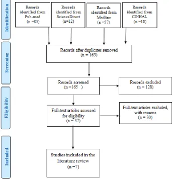
A number of electronic search engines including ScienceDirect, Pub-MED, CINAHL and Medline were used. A comprehensive strategy was adopted, whereby all possible and related articles that meet the criteria of this review were included. Figure 1 elaborates on the strategies used in the search. Based on the objective of the study, the keywords were “repetition rate” OR “digital radiography”.
These keyword were selected to come out with specific articles that discuss the repetition on the digital radiography rather than conventional radiography, since the digital radiography become the common equipment being installed in radiology department in the last decade. Studies were included if they were free full text articles provided, written in English
language and if they were published between 2009 to January 2016. Free full text filter were utilized with Pub-Med, CINAHL and Midline while in ScienceDirect, open access articles filter is the filter type which was utilized. Articles were included if they reported studies done in hospitals, diagnostic department, routine, or digital radiography, and if they focus on repetition rate measurement. Articles with studies carried out in other branches of the diagnostic imaging department, such as Computer Tomography, Ultrasound, Magnetic Resonance Imaging, Dental Imaging, Nuclear Medicine, or Mammography, were removed. Studies conducted on disease, radiological equipment and radiation protection were also excluded due to the scope of the study.
RESULTS
After removal of duplication, the total numbers of articles that were extracted from all electronic search engines were 165 articles. The total of 37 articles were checked based on the eligibility of inclusion, of which, 7 articles were found to have met the inclusion criteria and to be suitable for the review. Figure 1 explain the process of search regarding PRISMA flow chart.
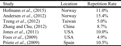
From the seven articles, three were cross-sectional studies, while the other four were intervention studies which compare the repetition rate before and after the intervention or during study period. Table 1 provides more details concerning the selected studies, arranged by the year of the study, from the most recent to the oldest.
[image:2.595.129.474.342.699.2]46326 Abdullah A. Almalki et al. A systematic review on repetition rate of routine digital radiography
Table 1. Studies included in the literature review
Authors Study Design Study Population Variables Results
(Hofmann, Rosanowsky, Jensen, & Wah, 2015)
Cross-sectional Study
All exposed images at two direct digital laboratories at a hospital in Norway were reviewed in January 2014. The type of examination, number of exposed images, and number of deleted images were registered. Each deleted image was separately analysed and the grounds for deleting the image was recorded
Repetition rate From the total of 5417 exposed images, 596 were repeated, giving a repetition rate of 11%. A total of 51.3% were repeated following positioning errors and 31.0% due to error in centring. The examinations with the highest percentage of deleted images were the knee, hip, and ankle, which were 20.6%, 18.5%, and 13.8% respectively. This study only utilized descriptive statistics
(Andersen et al., 2012) Cross-sectional Study
From January 2010 and for 3 months, images from two direct digital equipment in two centres was examined. These two radiology centres have performed 27,284 images.
Repetition rate Out of the 27,284 acquired images, 3206 were repeated, yielding an overall repetition rate of 12%. Highest repetition rates were discovered for the examination of knees, shoulders, and wrist. In all, 77% of the repeated mages arose from positioning errors. Repetition rates for the pelvis, hip, hand, and thorax were not significantly different from the entire repetition rate (P. >0.05). There was no correlation between the number of examinations and the percentage of repeated imaging.
(Tzeng et al., 2012)
Intervention Study
1300 bed hospital conducted 249,215 general radiography examinations every year, and it produces an average of 384,194 images was understudy. The study was conducted between January and December 2008 in Taiwan.
Repetition rate Repeat images decreased from 5% to 3%, and the achievement was through radiographers’ compliance from 60% to 70% to almost 100%. The most common reasons for repeat images were position errors, artefact, and patient movement, which accounted for 79.93% of the repeat images. Repeat radiography a adopted that approach at p<0.05).
(Zhang & Chu, 2012)
Intervention Study
Patient radiation dose and image repetition rate were evaluated in one hospital in China for 2 months before and after the phase of Optimization (OT). In total, 5505 images were evaluated before the intervention and 5119 after intervention.
Dose area product (DAP), Entrance Surface Dose (ESD) and repetition rate
For every radiographic procedure, t tests demonstrated significant difference in average ESD and DAP pre- and post-Optimization (OT) (p<0.005). The ESDs from most examinations before OT were three times higher than that after the optimization. For DAPs, the difference is more significant. Image repetition rate after OT is strikingly lower than that before OT (χ2=36.5, p<0.005). The substantial reductions of dose after OT stemmed from exposure field that was deemed appropriate.
(Jones, Polman, Willis & Shepard, 2011)
Intervention Study
All routine radiological examinations performed from April 2007 to March 2008 were analysed after implementing a central server system to accumulate and archive the data of repeated radiography and exposure indicator.
Repetition rate
From the total of 66063 images undertaken in the given period, 6002 were repeated. The primary reason of repetition was patient poisoning by 77%. The exposure factor was the second reason of repetition by 9.3%. The data stratification by clinical area revealed that areas where Computed Radiography (CR) is scarcely used suffer from higher repetition rates than areas where it is used often.
(Foos et al., 2009) Cross-sectional Study
Images of two computer radiographies in different locations were examined. The periods of study were 435 days for the community hospital (CH) and 275 days for the university hospital (UH). The study was conducted in the United States of America (USA).
Repetition rate From the total of 2,888,000 images that were performed in the two hospitals, the repetition rate was reported as 4.4% at UH and 4.9 % at CH. In the UH, the facial bone has the highest repeated image and portable chest x ray was the lowest. In CH hospital, the abdomen showed the highest repetition rate, while portable chest x ray showed the lowest (Pvalue < 0.05, CI (95%).)
(Prieto et al., 2009) Intervention Study
A total of 3,742 abdomen and 8,636 chest CR images were archived in the Picture Achieving Communication System (PACS) in 3 months, and in this period, 1,893 abdomen and 4,369 chest images for one month were sampled in this study. The study was conducted at San Carlos University Hospital in Madrid, Spain.
Table 2. Differences between repetition rates from various studies
Study Location Repetition Rate
Hofmann et al., (2015) Norway 11.0% Andersen et al., (2012) Norway 15.4%
Tzeng et al., (2012) Taiwan 5.0%
Zhang and Chu, (2012) China 8.7%
Jones et al., (2011) USA 10.0%
Foos et al., (2009) USA 4.9%
Prieto et al., (2009) Spain 10.5%
DISCUSSION
Quality control approach becomes essential in any department as it can help to achieve desired goals. It facilitates the improvement of the services quality of the department or organization through standard compliance. Moreover, quality standards should be predetermined by policy makers to overcome the prevalence of the repetition rate issues. As presented in Table 1, seven studies from various countries are discussed from various aspects of radiography services quality, focusing on the repetition rate, the causes of repetition, drawing in comparison the repetition rate between modalities and radiological examinations type. Table 2 shows the difference of the repetition rate between different studies. Those measurements explored in the table for some of the studies were obtained prior to the intervention. Moreover, in terms of the differences in percentage in Table 2, the work done by (Prieto et al., 2009) compared the chest and abdomen before and after a new technology was installed into the system. The initial repetition rate was 15.4% and 4.5% for the abdomen and chest respectively. New techniques called Digital Imaging and Communication in medicine (DICOM) and it helps to decrease the repetition rate from 15.4 % to 6.6% in abdomen and from 4.5 % to 3.3% in chest.
In the study conducted by Prieto et al., (2009), the one-month cases were selected and examined by professionals, which revealed a reduction by 3.3% and 0.90% for abdomen and chest respectively. Meanwhile, the study by Zhang and Chu, (2012) introduced a comprehensive optimization program to measure both the radiation dose and repetition rate. They probed into the digital equipment and trained the radiographers and patients. The earlier result was 8.72% and post intervention saw it decreased to 5.84%. To add, the study of Tzeng et al., (2012) implemented the system approach aided by the information technology, then the initial repetition rate started with 5% and within the period of the study and with increasing of radiographers’ compliance it came down to 3%. Also, the difference between the studies of Prieto et al., (2009); Tzeng et al., (2012) and Zhang and Chu, (2012) is that the repetition rate during their intervention was quite high and intervention was effective to reduce the repetition. In addition to repetition rate, Jones et al., (2011) also calculated the exposure and stressed on the dose area product and entrance surface dose.
The work by Zhang and Chu, (2012) had compared the pre and post intervention while the work of Jones et al., (2011) focused on the introduction of their technique during the period of the study. The reduction of radiation dose was clear within both the abovementioned studies. Studies by Andersen et al., (2012); Foos et al., (2009); Hofmann et al., (2015); Jones et al., (2011);Prieto et al., (2009) illustrated that there was a difference in the sample and period of study. They discussed the repetition rate and its causes. Studies of Andersen et al.,
(2012); Hofmann et al., (2015); Zhang and Chu, (2012) examined the repetition rate in direct digital radiography while the studies of Jones et al., (2011); Foos et al. Foos et al., (2009); Prieto et al., (2009) deal with computed radiography. Most studies look into the relationship between the repetition rate and different causes of repetition such as patient’s movement, patients’ position, artefacts, and exposure technique and other causes lead to the repetition of the radiograph, based on that causes of repetition are mentioned by radiology or by radiation protection agencies which the study follow. In reference to studies of Andersen et al., (2012); Foos et al., (2009); Hofmann et al., (2015); Jones et al., (2011); Tzeng et al., (2012); Zhang and Chu, (2012) the patients’ positioning error contributed most to the issue. Position fault considers the main cause of the previous mentioned studies by: 51%, 77%, 35%, 59%, 77.3%, 56%, and 50% respectively of all causes of repetition while study of Prieto et al., (2009) shows patients identification is the main causes of repetition. With regards to the positioning error, a radiographer or technologist plays a crucial role in that reason. Additionally, exposure factor or in other words under or over exposure was also the second cause mentioned in several studies such as Foos et al., (2009) and Jones et al. Jones et al., (2011). Patient movement was mentioned in studies by Tzeng et al. Tzeng et al., (2012);Zhang and Chu, (2012) as the second cause, whereas positioning error was the second most common error in the study by Prieto et al., (2009) that leads to repeat radiographs. Those causes were shown to be statistically significant with p<0.0.5.
Radiological examination types were also one of the variables that were included in the studies but the types differ from one study to another. Some of the studies have stratified the radiological examination into; chest, abdomen, pelvis, skull, spine, and extremities. Jones et al., (2011) showed that high repetition rate increases the radiation to body organs. Radiological examinations indicate which part of the body has high repetition. Recognising the high repetition rate related to body parts and causes can help arrange the solution. Furthermore, studies of Andersen et al. Andersen et al., (2012); Hofmann et al., (2015) reported that the examination of chest shows higher repetition rate while Foos et al., (2009) stated that the skull is one of the common parts that have high repetition rate. Studies of Jones et al., (2011); Prieto et al., (2009) have demonstrated that the abdomen is the highest to have repetition rate than chest. According to Tzeng et al., (2012) thoracic spine was the body organ with high repetition rate followed by the pelvis, abdomen, and chest. With regards to chest, it was noted that the chest radiography is the most radiological examination that performed in routine digital radiography. There were four intervention studies out of the seven articles reviewed. The intervention differed from study to another. The study carried out by Zhang and Chu, (2012) included the final report of the IAEA 2004 in a designated department with specific examination and digital equipment. Meanwhile, the study of Tzeng et al., (2012) started to fix new strategy that has to do with the repeat image. The difference between the study of Tzeng et al., (2012) and the one conducted by Zhang and Chu, (2012) is the later had adopted a comprehensive strategy to decrease the dose of radiation. The study conducted by Jones et al., (2011) used Server-Based System on computed radiography to archive, sort of repeated images and exposure indicator automatically. In addition, the intervention were conducted without control group and studies of Jones et al., (2011);Tzeng et al., (2012) were not dealing
with measurement before their intervention. The period of study and number of cases collected to measure the repetition rate of radiography also varied between the aforementioned studies since there was no randomization or probability sampling technique. This methods is recommended and used with most of studies that concern of repetition measurement Jones et al., (2015). Since the studies collected their data based on time period, radiographers were not included in those studies. Additionally, those studies utilized different types of technical intervention while there yet to be any written protocol that addresses the issue of repetition and takes correction action based on the analysis. Furthermore, radiographers or radiological technologist were not considered in the aforementioned studies since different studies has proved the important role in repetition issue.
Conclusion and Recommendation
Repetition rate is considered as critical event as it deals with practitioners and decision maker in a Diagnostic Imaging department. One can admit that there are varying strategies that might be applicable based on organizations, modalities and their capabilities to improve the outcome, further decreasing overall radiation to both staff and patient, and contributing to improved level of internal and external customers’ satisfaction. Different research has been delving into this issue through different aspects. Technical intervention were conducted in the intervention studies and show its positive impact on repetition. In addition, more emphasis must be placed on examining the various methods and strategies to diminish the repetition rate in routine digital radiography rather than merely describing the problem. Future research must take advantage of the previous research to try and overcome the issue. Additionally, quality control protocol with periodic measurement may help to improve the services. That intervention must be planned, developed, implemented, and tested to ensure that the impact is well evaluated.
REFERENCES
Andersen, E. R., Jorde, J., Taoussi, N., Yaqoob, S. H., Konst, B., and Seierstad, T. 2012. Reject Analysis in Direct Digital Radiography. Acta Radiologica (Stockholm, Sweden : 1987, 53(2), 174–8. http://doi.org/10.1258/ ar.2011.110350
Arbique, G. 2009. Reject Analysis in a Digital Radiography (DR) Quality Assurance (QA) Program. Retrieved from http://amos3.aapm.org/abstracts/pdf/77-22539-313436-92756.pdf
Brady, A., and Ryan, S. 2011. Requirements for Clinical Audit in Medical Radiological Practices (Diagnostic Radiology, Radiotherapy and Nuclear Medicine). Retrieved from http://www.radiology.ie/wp-content/uploads/2012/01/ Requirements-for-Clinical-Audit-in-Radiology-Jan-2011.pdf
Candido, S., Ribeiro, L. L. P. V. L., Ribeiro, A. A. M. A., Abrantes, A. F. L., Pinheiro, J. P., Almeida, R. P. P., … Azevedo, K. B. 2013. Image Quality Control in Digital Radiology. American Journal of Biomedical Engineering,
3(6A), 8–14. Retrieved from http://article.sapub.org/ 10.5923.s.ajbe.201310.02.html
Conference of Radiation Control Program Directors (CRCPD). 2009. Repeat Analysis. Retrieved from http://c.ymcdn.com/ sites/www.crcpd.org/resource/resmgr/docs/QA_Collectible s/RepeatAnalysisQAC.pdf
Eze, C. U., Olajide, B. O., Ohagwu, C. C., and Abonyi, L. C. 2012. Analysis of Film Reject Rate in the Diagnostic X-ray Facility of a Tertiary Health Institution in Benin, Nigeria. .
Nigerian Quarterly Journal, 201257–54 ,(1)23 ,. Retrieved from http://europepmc.org/abstract/med/24579496
Foos, D. H. D. D. H., Sehnert, W. J. W. J., Reiner, B., Siegel, E. L., Segal, A., and Waldman, D. L. 2009. Digital Radiography Reject Analysis: Data Collection Methodology, Results, and Recommendations from an In-depth Investigation at Two Hospitals. Journal of Digital Imaging, 22(1), 89–98. http://doi.org/10.1007/s10278-008-9112-5
Hofmann, B., Rosanowsky, T. B., Jensen, C., and Wah, K. H. 2015. Image Rejects in Reneral Direct Digital Radiography. Acta Radiologica Open, 4(10), 2058460115604339.
http://doi.org/http://dx.doi.org/10.1177/2058460115604339 Honea, R., Elissa Blado, M., and Ma, Y. 2002. Is Reject Analysis Necessary after Converting to Computed Radiography? Journal of Digital Imaging, 15 Suppl 1, 41– 52. http://doi.org/10.1007/s10278-002-5028-7
Ip, I. K., Mortele, K. J., Prevedello, L. M., and Khorasani, R. 2012. Repeat Abdominal Imaging Examinations in a Tertiary Care Hospital. American Journal of Medicine,
125(2), 155–161.
http://doi.org/10.1016/j.amjmed.2011.03.031
Jones, A. K., Heintz, P., Geiser, W., Goldman, L., Jerjian, K., Martin, M., … Yorkston, J. 2015. Ongoing Quality Control in Digital Radiography: Report of AAPM Imaging Physics Committee Task Group 151; Ongoing Quality Control in Digital Radiography: Report of AAPM Imaging Physics
Committee Task Group 151. AAPM (Vol. 42). 6670.
Retrieved from https://www.aapm.org/pubs/reports/ RPT_151.pdf
Jones, A. K., Polman, R., Willis, C. E., and Shepard, S. J. 2011. One year’s Results from a Server-Based System for Performing Reject Analysis and Exposure Analysis in Computed Radiography. Journal of Digital Imaging, 24(2), 243–255. http://doi.org/10.1007/s10278-009-9236-2 Monfared, A., Abdi, R., and Saber, M. 2007. Repeat Analysis
Program in Radiology Depatments in Mazandaran Province-Iran; Impact on Population Radiation Dose . Iran J Radiat Res, 2007240–237 ,(1)5 ,. Retrieved from http://citeseerx.ist.psu.edu/viewdoc/download?doi=10.1.1.6 14.1394andrep=rep1andtype=pdf
Prieto, C., Vano, E., Ten, J. I., Fernandez, J. M., Iñiguez, A. I., Arevalo, N., Martinez, D. 2009. Image Retake Analysis in Digital Radiography Using DICOM Header Information.
Journal of Digital Imaging, 22(4), 393–399. http://doi.org/ 10.1007/s10278-008-9135-y
Rajani, A., Sajjad, Z., Masroor, I., Parveen, A., and Naqvi, M. 2016. “Should we Stop Improving when Benchmarks are Met?” PJR, 18(1), 18–19. Retrieved from http://www. pakjr.com/ojs/index.php/PJR/article/view/298/330
Teferi, S., Zewdneh, D., Admassie, D., Nigatu, B., and Kebeta, K. 2012. X-ray Film Reject Rate Analysis at Eight Selected Government Hospitals in Addis Ababa, Ethiopia, 2010.
Ethiopian Journal of Health Development, 26(1), 54–59. http://doi.org/10.4314/ejhd.v26i1.
Tzeng, W. W.-S., Kuo, K. K.-M. K., Liu, C.-F. C. C.-F., Yao, H. H.-C. H., Chen, C. C.-Y. C., and Lin, H.-W. 2012. Managing Repeat Digital Radiography Images—A Systematic Approach and Improvement. Medical Systems,
Winston, J., Jackson, D., Wozniak, D., Zeisler, J., Farish, S., and Thoma, P. 2001. Quality Control Recommendations for Diagnostic Radiography. Radiography, 3(July), 72. Retrieved from http://www.crcpd.org/Pubs/QC-Docs/QC-Vol3-Web.pdf
Zhang, M., and Chu, C. 2012. Optimization of the Radiological Protection of Patients Undergoing Digital Radiography. Journal of Digital Imaging, 25(1), 196–200. http://doi.org/10.1007/s10278-011-9395-9