ISSN Online: 2164-2869 ISSN Print: 2164-2842
DOI: 10.4236/ojneph.2019.91001 Jan. 23, 2019 1 Open Journal of Nephrology
Changes in Brachial and Central Blood Pressure
after Short Term Continuous Positive Airway
Pressure Treatment of Patients with
Moderate-to-Severe Obstructive Sleep Apnoea
and Impaired Renal Function
Bodil G. Hornstrup
1*, Pia H. Gjørup
2, Jost Wessels
2, Thomas G. Lauridsen
1,2,
Erling B. Pedersen
1, Jesper N. Bech
1,21University Clinic in Nephrology and Hypertension, Regional Hospital West Jutland and Aarhus University, Aarhus, Denmark 2Department of Medicine, Regional Hospital West Jutland, Holstebro, Denmark
Abstract
Background: Previous studies of continuous positive airway pressure (CPAP) treatment for obstructive sleep apnoea (OSA) have shown conflicting results on the effect on blood pressure (BP), and patients with chronic kidney disease (CKD) have not been included in these studies. As OSA is a frequent comor-bidity in patients with CKD, it is of relevance to evaluate the effect of CPAP treatment on BP in this population. Aim: In this prospective follow-up study, we measured the effect of short term CPAP treatment of moderate-to-severe OSA on brachial and central BP, plasma level of syndecan-1 and vasoactive hormones, renal handling of sodium, subjective sleepiness, and quality of life in patients with impaired renal function. Methods: From December 2015 un-til March 2017, 25 patients were invited to participate in the study at the University Clinic in Nephrology and Hypertension, Aarhus University and Holstebro Hospital. At baseline and at follow-up after three to four months of CPAP treatment, we performed 24 h brachial and central ambulatory BP measurement, blood sampling measurements of plasma concentrations of syndecan-1, renin, angiotensin II, aldosterone, vasopressin, creatinine, hae-moglobin A1c, and cholesterol, cardio respiratory monitoring, 24 h urine collection for measurement of urinary excretion of albumin, aquaporin-2, and epithelial sodium channel, Epworth Sleepiness Scale (ESS), and SF-36 (quality of life). Results: At follow-up, the 17 included patients with mean baseline estimated glomerular filtration rate 66 mL/min/1.73 m2 had a
sig-How to cite this paper: Hornstrup, B.G., Gjørup, P.H., Wessels, J., Lauridsen, T.G., Pedersen, E.B. and Bech, J.N. (2019) Changes in Brachial and Central Blood Pressure after Short Term Continuous Positive Airway Pressure Treatment of Patients with Moderate-to-Severe Obstruc-tive Sleep Apnoea and Impaired Renal Function. Open Journal of Nephrology, 9, 1-19.
https://doi.org/10.4236/ojneph.2019.91001
Received: December 12, 2018 Accepted: January 20, 2019 Published: January 23, 2019
Copyright © 2019 by author(s) and Scientific Research Publishing Inc. This work is licensed under the Creative Commons Attribution International License (CC BY 4.0).
http://creativecommons.org/licenses/by/4.0/
DOI: 10.4236/ojneph.2019.91001 2 Open Journal of Nephrology nificant decrease in systolic office-, 24 h- and daytime-BP (13, 7, and 8 mmHg, respectively, p < 0.05), a non-significant reduction of nocturnal BP (6 mmHg). No changes was measured in frequency of non-dipping or in central 24 h-, day- and nighttime-BP. Renal function remained unchanged, but urinary al-bumin excretion fell. ESS was unchanged. Quality of life improved. Conclu-sion: Short-term CPAP treatment of patients with moderate-to-severe OSA and reduced renal function decreased 24 h- and daytime-BP significantly and reduced urinary albumin excretion. Our results underline the importance of treatment of OSA in hypertensive patients with impaired renal function.
Keywords
Chronic Kidney Disease, Nocturnal Blood Pressure, Obstructive Sleep Apnoea, Central Blood Pressure, Continuous Positive Airway Pressure
1. Introduction
Obstructive sleep apnoea (OSA) is a frequent comorbidity in hypertension and chronic kidney disease (CKD) [1][2]. OSA is characterised by obstructions of the upper airways during nighttime sleep causing repetitive pauses in breathings despite respiratory efforts. Termination of the obstructions requires arousal, which leads to poor sleep quality and daytime sleepiness in OSA patients. OSA patients show early signs of atherosclerosis and are of increased risk of stroke independent of other known risk factors such as hypertension, diabetes, smok-ing, and body mass index (BMI) [3][4]. Furthermore, the nocturnal hypoxemia seen in OSA is associated to progression of renal failure [5][6].
Continuous positive airway pressure (CPAP) has been an established treat-ment for OSA for many years with a docutreat-mented improving effect on sleep, quality of life, and nocturnal hypoxemia [7] [8][9]. High CPAP compliance is associated with lower risk of cardiovascular disease and with a renoprotective effect independent of blood pressure (BP) reduction [10] [11]. Different effects of CPAP treatment on BP levels have been reported; some studies show signifi-cant lowering effect [7], whereas other studies demonstrate sparse effect [12] [13][14]. No studies have analysed the effect of CPAP treatment in patients with CKD; hence, short term effects (three to four months) of CPAP treatment of moderate-to-severe OSA in patients with CKD are unknown with respect to brachial BP, quality of life, and sleep symptoms.
Central aortic systolic pressure (CASP) may provide additional information on the level of arteriosclerosis and cardiovascular disease associated end organ damage [15][16]. CPAP treatment has been shown to lower the central systolic pressure using twice-a-day measurements in normo- and uncomplicated essen-tial hypertensive patient with OSA [17]. However, the effect of CPAP treatment on CASP has not been clarified in CKD patients.
DOI: 10.4236/ojneph.2019.91001 3 Open Journal of Nephrology the glycocalyx protection layer as a response to cardiovascular stress [18]. We have previously found increased syndecan-1 levels in hypertensive patients compared with healthy controls [19]. However, it is not known, whether CPAP treatment can alter plasma levels of syndecan-1, and thereby be an indicator of reduced shedding as a response to CPAP treatment.
The aim of this intervention study was to evaluate the effect of short term CPAP treatment in subjects with moderate-to-severe OSA (apnoea hypopnoea index (AHI) >15) and impaired renal function (estimated glomerular filtration rate (eGFR) 15 - 89 mL/min/1.73 m2 at sampling time) on 1) brachial and central BP, 2) p-syndecan-1, 3) plasma levels of renin, angiotensin II, aldosterone, and vasopressin, 4) eGFR and urinary excretion of albumin, aquaporin-2, and a fraction of the epithelial sodium channel, 5) quality of life, and 6) subjective sleepiness.
2. Materials and Methods
2.1. Design
The project was carried out as a prospective intervention study. Patients were included at the time of start of CPAP treatment and followed up after approxi-mately three to four month.
2.2. Study Settings
The study was conducted at the University Clinic in Nephrology and Hyperten-sion, Aarhus University and Holstebro Hospital, and the Sleep Apnoea Clinic, Department of Medicine, Holstebro Hospital. The recruitment period was from December 2015 until March 2017.
2.3. Patients
Patients were recruited from the Renal Outpatient Clinic, Holstebro Hospital (eGFR 15 - 59 mL/min/1.73 m2) or from a population study in Holstebro County (diagnosis of hypertension and eGFR 60 - 89 mL/min/1.73 m2) [20]. All patients were diagnosed with moderate-to-severe OSA and had not received CPAP treatment previously. The patients were examined for OSA during participation in two other projects by the same authors [19] [21]. They were offered CPAP treatment according to usual clinical practice.
Inclusion criteria: men and women, age 18 - 80, eGFR 15 to 89 mL/min/1.73
DOI: 10.4236/ojneph.2019.91001 4 Open Journal of Nephrology
Number of subjects: the minimal relevant difference in mean 24 h systolic BP
(SBP) was 10 mmHg with standard deviation (SD) 10. With a statistical power of 80% and a significance level of 5%, it was calculated that the number of subjects should be at least 16.
2.4. Ethics
This study was reviewed and approved by the Central Denmark Region Com-mittees on Health Research Ethics (j.no.: M-2013-285-13 and M-2013-304-13) and by the Danish Data Protection Agency (j.no.: 1-16-02-399-13 and 1-16-02-458-13). The study was carried out in accordance with the Helsinki Declaration. All study patients received oral and written information about the project and provided informed written consent prior to study enrolment. ClinicalTrials.gov registration identification was NCT01951248 and NCT02078778.
2.5. End Points
The primary end point was change in brachial 24 h SBP after three to four months of CPAP treatment.
The secondary end points were changes in: 24 h central BP, relative nocturnal brachial SBP and CASP decrease, urinary excretion rate of aquaporin-2 (u-AQP2) and epithelial sodium channel fraction γ (u-ENaCγ), 24 h urine excretion of al-bumin (u-alal-bumin), p-syncecan-1, vasoactive hormones (plasma renin concen-tration, plasma aldosterone, plasma angiotensin II, plasma arginine vasopressin (p-AVP)), reporting of subjective sleepiness (Epworth Sleepiness Scale, ESS), and quality of life (by SF-36 questionnaire).
2.6. Blood Pressure
Twenty-four hour ambulatory BP measurement (ABPM) was carried out using an oscillometric device, A&D TM-2430 (A&D Company Limited, Tokyo, Japan). An appropriate size cuff was chosen after measuring the upper arms circumfer-ence and placed on the right side. Twenty-four hour CASP was measured using applanation tonometry by BPro Health Stat (BPro, HealthSTATS, Singapore). The BPro device was placed on the left wrist after being calibrated with a mean of the last three of four consecutive BP measurements on the left arm derived from the A&D device used for 24 h brachial ABPM on the same subject.
BP was measured every 15 min and every 30 min during day- and nighttime, respectively, by the A&D device, and every 15 min by the BPro device through-out the 24 hours. ABPM were considered satisfactory with 14 or more daytime recordings (fixed daytime setting at 6 am to11 pm) and seven or more nighttime recordings (fixed nighttime setting at 11 pm to 6 am).
A semiautomatic oscillometric device, Omron 705IT (Omron Matsusaka CO, Ltd., Matsusaka City, Japan) was used for bilateral brachial BP measurements with the subject sitting in upright position after minimum ten minutes rest.
DOI: 10.4236/ojneph.2019.91001 5 Open Journal of Nephrology 80 mmHg diastolic, non-dipping was defined as relative nocturnal systolic BP decrease ≤10%, and resistant hypertension was defined as hypertension on three antihypertensive drugs, one of these being a diuretic. These definitions are ac-cording to most recent guidelines from the European Society of Hypertension/ European Society of Cardiology [22].
2.7. Sleep Apnoea
An ambulatory cardio respiratory monitoring (CRM) was performed using Em-bletta Gold (Natus Medical Incorporated, USA) in all participants at baseline and follow-up. RemLogic-E Software was used for analysing and storing data. The sleep report was generated from sleep time and consisted of continuous re-cordings from the Embletta monitor. During sleep, this monitor recorded air flow from a nasal pressure transducer, arterial oxygen saturation from a pulse oximeter, respiratory effort from thoracic and abdominal impedance belts, and body position from sensors. An apnoeic event was defined as a cessation of nasal airflow accompanied by a drop of the signal below 10% of the reference ampli-tude for an interval of 10 seconds. A hypopnoeic event was defined as a reduc-tion of the signal below 70% of the reference amplitude for an interval of 10 seconds with a subsequent desaturation event no later than 20 seconds after the start of the event. An oxygen desaturation event was observed by a decrease in oxygen saturation by at least 4%. Events lasting longer than 120 seconds (ap-noea, hypopnea, or desaturation) were excluded.
Apnoea hypopnoea index (AHI) was defined as the sum of apnoeas and hy-popnoeas per hour of registered sleep. Oxygen desaturation index (ODI) was de-fined as oxygen desaturation events pr. hour of sleep. OSA was dede-fined as AHI ≥ 5, moderate-to-severe OSA ≥ 15. Definitions above are according to recommen-dations from the American Academy of Sleep Medicine (AASM) [23].
2.8. Continuous Positive Airway Pressure (CPAP)
OSA was treated with AirSense 10 AutoSet (ResMed/Maribo Medico) or REM-star Auto with A-Flex (Phillips/Respironics). Treatment was REM-started and fol-lowed up by trained nurses in the Sleep Apnoea Clinic, Department of Medicine, Holstebro Hospital, according to standard procedure. A mask for the CPAP treatment was chosen to the best fit for the patient, either nasal or full face mask. The two systems had identical functionality and were self-adjusting; the pressure was automatically adjusted in response to inhalation flow, snoring, and apnoeas. The subjects were instructed in correct use of the system, change of air filter, and daily maintenance/cleaning of the system. Humidifier was added to the system if needed. Adherence was defined from the last month of treatment as percentage of 4 hours every night; the patient had been using the equipment.
2.9. Experimental Procedures
in-DOI: 10.4236/ojneph.2019.91001 6 Open Journal of Nephrology formation meeting was set up, and after informed written consent, life style questionnaire (SF-36) was filled in by the patient alone after instructions were given, and follow-up meeting was planned. Baseline information on ABPM, CRM, blood and urine samples, medical treatment, sleep symptoms (Epworth Sleepiness Scale (ESS)), and medical history were collected from the previous project participation.
While participating in this study, patients followed their usual consultations in the Renal Outpatient Clinic or general practitioners with regard to their renal disease or hypertensive disease. At these consultations, antihypertensive medica-tions were adjusted if necessary. This was assessed by the patient’s physician in the Outpatient Clinic or the general practitioner.
At baseline and at follow-up after approximately three to four months of CPAP treatment, subjects were examined as described in the following section. Blood samples were drawn after 20 min of rest in supine position. Twenty-four hour brachial and central ABPM were carried-out simultaneously, and 24 h urine collection was performed and returned at the latest 4 hour after comple-tion. CRM, as described above, was completed, and at follow-up the CRM was carried-out simultaneously with CPAP treatment, and adherence was registered from the CPAP device. Information on changes in use of medication and even-tual new medical events were obtained by the electronic patient record and questionnaire, respectively. Current quality of life and subjective sleepiness were reported by SF-36 and ESS, respectively. These questionnaires were filled in by the participant alone after a short introduction.
2.10. Biochemical Analyses
Blood samples were centrifuged at 4˚C for 10 min at 2200× g. Plasma samples were kept frozen at −20˚C (P-Angiotensin II) and −80˚C (Syndecan, plasma renin concentration, P-aldosterone, and p-AVP), and urine samples at −20˚C until assayed.
Urinary and plasma osmolality were measured by freezing-point depression (Advanced Model 3900 multisampling osmometer).
Plasma renin concentration was determined by radioimmunoassay using a kit from CIS Bio International, Gif-Sur-Yvette Cedex, France. Minimal detection level was 1 pg/mL. The coefficients of variations were 14.5% (inter-assay) and 4.5% (intra-assay).
P-angiotensin II and p-AVP were extracted from plasma with C18 Sep-Pak (Water associates, Milford, MA, USA), and subsequently determined by radio-immunoassay [24][25]. The antibody against Ang II was obtained from the De-partment of Clinical Physiology, Glostrup Hospital, Denmark. Minimal detec-tion level was 2 pmol/L. The coefficients of variadetec-tion were 12% (inter-assay) and 8% (intra-assay). The antibody against AVP was a gift from Professor Jacques Dürr, Miami, FL., USA. The coefficients of variation were 13% (inter-assay) and 9% (intra-assay). Minimal detection level was 0.5 pmol/L.
DOI: 10.4236/ojneph.2019.91001 7 Open Journal of Nephrology Demeditec Diagnostics GmbH, Kiel, Germany. The coefficients of variations were 17.2% (inter-assay) and 12.6% (intra-assay). Minimal detection level was 3.99 pmol/L.
P-syncecan-1 was analysed on EDTA-plasma using a human ELISA kit from Abcam plc, Cambridge, United Kingdom. The coefficients of variations were 10.2% (inter-assay) and 6.2% (intra-assay). Minimal detection level was 4.94 ng/mL.
U-AQP2 was determined by RIA as previously described [26][27]. Rabbit an-ti-AQP2 antibodies were a gift from Professor Soren Nielsen and Professor Ro-bert Fenton, the Water and Salt Research Center, Aarhus University, Denmark. Coefficients of variation: 11.7% (inter-assay) and 5.9% (intra-assay). Minimal detection level: 32 pg/tube.
U-ENaCγ was measured by RIA as described previously [28][29]. ENaCγ was synthesized and purchased by Lofstrand, Gaithersburg, Maryland, USA. The ENaCγ antibody was a gift from Professor Soren Nielsen and Professor Robert Fenton, the Water and Salt Center, Aarhus University. Coefficients of variation: 10% at a mean level of 338 pg/tube (inter-assay), 9% at a mean level of 743 pg/tube (inter-assay), 5.0% in the range 125 - 135 pg/tube (intra-assay), and 5.6% in the range 290 - 380 pg/tube (intra-assay). Minimal detection level: 35 pg/tube.
Plasma levels of creatinine, haemoglobin A1c (HbA1c), cholesterols, and uri-nary concentrations of albumin, creatinine, and sodium were measured using routine methods at the Department of Clinical Biochemistry, Holstebro Hospi-tal, Denmark. eGFR was calculated by the MDRD-equation.
2.11. Statistical Methods
Statistical analyses were performed by the authors using IBM SPSS statistics ver-sion 22 (IBM Corp.; Armonk, NY, United States). All data were tested for nor-mality and variance equality. The statistical level of significance was p < 0.05 in all analyses.
Continuous variables were reported as means with SD or as median with in-terquartile rang [25; 75] depending on whether the data were normally distrib-uted or not. Categorical variables were reported as percentages with number. Paired t-tests were used for paired continuous variables with normally distrib-uted differences; otherwise Wilcoxon signed rank test was used. McNemars test was used for paired categorical data. Univariate analyses were performed using Pearson’s test or Spearman Rho test on normally distributed or non-normally distributed continuous variables, respectively.
The SF36 questionnaire filled out by the patients was scored using the proce-dure from International Resource Center for Health Care Assessment (Boston, MA), called RAND 36-Item Health Survey 1.0 [30].
3. Results
3.1. Demographics
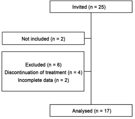
ex-DOI: 10.4236/ojneph.2019.91001 8 Open Journal of Nephrology cluded; four due to early discontinuation of treatment (within one month), and two due to uncompleted follow-up examination (incomplete data). Seventeen participants completed follow-up examination (Figure 1).
Baseline clinical, laboratory, and sleep characteristics of the included 17 pa-tients are presented in Table 1. Mean eGFR were 66 mL/min/1.73 m2, and 36% (n = 6) were diagnosed with diabetes. All participants were diagnosed with hy-pertension, and 76% (n = 13) received antihypertensive medication. Of them, 23% (n = 3) were controlled hypertensive, and 54% (n = 7) had resistant hyper-tension.
At baseline and follow-up, subjects received a mean of 3.7 ± 4.4 and 3.8 ± 4.5 defined daily doses (DDD) of BP lowering drugs, respectively (p > 0.05). Twelve subjects’ DDD remained unchanged, one subject’s DDD decreased (0.25 DDD), and four subjects’ DDD increased (mean change 0.8 DDD). Forty-two percent (n = 7) received statins throughout the follow-up period.
3.2. Brachial Blood Pressure
Table 2 shows baseline and follow-up brachial BP; brachial systolic and diastolic
office, 24 h and daytime BP decreased significant at follow-up. No changes were observed in nocturnal BP or nocturnal BP decrease. The frequency of non-dipping was unchanged (at baseline 47% (n = 8) and follow-up 53% (n = 9), p = 1.0 (Figure 2(a)).
3.3. Central Arterial Systolic Pressure
[image:8.595.259.490.507.710.2]Two patients’ baseline and one patient’s follow-up CASP measurement was ex-cluded from analysis due to few measurements Therefore, CASP analyses in-cluded 14 patients’ measurements. There was a non-significant decrease in 24 h, day and nighttime CASP (Table 2). Non-dipping was seen in 64% (n = 9) at baseline, and 86% (n = 12) at follow-up (p = 0.45) (Figure 2(b)).
DOI: 10.4236/ojneph.2019.91001 9 Open Journal of Nephrology
Figure 2. Percentage of patients with dipping/non-dipping blood pressure pattern at
baseline and follow-up in brachial (a) and CASP (b) values. Percentage (number) of sub-jects with nocturnal systolic blood pressure decrease </≥10% of day time blood pressure as non-dippers/dippers, respectively. Abbreviations. CASP: central aortic systolic pres-sure. Brachial: p = 1.0, CASP: p = 0.45. Statistics were performed using McNemars test.
Table 1. Demographics and sleep characteristics at baseline.
Demographic Characteristics N = 17
Age, years 65 (7)
Gender, male, % (n) 77 (13)
Body mass index, kg/m2 34 (7)
Estimated glomerular filtration rate, ml/min/1.73 m2 66 (23)
U-albumin, mg/24 hour 11 [7; 138]
Haemoglobin A1c, mmol/mol 49 (15)
Total cholesterol, mmol/L 4.9 (1.0)
High density lipoprotein cholesterol, mmol/L 1.1 (0.3)
Triglyceride, mmol/L 2.2 (1.1)
Sleep Characteristics
Epworth Sleepiness Scale (ESS), 0 - 24a 5.0 [3.5; 8.5]
Apnoea hypopnoea index (AHI), events pr. hour 33 [20; 37]
Oxygen desaturation index (ODI), events/hour 36 [17; 51]
Mean oxygen saturation, % 92 (2)
Baseline demographic characteristics and baseline sleep data from all 17 patients. Data are presented as mean (SD) except u-albumin, Epworth Sleepiness Scale, Apnoea hypopnoea index, and oxygen desaturation
index, which are presented as median with interquartile range [25%; 75%]. aFrom Epworth Sleepiness Scale,
self-reported data.
The changes in brachial and central BP from baseline to follow-up were simi-lar (data not shown).
3.4. Sleep Examination
[image:9.595.205.539.304.544.2]DOI: 10.4236/ojneph.2019.91001 10 Open Journal of Nephrology
Table 2. Brachial blood pressure and central aortic systolic pressure at baseline and at
follow-up.
Brachial (n = 17) CASP (n = 14)
Baseline Follow-up Baseline Follow-up
Office BP systolic, mmHg 149 (13) 136 (15)*
Office BP diastolic, mmHg 87 (7) 79 (12)*
24 hour BP, systolic, mmHg 143 (11) 136 (8)* 125 (16) 117 (16)
24 hour BP, diastolic, mmHg 81 (7) 78 (7)**
24 hour heart rate, beats/min 68 (7) 68 (7)
Daytime BP, systolic, mmHg 146 (11) 138 (9)* 127 (16) 118 (16)
Daytime BP, diastolic, mmHg 83 (8) 79 (7)*
Nighttime BP, systolic, mmHg 131 (16) 125 (14) 121 (17) 112 (16)
Nighttime BP, diastolic, mmHg 74 (6) 72 (7)
Absolute nocturnal BP decrease, mmHg 15 (12) 13 (14) 7 (9) 7 (8)
Relative nocturnal BP decrease, %a 10 (8) 9 (10) 5 (7) 6 (7)
Abbreviations: BP: blood pressure. CASP: central aortic systolic pressure. Office blood pressure obtained from baseline and follow-up examinations. 24 h, day and nighttime brachial and CASP data obtained from 24 h ABPM. CASP measurements only consist of systolic values from 24 h central ABPM. Data are pre-sented as mean (SD). aRelative systolic decrease from day time to night time. Statistics were performed us-ing paired t-test. *p < 0.05, **p < 0.001.
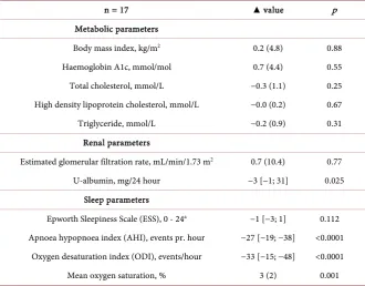
Table 3. Changes in body mass index, laboratory results, and sleep parameters at
fol-low-up.
n = 17 ▲ value p
Metabolic parameters
Body mass index, kg/m2 0.2 (4.8) 0.88
Haemoglobin A1c, mmol/mol 0.7 (4.4) 0.55
Total cholesterol, mmol/L −0.3 (1.1) 0.25
High density lipoprotein cholesterol, mmol/L −0.0 (0.2) 0.67
Triglyceride, mmol/L −0.2 (0.9) 0.31
Renal parameters
Estimated glomerular filtration rate, mL/min/1.73 m2 0.7 (10.4) 0.77
U-albumin, mg/24 hour −3 [−1; 31] 0.025
Sleep parameters
Epworth Sleepiness Scale (ESS), 0 - 24a −1 [−3; 1] 0.112
Apnoea hypopnoea index (AHI), events pr. hour −27 [−19; −38] <0.0001
Oxygen desaturation index (ODI), events/hour −33 [−15; −48] <0.0001
Mean oxygen saturation, % 3 (2) 0.001
[image:10.595.207.538.424.682.2]DOI: 10.4236/ojneph.2019.91001 11 Open Journal of Nephrology treatment was median 73% [57; 93], total range from 20% to 100%. There was no correlation between changes in BP parameters and adherence or follow-up time (data not shown). When excluding subjects with CPAP adherence below 50% (n = 3), BP changes on all parameters were the same as for the whole group (data not shown).
Univariate correlation analysis showed no association between baseline AHI and changes in BP. Baseline AHI was correlated to changes in ESS (r = 0.62, p = 0.008), but not to baseline ESS or adherence to CPAP treatment.
3.5. Renal and Metabolic Parameters
Changes in metabolic and renal characteristics at follow-up are shown in Table 3; no significant changes were observed in BMI, HbA1c, or cholesterols. Renal function remained unchanged, whereas albuminuria decreased significantly 3 [−1; 31] mg/24h (p = 0.025). There was no association between changes in BP and eGFR at baseline or at follow-up, and the reduction in albuminuria was not related to changes in any of the BP parameters, eGFR (at baseline or at fol-low-up), or mean SaO2 at follow-up(tested by univariate correlation analysis, data not shown).
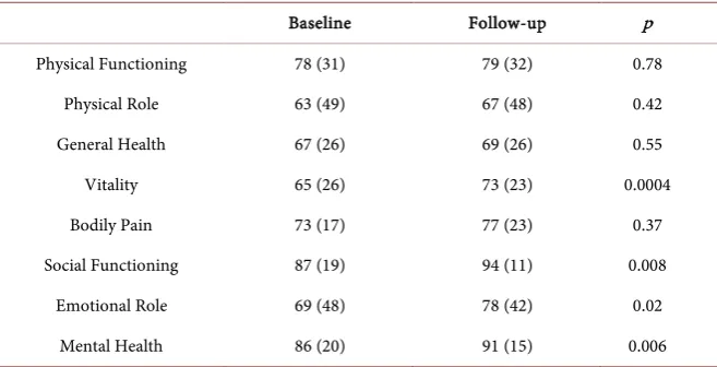
3.6. SF36-Questionnaire on Quality of Life
Patients experienced improved quality of life with regard to vitality, social func-tioning, emotional role, and mental health, whereas no significant changes in physical functioning, physical role, general health, or bodily pain were seen
(Table 4). Baseline eGFR was significantly associated to follow-up SF-36 score (r
[image:11.595.209.538.512.680.2]= 0.50, p = 0.042), but not baseline SF-36 score (r = 0.48, p = 0.052). No correla-tion of SF-36 baseline or follow-up and AHI or Epworth at baseline or fol-low-up.
Table 4. SF36 scores at baseline and follow-up.
Baseline Follow-up p
Physical Functioning 78 (31) 79 (32) 0.78
Physical Role 63 (49) 67 (48) 0.42
General Health 67 (26) 69 (26) 0.55
Vitality 65 (26) 73 (23) 0.0004
Bodily Pain 73 (17) 77 (23) 0.37
Social Functioning 87 (19) 94 (11) 0.008
Emotional Role 69 (48) 78 (42) 0.02
Mental Health 86 (20) 91 (15) 0.006
DOI: 10.4236/ojneph.2019.91001 12 Open Journal of Nephrology
3.7. Plasma Levels of Syndecan-1
There was no change in p-syncecan-1 from baseline to follow-up (18 [15; 25] vs. 19 [16; 23] ng/mL, p = 0.41).
3.8. Plasma Levels of Vasoactive Hormones
No changes were seen from baseline to follow-up in plasma levels of the vasoac-tive hormones; P-aldosterone (159 [131; 259] vs. 183 [115; 268] pmol/L, p = 0.87), P-angiotensin II (13 [7; 55] vs. 11 [7; 30] pg/mL, p = 0.11), plasma renin concentration (26 [8; 67] vs. 16 [10; 48] pg/mL, p = 0.83), or AVP (0.4 [0.4; 0.7] vs. 0.5 [0.3; 0.8] pmol/L, p = 0.28).
3.9. Urinary Excretion of AQP2 and ENaC
ɣ
From baseline to follow-up, no significant changes were seen in urinary excre-tion of AQP2 (1.4 [1.1; 1.8] vs. 1.3 [1.1; 2.6] ng/min, p = 0.88) or ENaCγ (1.2 [1.0; 1.6] vs. 1.2 [0.9; 1.8] ng/min, p = 0.54).
4. Discussion
The main findings of this study were a statistically significant and clinically rele-vant decrease in 24 h and daytime BP after short term CPAP treatment in pa-tients with impaired renal function (mean eGFR 66 mL/min/1.73 m2) and a re-duction in albuminuria. The decrease in nocturnal BP was not statistically sig-nificant, and there was no difference in nocturnal BP decrease or frequency of non-dipping. Renal function remained unchanged.
Previous studies reported different results of the BP response to CPAP. Becker et al. found a significant (>10 mmHg decrease) in systolic and diastolic using 24 hours BP measuring, both during day- and nighttime [31], Hermida et al. found a small (<3 mmHg) and statistically non-significant decrease in 24 h BP [32], whereas Durán-Cantolla et al. [33] measured a similar and significant fall in 24 h, day and nighttime BP. In the latter study, the number of non-dippers was re-duced after treatment [33]. All studies comprised mainly hypertensive patients diagnosed with OSA, whereas no information was given about the patients’ renal function and eventually diabetes. Hence, differences in baseline characteristics between studies may explain some of the divergent findings of BP response. Moreover, patients in the studies had different baseline levels of BP, AHI, and ESS, which may also influence the response to CPAP.
In the present study, five patients had changed their antihypertensive treat-ment. However, the difference in mean DDD at baseline and follow-up was less than 3%. We do not believe that such a small change in DDD can solely explain the BP response at follow-up.
DOI: 10.4236/ojneph.2019.91001 13 Open Journal of Nephrology compared with previous studies. However, in the present population, we dem-onstrated that it is possible to lower BP in patients with moderate-to-severe OSA using CPAP treatment. A previous meta-analysis of CKD patients demonstrated that a 5 mm Hg reduction in office BP reduced the risk of major cardiovascular events with 17%. Thus, the reduction we demonstrated in this population is of clinical relevance. One prevalence study found that as many as 40% of CKD3-4 patients suffered from moderate-to-severe OSA [1], whereas we in a preceding study only found moderate-to-severe OSA present in 22% of CKD3-4 patients
[21]. However, based on our studies, we find it reasonable to suggest that pa-tients with CKD should be examined for presence of OSA and treated, if moder-ate-to-severe OSA is present.
We also investigated the effects of CPAP treatment on central blood pressure derived from applanation tonometry. To our knowledge, no data have been re-ported on 24 h ambulatory central BP changes as response to CPAP treatment. Prior to our study, Litvin et al. [34] and Hoyos et al. [35] reported a decrease in central BP measured as spot measurements in an in-office set-up after three and eight weeks CPAP treatment, respectively. Both studies found a similar decrease in mmHg in central and peripheral BP after treatment. In agreement with the previous studies, we found a tendency to a decrease in central BP, however, in our study not statistically significant.
As expected, we found that AHI and ODI decreased, and mean oxygen satura-tion increased at follow-up in good agreement with a Cochrane review [7]. These findings also documented that patients adhered to the treatment.
DOI: 10.4236/ojneph.2019.91001 14 Open Journal of Nephrology In the present study, we did not demonstrate any significant change in porting of sleep symptoms (ESS). Self-reported quality of life (SF-36) was re-ported higher on mental parameters at follow-up. Other studies have demon-strated less pronounced sleep symptoms after CPAP treatment [7] [37] [38]. These studies comprised subjects with both higher and identical levels of sleep symptoms as in our study. However, none of the participants in these studies suffered from CKD. Hence, in our population, sleep symptoms may be more re-lated to renal disease than OSA, blunting the effect of CPAP treatment. The changes in ESS were correlated to AHI. This implies that the effect on sleep symptoms is more well-defined in more severe degrees of OSA.
Although patients in the present study suffered from renal failure and had hypertension and diabetes as frequent comorbidities, we demonstrated im-proved quality of life with respect to vitality (energy and fatigue) and mental pa-rameters with no changes in physical papa-rameters. Two previous studies have re-ported similar or more pronounced improvements of quality of life after 4 - 6 weeks CPAP treatment [8] [39]. In one of these studies, participant suffered from more severe OSA, and in both, patients reported more severe sleep symp-toms. Interestingly, we observed an improvement of quality of life in a heavier diseased population with less severe OSA and sleep symptoms. However, eGFR was correlated to overall quality of life at follow-up, which suggest that presence of renal disease is an important factor for quality of life in our population. This may be one explanation for the lack of effect on the patient’s physical health pa-rameters in this study.
We did not demonstrate any changes in p-syndecan-1. In vascular diseases, syndecans are shedded from the glycocalyx protection layer in the cardiovascular system [18]. In a previous study, we demonstrated higher plasma levels of syncecan-1 in a hypertensive population with sparse disease burden compared to healthy controls [19]. Other studies have reported higher levels of syndecans as-sociated to declining renal function in a CKD population [40] and to overt heart failure in hypertensive patients [41]. We did not demonstrate any changes in syncecan-1 levels after CPAP treatment. We included only a small population with other cardiovascular risk factors. The over-all cardiovascular risk profile may be a stronger negative influence than an eventual positive influence of CPAP treatment.
hor-DOI: 10.4236/ojneph.2019.91001 15 Open Journal of Nephrology mones and renal handling of sodium and water as an explanation for BP re-sponse. The lack of changes in plasma levels of hormones in the present study may be related treatment with antihypertensive agent blocking the RAAS sys-tem.
Strength of this study is that we at follow-up, in addition to adherence regis-trations, completed sleep examination in all patients to confirm the use of CPAP-treatment on improved sleep quality (AHI, ODI, and oxygen saturation).
The response on central BP was evaluated from 24 h measurements, and al-buminuria was evaluated from 24 h urine collection instead of spot urine meas-urement. We included a relatively new plasma marker of cardiovascular stress, syndecan-1.
It is a weakness that time from baseline to follow-up time varied from 72 to 139 day. However, we aimed to evaluate the effect short term treatment and de-fined that as approximately three to four months of intervention, and we did not expect amplification of findings within the present used timeframe. Another weakness of the study is that a few subjects experienced changes in antihyper-tensive treatment. However, it was not ethically justified not to treat these pa-tients with usual care, and the change in mean DDD was only minor. We did not include a sham or placebo intervention to eliminate the effect of biases or to ad-dress the Hawthorne effect.
5. Conclusion
CPAP seems to be an effective blood pressure lowering treatment in patients with mean eGFR 66 mL/min/1.73 m2 and sleep apnoea with beneficial effects on albuminuria. Larger studies are needed to verify this finding and explore poten-tial effects on the progression of renal failure.
Acknowledgements
The authors greatly acknowledge the skillful assistance of our laboratory techni-cians: Anne Mette Ravn, Kirsten Nygaard, and Henriette Vorup Simonsen.
The authors also greatly acknowledge the skilful assistance from Nurses Marianne Kirkegaard and Anja Mailund Mikkelsen in the Sleep Apnoea Clinic, Department of Medicine, Holstebro Hospital.
The authors thank the Department of Clinical Biochemistry, Holstebro Hos-pital for help in routine analyses.
Conflicts of Interest
The authors have no conflicts of interest.
Funding
DOI: 10.4236/ojneph.2019.91001 16 Open Journal of Nephrology
References
[1] Nicholl, D.D., Ahmed, S.B., Loewen, A.H., Hemmelgarn, B.R., Sola, D.Y., Beecroft, J.M., Turin, T.C. and Hanly, P.J. (2012) Declining Kidney Function Increases the Prevalence of Sleep Apnea and Nocturnal Hypoxia. Chest, 141, 1422-1430.
https://doi.org/10.1378/chest.11-1809
[2] Worsnop, C.J., Naughton, M.T., Barter, C.E., Morgan, T.O., Anderson, A.I. and Pierce, R.J. (1998) The Prevalence of Obstructive Sleep Apnea in Hypertensives.
American Journal of Respiratory and Critical Care Medicine, 157, 111-115.
https://doi.org/10.1164/ajrccm.157.1.9609063
[3] Yaggi, H.K., Concato, J., Kernan, W.N., Lichtman, J.H., Brass, L.M. and Mohsenin, V. (2005) Obstructive Sleep Apnea as a Risk Factor for Stroke and Death. The New England Journal of Medicine, 353, 2034-2041.
https://doi.org/10.1056/NEJMoa043104
[4] Drager, L.F., Bortolotto, L.A., Lorenzi, M.C., Figueiredo, A.C., Krieger, E.M. and Lorenzi-Filho, G. (2005) Early Signs of Atherosclerosis in Obstructive Sleep Apnea.
American Journal of Respiratory and Critical Care Medicine,172, 613-618.
https://doi.org/10.1164/rccm.200503-340OC
[5] Sakaguchi, Y., Hatta, T., Hayashi, T., Shoji, T., Suzuki, A., Tomida, K., Okada, N., Rakugi, H., Isaka, Y. and Tsubakihara, Y. (2013) Association of Nocturnal Hypox-emia with Progression of CKD. Clinical Journal of the American Society of Neph-rology, 8, 1502-1507.https://doi.org/10.2215/CJN.11931112
[6] Marrone, O., Battaglia, S., Steiropoulos, P., Basoglu, O.K., Kvamme, J.A., Ryan, S., Pepin, J.L., Verbraecken, J., Grote, L., Hedner, J., Bonsignore, M.R. and ESADA Study Group (2016) Chronic Kidney Disease in European Patients with Obstructive Sleep Apnea: The ESADA Cohort Study. Journal of Sleep Research, 25, 739-745.
https://doi.org/10.1111/jsr.12426
[7] Giles, T.L., Lasserson, T.J., Smith, B.H., White, J., Wright, J. and Cates, C.J. (2006) Continuous Positive Airways Pressure for Obstructive Sleep Apnoea in Adults.
Cochrane Database of Systematic Reviews,25, CD001106.
[8] Montserrat, J.M., Ferrer, M., Hernandez, L., Farre, R., Vilagut, G., Navajas, D., Ba-dia, J.R., Carrasco, E., De Pablo, J. and Ballester, E. (2001) Effectiveness of CPAP Treatment in Daytime Function in Sleep Apnea Syndrome: A Randomized Con-trolled Study with an Optimized Placebo. American Journal of Respiratory and Critical Care Medicine, 164, 608-613.https://doi.org/10.1164/ajrccm.164.4.2006034 [9] Sullivan, C.E., Issa, F.G., Berthon-Jones, M. and Eves, L. (1981) Reversal of
Ob-structive Sleep Apnoea by Continuous Positive Airway Pressure Applied through the Nares. The Lancet, 317, 862-865.
https://doi.org/10.1016/S0140-6736(81)92140-1
[10] Puckrin, R., Iqbal, S., Zidulka, A., Vasilevsky, M. and Barre, P. (2015) Renoprotec-tive Effects of Continuous PosiRenoprotec-tive Airway Pressure in Chronic Kidney Disease Pa-tients with Sleep Apnea.International Urology and Nephrology, 47, 1839-1845.
https://doi.org/10.1007/s11255-015-1113-y
[11] Schipper, M.H., Jellema, K., Thomassen, B.J.W., Alvarez-Estevez, D., Verbraecken, J. and Rijsman, R.M. (2017) Stroke and Other Cardiovascular Events in Patients with Obstructive Sleep Apnea and the Effect of Continuous Positive Airway Pres-sure.Journal of Neurology, 264, 1247-1253.
https://doi.org/10.1007/s11255-015-1113-y
DOI: 10.4236/ojneph.2019.91001 17 Open Journal of Nephrology
Airway Pressure on BP in Patients with Hypertension and Sleep Apnea. Chest, 132, 1847-1852.https://doi.org/10.1378/chest.07-1478
[13] Campos-Rodriguez, F., Grilo-Reina, A., Perez-Ronchel, J., Merino-Sanchez, M., Gonzalez-Benitez, M.A., Beltran-Robles, M. and Almeida-Gonzalez, C. (2006) Effect of Continuous Positive Airway Pressure on Ambulatory BP in Patients with Sleep Apnea and Hypertension: A Placebo-Controlled Trial. Chest, 129, 1459-1467.
https://doi.org/10.1378/chest.129.6.1459
[14] Bazzano, L.A., Khan, Z., Reynolds, K. and He, J. (2007) Effect of Nocturnal Nasal Continuous Positive Airway Pressure on Blood Pressure in Obstructive Sleep Ap-nea. Hypertension, 50, 417-423.
https://doi.org/10.1161/HYPERTENSIONAHA.106.085175
[15] Williams, B., Lacy, P.S., Yan, P., Hwee, C.N., Liang, C. and Ting, C.M. (2011) De-velopment and Validation of a Novel Method to Derive Central Aortic Systolic Pressure from the Radial Pressure Waveform Using an N-Point Moving Average Method.Journal of the American College of Cardiology, 57, 951-961.
https://doi.org/10.1016/j.jacc.2010.09.054
[16] Roman, M.J., Devereux, R.B., Kizer, J.R., Lee, E.T., Galloway, J.M., Ali, T., Umans, J.G. and Howard, B.V. (2007) Central Pressure More Strongly Relates to Vascular Disease and Outcome than Does Brachial Pressure: The Strong Heart Study.
Hypertension, 50, 197-203.
https://doi.org/10.1161/HYPERTENSIONAHA.107.089078
[17] Korcarz, C.E., Benca, R., Barnet, J.H. and Stein, J.H. (2016) Treatment of Obstruc-tive Sleep Apnea in Young and Middle-Aged Adults: Effects of PosiObstruc-tive Airway Pressure and Compliance on Arterial Stiffness, Endothelial Function, and Cardiac Hemodynamics. Journal of the American Heart Association, 5, e002930.
https://doi.org/10.1161/JAHA.115.002930
[18] Tarbell, J.M. and Cancel, L.M. (2016) The Glycocalyx and Its Significance in Hu-man Medicine. Journal of Internal Medicine, 280, 97-113.
https://doi.org/10.1111/joim.12465
[19] Hornstrup, B.G. (2018) Nocturnal Blood Pressure Decrease in Hypertensive Pa-tients and Normotensives-Association with Obstructive Sleep Apnoea and Renal Function. The Open Hypertension Journal, 10, 28-40.
https://doi.org/10.2174/1876526201810010028
[20] Hoffmann-Petersen, N., Lauritzen, T., Bech, J.N. and Pedersen, E.B. (2017) Short-Term Telemedical Home Blood Pressure Monitoring Does Not Improve Blood Pressure in Uncomplicated Hypertensive Patients. Journal of Human Hyper-tension, 31, 93-98.https://doi.org/10.1038/jhh.2016.43
[21] Hornstrup, B.G., Gjoerup, P.H., Wessels, J., Lauridsen, T.G. and Pedersen, E.B. (2018) Nocturnal Blood Pressure Decrease in Patients with Chronic Kidney Disease and in Healthy Controls—Significance of Obstructive Sleep Apnea and Renal Func-tion. International Journal of Nephrology and Renovascular Disease, 11, 279-290. https://doi.org/10.2147/IJNRD.S176606
[22] ESH/ESC Task Force for the Management of Arterial Hypertension (2013) Practice Guidelines for the Management of Arterial Hypertension of the European Society of Hypertension (ESH) and the European Society of Cardiology (ESC): ESH/ESC Task Force for the Management of Arterial Hypertension. Journal of Hypertension, 31, 1925-1938.https://doi.org/10.1097/HJH.0b013e328364ca4c
DOI: 10.4236/ojneph.2019.91001 18 Open Journal of Nephrology
Ward, S.L. and Tangredi, M.M. (2012) American Academy of Sleep Medicine: Rules for Scoring Respiratory Events in Sleep: Update of the 2007 AASM Manual for the Scoring of Sleep and Associated Events. Deliberations of the Sleep Apnea Defini-tions Task Force of the American Academy of Sleep Medicine. Journal of Clinical Sleep Medicine, 8, 597-619.
[24] Pedersen, E.B., Eiskjaer, H., Madsen, B., Danielsen, H., Egeblad, M. and Nielsen, C.B. (1993) Effect of Captopril on Renal Extraction of Renin, Angiotensin II, Atrial Natriuretic Peptide and Vasopressin, and Renal Vein Renin Ratio in Patients with Arterial Hypertension and Unilateral Renal Artery Disease. Nephrology Dialysis Transplantation, 8, 1064-1070.
[25] Pedersen, E.B., Danielsen, H. and Spencer, E.S. (1984) Effect of Indapamide on Renal Plasma Flow, Glomerular Filtration Rate and Arginine Vasopressin in Plasma in Essential Hypertension. European Journal of Clinical Pharmacology, 26, 543-547. https://doi.org/10.1007/BF00543482
[26] Pedersen, R.S., Bentzen, H., Bech, J.N. and Pedersen, E.B. (2001) Effect of Water Deprivation and Hypertonic Saline Infusion on Urinary AQP2 Excretion in Healthy Humans. American Journal of Physiology-Renal Physiology, 280, F860-F867. https://doi.org/10.1152/ajprenal.2001.280.5.F860
[27] Graffe, C.C., Bech, J.N. and Pedersen, E.B. (2012) Effect of High and Low Sodium Intake on Urinary Aquaporin-2 Excretion in Healthy Humans. American Journal of Physiology-Renal Physiology, 302, F264-F275.
https://doi.org/10.1152/ajprenal.00442.2010
[28] Matthesen, S.K., Larsen, T., Vase, H., Lauridsen, T.G., Jensen, J.M. and Pedersen, E.B. (2013) Effect of Amiloride and Spironolactone on Renal Tubular Function and Central Blood Pressure in Patients with Arterial Hypertension during Baseline Conditions and after Furosemide: A Double-Blinded, Randomized, Place-bo-Controlled Crossover Trial. Clinical and Experimental Hypertension, 35, 313-324.https://doi.org/10.3109/10641963.2012.721843
[29] Al Therwani, S., Malmberg, M.E.S., Rosenbaek, J.B., Bech, J.N. and Pedersen, E.B. (2017) Effect of Tolvaptan on Renal Handling of Water and Sodium, GFR and Cen-tral Hemodynamics in Autosomal Dominant Polycystic Kidney Disease during In-hibition of the Nitric Oxide System: A Randomized, Placebo-Controlled, Double Blind, Crossover Study. BMC Nephrology, 18, 268.
[30] Hays, R.D., Sherbourne, C.D. and Mazel, R.M. (1993) The RAND 36-Item Health Survey 1.0. Health Economics, 2, 217-227.https://doi.org/10.1002/hec.4730020305 [31] Becker, H.F., Jerrentrup, A., Ploch, T., Grote, L., Penzel, T., Sullivan, C.E. and Peter,
J.H. (2003) Effect of Nasal Continuous Positive Airway Pressure Treatment on Blood Pressure in Patients with Obstructive Sleep Apnea. Circulation, 107, 68-73. https://doi.org/10.1161/01.CIR.0000042706.47107.7A
[32] Hermida, R.C., Zamarron, C., Ayala, D.E. and Calvo, C. (2004) Effect of Conti-nuous Positive Airway Pressure on Ambulatory Blood Pressure in Patients with Obstructive Sleep Apnoea. Blood Pressure Monitoring, 9, 193-202.
https://doi.org/10.1097/00126097-200408000-00004
DOI: 10.4236/ojneph.2019.91001 19 Open Journal of Nephrology
[34] Litvin, A.Y., Sukmarova, Z.N., Elfimova, E.M., Aksenova, A.V., Galitsin, P.V., Ro-goza, A.N. and Chazova, I.E. (2013) Effects of CPAP on “Vascular” Risk Factors in Patients with Obstructive Sleep Apnea and Arterial Hypertension. Vascular Health and Risk Management, 9, 229-235.https://doi.org/10.2147/VHRM.S40231
[35] Hoyos, C.M., Yee, B.J., Wong, K.K., Grunstein, R.R. and Phillips, C.L. (2015) Treatment of Sleep Apnea with CPAP Lowers Central and Peripheral Blood Pres-sure Independent of the Time-of-Day: A Randomized Controlled Study. American Journal of Hypertension, 28, 1222-1228.https://doi.org/10.1093/ajh/hpv023 [36] Koga, S., Ikeda, S., Yasunaga, T., Nakata, T. and Maemura, K. (2013) Effects of
Nas-al Continuous Positive Airway Pressure on the Glomerular Filtration Rate in Pa-tients with Obstructive Sleep Apnea Syndrome. Internal Medicine, 52, 345-349. https://doi.org/10.2169/internalmedicine.52.8468
[37] Barbe, F., Duran-Cantolla, J., Capote, F., de la Pena, M., Chiner, E., Masa, J.F., Gonzalez, M., Marin, J.M., Garcia-Rio, F., de Atauri, J.D., Teran, J., Mayos, M., Monasterio, C., del Campo, F., Gomez, S., de la Torre, M.S., Martinez, M., Mont-serrat, J.M., Spanish Sleep and Breathing Group (2010) Long-Term Effect of Con-tinuous Positive Airway Pressure in Hypertensive Patients with Sleep Apnea.
American Journal of Respiratory and Critical Care Medicine, 181, 718-726. https://doi.org/10.1164/rccm.200901-0050OC
[38] Zhang, J., Wang, C., Gong, W., Ye, Z., Tang, Y., Zhao, W., Peng, H. and Lou, T. (2016) Poor Sleep Quality Is Responsible for the Non-Dipper Pattern in Hyperten-sive But Not in NormotenHyperten-sive Chronic Kidney Disease Patients. Nephrology ( Carl-ton), 22, 690-698.
[39] Jenkinson, C., Davies, R.J., Mullins, R. and Stradling, J.R. (1999) Comparison of Therapeutic and Subtherapeutic Nasal Continuous Positive Airway Pressure for Obstructive Sleep Apnoea: A Randomised Prospective Parallel Trial. The Lancet, 353, 2100-2105.https://doi.org/10.1016/S0140-6736(98)10532-9
[40] Padberg, J.S., Wiesinger, A., di Marco, G.S., Reuter, S., Grabner, A., Kentrup, D., Lukasz, A., Oberleithner, H., Pavenstadt, H., Brand, M. and Kumpers, P. (2014) Damage of the Endothelial Glycocalyx in Chronic Kidney Disease. Atherosclerosis, 234, 335-343.https://doi.org/10.1016/j.atherosclerosis.2014.03.016
[41] Bielecka-Dabrowa, A., Michalska-Kasiczak, M., Gluba, A., Ahmed, A., Gerdts, E., von Haehling, S., Rysz, J. and Banach, M. (2015) Biomarkers and Echocardiograph-ic PredEchocardiograph-ictors of Myocardial Dysfunction in Patients with Hypertension. Scientific Reports, 5,Article No. 8916.https://doi.org/10.1038/srep08916
[42] Gaddam, K., Pimenta, E., Thomas, S.J., Cofield, S.S., Oparil, S., Harding, S.M. and Calhoun, D.A. (2010) Spironolactone Reduces Severity of Obstructive Sleep Apnoea in Patients with Resistant Hypertension: A Preliminary Report. Journal of Human Hypertension,24, 532-537.https://doi.org/10.1038/jhh.2009.96
[43] Follenius, M., Krieger, J., Krauth, M.O., Sforza, F. and Brandenberger, G. (1991) Obstructive Sleep Apnea Treatment: Peripheral and Central Effects on Plasma Re-nin Activity and Aldosterone. Sleep, 14, 211-217.