organic papers
o860
Eriksson and Widmalm C20H33NO14 doi:10.1107/S1600536805006227 Acta Cryst.(2005). E61, o860–o862 Acta Crystallographica Section E
Structure Reports Online
ISSN 1600-5368
Amygdalin trihydrate
Lars Erikssona* and Go¨ran
Widmalmb
a
Division of Structural Chemistry, Arrhenius Laboratory, Stockholm University, S-106 91 Stockholm, Sweden, andbDepartment of
Organic Chemistry, Arrhenius Laboratory, Stockholm University, S-106 91 Stockholm, Sweden
Correspondence e-mail: [email protected]
Key indicators
Single-crystal synchrotron study
T= 110 K
Mean(C–C) = 0.002 A˚
Rfactor = 0.037
wRfactor = 0.097
Data-to-parameter ratio = 15.9
For details of how these key indicators were automatically derived from the article, see http://journals.iucr.org/e.
#2005 International Union of Crystallography Printed in Great Britain – all rights reserved
Extensive hydrogen bonding is present in the crystal structure
of the title compound [systematic name: (R
)-1-cyano-1-(phenylmethyl)--d-glucopyranosyl-(1!6)--d
-glucopyran-oside], C20H27NO113H2O, involving all of the hydroxy groups.
The water molecules are also involved in the hydrogen bonding; in particular, one of them acts as a bridge between the endocyclic O atoms of the two sugar residues. The overall
conformation of the disaccharide is described by the exo
-anomeric conformations, ’00
H = 26 and ’0H = 38, together
with an antiperiplanar extended conformation of the
consti-tuent sugar residues, with 00= 155.
Comment
Amygdalin is a cyanogenic glycoside found in the kernels of, for example, apples, almonds and peaches (Jones, 1998). More than 2500 plant species have been identified as containing
cyanogenic compounds (Moller et al., 1999). The title
compound is a diglycosyl glycoside from which cytotoxic hydrogen cyanide can be released, leading to acute cyanide
poisoning. This (R)-1-cyano-1-(phenylmethyl)--d
-glucopyranosyl-(1!6)--d-glucopyranoside, (I), has been
studied in solution by NMR spectroscopy (Ribeiro, 1990) and recently also in the solid state as a function of temperature
(Widmalm et al., 2003). It was found that the phenyl ring
exhibited thermally activated dynamics about the para axis,
whereas the glucosyl residues remained static. We present here the crystal structure of amygdalin trihydrate, and analyze a number of important degrees of freedom with respect to its torsion angles and the possibility of forming a complex hydrogen-bonding network due to the presence of three ordered water molecules per amygdalin molecule.
The conformation of an oligosaccharide is described by the
torsion angles’and at the glycosidic linkage between the
constituent monosaccharides. The ’ and angles for the
present compound are defined by’0 0(O500—C100—O60—C60),
’0 (O50—C10—O7—C7), 00 (C100—O60—C60—C50) and 0
(C10—O7—C7—C1). Selected torsion angles are shown in
Table 1.
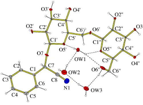
The additional major degree of freedom in a hexopyranosyl residue is the conformational preference of the hydroxy-methyl group. In amygdalin this is described by the torsion angles !00 (O500—C500—C600—O60 0) and !0(O50—C50—C60—
O60), the latter being part of the (1!6) linkage. The two!
torsion angles both correspond to theggconformation (Fig. 1),
which is one of the two found to be significantly populated in
solution. The two’Htorsion angles are in the vicinity of the
exo-anomeric conformation (’H ’50), as indicated by the
torsion angles’00 H(H1
00—C100—O60—C60) and’0 H(H1
0—C10—
O7—C7) (Table 1). For the-(1!6) linkage the value of 00
leads to an extended antiperiplanar conformation, also found in solutions of similar compounds, on the basis of NMR
spectroscopic and computer simulation studies (Lycknert et
al., 2004).
The torsion angle at the glycosidic linkage to the aglycone,
0, results in spatial proximity of atoms H10 and H7 and, in
oligosaccharide terminology, a syn conformation. For the
aglyconic part, the torsion angle (C2—C1—C7—O7)
describes the orientation of the phenyl group. The latter is
planar within less than 0.01 (1) A˚ , with atom C7 deviating by
0.023 (2) A˚ from the least-squares plane defined by the phenyl
ring. The plane defined by atoms C1, C7 and C8 is inclined by
14.0 (2) to the phenyl ring plane. Atom N1 is displaced by
0.060 (4) A˚ from the C1/C7/C8 plane.
The Cremer & Pople (1975) parameters for the two sugar
rings areQ= 0.5915 (11) A˚ ,= 3.85 (11)and’= 221.4 (15)
for the O500 ! C500 direction, and Q = 0.5607 (10) A˚ , =
12.68 (10) and’ = 345.5 (6) for the O50 ! C50 direction.
Both of the sugar residues have the anticipated chair confor-mation,i.e.4C1.
All hydroxy groups act as donors in intermolecular
hydrogen bonds. The O300hydroxy group is also involved in an
intramolecular hydrogen bond to O400. All hydroxy groups,
except O40, also act, at least once, as hydrogen-bond acceptors.
Furthermore, atoms O400 and O20 are dual hydrogen-bond
acceptors. The three water molecules (OW1, OW2 and OW3)
act as dual hydrogen-bond donors. In fact, OW1 provides
donor H atoms for three hydrogen bonds,viz.one simple and
one bifurcated. Two of the water molecules are single
accep-tors and one (OW1) is a dual hydrogen-bond acceptor.
Interestingly, OW1 mediates the intramolecular hydrogen
bonding between O50and O500. A view of the hydrogen-bond
pattern around the three water molecules is shown in Fig. 1. In addition, atom N1 is an acceptor in the hydrogen-bond
pattern. The sugar residues pack in the ab plane and the
phenyl rings are involved in–interactionsviaa T-shaped
edge-to-face arrangement (Meyer et al., 2003). The angle
between the least-squares planes of the phenyl rings at (x,y,z)
and (2x,1
2+y, 2z) is 77.02 (4)
. The distance between the
centers of gravity of these rings is 4.930 (1) A˚ , comparable to
that in the crystal structure of edge-to-face oriented benzene
(5.025 A˚ ). A diagram of the packing, viewed along theaaxis,
is shown in Fig. 2. C—H interactions are present (Table 2),
similar to the aromatic interaction described above. Although
weak, the C—H interactions play important
structure-stabilizing roles, in particular for carbohydrate–protein complexes.
Experimental
Amygdalin [or (R)-1-cyano-1-(phenylmethyl)--d
-glucopyranosyl-(1!6)--d-glucopyranoside] was obtained from C. A. F. Kahlbaum
GmbH, Berlin, Germany. The compound was crystallized by slow evaporation of a mixture of water, ethanol and acetonitrile (1:1:1) at ambient temperature, yielding needles which were mounted with epoxy glue on to glass fibres. The scattering power of the crystals was weak; thus it was decided to collect data with synchrotron radiation at beamline I711 at the Swedish synchrotron radiation facility MAXLAB, Lund, Sweden.
Crystal data
C20H33NO14
Mr= 511.47
Monoclinic,P21
a= 9.4794 (11) A˚
b= 7.9025 (9) A˚
c= 16.2016 (19) A˚
= 94.089 (3)
V= 1210.6 (2) A˚3
Z= 2
Dx= 1.403 Mg m
3
Synchrotron radiation
= 0.891 A˚
Cell parameters from 999 reflections
= 3.0–29.0
= 0.12 mm1
T= 110 (2) K Plate, colorless 0.200.100.02 mm
Data collection
Bruker SMART 1K area-detector diffractometer
!scans at different’and 2
Absorption correction: multi-scan (SADABS; Sheldrick, 2002)
Tmin= 0.98,Tmax= 1.00 20604 measured reflections
5454 independent reflections 5010 reflections withI> 2(I)
Rint= 0.031
max= 45.9
h=15!14
k=10!12
l=16!25
organic papers
Acta Cryst.(2005). E61, o860–o862 Eriksson and Widmalm C
[image:2.610.44.294.70.255.2]20H33NO14
o861
Figure 1Refinement
Refinement onF2 R[F2> 2(F2)] = 0.037
wR(F2) = 0.097
S= 1.04 5454 reflections 343 parameters
H atoms treated by a mixture of independent and constrained refinement
w= 1/[2
(Fo2) + (0.0714P)2] whereP= (Fo2+ 2Fc2)/3 (/)max< 0.001
max= 0.50 e A˚
3
min=0.55 e A˚
3
Extinction correction:SHELXL97
Extinction coefficient: 0.051 (3)
Table 1
Selected torsion angles ().
C2—C1—C7—O7 69.87 (16) C1—C7—O7—C10
145.08 (11) O7—C7—C8—N1 172 (3) C7—O7—C10—O50 83.22 (12)
O50—C50—C60—O60 74.07 (12)
C50
—C60
—O60
—C10 0
154.93 (10)
C60—O60—C10 0—O50 0 94.69 (11)
O50 0
—C50 0
—C60 0
—O60 0
61.27 (13) H10 0
—C10 0
—O60
—C60
25.7 H10—C10—O7—C7 37.8
[image:3.610.44.296.354.520.2]C10—O7—C7—H7 28.1
Table 2
Hydrogen-bond geometry (A˚ ,).
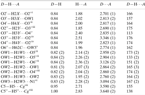
Cgis the centroid of the phenyl ring.
D—H A D—H H A D A D—H A
O20—H2X O20 0i
0.84 1.88 2.701 (1) 166 O30
—H3X OW1 0.84 2.02 2.813 (2) 157 O40
—H4X O30ii
0.84 2.00 2.817 (1) 164 O20 0—H2Y O30 0iii
0.84 1.85 2.690 (1) 175 O30 0
—H3Y O40 0
0.84 2.40 2.835 (1) 113 O30 0
—H3Y O20iv
0.84 2.51 3.346 (1) 176 O40 0—H4Y O20iv
0.84 1.99 2.823 (1) 174 O60 0—H62C OW3v
0.84 1.96 2.774 (1) 162 OW1—H1W1 O50ii
0.82 (2) 2.14 (2) 2.958 (2) 173 (2) OW1—H2W1 O50 0ii
0.84 (2) 2.26 (2) 2.904 (1) 133 (2) OW1—H2W1 O60 0ii
0.84 (2) 2.36 (2) 3.126 (2) 151 (2) OW2—H1W2 OW1 0.81 (3) 2.07 (3) 2.864 (2) 165 (3) OW2—H2W2 O40 0iii
0.82 (2) 2.04 (2) 2.860 (2) 174 (2) OW3—H1W3 OW2 0.83 (2) 1.95 (2) 2.760 (2) 164 (2) OW3—H2W3 N1vi
0.83 (2) 2.28 (2) 3.094 (2) 167 (2) C5—H5 Cgvii
0.95 2.71 3.590 (2) 155 C50 0
—H50 0 Cgviii
1.00 2.83 3.640 (2) 138
Symmetry codes: (i)xþ1;y;z; (ii)xþ1;yþ1
2;zþ1; (iii)x;yþ 1
2;zþ1; (iv)
x1;y1;z; (v) xþ1;y1
2;zþ1; (vi) x;y;z1; (vii)x3;y 1 2;zþ2;
(viii)x1;y;z.
H atoms were positioned geometrically and allowed to ride on their parent atoms, with CH, CH2and aromatic C—H bonds set equal
to 1.00, 0.99 and 0.95 A˚ , respectively, andUiso(H) = 1.2Ueq(C) and
1.5Ueq(O). A ‘rigid-bond’ restraint was applied toU
ij
values (Rollett, 1970). The Flack (1983) parameter was inconclusive, but the absolute configuration was set by thea prioriknowledge that the sample is a known natural product. In the absence of significant anomalous scattering effects, the 5001 Friedel pairs were merged.
Data collection: SMART (Siemens, 1998); cell refinement:
SMART; data reduction:SAINT(Siemens, 1998); program(s) used to solve structure: SHELXS97(Sheldrick, 1997); program(s) used to refine structure:SHELXL97(Sheldrick, 1997); molecular graphics:
DIAMOND(Bergerhoff, 1996); software used to prepare material for publication:PLATON(Spek, 2003).
This work was supported by a grant from the Swedish Research Council (VR).
References
Bergerhoff, G. (1996).DIAMOND. Gerhard-Domagk-Straße 1, 53121 Bonn, Germany.
Cremer, D. & Pople, J. A. (1975).J. Am. Chem. Soc.97, 1354–1358. Flack, H. D. (1983).Acta Cryst.A39, 876–881.
Jones, D. A. (1998).Phytochemistry,47, 155–162.
Lycknert, K., Edblad, M., Imberty, A. & Widmalm, G. (2004).Biochemistry,
43, 9647–9654.
Meyer, E. A., Catellano, R. K. & Diederich, F. (2003).Angew. Chem. Int. Ed. 42, 1210–1250.
Moller, B. L. & Siegler, D. S. (1999).Plant Amino Acids: Biochemistry and Biotechnology, edited by B. K. Singh, pp. 563–609. New York: Marcel Dekker Inc.
Ribeiro, A. A. (1990).Magn. Reson. Chem.28, 765–773.
Rollett, J. S. (1970).Crystallographic Computing, edited by F. R. Ahmed, S. R. Hall & C. P. Huber, pp. 167–181. Copenhagen: Munksgaard.
Sheldrick, G. M. (1997). SHELXS97 and SHELXL97. University of Go¨ttingen, Germany.
Sheldrick, G. M. (2002).SADABS. Version 2.03. University of Go¨ttingen, Germany.
Siemens (1998).SMARTandSAINT. Siemens Analytical X-ray Instruments Inc., Madison, Wisconsin, USA.
Spek, A. L. (2003).J. Appl. Cryst.36, 7–13.
Widmalm, G., Jansson, K., Pellijeff, G. & Sanstro¨m, D. (2003).J. Phys. Chem. B,107, 11794–11798.
organic papers
o862
Eriksson and Widmalm C20H33NO14 Acta Cryst.(2005). E61, o860–o862
Figure 2
supporting information
sup-1 Acta Cryst. (2005). E61, o860–o862
supporting information
Acta Cryst. (2005). E61, o860–o862 [https://doi.org/10.1107/S1600536805006227]
Amygdalin trihydrate
Lars Eriksson and G
ö
ran Widmalm
(R)-1-cyano-1-(phenylmethyl)-β-D-glucopyranosyl-(1→ 6)-β-D– glucopyranoside
Crystal data
C20H33NO14 Mr = 511.47 Monoclinic, P21
Hall symbol: P 2yb
a = 9.4794 (11) Å
b = 7.9025 (9) Å
c = 16.2016 (19) Å
β = 94.089 (3)°
V = 1210.6 (2) Å3
Z = 2
F(000) = 544
Dx = 1.403 Mg m−3
Synchrotron radiation, λ = 0.891 Å Cell parameters from 999 reflections
θ = 3.0–29.0°
µ = 0.12 mm−1 T = 110 K Plate, colorless 0.20 × 0.10 × 0.02 mm
Data collection
Bruker SMART 1K area-detector diffractometer
Radiation source: Beamline I711, Maxlab, Lund, Sweden
Silicon monochromator
Detector resolution: 10 pixels mm-1 ω scan at different φ and 2θ
Absorption correction: part of the refinement model (ΔF)
(SADABS; Sheldrick, 2002)
Tmin = 0.98, Tmax = 1.00 20604 measured reflections 5454 independent reflections 5010 reflections with I > 2σ(I)
Rint = 0.031
θmax = 45.9°, θmin = 1.6°
h = −15→14
k = −10→12
l = −16→25
Refinement
Refinement on F2
Least-squares matrix: full
R[F2 > 2σ(F2)] = 0.037 wR(F2) = 0.097 S = 1.04 5454 reflections 343 parameters 10 restraints
Primary atom site location: structure-invariant direct methods
Secondary atom site location: difference Fourier map
Hydrogen site location: inferred from neighbouring sites
H atoms treated by a mixture of independent and constrained refinement
w = 1/[σ2(Fo2) + (0.0714P)2]
where P = (Fo2 + 2Fc2)/3
(Δ/σ)max < 0.001
Δρmax = 0.50 e Å−3
Δρmin = −0.55 e Å−3
Extinction correction: SHELXL97, Fc*=kFc[1+0.001xFc2λ3/sin(2θ)]-1/4
supporting information
sup-2 Acta Cryst. (2005). E61, o860–o862
Special details
Experimental. Absolute structure known as it is a natural product.
Geometry. All e.s.d.'s (except the e.s.d. in the dihedral angle between two l.s. planes) are estimated using the full covariance matrix. The cell e.s.d.'s are taken into account individually in the estimation of e.s.d.'s in distances, angles and torsion angles; correlations between e.s.d.'s in cell parameters are only used when they are defined by crystal symmetry. An approximate (isotropic) treatment of cell e.s.d.'s is used for estimating e.s.d.'s involving l.s. planes.
Refinement. Refinement of F2 against ALL reflections. The weighted R-factor wR and goodness of fit S are based on F2,
conventional R-factors R are based on F, with F set to zero for negative F2. The threshold expression of F2 > σ(F2) is used
only for calculating R-factors(gt) etc. and is not relevant to the choice of reflections for refinement. R-factors based on F2
are statistically about twice as large as those based on F, and R- factors based on ALL data will be even larger.
Fractional atomic coordinates and isotropic or equivalent isotropic displacement parameters (Å2)
x y z Uiso*/Ueq
supporting information
sup-3 Acta Cryst. (2005). E61, o860–o862
H6XA 0.2501 0.4326 0.7292 0.009* H6XB 0.1949 0.5305 0.6462 0.009* O6′ 0.22795 (8) 0.28318 (12) 0.62937 (6) 0.00766 (16) C1′′ 0.12150 (10) 0.18916 (15) 0.66312 (7) 0.00554 (18) H1′′ 0.0560 0.2653 0.6915 0.007* C2′′ 0.04170 (10) 0.09484 (14) 0.59197 (7) 0.00477 (18) H2′′ 0.1105 0.0305 0.5600 0.006* O2′′ −0.03409 (8) 0.21247 (12) 0.53828 (6) 0.00786 (16) H2Y 0.0218 0.2577 0.5069 0.012* C3′′ −0.06282 (10) −0.02798 (14) 0.62669 (7) 0.00450 (18) H3′′ −0.1337 0.0371 0.6567 0.005* O3′′ −0.13500 (8) −0.12627 (12) 0.56258 (6) 0.00721 (15) H3Y −0.1399 −0.2274 0.5781 0.011* C4′′ 0.01806 (11) −0.14764 (14) 0.68725 (7) 0.00527 (18) H4′′ 0.0898 −0.2115 0.6573 0.006* O4′′ −0.07501 (9) −0.26505 (12) 0.72259 (6) 0.00915 (16) H4Y −0.0967 −0.3420 0.6882 0.014* C5′′ 0.09444 (11) −0.04260 (15) 0.75680 (7) 0.00632 (19) H5′′ 0.0229 0.0230 0.7861 0.008* O5′′ 0.18930 (8) 0.07326 (12) 0.72073 (6) 0.00724 (16) C6′′ 0.18368 (12) −0.14633 (17) 0.81940 (8) 0.0103 (2) H6YA 0.2304 −0.0707 0.8617 0.012* H6YB 0.1227 −0.2262 0.8476 0.012* O6′′ 0.28803 (11) −0.23813 (15) 0.77847 (7) 0.0182 (2) H62C 0.3464 −0.2812 0.8139 0.027* OW1 0.51979 (10) 0.50771 (13) 0.31483 (7) 0.01277 (18) H1W1 0.510 (2) 0.610 (2) 0.3052 (14) 0.019* H2W1 0.587 (2) 0.476 (3) 0.2876 (13) 0.019* OW2 0.30569 (13) 0.3800 (2) 0.19742 (9) 0.0294 (3) H1W2 0.357 (3) 0.432 (4) 0.2304 (15) 0.044* H2W2 0.238 (2) 0.346 (4) 0.2215 (16) 0.044* OW3 0.48522 (12) 0.18508 (19) 0.11009 (8) 0.0241 (3) H1W3 0.429 (2) 0.254 (4) 0.1276 (15) 0.036* H2W3 0.487 (3) 0.205 (4) 0.0598 (12) 0.036*
Atomic displacement parameters (Å2)
U11 U22 U33 U12 U13 U23
supporting information
sup-4 Acta Cryst. (2005). E61, o860–o862
C1′ 0.0043 (4) 0.0072 (5) 0.0077 (5) 0.0003 (3) −0.0026 (3) −0.0010 (4) C2′ 0.0033 (3) 0.0060 (5) 0.0082 (5) 0.0002 (3) −0.0015 (3) −0.0010 (4) O2′ 0.0021 (3) 0.0119 (4) 0.0170 (5) −0.0011 (3) −0.0007 (3) −0.0040 (4) C3′ 0.0038 (3) 0.0051 (4) 0.0067 (5) −0.0004 (3) −0.0006 (3) 0.0007 (4) O3′ 0.0083 (3) 0.0122 (4) 0.0056 (4) 0.0023 (3) 0.0009 (3) 0.0006 (3) C4′ 0.0036 (3) 0.0047 (4) 0.0067 (5) −0.0002 (3) −0.0013 (3) 0.0005 (4) O4′ 0.0065 (3) 0.0074 (4) 0.0110 (4) 0.0012 (3) −0.0045 (3) 0.0023 (3) C5′ 0.0039 (3) 0.0043 (4) 0.0085 (5) −0.0005 (3) −0.0002 (3) −0.0010 (4) O5′ 0.0054 (3) 0.0078 (4) 0.0081 (4) −0.0004 (3) −0.0019 (3) 0.0012 (3) C6′ 0.0048 (4) 0.0054 (5) 0.0134 (6) −0.0018 (3) 0.0024 (4) −0.0032 (4) O6′ 0.0070 (3) 0.0059 (4) 0.0104 (4) −0.0044 (3) 0.0035 (3) −0.0029 (3) C1′′ 0.0039 (3) 0.0053 (4) 0.0075 (5) −0.0012 (3) 0.0004 (3) 0.0000 (4) C2′′ 0.0031 (3) 0.0049 (4) 0.0061 (5) −0.0003 (3) −0.0009 (3) 0.0002 (3) O2′′ 0.0050 (3) 0.0083 (4) 0.0102 (4) 0.0007 (3) −0.0001 (3) 0.0048 (3) C3′′ 0.0024 (3) 0.0053 (4) 0.0056 (5) −0.0007 (3) −0.0014 (3) −0.0007 (4) O3′′ 0.0066 (3) 0.0060 (4) 0.0085 (4) −0.0015 (3) −0.0032 (3) −0.0015 (3) C4′′ 0.0044 (3) 0.0047 (4) 0.0065 (5) −0.0007 (3) −0.0009 (3) 0.0005 (4) O4′′ 0.0102 (3) 0.0074 (4) 0.0097 (4) −0.0045 (3) −0.0005 (3) 0.0020 (3) C5′′ 0.0058 (4) 0.0067 (4) 0.0064 (5) −0.0011 (3) −0.0002 (3) 0.0002 (4) O5′′ 0.0042 (3) 0.0079 (4) 0.0092 (4) −0.0020 (3) −0.0026 (3) 0.0018 (3) C6′′ 0.0111 (4) 0.0112 (5) 0.0080 (5) 0.0019 (4) −0.0028 (4) 0.0001 (4) O6′′ 0.0180 (4) 0.0221 (6) 0.0132 (5) 0.0130 (4) −0.0069 (3) −0.0056 (4) OW1 0.0103 (4) 0.0129 (4) 0.0153 (5) 0.0006 (3) 0.0025 (3) 0.0023 (4) OW2 0.0191 (5) 0.0419 (8) 0.0276 (7) −0.0114 (5) 0.0035 (4) −0.0059 (6) OW3 0.0215 (5) 0.0312 (7) 0.0191 (6) −0.0058 (5) −0.0030 (4) −0.0008 (5)
Geometric parameters (Å, º)
supporting information
sup-5 Acta Cryst. (2005). E61, o860–o862
C2′—O2′ 1.4235 (13) C5′′—O5′′ 1.4364 (14) C2′—C3′ 1.5304 (15) C5′′—C6′′ 1.5145 (17) C2′—H2′ 1.0000 C5′′—H5′′ 1.0000 O2′—H2X 0.8400 C6′′—O6′′ 1.4276 (16) C3′—O3′ 1.4330 (15) C6′′—H6YA 0.9900 C3′—C4′ 1.5300 (14) C6′′—H6YB 0.9900 C3′—H3′ 1.0000 O6′′—H62C 0.8400 O3′—H3X 0.8400 OW1—H1W1 0.824 (18) C4′—O4′ 1.4315 (14) OW1—H2W1 0.843 (17) C4′—C5′ 1.5311 (17) OW2—H1W2 0.811 (19) C4′—H4′ 1.0000 OW2—H2W2 0.821 (19) O4′—H4X 0.8400 OW3—H1W3 0.832 (19) C5′—O5′ 1.4428 (14) OW3—H2W3 0.832 (19) C5′—C6′ 1.5196 (15)
supporting information
sup-6 Acta Cryst. (2005). E61, o860–o862
O2′—C2′—C3′ 110.41 (9) C3′′—C4′′—C5′′ 108.94 (9) C1′—C2′—C3′ 112.17 (9) O4′′—C4′′—H4′′ 109.1 O2′—C2′—H2′ 108.6 C3′′—C4′′—H4′′ 109.1 C1′—C2′—H2′ 108.6 C5′′—C4′′—H4′′ 109.1 C3′—C2′—H2′ 108.6 C4′′—O4′′—H4Y 109.5 C2′—O2′—H2X 109.5 O5′′—C5′′—C6′′ 106.35 (9) O3′—C3′—C4′ 110.48 (9) O5′′—C5′′—C4′′ 108.75 (9) O3′—C3′—C2′ 106.00 (9) C6′′—C5′′—C4′′ 114.18 (10) C4′—C3′—C2′ 112.15 (9) O5′′—C5′′—H5′′ 109.1 O3′—C3′—H3′ 109.4 C6′′—C5′′—H5′′ 109.1 C4′—C3′—H3′ 109.4 C4′′—C5′′—H5′′ 109.1 C2′—C3′—H3′ 109.4 C1′′—O5′′—C5′′ 114.16 (8) C3′—O3′—H3X 109.5 O6′′—C6′′—C5′′ 109.66 (10) O4′—C4′—C3′ 110.94 (9) O6′′—C6′′—H6YA 109.7 O4′—C4′—C5′ 111.37 (9) C5′′—C6′′—H6YA 109.7 C3′—C4′—C5′ 112.06 (9) O6′′—C6′′—H6YB 109.7 O4′—C4′—H4′ 107.4 C5′′—C6′′—H6YB 109.7 C3′—C4′—H4′ 107.4 H6YA—C6′′—H6YB 108.2 C5′—C4′—H4′ 107.4 C6′′—O6′′—H62C 109.5 C4′—O4′—H4X 109.5 H1W1—OW1—H2W1 105.6 (19) O5′—C5′—C6′ 107.00 (10) H1W2—OW2—H2W2 108 (2) O5′—C5′—C4′ 108.12 (9) H1W3—OW3—H2W3 106 (2) C6′—C5′—C4′ 112.37 (9)
supporting information
sup-7 Acta Cryst. (2005). E61, o860–o862
O2′—C2′—C3′—C4′ −164.35 (9) C3′′—C4′′—C5′′—C6′′ −177.68 (9) C1′—C2′—C3′—C4′ −43.34 (13) O6′—C1′′—O5′′—C5′′ −175.61 (9) O3′—C3′—C4′—O4′ −70.91 (12) C2′′—C1′′—O5′′—C5′′ −58.88 (12) C2′—C3′—C4′—O4′ 171.08 (9) C6′′—C5′′—O5′′—C1′′ −176.51 (9) O3′—C3′—C4′—C5′ 163.92 (9) C4′′—C5′′—O5′′—C1′′ 60.09 (12) C2′—C3′—C4′—C5′ 45.90 (13) O5′′—C5′′—C6′′—O6′′ −61.27 (13) O4′—C4′—C5′—O5′ 178.90 (8) C4′′—C5′′—C6′′—O6′′ 58.66 (13) C3′—C4′—C5′—O5′ −56.16 (12) H1′′—C1′′—O6′—C6′ 25.7 O4′—C4′—C5′—C6′ 61.05 (12) H1′—C1′—O7—C7 37.8 C3′—C4′—C5′—C6′ −174.02 (10) C1′—O7—C7—H7 −28.1
Hydrogen-bond geometry (Å, º)
D—H···A D—H H···A D···A D—H···A
O2′—H2X···O2′′i 0.84 1.88 2.701 (1) 166
O3′—H3X···OW1 0.84 2.02 2.813 (2) 157 O4′—H4X···O3′ii 0.84 2.00 2.817 (1) 164
O2′′—H2Y···O3′′iii 0.84 1.85 2.690 (1) 175
O3′′—H3Y···O4′′ 0.84 2.40 2.835 (1) 113 O3′′—H3Y···O2′iv 0.84 2.51 3.346 (1) 176
O4′′—H4Y···O2′iv 0.84 1.99 2.823 (1) 174
O6′′—H62C···OW3v 0.84 1.96 2.774 (1) 162
OW1—H1W1···O5′ii 0.82 (2) 2.14 (2) 2.958 (2) 173 (2)
OW1—H2W1···O5′′ii 0.84 (2) 2.26 (2) 2.904 (1) 133 (2)
OW1—H2W1···O6′′ii 0.84 (2) 2.36 (2) 3.126 (2) 151 (2)
OW2—H1W2···OW1 0.81 (3) 2.07 (3) 2.864 (2) 165 (3) OW2—H2W2···O4′′iii 0.82 (2) 2.04 (2) 2.860 (2) 174 (2)
OW3—H1W3···OW2 0.83 (2) 1.95 (2) 2.760 (2) 164 (2) OW3—H2W3···N1vi 0.83 (2) 2.28 (2) 3.094 (2) 167 (2)
C5—H5···Cgvii 0.95 2.71 3.590 (2) 155
C5′′—H5′′···Cgviii 1.00 2.83 3.640 (2) 138