Involvement of Human Primary Somatosensory Cortex in
Vibrotactile Detection Depends on Task Demand
Luigi Tamè1,2,3 and Nicholas P Holmes4
1Centre for Integrative Neuroscience & Neurodynamics, School of Psychology & Clinical Language Sciences, University of Reading, Reading, UK
2Center for Mind/Brain Sciences, University of Trento, Rovereto, Italy
3 Department of Psychological Sciences, Birkbeck, University of London, London UK 4School of Psychology, University of Nottingham, Nottingham, UK
Address for correspondence: Luigi Tamè
Department of Psychological Sciences Birkbeck, University of London
Malet Street, London WC1E 7HX, United Kingdom E-Mail: [email protected]
Pages: 28; Figures: 6; Tables: 2; Abstract: 223/250; Significant Statement: 120/120
Abstract
Significant Statement
1.
Introduction
The contribution of the primary somatosensory cortex (SI) to the perception of tactile stimuli on the body remains a debated topic. Neurophysiological (Romo et al., 2002) and lesion (LaMotte and Mountcastle, 1979) studies on monkeys, in accordance with brain stimulation reports in humans (Morley et al., 2007), agree on the critical role played by SI in tactile discrimination tasks. However, they disagree on the contribution of SI in simpler tactile tasks such as detection. Neuroimaging studies in humans suggest a broader role of SI. Responses in somatosensory brain areas are well correlated with participants' behavioural performance (Jones et al., 2007), suggesting that human SI is required for detecting the mere presence of tactile stimuli, and for generating a conscious perception of those stimuli. This is further supported by studies reporting that neural activity (Palva et al., 2005) and blood-oxygenation level-dependent (BOLD) responses (Moore et al., 2013) in the somatosensory cortex correlate with conscious tactile perception. By contrast, single unit recording studies in monkeys suggest that neurons in SI are primarily devoted to signalling the physical presence of a stimulus on the skin yet, paradoxically, their activity fails to predict the monkeys' behavioural performance (i.e., hits and false alarms) on judgments of the very same stimulus’ presence (Romo and de Lafuente, 2012). Instead, areas higher in the
Studies using transcranial magnetic stimulation (TMS) in humans suggest that SI is necessary for conscious detection of touch on the fingers. Cohen and colleagues (1991) reported that detection of electrical stimuli on the index finger was attenuated or completely abolished when single pulse (sp) TMS was applied over contralateral sensorimotor cortex between 200 milliseconds (ms) before and 20ms after the tactile stimulus (Cohen et al., 1991). Subsequent studies using spTMS during tactile detection (McKay et al., 2003), localization (Seyal et al., 1997), or discrimination (Morley et al., 2007) tasks have shown similarly disruptive effects of TMS on performance, namely the ability to detect, localize, and discriminate tactile stimuli (Zangaladze et al., 1999; for review, see Song et al., 2011).
Despite the substantial literature from neurophysiological, lesion, neuroimaging, and neurostimulation studies in monkeys and humans, it remains unclear whether, and to what extent, human SI contributes to detection of tactile stimuli. In particular, the role played by SI in tactile detection remains unresolved, despite some apparently clear-cut previous results (Cohen et al., 1991; Jones et al., 2007; McKay et al., 2003; Song et al., 2011), while in monkeys, SI is not as critical as it is for tactile discrimination (LaMotte and Mountcastle, 1979, 1975). The aim of the present work is primarily to resolve this long-outstanding issue. By tactile detection, we mean the ability of observers to report the presence of a tactile stimulus on the skin better than chance. Statistically reliable detection of stimuli may not be the same as explicit conscious awareness of those stimuli (e.g., Libet et al., 1991), and the experimental designs in these kinds of studies can be critical (e.g., Azzopardi and Cowey, 1997). For the purposes of the present work, we focus only on whether observers are able to report the presence of a vibrotactile stimulus better than chance.
communis, EDC). Similar to most previous TMS studies, our first experiment used a one-interval forced choice (1IFC) design (a 'yes'/'no' task), but, different from them, we measured both the perceptual (i.e., d-prime) and decision (i.e., criterion) components of tactile detection. Furthermore, in subsequent experiments, we used a Bayesian adaptive staircase procedure (QUEST), in a two interval forced-choice (2IFC) design (Tamè et al., 2014) to evaluate whether TMS over SI is equally disruptive for participants' performance when using a task with lower dependence on response criteria, and lower memory load. These two different designs (i.e., 1IFC vs. 2IFC) enabled us to test the contribution of SI in tactile detection and discrimination under different task demands.
We predicted that participants’ performance will be significantly impaired when TMS
is applied over SI relative to the control site IPL, if SI is critical to perform a certain task. By contrast, participants’ performance will be comparable with TMS over SI and IPL in tasks in
which SI is not required.
2. General Materials and Methods
2.1 Participants
A total of 20 healthy adults participated in seven experiments. All participants reported normal or corrected vision and normal somatosensation and gave their written informed consent prior to participation. The study was carried out according to the principles of the Declaration of Helsinki (as of 2008), and under local institutional approval. Participants were reimbursed for their participation in the studies at a rate of £7.50 per hour. We used ARM, LabMan, and the HandLabToolbox to document and control experiments and analyze
data. The associated repositories are or will be freely available at
https://github.com/TheHandLaboratory.
Design. Each participant performed one experimental run for each of the ten digits on their hands, tested as part of a larger, ongoing study. Each run consisted of 10x11.5s ‘task’ blocks, interleaved with 10x12.5s ‘rest’ blocks, for a total of 280s, including 20s at the start
and end for scanner and signal equilibration. Each block included 8 cycles ON (1s) and OFF (0.5s) of vibrotactile stimulation, while the participant was lying in the scanner with no task.
Stimuli. Vibrotactile stimuli (sinusoidal waveforms at ~100Hz) were delivered to each fingertip using a custom-built MR-compatible piezoelectric-ceramic stimulator (with a plastic rod actuator, surface area 19.6mm2) driven by a custom waveform generator. We used a vibrotactile stimulus at 100Hz for two reasons: First, the MRI compatible stimulation apparatus that we were using worked optimally at this frequency, being in the middle of its sensitivity range, producing a very strong, clearly supra-threshold stimulus. Second, it is a frequency with which we can stimulate a range of mechanoreceptors. Ferrite low-pass filters surrounded the cables entering the magnet. Instructions and a fixation point were presented on video goggles inside the bore. Stimuli were presented using E-Prime software (Psychological Software Tools Inc.). Earplugs and closed-ear headphones reduced noise caused by the scanner and made the stimulators inaudible.
MRI acquisition. MR scans were acquired using a 3T Siemens TIM Trio MRI scanner and a twelve-channel head coil. Functional scans were acquired using T2*-weighted EPI scans with almost whole-brain coverage (37 slices, TR=2000ms, 3×3×3mm, TE=30ms, flip angle=90°, FOV=192×192mm). We also acquired T1-weighted anatomical scans (MPRAGE, 176 slices, TR=2020ms, 1×1×1mm, TE=2.90ms, TI=1100ms, FOV=256×256mm).
MRI analysis. Raw DICOM files were converted to NIfTI format files using dcm2nii in MRICron (http://www.mccauslandcenter.sc.edu/mricro/mricron/install.html). MRI data processing was carried out using FEAT (FMRI Expert Analysis Tool) version 5.0.6, part of FSL (FMRIB’s Software Library, www.fmrib.ox.ac.uk/fsl). For each run, the following were
2002); 3D spatial smoothing using an isotropic Gaussian kernel of 5mm full width at half maximum (FWHM); Global (volumetric) multiplicative mean intensity renormalization; 100s highpass temporal filtering. Time-series statistical analysis was carried out using FILM with local autocorrelation (Woolrich et al., 2001). Registration of each participant’s functional
T2*-weighted to high resolution T1- T2*-weighted scans and subsequently to the MNI52 standard brain template was carried out using FLIRT, with 6 and 12 degree-of freedom linear transforms, respectively (Jenkinson et al., 2002; Jenkinson and Smith, 2001).
For each participant, the time-series data for finger stimulation were modelled with a single condition boxcar block design (11.5s ON, 12.5s OFF) convolved with a canonical (double-gamma) haemodynamic response function. The six motion correction parameters obtained during preprocessing were entered as regressors of no interest. First-level GLM analysis was carried out for each run. Each participant's structural and functional data were used for their own TMS experiment (i.e., no group averaging of data was performed, apart from to produce the functional volume of interest for illustrative purposes only, in Figure 3. These volumes are group averages (n=20) of the stimulation>rest contrast for all fingers on each hand, separately for left and right hands. The group data displayed used a cluster-creation threshold of Z>2.33, with no whole-brain correction. After applying corrections, only contralateral SI and bilateral SII clusters were sufficiently large to meet the statistical criteria for both hands.)
2.3 SI Localisation
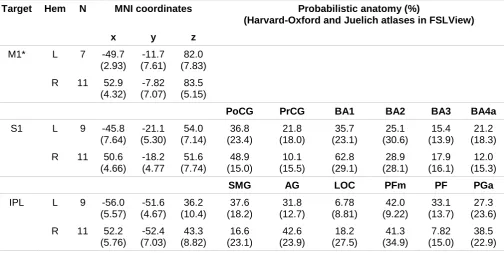
site for both the SI and control IPL brain regions using both anatomical (i.e., the postcentral gyrus or the IPL respectively) and functional criteria (i.e., a vibrotactile activation cluster, or the absence of activation respectively) from fMRI localizer scans, separately for each participant. This procedure allowed greater confidence than all previous reports that the functionally-defined SI hand area was stimulated in each participant. For the left hemisphere the mean±SD distance of participants' coordinates (x,y,z) of the SI site was 11.4±7.89mm lateral, 7.00±5.32mm posterior, and 11.1±6.91mm inferior (Euclidean distance=19.3±7.68mm) to the functionally-defined motor hotspot, as measured on participants’ MRI scans in scanner anatomical space. For the right hemisphere the mean
distances were 14.2±5.15mm lateral, 7.64±6.30mm posterior, and 15.0±7.46mm inferior (Euclidean distance=23.4±7.18mm). The location of SI in the two hemispheres, relative to the coordinate system origin, was different, compatible with the results of previous fMRI studies that identified SI using unilateral median nerve stimulation (Nihashi et al., 2005) or vibrotactile mechanical stimulation at the fingers (Tamè et al., 2012).
2.4 TMS experiments
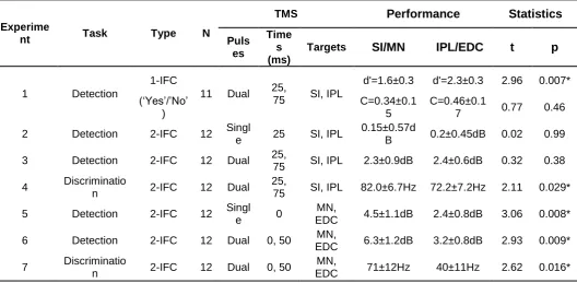
2-interval tasks. The interval containing the target was randomized. A summary of the different experiments is presented in Table 1.
Apparatus & stimuli. In Experiments 2, 3, and 4, Electromyographic (EMG) activity was recorded using Ag-AgCl surface electrodes placed over the first dorsal interosseus (FDI) muscle of the hand contralateral to the hemisphere stimulated. The EMG signal was sampled, digitised, and stored by the BrainSight software for off-line data analysis.
TMS was presented with a Mag&More PowerMag Research 100 stimulator via a 10cm diameter, flat, figure-of-8 shaped coil. BrainSight 2.2.6 (Rogue Research Inc., Montreal, QC) frameless stereotaxy was used for neuronavigation. Vibrotactile stimuli were delivered with bone conducting vibrators (Oticon, Xiamen, BC461-1 polarized), driven by a standard computer’s audio card (Vinyl AC’97 Audio wave). Vibrotactile stimulation consisted
of sinusoidal waves (200Hz, 50ms, linear rise and fall of 5ms). We used 200Hz for the vibrotactile stimulation because this is an ideal frequency to induce responses from the Pacinian corpuscle, which has the highest vibration sensitivity and lowest thresholds. Tactile stimulators gently pressed on the finger(s). Levers, cords, and pulleys ensured constant pressure, despite finger movement.
Two light-emitting diodes (LEDs) were placed in front of the participant to indicate the stimulation intervals and to provide feedback. Two response pedals were positioned one under each of the participants' feet. Stimulus presentation and response collection were controlled by custom scripts written using MATLAB (Mathworks, Natick) and PsychToolBox 3 libraries (Brainard, 1997). White noise was presented over two loudspeakers positioned in front of the participant to mask any sounds made by the tactile stimulators.
presented in Figure 3.
For each participant, we first obtained the approximate resting motor threshold (RMT) from biphasic stimulation over M1 (by finding the minimum stimulator intensity that produced motor evoked potentials of 50μV or higher on 5 or more out of 10 trials with the hand relaxed
was reduced to a minimum of 110% (i.e., for three participants in one of the experiments). General procedure. The behavioural methods were very similar to those of Tamè and colleagues (2014). Briefly, participants performed about 20 practice trials starting from the maximum possible intensity of the tactile stimulus. The starting intensity for experimental blocks was set for all participants at 0.5, half of the maximum possible intensity of the Personal Computer's (PC) audio output. Stimulus amplitude was adjusted on each trial in accordance with the QUEST algorithm (Watson and Pelli, 1983). The suggested default settings of QUEST in PsychToolBox3 were used (beta=3.5, percent correct at threshold=82%). Participants kept their eyes open, could see their hands, and were instructed to fixate between two LEDs (Harrar and Harris, 2009), and to keep two foot-pedals pressed, unless indicating their response to a target stimulus. Response accuracy was emphasized, whereas speed was not. Following correct responses, the LEDs were extinguished until the start of the next trial. Following an incorrect response, the LEDs flashed twice briefly before the trial ended in all Experiments except for Experiment 1, in which no feedback was provided. Participants were offered short breaks and water between blocks. Two experimenters remained in the room throughout the session to hold and position the coil, monitor participant safety, and ensure compliance with the instructions.
The number of trials used to calculate threshold was the same for all participants and sessions. In Experiments 2 and 5, each of two blocks per TMS site had 48 trials with two intervals, each with one TMS pulse (96 pulses). In Experiments 3, 4, 6, and 7, it was the same, but with 2 TMS pulses per interval (192 pulses). Each experiment/session last approximately 1 hour. While we did not explicitly control for baseline sensitivity between sessions or days, counterbalancing ensures that differences between TMS over SI and IPL cannot be systematically affected by between-session differences; all our critical comparisons are within-session.
Behaviour. The detection threshold values for experimental and control TMS conditions were converted to decibels (dB) relative to the no TMS condition using the following formula: dB=10*log10(TMS threshold/no TMS threshold) (D'Amour and Harris, 2014; Tamè et al., 2014). The discrimination threshold values were obtained by subtracting the no TMS condition from the experimental and TMS control conditions. Hypothesis driven one-tailed t-tests were used for all planned comparisons, with the prediction that TMS over the target site (SI) should impair performance relative to the control site (IPL). The raw data for Experiments 2-7 including the no TMS, SI, and IPL conditions are reported in Table S2, whereas the statistical comparisons between the conditions are reported in Table S3 of the Supplementary Materials.
MEPs. The peak-to-peak MEP amplitude was determined automatically by finding the minimum and maximum values within a search window, starting from 15 ms and ending ~60 ms after TMS, while excluding TMS-related artefacts. All of the experimental programs, the raw data, and analysis scripts are or will be available on the laboratory's webpages (http://neurobiography.info).
3. Experiment 1: TMS over SI impairs vibrotactile detection and prevents improvement over time
stimulus was present or absent. The target stimulus was randomly present 50% of the time. As in previous reports, no feedback about accuracy was provided. Participants’ performance
was measured in accordance with signal detection theory to distinguish changes in tactile sensitivity (d’) and decision criterion (C, Green and Swets, 1966; Macmillan and Creelman,
1991), well-established measures of sensory perception (see, especially in the present context, Azzopardi and Cowey, 1997).
3.1 Participants
Eleven participants (mean±SD age=28.4±6.24 years, 7 females) took part in the study. Nine were right-handed by self-report, two were left-handed.
3.2 Methods
Vibrotactile detection task, 1IFC ('yes'/'no') design. Participants were instructed to indicate whether the target finger (i.e., index or middle, counterbalanced between participants) was stimulated on each trial. Participants raised their left foot to report 'no' (target absence), and their right to report 'yes' (target presence). There was only one temporal interval and the target was present on half of the trials. No feedback about accuracy was provided (Tamè et al., 2014). Pulses of TMS were applied at 25 and 75ms after stimulus onset. Each trial started with the left LED flashing for 250ms indicating the potential occurrence, after 500ms, of the tactile stimulation. All participants in this experiment had just taken part in Experiment 3. We used the mean threshold obtained in the last 3 blocks of Experiment 3 (i.e., the mean across no TMS, TMS over SI, and TMS over IPL) as the target intensity for Experiment 1. We are reporting the order of experiments non-chronologically for narrative clarity.
3.3 Results
when TMS was applied over SI (M±SE d’=1.59±0.285) compared to over IPL (M±SE d’=2.27±0.306; t(10)=2.96, p=.007), and compared to no TMS (M±SE d’=2.34±0.323;
t(10)=2.45, p=.028). When TMS was applied over IPL, sensitivity was comparable to no TMS (t(10)=0.406, p=.69). Moreover, participants' decision criteria were also significantly increased when TMS was applied over SI (M±SE C=0.343±0.154, t(10)=-2.57, p=.024) or over IPL (M±SE C=0.460±0.168, t(10)=-2.65, p=.028) as compared to no TMS (M±SE C=0.0872±0.165). The numbers of hits and false alarms used to calculate d-prime and criterion values of each participant are reported in Supplementary Materials (Table S1).
Following previous reports on the role of SI in tactile working memory (Harris et al., 2002, 2001), and to explore the possibility that the effects of TMS over SI may build up over time, we also analyzed the first half (i.e., first 30 trials) and second half (i.e., second 30 trials) of the data separately. Results of the first half showed no differences in the participants’ sensitivity (d’) when TMS was applied over SI (M±SE d’=1.84±0.384) compared either to TMS over IPL (M±SE d’=2.29±0.384; t(10)=1.10, p=.150), or to no TMS (M±SE d’=1.97±0.454; t(10)=0.257, p=.401), and sensitivity when TMS was applied over IPL was
not different to no TMS (t(10)=-1.23, p=.124). Conversely, in the second half of the trials, participants' sensitivity (d’) was significantly lower with TMS over SI (M±SE d’=1.62±0.321) compared to both IPL (M±SE d’=2.60±0.315; t(10)=3.72, p=.002), and no TMS (M±SE d’=3.17±0.236; t(10)=4.40, p=.0005). On further exploration, these effects seemed to build
up over time for sensitivity (Figure 4A), but stay relatively constant for criterion (Figure 4B). In the second half of the trials, sensitivity when TMS was applied over IPL was not different to no TMS (t(10)=1.41, p=.092).
3.4 Discussion
These results concerning participants’ sensitivity are compatible with previous reports
previous reports, we also found a significant difference in the participants' decision criteria, namely a more conservative criterion when TMS was delivered over both SI and IPL compared to no TMS. With TMS, participants were more likely to report that the stimulus was absent, even if their ability to detect that stimulus was unaffected (compare IPL with no TMS). Overall, the results of our first experiment showed that individual fMRI-guided TMS over SI interferes with tactile detection at the fingers, seems to prevent improvement in performance over time (Figure 4A), and that TMS over both SI and IPL affects participants’
decision criteria, increasing their reports of 'no stimulus present' (Figure 4B), independently of changes in sensitivity.
4.1 Participants
Twelve participants (mean±SD age=30.3±10.4 years, 9 females) took part in Experiment 2 and twelve took part (mean±SD age=29.4±6.82 years, 8 females) in Experiment 3. Eleven were right-handed by self-report, one was left-handed.
4.2 Methods
Vibrotactile detection task, 2IFC design (Experiments 2 & 3). Participants were informed that they had to perform a two-interval forced choice task to indicate in which of two temporal intervals the target finger had been stimulated. Tactile stimulation was always present in only one interval (i.e., first or second). Each trial started with the left LED flashing for 250ms indicating the potential occurrence, after 500ms, of the first tactile stimulation (1st interval). 500ms after the end of the first stimulation the right LED flashed (250ms), indicating the potential occurrence, after 500ms, of the second tactile stimulation (2nd interval). 500ms after the second interval, both LEDs switched on until the participant gave their response. In Experiment 2, a single pulse of TMS was applied 25ms after target onset (i.e., 25ms after the time at which the tactile stimulus was or would have been presented, since in one of the intervals there was no target). Experiment 3 was the same as Experiment 2, with the exception that TMS was applied with two pulses per interval, one at 25 and one at 75ms after target onset.
4.3 Results
Figure 5A). Moreover, in Experiment 3 we applied two pulses of TMS, at 25ms and 75ms after stimulus onset, in place of the single pulse. The results of Experiment 3 showed a general and significant increase of detection thresholds relative to no TMS both for the experimental (SI, M±SE=2.30±0.848dB, t(11)=-2.72, p=.0100), and control sites (IPL, M±SE=2.42±0.635dB, t(11)=-3.81, p=.001), but again detection thresholds were not significantly greater for TMS over SI than over IPL (t(11)=-0.357, p=.377; Figure 5B).
4.4 Discussion
These two negative results are very surprising, considering both the results of Experiment 1, and substantial published evidence showing the disruptive effect of single pulse TMS in simple tactile detection tasks (reported effect sizes for comparable experimental conditions range, for example, from d=0.356 in Seyal et al., 1997; to d=5.22 in Hannula et al., 2005, and the infinite effect size corresponding to total blocking of sensation in Cohen et al., 1991). Our sample size of 12 gave us 80% power to detect effect sizes of d>0.76. Almost all previous studies of the effects of TMS on somatosensory perception had sample sizes smaller than 12. The lack of an effect might be explained by the single pulse TMS being too weak or short-lived to interfere with tactile processing in SI in this task (although see Cohen et al., 1991; Seyal et al., 1992 for evidence to the contrary). The results of our Experiment 3 show that, while dual pulse TMS produced a general interference effect by elevating 2IFC tactile detection thresholds, this was not specific to SI as might have been expected from previous reports (Cohen et al., 1991; Seyal et al., 1997, 1992).
To further investigate whether the lack of difference between SI and IPL in tactile thresholds for Experiments 2 and 3 was statistically reliable, we calculated Bayes’ factor
was 0.301 for Experiment 2 and 0.139 for Experiment 3. Therefore, for both Experiments 2 and 3, we found substantial evidence in support of no difference between the two conditions (i.e., TMS over SI and over IPL). Finally, in both Experiments 2 and 3 we can rule out the possibility that the lack of a difference was due to the ineffectiveness of our TMS protocol because of the significantly non-zero MEPs that we recorded from the hand contralateral to the TMS sites – TMS was strong enough to excite M1, resulting in MEPs recorded from the
contralateral hand (see Supplementary Materials).
5. Experiment 4. TMS over SI impairs 2IFC frequency discrimination
In Experiment 4, we tested whether tactile 2IFC frequency discrimination thresholds are susceptible to dual pulse TMS delivered, as in the previous experiments, 25ms and 75ms after stimulus onset, over SI. We expected an interference effect of TMS over SI on the basis of similar previous reports in both monkeys (LaMotte and Mountcastle, 1979) and humans (Knecht et al., 2003; Morley et al., 2007). This experiment served as a positive control for the general effectiveness of our TMS protocols.
5.1 Participants
Twelve participants (mean±SD age=27.7±6.04 years, 9 females) took part in the study. Eleven were right-handed by self-report, one was left-handed.
5.2 Methods
5.3 Results
As expected, dual pulse TMS over SI increased frequency discrimination thresholds (M±SE=82.0±6.73Hz) relative both to the no TMS control (M±SE=63.5±6.39Hz, t(11)=-3.26, p=.004), as well as to the full TMS control condition (IPL: M±SE=72.3±7.17Hz, t(11)=-2.11, p=.0291). Discrimination thresholds did not differ significantly between no TMS and TMS over IPL, t(11)=-1.57, p=.0727.
5.4 Discussion
In contrast to the simple detection thresholds investigated in Experiments 2 and 3, using the same pool of participants and near-identical stimuli and methods, TMS specifically and significantly increased discrimination thresholds when applied over SI relative to over IPL (Figure 5C). Although we manipulated target frequency, changes in frequency can be accompanied by changes in perceived intensity, so participants may have used either cue to perform the task. Nevertheless, our data clearly support the hypothesis of a contribution of SI in tactile frequency and/or intensity discrimination, in agreement with previous monkey (de Lafuente and Romo, 2005; LaMotte and Mountcastle, 1979) and human studies (Knecht et al., 2003; Morley et al., 2007; Porro et al., 2007).
6. Experiments 5, 6, & 7: Magnetic stimulation over the somatosensory periphery impairs both detection and discrimination
cannot easily be interfered with by TMS over SI. Experiments 5-7 provide further positive controls for the general effectiveness of our TMS protocols, and followed the same design as Experiments 2-4.
6.1 Participants
The same twelve participants (mean±SD age=27.4±6.11 years, 6 females) took part in Experiments 5, 6 and 7. Eleven were right-handed by self-report, one was left-handed.
6.2 Methods
In Experiments 5, 6 and 7, the pulses of TMS (TMS intensity M±SE=28.7±1.24% of maximum stimulator output), at 0 and 50ms after stimulus onset, were applied over the median nerve at the wrist (experimental site), or over the EDC of the proximal forearm (control site), or with no TMS (the active coil held ~30 cm from forearm). The coil handle was oriented proximally along the forearm. The same 2IFC design for the detection and discrimination tasks were used as for the previous Experiments 2-4.
6.3 Results
interfering with tactile thresholds at the fingers in a 2IFC detection task.
6.4 Discussion
Experiments 5, 6, and 7 replicated Experiments 2, 3, and 4, with the exception that stimulation was applied 25ms earlier, and over the MN at the wrist (in place of SI), and over the EDC muscles of the forearm (control site, in place of IPL). The results showed that both single- and dual-pulse TMS over the MN increased detection and discrimination thresholds compared to the muscle control site (EDC).
7. General Discussion
We investigated the contribution of human SI to tactile detection and discrimination using individually-tailored fMRI-guided TMS, in both 1IFC ('yes'/'no') and 2IFC designs (Tamè et al., 2014). Our results showed that both the perceptual and decisional components of tactile detection are affected by TMS. When applied over SI, TMS seems to interfere with the maintenance of a representation of the tactile stimulus over time (Libet et al., 1991; Morgan et al., 2000; Recanzone et al., 1992a, 1992b; Wolpert et al., 1998). This 1IFC approach is known to have a greater memory component with respect to the 2IFC design (Harris et al., 2006). Conversely, when tactile detection was tested in the 2IFC design, a task with a lower memory load, and which is more sensitive to sensory inputs (Harris et al., 2006) as well as other task demands, participants’ performance was not modulated selectively by
TMS over SI. In addition, the reported criterion shift demonstrates that TMS can also bias participants’ decision about the presence of a tactile stimulus on the fingers, an issue that,
this effect. Indeed, we believe that increases in response criterion have strong implications for the interpretation of many previous reports of TMS interference with tactile perception. Most importantly, criterion shifts should be taken into account in the design and data analyses of all relevant future TMS studies.
Previous reports that investigated the role of SI in tactile detection arrived at different conclusions. Electrophysiological and lesion studies in the macaque brain suggested that SI (indeed, the whole contralateral parietal cortex) is not required for simple detection of tactile stimuli (LaMotte and Mountcastle, 1979, 1975), whereas studies in humans using single-pulse TMS over sensorimotor cortex showed near-complete disruption of detection (Cohen et al., 1991). The present work suggests a resolution to this discrepancy (see also a similar argument in the blindsight literature, Azzopardi & Cowey, 1997), and reconciles monkey and human studies of the role of SI, by showing that SI is differentially involved in tactile detection as a function of task demand. We speculate that SI is not passively involved in simple detection of incoming inputs, but rather is actively involved in the formation and maintenance of representations of the tactile signal (for example it’s frequency and/or
intensity) over time (Harris et al., 2002; Katus et al., 2014; Libet et al., 1991; Morgan et al., 2000).
feels, and on each trial, to compare the potential stimulus with this memory or 'internal standard' (Libet et al., 1991; Morgan et al., 2000). Furthermore, an internal criterion must be set and used to decide whether there was or wasn't a stimulus (Azzopardi and Cowey, 1997). Other forms of tasks include tactile localization (e.g., Braun et al., 2005) and discrimination, which require the participant to report which stimulus was presented, instead of noticing only whether or when it occurred. Such tactile discrimination tasks may entail more complex processing as compared to simple detection tasks. This last paradigm is typically used to investigate tactile learning (e.g., Harris et al., 2001; Sathian and Zangaladze, 1997). In sum, the specific task that participants perform may determine the amount of resources necessary to complete it and the recruitment of specific brain regions (for a discussion on the difference between tactile detection and discrimination see Romo et al., 2012). In the following, we examine the demands of 1IFC and 2IFC tasks in more detail.
7.1 Tactile detection in a 1IFC design
Most studies in humans similar to the present one have shown that detection of tactile stimuli on the fingers is impaired when TMS is applied in close temporal proximity with the target over contralateral sensorimotor cortex. Cohen and colleagues (1991) found that detection of electrical stimuli on the index finger was attenuated when spTMS was applied 200ms before stimulus onset, and blocked it completely when spTMS was delivered simultaneously to and 20ms after it (Cohen et al., 1991). Similarly, McKay and colleagues (2003) applied trains of electro-cutaneous ulnar nerve stimuli, and reported that when TMS was delivered over the contralateral sensorimotor cortex 100ms before or 20ms after stimulus onset, perception was strongly suppressed (McKay et al., 2003). Our findings are compatible with these results in showing attenuation in participants’ tactile detection
2006). As suggested by Koch and colleagues (2006), their lack of effect may be because they applied TMS over the somatosensory cortex (Koch et al., 2006) rather than over the combined sensorimotor cortex (Cohen et al., 1991; Pascual-Leone et al., 1994; Seyal et al., 1992). Indeed, concurrent stimulation of sensory and motor cortices may have evoked responses in finger muscles that in turn influenced perception (Koch et al., 2006). Here, we targeted SI using individually-tailored fMRI-guided neuronavigation, and by recording EMG responses, showed a dissociation in motor responses and sensory thresholds (Supplementary Materials, Figures S1-S2).
Our findings extend previous results by showing that the reduction in participants’
sensitivity with TMS over SI relative to over IPL and no TMS was not constant, but built up over time, perhaps due to a learning effect (Seitz and Dinse, 2007). Indeed, as shown in Figure 3A, participants’ performance significantly improved across trials when TMS is not
applied over SI, as if the participants are still learning trial-by-trial how to detect the stimulus, and improving their performance. Repeated presentation of the target stimulus allows participants to extract more and more information about it. Yet, with TMS over SI, this learning effect was not seen. This further supports our argument that SI may be extracting and maintaining information about tactile stimuli over trials, rather than merely passively responding to them. When TMS was applied over SI there was no improvement in participants' performance over time, unlike the behavioural improvement seen with no TMS or TMS over IPL. This is compatible with a previous report (Knecht et al., 2003) in which tactile frequency discrimination performance increased across blocks of trials with sham or no TMS. The effect of TMS on this short-term tactile ‘learning’ effect contrasts with previous
unaffected by TMS over SI.
7.2 Tactile detection in a 2IFC design
In Experiments 2-3, we used a 2IFC task known to be less criterion dependent and with lower, and minimal memory demand (Harris et al., 2006; Lloyd et al., 2013). Differently from Experiment 1, this showed that TMS over SI versus IPL did not affect participants' detection performance either when single (Figure 5A) or double (Figure 5B) TMS pulses were applied. This suggests that TMS over SI is not effective in interfering with simple tactile detection when the task is less criterion-dependent and/or requires lower memory load (Harris et al., 2006). In agreement with neurophysiological studies on monkeys (LaMotte and Mountcastle, 1979), SI, under certain circumstances, may not be necessary for detecting a tactile stimulus on the fingers. The fact that the effect of TMS over SI on tactile detection depends on the task (1IFC vs. 2IFC), suggests that SI plays a different role than the simple detection of stimuli (see also (Harris et al., 2006, 2004; Vroomen and Stekelenburg, 2014). Another difference between our first and second experiments was the number of TMS pulses, but we ruled out this potential explanation in Experiment 3, in which doubling the number of TMS pulses still did not impair 2IFC tactile detection for SI relative to the control site.
7.3 Role of the primary somatosensory cortex in tactile detection
storage for working memory is provided by Harris and coworkers (Harris et al., 2002). They found that participants' performance was significantly impaired when TMS was delivered over contralateral SI at specific times in the interval between stimuli to be compared. The direct involvement of SI when a memory component is required, as proposed by Harris and colleagues (2002), is compatible with the profile of results of our Experiments 1, 2, and 3. In particular, in Experiment 1 the inter-stimulus interval, given that trials with a target stimulus were randomly presented among trials with no target, could be very long (i.e., up to 30 seconds or even more), thus enhancing the memory requirement of the task – to remember what the target feels like. By contrast, in Experiments 2 and 3, a tactile stimulus was always present in each trial, and participants needed only to compare the two intervals directly (Libet et al., 1991). These differences in the inter-stimulus temporal profile in the two designs, we think, can account for the different effectiveness of TMS over SI, and in turn highlight its active role in the processing of tactile stimuli at the fingers. Electrophysiological studies of delayed match-to-sample tasks in humans have also shown the active recruitment of SI for working memory (Katus et al., 2015). These authors reported sustained activity over the contralateral primary somatosensory cortex during the retention interval, and the amplitude of this component increased with the memory load and reflected the individual memory capacity. Katus and colleagues interpreted this as evidence for the maintenance of tactile information in SI (Katus et al., 2015; see also Haegens et al., 2010; Spitzer and Blankenburg, 2011). The present results are consistent with these findings. We cannot rule out the possibility that attentional factors may also, to some extent, contribute to our effects, given that prior expectation has been shown to bias sensory representation at the early stages of the sensory processing, at least in the visual domain (Kok et al., 2013).
it is possible that when SI capacity is disrupted (e.g., by TMS) SII executes some of its functions, though this is less likely considering LaMotte and Mountcastle's results (1979). Another possibility is that ipsilateral SI - with respect to the stimulated finger - is executing some of the functions of contralateral SI. Indeed, there is evidence in humans that vibrotactile stimuli elicit responses in ipsilateral SI quite early in time (for a review see Tamè et al., in press). Moreover, subcortical regions, such as, for instance, the thalamus (e.g., Vázquez et al., 2013) or the cuneate nucleus (e.g., Jörntell et al., 2014), may also significantly contribute to the processing of simple vibrotactile signals. In this respect, Libet and colleagues (1991) demonstrated that a tactile signal delivered at the level of the ventrobasal thalamus via electrodes – implanted in patients with intractable pain - can be correctly detected (more than 50% of the time), with little conscious awareness of the stimulus (Libet et al., 1991). Increasing the duration of the tactile stimuli allowed participants to be consciously aware of them. Likewise, our results suggest that, while SI is not necessarily involved in simple tactile detection (i.e., reporting stimulus presence better than chance), it may still be involved in conscious awareness of the same tactile stimuli.
7.4 Conclusion
Our results support the notion of a critical role of SI in tactile discrimination and a task-dependent role in tactile detection. Specifically, SI is required for tactile detection in the 1-IFC design, most likely because it is actively involved in maintaining a representation of the stimulus over time. This is in agreement with the “sensory recruiting” model of working
Acknowledgements
References
Azzopardi, P., Cowey, A., 1997. Is blindsight like normal, near-threshold vision? Proc. Natl. Acad. Sci. U. S. A. 94, 14190–14194.
Bensmaia, S.J., Denchev, P.V., Dammann, J.F., Craig, J.C., Hsiao, S.S., 2008. The representation of stimulus orientation in the early stages of somatosensory processing. J. Neurosci. 28, 776–786. doi:10.1523/JNEUROSCI.4162-07.2008
Brainard, D.H., 1997. The Psychophysics Toolbox. Spat. Vis. 10, 433–436.
Braun, C., Hess, H., Burkhardt, M., Wühle, A., Preissl, H., 2005. The right hand knows what the left hand is feeling. Exp. Brain Res. 162, 366–373. doi:10.1007/s00221-004-2187-4
Cohen, L.G., Bandinelli, S., Sato, S., Kufta, C., Hallett, M., 1991. Attenuation in detection of
somatosensory stimuli by transcranial magnetic stimulation. Electroencephalogr. Clin. Neurophysiol. 81, 366–376.
D’Amour, S., Harris, L.R., 2014. Contralateral tactile masking between forearms. Exp. Brain Res. 232, 821–826. doi:10.1007/s00221-013-3791-y
de Lafuente, V., Romo, R., 2006. Neural correlate of subjective sensory experience gradually builds up across cortical areas. Proc. Natl. Acad. Sci. U. S. A. 103, 14266–14271.
de Lafuente, V., Romo, R., 2005. Neuronal correlates of subjective sensory experience. Nat. Neurosci. 8, 1698–1703. doi:10.1038/nn1587
D’Esposito, M., 2007. From cognitive to neural models of working memory. Philos. Trans. R. Soc. Lond. B. Biol. Sci. 362, 761–772. doi:10.1098/rstb.2007.2086
Dienes, Z., 2014. Using Bayes to get the most out of non-significant results. Front. Psychol. 5, 781. doi:10.3389/fpsyg.2014.00781
Green, D., Swets, J., 1966. Signal detection theory and psychophysics. Wiley & Sons, Inc., New York. Haegens, S., Osipova, D., Oostenveld, R., Jensen, O., 2010. Somatosensory working memory performance in humans depends on both engagement and disengagement of regions in a distributed network. Hum. Brain Mapp. 31, 26–35. doi:10.1002/hbm.20842
Hamada, M., Murase, N., Hasan, A., Balaratnam, M., Rothwell, J.C., 2013. The role of interneuron networks in driving human motor cortical plasticity. Cereb. Cortex N. Y. N 1991 23, 1593–1605. doi:10.1093/cercor/bhs147
Hannula, H., Neuvonen, T., Savolainen, P., Tukiainen, T., Salonen, O., Carlson, S., Pertovaara, A., 2008. Navigated transcranial magnetic stimulation of the primary somatosensory cortex impairs perceptual processing of tactile temporal discrimination. Neurosci. Lett. 437, 144–147.
doi:10.1016/j.neulet.2008.03.093
Hannula, H., Ylioja, S., Pertovaara, A., Korvenoja, A., Ruohonen, J., Ilmoniemi, R.J., Carlson, S., 2005. Somatotopic blocking of sensation with navigated transcranial magnetic stimulation of the primary somatosensory cortex. Hum. Brain Mapp. 26, 100–109.
Harrar, V., Harris, L.R., 2009. Eye position affects the perceived location of touch. Exp. Brain Res. 198, 403–410.
Harris, J.A., Harris, I.M., Diamond, M.E., 2001. The topography of tactile working memory. J. Neurosci. Off. J. Soc. Neurosci. 21, 8262–8269.
Harris, J.A., Karlov, L., Clifford, C.W.G., 2006. Localization of tactile stimuli depends on conscious detection. J. Neurosci. 26, 948–952.
Harris, J.A., Miniussi, C., Harris, I.M., Diamond, M.E., 2002. Transient storage of a tactile memory trace in primary somatosensory cortex. J. Neurosci. 22, 8720–8725.
Harris, J.A., Thein, T., Clifford, C.W.G., 2004. Dissociating detection from localization of tactile stimuli. J. Neurosci. 24, 3683–3693. doi:10.1523/JNEUROSCI.0134-04.2004
Harrison, S.A., Tong, F., 2009. Decoding reveals the contents of visual working memory in early visual areas. Nature 458, 632–635. doi:10.1038/nature07832
Jenkinson, M., Bannister, P., Brady, M., Smith, S., 2002. Improved optimization for the robust and accurate linear registration and motion correction of brain images. NeuroImage 17, 825–841. Jenkinson, M., Smith, S., 2001. A global optimisation method for robust affine registration of brain images. Med. Image Anal. 5, 143–156.
Jones, S.R., Pritchett, D.L., Stufflebeam, S.M., Hämäläinen, M., Moore, C.I., 2007. Neural correlates of tactile detection: a combined magnetoencephalography and biophysically based computational modeling study. J. Neurosci. 27, 10751–10764.
doi:10.1016/j.neuron.2014.07.038
Katus, T., Grubert, A., Eimer, M., 2014. Electrophysiological Evidence for a Sensory Recruitment Model of Somatosensory Working Memory. Cereb. Cortex.
Knecht, S., Ellger, T., Breitenstein, C., Bernd Ringelstein, E., Henningsen, H., 2003. Changing cortical excitability with low-frequency transcranial magnetic stimulation can induce sustained disruption of tactile perception. Biol. Psychiatry 53, 175–179.
Koch, G., Franca, M., Albrecht, U.-V., Caltagirone, C., Rothwell, J.C., 2006. Effects of paired pulse TMS of primary somatosensory cortex on perception of a peripheral electrical stimulus. Exp. Brain Res. 172, 416–424.
Kok, P., Brouwer, G.J., van Gerven, M.A.J., de Lange, F.P., 2013. Prior expectations bias sensory representations in visual cortex. J. Neurosci. Off. J. Soc. Neurosci. 33, 16275–16284.
doi:10.1523/JNEUROSCI.0742-13.2013
LaMotte, R.H., Mountcastle, V.B., 1979. Disorders in somesthesis following lesions of parietal lobe. J. Neurophysiol. 42, 400–419.
LaMotte, R.H., Mountcastle, V.B., 1975. Capacities of humans and monkeys to discriminate vibratory stimuli of different frequency and amplitude: a correlation between neural events and psychological measurements. J. Neurophysiol. 38, 539–559.
Libet, B., Pearl, D.K., Morledge, D.E., Gleason, C.A., Hosobuchi, Y., Barbaro, N.M., 1991. Control of the transition from sensory detection to sensory awareness in man by the duration of a thalamic stimulus. The cerebral “time-on” factor. Brain J. Neurol. 114 ( Pt 4), 1731–1757.
Lloyd, D.A., Abrahamyan, A., Harris, J.A., 2013. Brain-stimulation induced blindsight: unconscious vision or response bias? PloS One 8, e82828.
Macmillan, N.A., Creelman, C.D., 1991. Detection theory: a user’s guide. Cambridge University Press, Cambridge.
McKay, D.R., Ridding, M.C., Miles, T.S., 2003. Magnetic stimulation of motor and somatosensory cortices suppresses perception of ulnar nerve stimuli. Int. J. Psychophysiol. Off. J. Int. Organ. Psychophysiol. 48, 25–33.
Meehan, S.K., Legon, W., Staines, W.R., 2008. Paired-pulse transcranial magnetic stimulation of primary somatosensory cortex differentially modulates perception and sensorimotor transformations. Neuroscience 157, 424–431. doi:10.1016/j.neuroscience.2008.09.008
Moore, C.I., Crosier, E., Greve, D.N., Savoy, R., Merzenich, M.M., Dale, A.M., 2013. Neocortical correlates of vibrotactile detection in humans. J. Cogn. Neurosci. 25, 49–61.
doi:10.1162/jocn_a_00315
Morgan, M.J., Watamaniuk, S.N., McKee, S.P., 2000. The use of an implicit standard for measuring discrimination thresholds. Vision Res. 40, 2341–2349.
Morley, J.W., Vickery, R.M., Stuart, M., Turman, A.B., 2007. Suppression of vibrotactile discrimination by transcranial magnetic stimulation of primary somatosensory cortex. Eur. J. Neurosci. 26, 1007– 1010. doi:10.1111/j.1460-9568.2007.05729.x
Nihashi, T., Naganawa, S., Sato, C., Kawai, H., Nakamura, T., Fukatsu, H., Ishigaki, T., Aoki, I., 2005. Contralateral and ipsilateral responses in primary somatosensory cortex following electrical median nerve stimulation--an fMRI study. Clin. Neurophysiol. Off. J. Int. Fed. Clin. Neurophysiol. 116, 842– 848. doi:10.1016/j.clinph.2004.10.011
Olney, R.K., So, Y.T., Goodin, D.S., Aminoff, M.J., 1990. A comparison of magnetic and electrical stimulation of peripheral nerves. Muscle Nerve 13, 957–963. doi:10.1002/mus.880131012 Palva, S., Linkenkaer-Hansen, K., Näätänen, R., Palva, J.M., 2005. Early neural correlates of conscious somatosensory perception. J. Neurosci. 25, 5248–5258. doi:10.1523/JNEUROSCI.0141-05.2005
Pascual-Leone, A., Cohen, L.G., Brasil-Neto, J.P., Valls-Solé, J., Hallett, M., 1994. Differentiation of sensorimotor neuronal structures responsible for induction of motor evoked potentials, attenuation in detection of somatosensory stimuli, and induction of sensation of movement by mapping of optimal current directions. Electroencephalogr. Clin. Neurophysiol. 93, 230–236.
Porro, C.A., Martinig, M., Facchin, P., Maieron, M., Jones, A.K.P., Fadiga, L., 2007. Parietal cortex involvement in the localization of tactile and noxious mechanical stimuli: a transcranial magnetic stimulation study. Behav. Brain Res. 178, 183–189. doi:10.1016/j.bbr.2006.11.011
Postle, B.R., 2006. Working memory as an emergent property of the mind and brain. Neuroscience 139, 23–38. doi:10.1016/j.neuroscience.2005.06.005
Recanzone, G.H., Jenkins, W.M., Hradek, G.T., Merzenich, M.M., 1992a. Progressive improvement in discriminative abilities in adult owl monkeys performing a tactile frequency discrimination task. J. Neurophysiol. 67, 1015–1030.
Recanzone, G.H., Merzenich, M.M., Schreiner, C.E., 1992b. Changes in the distributed temporal response properties of SI cortical neurons reflect improvements in performance on a temporally based tactile discrimination task. J. Neurophysiol. 67, 1071–1091.
Romo, R., de Lafuente, V., 2012. Conversion of sensory signals into perceptual decisions. Prog. Neurobiol. doi:10.1016/j.pneurobio.2012.03.007
Romo, R., Hernández, A., Zainos, A., Brody, C., Salinas, E., 2002. Exploring the cortical evidence of a sensory-discrimination process. Philos. Trans. R. Soc. Lond. B. Biol. Sci. 357, 1039–1051.
doi:10.1098/rstb.2002.1100
Romo, R., Lemus, L., de Lafuente, V., 2012. Sense, memory, and decision-making in the somatosensory cortical network. Curr. Opin. Neurobiol. doi:10.1016/j.conb.2012.08.002
Romo, R., Salinas, E., 2003. Flutter discrimination: neural codes, perception, memory and decision making. Nat. Rev. Neurosci. 4, 203–218. doi:10.1038/nrn1058
Rossini, P.M., Barker, A.T., Berardelli, A., Caramia, M.D., Caruso, G., Cracco, R.Q., Dimitrijević, M.R., Hallett, M., Katayama, Y., Lücking, C.H., 1994. Non-invasive electrical and magnetic stimulation of the brain, spinal cord and roots: basic principles and procedures for routine clinical application. Report of an IFCN committee. Electroencephalogr. Clin. Neurophysiol. 91, 79–92.
Sathian, K., Zangaladze, A., 1997. Tactile learning is task specific but transfers between fingers. Percept. Psychophys. 59, 119–128.
Seitz, A.R., Dinse, H.R., 2007. A common framework for perceptual learning. Curr. Opin. Neurobiol. 17, 148–153.
Seyal, M., Masuoka, L.K., Browne, J.K., 1992. Suppression of cutaneous perception by magnetic pulse stimulation of the human brain. Electroencephalogr. Clin. Neurophysiol. 85, 397–401.
Seyal, M., Siddiqui, I., Hundal, N.S., 1997. Suppression of spatial localization of a cutaneous stimulus following transcranial magnetic pulse stimulation of the sensorimotor cortex. Electroencephalogr. Clin. Neurophysiol. 105, 24–28.
Smith, S.M., 2002. Fast robust automated brain extraction. Hum. Brain Mapp. 17, 143–155. Song, S., Sandrini, M., Cohen, L.G., 2011. Modifying somatosensory processing with non-invasive brain stimulation. Restor. Neurol. Neurosci. 29, 427–437. doi:10.3233/RNN-2011-0614
Spitzer, B., Blankenburg, F., 2011. Stimulus-dependent EEG activity reflects internal updating of tactile working memory in humans. Proc. Natl. Acad. Sci. U. S. A. 108, 8444–8449.
Tamè, L., Braun, C., Lingnau, A., Schwarzbach, J., Demarchi, G., Li Hegner, Y., Farnè, A., Pavani, F., 2012. The contribution of primary and secondary somatosensory cortices to the representation of body parts and body sides: an fMRI adaptation study. J. Cogn. Neurosci. 24, 2306–2320.
Tamè, L., Moles, A., Holmes, N.P., 2014. Within, but not between hands interactions in vibrotactile detection thresholds reflect somatosensory receptive field organization. Front. Psychol. 5, 174. Vázquez, Y., Salinas, E., Romo, R., 2013. Transformation of the neural code for tactile detection from thalamus to cortex. Proc. Natl. Acad. Sci. U. S. A. 110, E2635-2644. doi:10.1073/pnas.1309728110 Vroomen, J., Stekelenburg, J.J., 2014. A bias-free two-alternative forced choice procedure to examine intersensory illusions applied to the ventriloquist effect by flashes and averted eye-gazes. Eur. J. Neurosci. doi:10.1111/ejn.12525
Watson, A.B., Pelli, D.G., 1983. QUEST: a Bayesian adaptive psychometric method. Percept. Psychophys. 33, 113–120.
Wolpert, D.M., Goodbody, S.J., Husain, M., 1998. Maintaining internal representations: the role of the human superior parietal lobe. Nat. Neurosci. 1, 529–533. doi:10.1038/2245
Woolrich, M.W., Ripley, B.D., Brady, M., Smith, S.M., 2001. Temporal autocorrelation in univariate linear modeling of FMRI data. NeuroImage 14, 1370–1386. doi:10.1006/nimg.2001.0931
Yeshurun, Y., Carrasco, M., Maloney, L.T., 2008. Bias and sensitivity in two-interval forced choice procedures: Tests of the difference model. Vision Res. 48, 1837–1851.
doi:10.1016/j.visres.2008.05.008
Zangaladze, A., Epstein, C.M., Grafton, S.T., Sathian, K., 1999. Involvement of visual cortex in tactile discrimination of orientation. Nature 401, 587–590. doi:10.1038/44139
Figure Legends
Figure 1. Schematic representation of the tactile stimulation conditions in the 2IFC detection (A) and discrimination (B) tasks. TMS pulses were applied over SI or IPL in the 2IFC design at 25 and 75ms after tactile stimulus onset (C). For the 1IFC design the same timings were used for a single interval only.
Figure 2. Activation from the functional localiser for each of 20 participants. FMRI
data show the contrast of contralateral index finger against rest, thresholded for display purposes at between p<.05 and p<.01, uncorrected for each participant. TMS sites were determined for each participant using both anatomical (postcentral gyrus) and functional (activation) criteria. The superimposed red dots represent the location of the primary motor cortex 'hotspot' (M1). Yellow dots represent the TMS site for the primary somatosensory cortex (SI: target site) and purple dots represent the TMS site for the inferior parietal lobe (IPL: control site). All participants had activation in the postcentral gyrus at least at the p<.05 level.
Figure 3. Somatosensory functional localiser and TMS target sites. Mean functional activation maps of 20 participants for contralateral finger stimulation contrasted against baseline (blue, all five fingers against baseline, Z>2.33, no whole-brain correction applied, but contralateral SI and bilateral SII clusters all passed family-wise error correction). The statistical maps are overlaid on the mean anatomical images of the 20 participants, transformed into the MNI template space. 95% confidence ellipsoids for the TMS target sites in MNI coordinate space (Table 2). Red: M1; Green: SI, Pink: IPL. fVOI: functional volume of interest.
Figure 4. Results of Experiment 1, 1IFC design. Participants' performance in the 1IFC ‘Yes’/’No’ detection task over time (trial window) expressed in terms of d-prime (A)
and criterion (B) for the three TMS conditions (i.e., no TMS, SI, and IPL).
Figure 5. Results of Experiments 2-7, 2IFC design. (A) Tactile detection thresholds
Tables
Table 1. Summary of the experimental designs
Experime
nt Task Type N
TMS Performance Statistics
Puls es
Time s (ms)
Targets SI/MN IPL/EDC t p
1 Detection
1-IFC
11 Dual 25,
75 SI, IPL
d'=1.6±0.3 d'=2.3±0.3 2.96 0.007* (‘Yes’/’No’
)
C=0.34±0.1 5
C=0.46±0.1
7 0.77 0.46 2 Detection 2-IFC 12 Singl
e 25 SI, IPL
0.15±0.57d
B 0.2±0.45dB 0.02 0.99 3 Detection 2-IFC 12 Dual 25,
75 SI, IPL 2.3±0.9dB 2.4±0.6dB 0.32 0.38 4 Discriminatio
n 2-IFC 12 Dual
25,
75 SI, IPL 82.0±6.7Hz 72.2±7.2Hz 2.11 0.029* 5 Detection 2-IFC 12 Singl
e 0
MN,
EDC 4.5±1.1dB 2.4±0.8dB 3.06 0.008* 6 Detection 2-IFC 12 Dual 0, 50 MN,
EDC 6.3±1.2dB 3.2±0.8dB 2.93 0.009* 7 Discriminatio
n 2-IFC 12 Dual 0, 50
MN,
EDC 71±12Hz 40±11Hz 2.62 0.016* Yes/No – participants made one response for target present, another for target absent; 2-IFC
Table 2. MNI coordinates and probabilistic anatomy of TMS target sites
Target Hem N MNI coordinates Probabilistic anatomy (%)
(Harvard-Oxford and Juelich atlases in FSLView)
x y z
M1* L 7 -49.7 (2.93)
-11.7 (7.61)
82.0 (7.83) R 11 52.9
(4.32)
-7.82 (7.07)
83.5 (5.15)
PoCG PrCG BA1 BA2 BA3 BA4a
S1 L 9 -45.8 (7.64) -21.1 (5.30) 54.0 (7.14) 36.8 (23.4) 21.8 (18.0) 35.7 (23.1) 25.1 (30.6) 15.4 (13.9) 21.2 (18.3) R 11 50.6
(4.66) -18.2 (4.77 51.6 (7.74) 48.9 (15.0) 10.1 (15.5) 62.8 (29.1) 28.9 (28.1) 17.9 (16.1) 12.0 (15.3)
SMG AG LOC PFm PF PGa
IPL L 9 -56.0 (5.57) -51.6 (4.67) 36.2 (10.4) 37.6 (18.2) 31.8 (12.7) 6.78 (8.81) 42.0 (9.22) 33.1 (13.7) 27.3 (23.6) R 11 52.2
(5.76) -52.4 (7.03) 43.3 (8.82) 16.6 (23.1) 42.6 (23.9) 18.2 (27.5) 41.3 (34.9) 7.82 (15.0) 38.5 (22.9) Values are means (SD in parentheses) Hem=hemisphere; N=number of participants; MNI=Montreal
Neurological Institute coordinates (MNI 152 brain, 2mm resolution, viewed in FSLView;
x,y,z=coordinates in MNI space; M1=primary motor cortex target; * Coil location, outside the brain; S1=primary somatosensory target; IPL=inferior parietal lobule target; PoCG=postcentral gyrus; PrCG=precentral gyrus; BA1=Brodmann's area 1; BA2=Brodmann's area 2; BA3=Brodmann's area 3; BA4a=Brodmann's area 4, anterior division; SMG=supramarginal gyrus; AG=angular gyrus;