SULFOLOBUS SOLFATARICUS
Michael J. Morten
A Thesis Submitted for the Degree of PhD
at the
University of St Andrews
2015
Full metadata for this item is available in
St Andrews Research Repository
at:
http://research-repository.st-andrews.ac.uk/
Please use this identifier to cite or link to this item:
http://hdl.handle.net/10023/7568
the single-stranded DNA binding protein from
Sulfolobus solfataricus
Michael J. Morten
This thesis is submitted in partial fulfilment for the degree of PhD
at the University of St Andrews
Table of Contents
1. Introduction ... 1
1.1. Double-stranded DNA ... 1
1.2. DNA damage and mutations ... 2
1.3. DNA repair ... 3
1.4. Single-stranded nucleic acids ... 5
1.5. Single-stranded DNA binding proteins ... 7
1.5.1. Oligonucleotide/oligosaccharide binding folds. ... 9
1.5.2. Loops ... 10
1.5.3. Human replication protein A ... 12
1.5.4. Bacterial SSB ... 19
1.6. Archaea ... 23
1.6.1. Archaeal SSB ... 26
1.7. Single molecule assays ... 31
1.8. Fluorescence ... 33
1.9. FRET ... 37
1.9.1. A brief history ... 38
1.9.2. FRET theory ... 42
1.9.3. FRET as a spectroscopic ruler ... 47
1.10. TIRF microscopy ... 49
1.12. Protein & DNA labelling... 58
1.13. Fluorophores... 59
1.14. Single molecule fluorescence investigations into SSBs ... 62
2. Materials and methods ... 64
2.1. SSB expression & purification ... 64
2.2. Protein Labelling ... 65
2.3. Labelling and annealing DNA ... 67
2.4. Fluorescence experiments ... 68
2.4.1. Ratio A ... 68
2.5. Binding to a one dimensional lattice ... 69
2.5.1. 1:1 binding ... 69
2.5.2. Cooperative binding ... 70
2.6. Wormlike chain model ... 75
2.7. Single molecule experiments ... 76
2.7.1. Cleaning of slides ... 76
2.7.2. Aminosilation of glass ... 76
2.7.3. PEGylation of amino-covered slides ... 77
2.7.4. Microchannel preparation ... 77
2.7.5. TIRF experiments ... 77
2.7.6. Rate analysis ... 78
2.9. Exosome degradation ... 79
3. Ensemble studies of SsoSSB ... 81
3.1. Introduction ... 81
3.2. Expression and purification ... 82
3.3. Tryptophan quenching ... 83
3.4. Dimensions of SSB/ssDNA complexes ... 84
3.5. ssDNA Labelling ... 87
3.6. Protein induced fluorescence enhancement ... 88
3.7. Protein labelling ... 90
3.8. FRET ... 91
3.9. Quenching ... 92
3.10. PELDOR ... 93
3.11. Persistence length of naked and decorated ssDNA ... 98
3.12. Dissociation constants and occupancy ... 101
3.13. Cooperativity parameters ... 108
3.14. Quenching suggests monomeric structure ... 110
3.15. Discussion ... 111
3.16. Conclusions ... 116
4. Single molecule analysis of SsoSSB ... 118
4.1. Non-specific binding to the slide ... 120
4.3. FRET ... 127
4.4. Full in-trace interpretation ... 129
4.5. Binding kinetics ... 132
4.6. Discussion ... 138
4.7. Conclusions ... 144
5. SsoSSB binds to RNA ... 146
5.1. Ensemble fluorescence results ... 152
5.2. Single molecule fluorescence results ... 154
5.3. Possible roles for RNA binding ... 156
5.4. Discussion ... 158
5.5. Conclusions ... 160
6. Further work ... 162
6.1. Applications in high temperature and salt concentrations ... 162
6.1.1. Ensemble fluorescence ... 163
6.1.2. Discussion ... 165
6.2. The role of the acidic C terminal tail ... 167
6.2.1. In silico model of SsoSSB tail binding to the OB fold ... 170
6.2.2. Ensemble measurement of koff ... 171
6.2.3. Discussion ... 172
6.3. Conclusions for further work ... 176
List of figures
Figure 1.1: Crystal structure of B DNA. ... 2
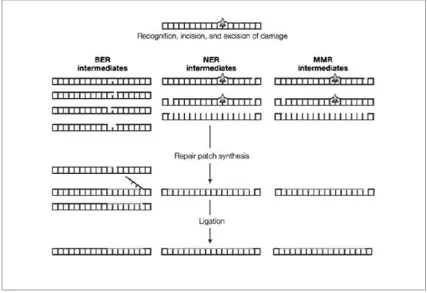
Figure 1.2: A simple diagram showing the pathways and the main intermediates of BER, NER and MMR. ... 5
Figure 1.3: The structures of RNA ... 7
Figure 1.4: Cartoon of an OB fold ... 10
Figure 1.5: The structure of an OB fold from RPA. ... 11
Figure 1.6: The structure and domain organisation of RPA. ... 13
Figure 1.7: A cartoon of RPA binding to ssDNA and exchanging its position with another RPA protein. ... 16
Figure 1.8: A schematic of the main steps involved NER... 18
Figure 1.9: A cartoon showing the tetrameric structure of EcoSSB bound to ssDNA. ... 20
Figure 1.10: A cartoon showing two of EcoSSBs binding modes. ... 22
Figure 1.11: Phylogenetic tree showing the distinction between the 16SRNA sequences of the archaea... 25
Figure 1.12: The crystal structure of a SsoSSB monomer and the relative electrostatic charges on its surface. ... 27
Figure 1.13: Part of the OB fold from SsoSSB. ... 28
Figure 1.14: A plot of the molecular potential energy against the internuclear separation during an electronic transfer to an excited state. ... 35
Figure 1.15: A simple Jablonksi plot. ... 36
Figure 1.17: Vectors that describe the transition dipoles involved in FRET between
donor and acceptor molecules. ... 43
Figure 1.18: A schematic showing how the relative intensities of donor and acceptor
fluorescent dyes, with R0 = 6 nm, change with respect to distance. ... 49
Figure 1.19: A schematic to show how light refracts as it travels into a medium with
a lower refractive index (n1>n2.) ... 50
Figure 1.20: Schematic of a prism based FRET microscope. ... 52
Figure 1.21: A schematic of a surface being passivated with a SAM. ... 54
Figure 1.22: ssDNA can be specifically immobilised on a quartz slide through a
biotin with an eight carbon linker to the 5’ end. ... 55
Figure 1.23: DNA curtains imaged using RPA tagged with m-Cherry. ... 56
Figure 1.24: Schematics of RecA binding to ssDNA inside a porous lipid
nanocontainer. ... 57
Figure 1.25: The structure of Cy3 and Cy5 dyes. ... 61
Figure 2.1: Curly arrow mechanism for the maleimide thiol coupling used for
attaching fluorescent dyes to exposed thiol groups on cysteine residues on the surface
of proteins. ... 66
Figure 2.2: Four fluorescence spectra to illustrate how the Ratio A value is
calculated. ... 69
Figure 2.3: Schematic of ssNA with a length of 7 nt (M=7) where a protein that has a
binding site of 5 nt (n=5), gives a total of 3 possible overlapping binding sites. ... 71
Figure 2.4: Schematic of two proteins with binding sites of 2 nt on a lattice of length
Figure 2.5: Schematic showing a free ligand binding to a) a singly and b) a doubly
contiguous binding site (n=2, M=6 and for (a) k=2,j=1 and (b) k=3, j=2.)... 72
Figure 3.1: The purification of wild type SsoSSB. ... 83
Figure 3.2: A cartoon to show the volumes that fluorescent dyes can occupy bound to
ssDNA. ... 85
Figure 3.3: Minimum energy model of two SsoSSB monomers, each bound to 10nt
ssDNA Cy3 with a gap of a single nucleotide between the two proteins. ... 87
Figure 3.4: The viscosity of the solution is a barrier to the rotation around the double
bond in the alkyl chain between the E and Z conformations cyanine dyes can adopt.
... 89
Figure 3.5: Labelling of SsoSSB with Alexa 647 in the presence of 8 M urea
increases the efficiency of the labelling reaction. ... 91
Figure 3.6: FRET can be used to track SsoSSB binding to ssDNA by using ensemble
spectroscopy. ... 92
Figure 3.7: MALDI-TOF analysis of A114C SsoSSB before (top) and after (bottom)
labelling with spin label MTSSL. ... 94
Figure 3.8: PELDOR can be used to verify the structure of proteins such as Influenza
A Virus NS1. ... 95
Figure 3.9: PELDOR data from SsoSSB MTSL experiments... 97
Figure 3.10: Ensemble FRET spectra of 50 nM 39 nt ssDNA Cy3 Cy5 in the absence
and presence of SsoSSB showing a decrease in ratioA, that is consistent with the
Figure 3.11: From the data from PELDOR and end to distance measurements this
suggests that SsoSSB binds as a monomer and produces a relatively unordered
nucleofilament. ... 101
Figure 3.12: Tryptophan quenching due to SsoSSB binding to ssDNA. ... 103
Figure 3.13: Enhancement of the fluorescence from 10 nM ssDNA Cy3 was
measured at different concentrations of unlabelled SsoSSB, and a two fold increase
in the intensity was observed. ... 106
Figure 3.14: Ensemble FRET titrations were also completed adding a SsoSSB Alexa
647 to 10 nM ssDNA Cy3, as shown in the cartoon. ... 107
Figure 3.15: A reverse titration adding ssDNA to 50 nM SsoSSB Alexa 647 was
completed in triplicate. ... 108
Figure 3.16: PIFE titrations could also be fitted to Epstein’s model that describes the
cooperative effects of SsoSSB binding to adjacent sites along ssDNA, taking into
account overlapping binding sites and the end effects of a short oligo. ... 109
Figure 3.17: Quenching of Alexa 647 fluorescence. ... 110
Figure 4.1: The fluorescence intensities from single molecules of 12C ssDNA Cy3
molecules. ... 119
Figure 4.2: Images taken from TIRF microscope of a clean PEG slide before and
after loading 100 nM SsoSSB labelled with Alexa 647 on to the surface. ... 121
Figure 4.3: The intensities of the fluorescence emitted from single molecules of 12C
ssDNA Cy3 immobilised on quartz slide have been normalised and plotted against
Figure 4.4: Histograms, each from approximately 1000 single molecule traces, which
show the fluorescence intensity of immobilised 12C ssDNA Cy3 on a quartz slide
being exposed to increasing concentrations of unlabelled SsoSSB. ... 124
Figure 4.5: Histograms and a single molecule trace to show that at high
concentrations of SsoSSB the traces showed no dynamics but were approximately
twice the intensity of 12C ssDNA Cy3 in the absence of protein. ... 125
Figure 4.6: Gaussian peaks fitted to histograms showing the frequency distribution of
the fluorescence intensity of 12C ssDNA Cy3 at 0 nM and 1 nM unlabelled SsoSSB
for approximately 200 molecules. ... 126
Figure 4.7: Single molecule traces showing SsoSSB Alexa 647 monomers binding to
12C ssDNA Cy3 as bursts of increased acceptor fluorescence accompanied by
quenching of the donor intensity, typical of highly efficient FRET events. ... 128
Figure 4.8: An example of a single molecule trace showing the changes in
fluorescence when one SsoSSB Alexa 647 monomer and two monomers bind to 12C
ssDNA Cy3. ... 130
Figure 4.9: An example of how the FRET, quenching and PIFE events can be
interpreted in terms of the number of SsoSSB Alexa 647 monomers bound to 12C
ssDNA Cy3. ... 131
Figure 4.10: Two SsoSSB monomers can bind to or dissociate from ssDNA either
sequentially or simultaneously. ... 132
Figure 4.11: The rates of koff and kon for the a single monomer binding to 12C
ssDNA Cy3 were calculated by measuring the time (a) immediately before a single
Figure 4.12: The dwell times of approximately 200 events showing a single SsoSSB
Alexa 647 monomer binding to 12C ssDNA Cy3 were measured at each
concentration (0.05, 2.5 and 10 nM) and plotted as histograms. ... 134
Figure 4.13: The rates of koff and kon for a second monomer binding to 12C ssDNA
Cy3 were calculated by measuring the time (a) immediately before the second
monomer bound to or (b) dissociated from ssDNA. ... 136
Figure 4.14: The dwell times of approximately 200 events showing a second SsoSSB
Alexa 647 monomer binding to 12C ssDNA Cy3 were measured at each
concentration (0.05, 2.5 and 10 nM) and plotted as histograms. ... 137
Figure 5.1: The results from ITC, performed by Dr Lisa Cubbedu, showing the
interaction of SsoSSB with ssDNA and RNA: with 21dA (a) and 21dU (b). ... 149
Figure 5.2: NMR analysis of SsoSSB binding to RNA and ssDNA by Dr Roland
Gamsjaeger. ... 150
Figure 5.3: Molecules of ssDNA and RNA bound to the OB fold of SsoSSB. Figure
made by Dr Roland Gamsjeager. ... 151
Figure 5.4: Ensemble fluorescence titrations describing the similarities between
SsoSSB binding to RNA and ssDNA. ... 153
Figure 5.5: Single molecule traces from (a) 12C ssDNA Cy3 and (b) 12C RNA Cy3
immobilised on to a microscope slide in the presence of SsoSSB monomers labelled
with Alexa 647. ... 155
Figure 5.6: SSoSSB binding to RNA may suggest a role in vivo such as acting as a
chaperone of RNA during transport or metabolism. ... 157
Figure 6.1: The enhanced fluorescence from 50 nM 20C ssDNA Cy3 was observed
Figure 6.2: PIFE was used again to study SsoSSB binding at temperatures closer to
the native environment of S. solfataricus. ... 165
Figure 6.3: Cartoons of the four C terminal tails of EcoSSB. ... 167
Figure 6.4: The end sequence of C terminal tails from the SSBs from various
organisms, showing that the acidic residues (D and E) are commonly found at the tip
of the disordered tails. ... 169
Figure 6.5: Left shows how SsoSSB has a 33 amino acid C terminal tail that is long
enough to allow the acidic tip to interact with the OB fold. Right shows a model
produced by Vina Autodock and MGL Tools. ... 171
Figure 6.6: The decrease in PIFE was used to track the dissociation of 50 nM
unlabelled SsoSSB from 25 nM 12C ssDNA Cy3 as it binds to a 1000 times excess
List of tables
Table 2.1: Names and sequences of nucleic acids used. ... 79
Table 3.1: Dissociation constants from tryptophan quenching experiments with
increasing lengths of ssDNA that can respectively allow up to 1, 2 and 4 monomers
to bind to them at any one time. Dissociation constants were calculated from a
modified one to one binding model. ... 104
Table 4.1: Table of observed association constants for the first and second SsoSSB
monomers and dimers to bind to ssDNA, represented as kon,1, kon,2 and kon,1.2
respectively. ... 136
Table 4.2: Table of observed dissociation constants for the first and second SsoSSB
monomers and dimers to bind to ssDNA, represented as koff,1, koff,2 and koff,1.2
respectively. ... 136
List of abbreviations
(SSB)35 SSB binding mode occluding 35 nt
APS Ammonium persulfate
ATP Adenosine triphophate
BER Base excision repair
bp Base pair
DBD DNA binding domain
DMPC 1,2-dimyristoyl-sn-glycero-3-phosphocholine
DNA Deoxyribonucleic acid
dsDNA Double stranded DNA
DTT Dithiothreitol
E. coli Escherichia coli
EcoSSB E. coli SSB
EDTA Ethylenediaminetetraacetic acid
EMCCD Electron multiplying charge coupled device
EPR Electron paramagnetic resonance
FRET Förster resonance energy transfer
FWHM Full width half maximum
HEPES 4-(2-hydroxyethyl)-1-piperazineethanesulfonic acid
HSQC NMR Heteronuclear single quantum coherence NMR
HtH Helix turn helix
ITC Isothermal caloimetery
L12 Loop connecting beta sheets 1 and 2
LB Luria-Bertani
MALDI-TOF Matrix associated laser desorption ionisation time of flight
MTSSL (1-Oxyl-2,2,5,5-tetramethylpyrroline-3-methyl) methanethiosulfonate
NA Nucleic acid
NER Nucleotide excision repair
NHS N-HydroxySuccinimide
NMR Nuclear magnetic resonance
nt Nucleotide
OB Oligonucleotide/oligosaccharide binding
PDB Protein database
PEG Polyethylene glycol
PELDOR Pulsed electron–electron double resonance
PIFE Protein induced fluroescence enhancement
RecA DNA recombination and repair protein A
RNA Ribonucleic acid
RPA Replication protein A
Rrp Ribosomal RNA processing protein
S. solfataricus Sulfolobus solfataricus
SAM Self assembled monolayer
SCOP Structural classification of proteins
smFRET single molecule FRET
smTIRF single molecule TIRF
ss Single stranded
SSB ssDNA binding protein
ssDNA single stranded DNA
ssNA single stranded NA
SsoSSB S. solfataricus SSB
STM Site targeted mutagenesis
TBE TRIS borate EDTA buffer
TEMED Tetramethylethylenediamine
TFB Transcription factor B
TIRF Total internal reflection fluoresence
Declarations:
1. Candidates’s declaration:
I, Michael Morten, hereby certify that this thesis, which is approximately 50,000
words in length, has been written by me, and that it is the record of work carried out
by me, or principally by myself in collaboration with others as acknowledged, and
that it has not been submitted in any previous application for a higher degree.
I was admitted as a research student in September 2011 and as a candidate for the
degree of PhD in September 2011; the higher study for which this is a record was
carried out in the University of St Andrews between 2011 and 2015.
Date:
Michael Morten
2. Supervisor’s declaration:
I hereby certify that the candidate has fulfilled the conditions of the Resolution and
Regulations appropriate for the degree of PhD in the University of St Andrews and
that the candidate is qualified to submit this thesis in application for that degree.
Date:
Prof. Malcolm White
Date:
3. Permission for publication: (to be signed by both candidate and supervisor) In submitting this thesis to the University of St Andrews I understand that I am
giving permission for it to be made available for use in accordance with the
regulations of the University Library for the time being in force, subject to any
copyright vested in the work not being affected thereby. I also understand that the
title and the abstract will be published, and that a copy of the work may be made and
supplied to any bona fide library or research worker, that my thesis will be
electronically accessible for personal or research use unless exempt by award of an
embargo as requested below, and that the library has the right to migrate my thesis
into new electronic forms as required to ensure continued access to the thesis. I have
obtained any third-party copyright permissions that may be required in order to allow
such access and migration, or have requested the appropriate embargo below.
The following is an agreed request by candidate and supervisor regarding the
publication of this thesis:
PRINTED COPY
ELECTRONIC COPY
Embargo on all of electronic copy for a period of 1 year on the following ground(s):
Publication would preclude future publication
Date:
Michael Morten
Date:
Prof. Malcolm White
Date:
Acknowledgements
First and foremost I would like to thank my supervisors Prof. Malcolm White and Dr
Carlos Penedo. They have both been incredibly supportive and have given me the
opportunity and encouragement to study an interesting and engaging project.
I am extremely grateful to the members of both the MFW and JCP groups for helping
me and keeping me going throughout the project. Special mentions go to Dr Jose
Peregrina and Biljana Petrovic-Stojanovska as they were the first ones to try and
teach me the techniques used in my PhD, however everyone in the labs was always
very kind with their time when I needed their help - so thank you!
With respect to the work presented in this thesis, I am very grateful for Dr Bela
Bodewho carried out the PELDOR work, Dr Elena Evguenieva-Hackenberg who
kindly donated the exosome, Dr Jose Peregrina who made the models of SsoSSB, Dr
Catherine Botting and Dr Sally Shirran who carried out the MALDI-TOF
experiments and I have also been fortunate enough to benefit from a collaboration on
the RNA work with Dr Liza Cubeddu and Dr Roland Gamsjaeger.
I would also like to thank my family and the pals in and out of the lab who made the
St Andrews experience so enjoyable, as well as Hannah for being so supportive
Abstract
Single-stranded DNA binding proteins (SSB) bind to single-stranded DNA (ssDNA)
that is generated by molecular machines such as helicases and polymerases. SSBs
play crucial roles in DNA translation, replication and repair and their importance is
demonstrated by their inclusion across all domains of life. The homotetrameric E.
coli SSB and the heterotrimeric human RPA demonstrate how SSBs can vary
structurally, but all fulfil their roles by employing oligonucleotide/oligosaccharide
binding (OB) folds. Nucleofilaments of SSB proteins bound to ssDNA sequester the
ssDNA strands, and in doing so protect exposed bases, keep the ssDNA in
conformations favoured by other proteins that metabolise DNA and also recruit other
proteins to bind to ssDNA.
This thesis focuses on the SSB from the archaeon S. solfataricus (SsoSSB), and has
found SsoSSB to be a monomer that binds cooperatively to ssDNA with a binding
site size of 4-5 nucleotides. Tagging ssDNA and SsoSSB with fluorescent labels
allowed the real time observation of single molecule interactions during the initial
nucleation event and subsequent binding of an adjacent SsoSSB monomer. This was
achieved by interpreting fluorescent traces that have recorded combinations of
FRET, protein induced fluorescent enhancement (PIFE) and quenching events. This
novel analysis gave precise measurements of the dynamics of the first and second
monomers binding to ssDNA, which allowed affinity and cooperativity constants to
SsoSSB was also found to have a similar affinity for RNA, demonstrating a
promiscuity not found in other SSBs and suggesting further roles for SsoSSB in the
cell - possibly exploiting its capacity to protect nucleic acids from degradation. The
extreme temperatures that S. solfataricus experiences and the strength of the
interaction with ssDNA and RNA make exploring the application of SsoSSB for
industrial uses an interesting prospect; and its rare monomeric structure provides an
opportunity to investigate the action of OB folds in a more isolated environment than
1.
Introduction
1.1. Double-stranded DNA
All cellular life depends on DNA – a macromolecule that is used as a code for an
organism’s genomic information. Its structure was famously solved by Franklin,
Watson and Crick in 1953 which sparked a rapid gain in knowledge about biological
systems, and the central dogma of biology of ‘DNA makes RNA makes proteins’
was soon coined.1, 2
The DNA molecule itself is a polymer and usually exists in a conformation that
comprises two strands woven around each other creating a double helix. The most
common form is B-DNA: a right handed double helix has a pitch of 34 Å and a
radius of 10 Å. Each DNA polymer consists of monomer units called nucleotides,
which are conjugated together through a sugar phosphate backbone that run around
the outside of the double stranded molecule. The covalent bonds that run through one
nucleotide to the next pass through the 3’ and 5’ carbon of the sugar ring, giving a
single strand of DNA an asymmetric characteristic and thus directionality. The
strands in the double helix run in opposite directions to each other, which allows
Figure 1.1: Crystal structure of B DNA.
The crystal structure of B DNA is shown on the left (pdb: 1bna) with the cartoon on the right showing the chemical structure of adenine, thymine, cytosine and guanine with hydrogen bonds between complementary base pairs drawn as dashed lines.
In the centre of the two strands lie aromatic bases that project inwards and lie normal
to the direction propagated by the backbones. The bases make up a four letter
alphabet that code for an organism’s genome, and in DNA they are cytosine (C),
guanine (G), thymine (T) and adenine (A). These bases may pair through specific
hydrogen bonds so that G-C and A-T pairs are formed. The complementary base
pairing contributes to holding the two strands together, and facilitates the replication
of genetic material – a fact that had not ‘escaped the notice’ of Watson and Crick in
1953.1 It since been shown that complementary base pairing of nucleic acids is also
integral in transcription, translation and repair processes.
1.2. DNA damage and mutations
The role of DNA in the cell is to faithfully store genomic information, however this
information also needs to be accessible, which requires the macromolecule to present
made during replication and gives opportunities for damaging reagents to interact
with DNA.
Naturally occurring reagents such as reactive oxygen, nitrogen, or carbonyl species
as well as lipid peroxidation products and alkylating reagents can produce abnormal
chemical structures of DNA which can disrupt and stall DNA replication and
transcription.3 Hydrolytic processes in the cell cause breakages in DNA strands
which can also be fatal for the cell.4 Mispaired bases, insertions and deletions can
arise from faulty DNA replication or spontaneous degradation and can also result in
irregular conformations of DNA, where bases can be partially flipped out of the
double helix.5 Exogenous causes of DNA damage include mutagenic chemicals,
toxins, thermal disruption and exposure to UV, X-ray and gamma radiation.3
A mutation is a heritable alteration to the original nucleotide sequence of a genome -
either through an insertion, deletion or substitution of one or more nucleotides. This
can occur through spontaneous molecular degradation of DNA; through error prone
translesion synthesis where replication proteins that can bypass DNA damage also
tend to offer low fidelity as they process along undamaged templates; through faulty
DNA repair; and through mutagenic chemicals and UV light.
1.3. DNA repair
Each day there could be up to 1 million events that damage DNA in a single human
cell.6 This still only affects a small fraction of the genome; however the damage to
repair DNA in multiple pathways to cope with the variation in types of damage.7
These pathways can be classed as either direct reversal of the DNA damage or
excision repair. Direct reversal of the chemical steps that occurred during DNA
damage can be more efficient than excision repair, however it is only possible to
repair a few particular types of damage using a direct reversal pathway. Examples of
direct reversal pathways include the removal of alkyl adducts from guanine bases
and the reversal of pyrimidine dimer formation, by methyl guanine methyl
transferase proteins and photolyases respectively.8, 9 Humans do not possess a
photolysase enzyme and rely on excision repair pathways to correct for these and
other damaged DNA structures.8
During excision repair processes the damage to the DNA is recognised, the damaged
DNA is removed and replaced with newly generated DNA. The three types of
excision repair are base excision repair (BER), nucleotide excision repair (NER) and
mismatch repair (MMR). As their names suggest BER involves the removal of a
damaged base, NER includes the removal of a damaged nucleotide and MMR
corrects undamaged but mismatched base pairs. The roles of each pathway overlap
with each other and as a result they can cooperate to keep the genomic information
Figure 1.2: A simple diagram showing the pathways and the main intermediates of BER, NER and MMR.
The damage or mismatch (arrow) is first recognised and a repair patch containing the damage is removed from DNA. The non-damaged strand is used as a template to re-synthesise the missing patch of DNA and the repair of DNA is completed by the ligation of the nascent patch to the DNA backbone of the now repaired strand. Figure modified from Cline and Hanawalt.10
1.4. Single-stranded nucleic acids
The planar arrangement of the aromatic bases in a dsDNA double helix lends itself to
π-π stacking where the overlap of the p orbitals contribute to the stability of the
double helix.11 The arrangement of the sugar-phosphate backbone on the outside of
the double helix provides a barrier between the bases and potential damaging agents,
which gives a certain amount of protection and ensures the fidelity of the genetic
information. For example, hydrolytic deamination of cytosine occurs three orders of
magnitude slower in dsDNA than ssDNA as the double helix structure prevents
solvent access to the bases.12 Unfortunately, the arrangement of dsDNA also presents
order to complete these vital processes, the two strands may have to be separated to
expose the bases and allow access to the genetic code.
Single-stranded nucleic acid (ssNA) are more flexible and, unlike double stranded
DNA, a helix is no longer the lowest energy conformation.13 A single strand of NA
collapses into less defined forms, but still adopts conformations that minimise like
charge interactions along the phosphate backbone.14, 15 As a result, a single strand of
NA will generally be more compact in high salt concentrations, allowing the
phosphates to exist in closer proximity to each other since ions in solution can shield
the electrostatic repulsion along the sugar phosphate backbone. Depending on the
sequence of the bases, complementary pairing between bases on the same strand may
be possible and this coupled with base stacking and the ions in solution could result
in the molecule being able to fold up to give a more defined structure. This allows
certain ssNA’s to perform cellular functions, as is the case for riboswitches, tRNA
and other hairpins and pseudoknots.16, 17
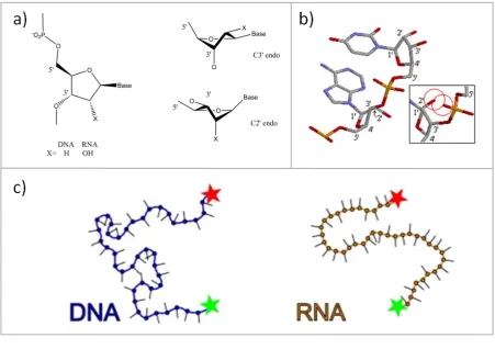
ssNA are present in the cell as single-stranded DNA (ssDNA) or RNA. RNA has a
similar structure to DNA and it is thought that RNA pre-dated DNA as a genomic
material in an earlier RNA/protein world.18 Each RNA nucleotide also consists of a
sugar-base nucleoside conjugated to a monophosphate, however, the sugar has an
extra hydroxyl group at the 2’ carbon of the ribose sugar. The base thymine is not
present in RNA which uses uracil (U) instead. The differences between the two NA
strands result in RNA being a shorter, stiffer polymer than its ssDNA analogue due
Figure 1.3: The structures of RNA
(a) The difference between ribose and deoxyribose sugars is the 2’ hydroxyl group. ssDNA can interconvert relatively easily between the C3’ endo and C2’ endo conformations. (b) RNA shows more of a preference towards the C3’ endo as shown by the 3D structure. The inset shows the clash between the 2’ hydroxyl and the phosphate group whilst the RNA adopts the C2’ endo conformation. (c) The increased base stacking due to the presence of uracil rather than thymine, and the barrier for the ribose sugar adopting the C2’ endo ring pucker results in polymers of RNA being shorter and stiffer than ssDNA analogues. This was observed by Chen et al. who measured the distance between fluorescence dyes (green and red stars) conjugated to the 3’ and 5’ ends of ssDNA and RNA.14
1.5. Single-stranded DNA binding proteins
The propensity of ssDNA to fold and adopt compact conformations could potentially
cause fatal stalls of molecule machinery involved in DNA repair, replication and
transcription.19 Breakages in ssDNA occur more frequently than in dsDNA and are
extremely disruptive to cellular life.20 Specialist proteins named single stranded DNA
binding proteins (SSB) are expressed to protect the ssDNA,21 to sequester the strands
conformation of the polymer that is conducive to other proteins that metabolise or
manipulate DNA.22
By definition SSBs have a high affinity for ssDNA and they form filaments that
decorate the single strands through sequence independent binding,23, 24 allowing them
to provide an essential role in a large number of processes. The high affinity of SSBs
for ssDNA rather than dsDNA or RNA, automatically gives the cell a marker for
ssDNA if it can somehow detect and exploit SSB’s presence on ssDNA.25, 26
This
potentially could enhance the efficacy of essential DNA processes as proteins with
lower affinity to ssDNA than SSB as an initial interaction with the SSB, rather than
the DNA, could increase the rate of binding and stabilise the protein DNA
interaction. SSBs must also be removed readily from ssDNA at some point in DNA
metabolism, either to allow other proteins access to the genetic code or simply for the
regeneration of the double helix at the end of replication, transcription or repair.
There are a great range of SSBs varying in structure and function, from monomers to
pentamers.27-32
Here a brief overview is presented of some of the common features shared amongst
SSBs as well as short overviews on two of the more well known SSBs: the human
replication protein A (RPA) and the SSB from E. coli (EcoSSB.) These two proteins
can both alternate between different ssDNA binding modes, to allow the
re-distribution of proteins along a nucleofilament, and also engage with other proteins
different strategies which each protein employs in order to carry out the same
functions.
1.5.1. Oligonucleotide/oligosaccharide binding folds.
SSBs are ubiquitous in all cellular life and there are also examples of viral SSBs,33
which reflects the vital roles these proteins fulfil.34 SSB sequence and overall
structure vary greatly between organisms but all bind to ssDNA through similar
oligonucleotide/oligosaccharide binding (OB) folds.35, 36 These folds all consist of
loops that can interact with the ssDNA as it enters the OB fold, forming hydrogen
bonds with the sugar phosphate backbone, anchoring the ssDNA in place. Aromatic
residues in the heart of the OB fold act like teeth in a zip, stacking between the bases
on the DNA strand, thus cementing the position of the OB fold along the strand.35
OB folds are not restricted to SSBs. Evolution has produced OB folds that have been
reported to be involved in inorganic pyrophosphatases in yeast, archaea and bacteria
to bind to a host of ligands - including RNA in anti-codon binding domains,
oligosaccharides in AB5 bacterial toxins, and proteins in superantigens.37, 38. A
schematic of an OB fold is shown in Figure 1.4, where the core of the OB fold is
made up of five β sheets typically forming a barrel with a Greek key motif,
sometimes capped by an α helix. This topology is conserved throughout nucleic acid
binding proteins and a total of sixteen Structural Classification of Proteins (SCOP)
superfamilies.39 This has led to the suggestion that OB folds are an example of an
ancient topology that is highly capable of withstanding a range of mutations,
folds can be tolerated in residues that are not integral to the structure of the β sheets,
away from the hydrophobic core. There is a wide variety observed in the length and
sequence of the hydrophilic loops that link the β sheets together that could account
for the exploitation of similar OB fold cores to fulfil a range of different functions.
The OB folds from the nucleic acid binding superfamily have been subjected to a
level of evolutionary pressure to remain unchanged since before the existence of the
last universal common ancestor, before the divergence of bacteria, eukaryotes and
archaea which could account for the similarity of these OB folds across all domains
of life.37
Figure 1.4: Cartoon of an OB fold
Five β sheets in Greek key topology (left) and folded to produce a cartoon example of an OB fold (right).
1.5.2. Loops
The rigid β strands of the OB fold barrel are linked via flexible hydrophilic loops,
which do not interfere with the hydrophobic core of the OB fold. Mutations in these
residues would not require the rearrangement of a significant number of atomic
contacts and are not as crucial during the folding of the protein structure.40 As a
of the β sheets, where the extension and sequence of the loops can determine which
ligand the OB fold may bind and therefore help define the protein’s function and
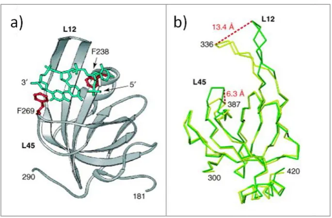
superfamily.36, 41 One example is seen in human RPA, where the L12 and L45 loops
are flexible enough to move closer to the ssDNA in order to form strong interactions,
[image:38.595.135.478.247.472.2]shown in Figure 1.5.35
Figure 1.5: The structure of an OB fold from RPA.
(a) Crystal structure of an OB fold from RPA showing the bound ssDNA as a green stick model. Aromatic residues are shown in red that take part in base stacking with the ssDNA. (b) Two Cα traces from crystal structure of RPA OB fold in DNA binding domain A (DBD-A) which is bound (yellow) and unbound (green) to ssDNA. The flexible loops L12 and L45 change position in order to interact
with the ssDNA, and the distance moved by each loop is labelled in red. Figure modified from Bochkarev et al.35
A variety of loops can be seen in members of the same group of proteins in the same
superfamily, and is exemplified by SSBs where loops are explicitly involved in
protein-protein interfaces and protein-ssDNA contacts outside the OB fold. As a
result, loops between β sheets are heavily implicated in determining the ssDNA
binding modes, the multimeric state of the SSB and the processes in which the SSB
1.5.3. Human replication protein A
A well studied example of a eukaryotic SSB is the Homo sapiens replication protein
A (RPA). No entire crystal structure of human RPA has been solved, however
structure containing the four OB folds`of an analogue RPA from Ustilago maydis,
bound to 32 nt ssDNA as shown in Figure 1.6(a).30 Crystal structures of truncated
human RPA have been solved, showing the first two OB folds from the bound to
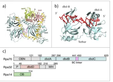
ssDNA.42 The heterotrimer of RPA consists of six OB folds in separate ssDNA
binding domains (DBD) spread across the three different subunits, as shown in
Figure 1.6(c). Each subunit is connected covalently to the next through flexible
linkers, and each is named RPA70, RPA32 and RPA14 to reflect their molecular
weight. Four OB folds are distributed around RPA70, one in RPA32 and another in
RPA14. There also is a winged helix-turn-helix (HtH) domain in RPA32 that assists
in binding to ssDNA but is primarily involved in protein-protein interactions.43, 44
RPA subunits bind sequentially to ssDNA in a 5’-3’ direction beginning with the two
OB folds found in ssDNA binding domain A (DBD-A) and DBD-B respectively, to
form the complex shown in Figure 1.6(b).42, 45, 46 DBD-A binds initially and is
tethered to DBD-B through a flexible loop, which results in the binding of DBD-B
quickly following DBD-A. NMR data suggest that these two domains tumble
independently of each other in solution and a thermodynamic analysis shows that
they bind to ssDNA in a non-cooperative fashion.47, 48 The tethering effectively
increases the concentration of ssDNA in DBD-B’s local environment and results in
an increased rate of binding once DBD-A has bound.47 RPA then binds ssDNA
cooperatively using two other OB-folds, DBD-C and DBD-D, found in the subunits
DBD-C occurs with a conformational change in the RPA structure where the zinc ribbon in
DBD-C stabilises the trimeric core and the interaction with ssDNA. The
conformational change also allows DBD-D to bind with ssDNA, with the whole
protein occluding 27 nt in the fully bound state. RPA14 and the OB fold found in the
N terminus of RPA70 lack two phenylalanines, the aromatic residues found that are
conserved in the other OB folds that are used to stack in between the bases of
ssDNA, and it is known the RPA14 does not contribute to ssDNA binding, and
participates in heterotrimer formation,49 but there is some controversy surrounding
[image:40.595.113.502.383.666.2]the N terminus of RPA70 and its role in ssDNA binding.50
Figure 1.6: The structure and domain organisation of RPA.
The different binding domains of RPA exhibit different affinities for ssDNA, with
the overall complex binding to ssDNA with a dissociation constant (Kd) that ranges
from picomolar to low nanomolar depending on the experimental conditions and
sequence and length of ssDNA,51, 52 which is two to three orders of magnitude lower
than DBD-A.47 The result of the conformational change when binding in the fully
bound state can be observed as the protein holds the ssDNA along a U-shaped path
through the four OB folds, as shown in Figure 1.6(a).30 Multiple trimers of RPA are
known to only bind to ssDNA with low cooperativity,52 which could be a result of
the high abundance of the protein inside the cell that negates any requirement for the
heterotrimers to prefer to bind adjacently to each other.52 The overall conformation
of an RPA-ssDNA nucleofilament is therefore relatively unordered, with kinks of
bare ssDNA in between the heterotrimers.53
A nucleofilament needs to both protect ssDNA and also allow other parts of the
cellular machinery access to the ssDNA. The proteins in the nucleofilament therefore
need to be easily redistributed or removed once they interact with other proteins
involved in transcription, replication or repair processes. The fully bound RPA is in
equilibrium with a partially bound state where only 8-10 nt are in contact with
DBD-A and DBD-B, which are the two DBDs with the highest affinity for ssDNDBD-A.42 These
two binding modes are shown in Figure 1.7, as well as a model of how they could be
exploited during the exchange of RPA heterotrimers. The different affinities of
RPA’s binding domains allow the protein to bind to ssDNA with different modes and
fulfil different roles in the cell, facilitating diffusion along ssDNA, melting dsDNA,
somewhat controlled through phosphorylation of RPA32, which can favour a
conformation of RPA that promotes the 8-10 nt binding mode over the fully bound
mode. RPA32 can be phosphorylated at a number of sites. The extent of
phosphorylation varies depending on the cell’s stage in mitosis and also in response
to different levels of DNA damage, leading to different affinities for ds and ssDNA.43
The winged helix and DBD-F primarily form contacts with other proteins in order to
fulfil RPA’s role to recruit proteins to ssDNA. The N terminus of RPA70 has been
shown to directly interact with proteins involved in DNA damage repair and
processes at replication checkpoints including p53, ATRIP, Mre11 and Rad9.56
RPA’s name comes from its role in replication that was identified when it was first
purified in 1988.57 In the initial replication phases, RPA’s high affinity for ssDNA
and its ability to melt dsDNA is used to initiate and recruit helicase and polymerase
alpha activity.58 During elongation, RPA recruits the polymerases delta and epsilon
to DNA through contacts with PCNA, as well as interacting with Dna2 during
Okazaki fragment processing.59, 60 Removal of RPA also allows FEN1 access to
Figure 1.7: A cartoon of RPA binding to ssDNA and exchanging its position with another RPA protein.
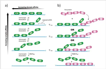
The different affinities for ssDNA of the different domains of RPA are exploited during exchange with other RPA heterotrimers and other proteins. The strength of RPA affinity increases with the number of DBDs bound to ssDNA. The affinity of each domain also differs, with DBD-A and DBD-B exhibiting the strongest affinity for ssDNA. RPA is modelled as dissociating from ssDNA as a sequence of microscopic dissociations of individual DBDs, shown on the left by the green protein. Similarly, the mechanism of RPA binding to ssDNA can also be broken down into the microscopic binding of each DBD. The exchange of two RPA proteins (one green, the other purple) can also be viewed in stages of DBDs binding and unbinding. As each green DBD dissociates, this exposes an increasing number of nucleotides available for other proteins to bind to, increasing the strength of interaction between the purple RPA and ssDNA as the affinity for ssDNA of the green RPA is weakened. Figure modified from Gibb et al.61
RPA also has roles in DNA repair, contributing towards NER, BER, MMR and
double strand break repair. For example, RPA displays a weaker affinity towards
damaged DNA, therefore during NER RPA preferentially binds to the undamaged
strand, protecting it from endonucleases (Figure 1.8).62, 63 The binding polarity,
coupled with the weaker affinity for damaged sites, helps to position the RPA to the
5’ side of the damaged strand which allows RPA to specifically recruit the nucleases
[image:43.595.90.511.85.361.2]64
Following the removal of the damaged section of DNA, RPA is poised to recruit
polymerases involved in DNA replication similar to the processes described above.65
The phosphorylation of RPA32 in response to DNA damage decreases RPA’s
affinity for ssDNA, which prevents RPA melting unnecessary lengths of dsDNA,
regulating and containing the nucleofilament structure around the DNA lesion. The
weakened binding also facilitates the removal of RPA by DNA polymerases at the
end of the repair process.66
RPA’s role in BER is less understood; however, its involvement is certainly critical
since substitution of mutants for wild-type RPA increases sensitivity to methyl
methane sulfonate, a lesion repaired through BER.67 Interactions between RPA and
DNA glycosylases, its capacity to enhance primer extension, unwind the downstream
strand at the 5’ end, and stimulate DNA ligase I all reinforce the importance of
RPA’s contributions to BER.68, 69
During MMR, RPA protects the template strand and has a role in stimulating and
regulating the nuclease activity that removes the mismatched base.70 The
phosphorylation of RPA32 leads to a decrease in RPA’s affinity for ssDNA, inducing
a conformation change where RPA32 interacts with DBD-F, moving DBD-C and D
away from the ssDNA.43, 71 Phosphorylation occurs before the re-annealing of DNA
Figure 1.8: A schematic of the main steps involved NER.
A repair response is triggered by the stalling of a transcription complex when it encounters damage or through global genomic repair (GGR) when a lesion is present on non-transcribed DNA. In transcription-coupled repair (TCR), the damage is encountered by the RNA polymerase II complex and the initiation of NER is carried out by CSB (also known as ERCC6). In GGR the lesion is detected by xeroderma pigmentosum complementation group E (XPE, also known as DDB1) or by XPC. RPA or XPA are involved in pre-incision events that precede the unwinding of a repair bubble by transcription factor II human (TFIIH). XPF and XPG are recruited to either side of the damage and incisions are made sequentially, initially by XPG at the 3’ site of the damage followed by XPF at the 5’ site. The section of nucleotides containing the lesion is then removed, with RPA able to assist in recruiting DNA polymerase (Polδ) and proliferating cell nuclear antigen, PCNA, with Polε to fill the gap with undamaged nucleotides. DNA ligase I (LIG1) is then able to complete the repair process by conjugating the new nucleotides to the original DNA strand. Figure modified from McKinnon.72
After 3’ overhangs are generated in response to a double strand break, RPA binds to
the ssDNA to protect and to prevent the formation of any unwanted secondary
structures.73 RPA recruits RAD52 through an interaction between the C terminus of
RPA32, stabilising the nucleoprotein complex, possibly through RAD52 interacting
with RAD52, coupled with the phosphorylation of RPA32 and subsequent change in
RPA conformation and resulting shift to the 8-10 bt binding mode, aids the exchange
between RAD51 and RPA on ssDNA.55, 75
1.5.4. Bacterial SSB
E. coli SSB (EcoSSB) is arguably the most intensively studied SSB with many
extensive reviews detailing its structure, functions and roles.76 It displays a well
characterised homotetramer structure with each 19 kDa monomer consisting of a
single OB fold, surrounded by extended loops that support contacts between the
monomers and other tetramers in addition to guiding the ssDNA around the
tetramer.32, 77, 78 Each monomer also has an acidic C terminal tail that has been shown
to be involved in protein recruitment, and also has a possible role in binding to
ssDNA although there are conflicting reports to the extent of its contribution.78-81 The
homotetramer structure is shown in Figure 1.9 which also displays the four
intrinsically disordered C terminal tails and the sequence of the acidic tips.
Similar to RPA, the multiple sites EcoSSB employs to bind to ssDNA allow the
diffusion and re-distribution of tetramers in a nucleofilament. Coupled with many
possible protein-protein interactions, this allows EcoSSB to assist in many different
roles in the cell. EcoSSB can wrap ssDNA around all four of its monomers, using
multiple points along the surface of EcoSSB both in and around the OB folds.32
These contacts provide both electrostatic and base stacking interactions, contributing
to a strong overall affinity towards the ssDNA. EcoSSB has multiple binding modes
cation concentrations, temperature and protein concentrations, with the (SSB)35 and
(SSB)65 modes being the two most prevalent, which are shown in Figure 1.10.82, 83
The number of nucleotides occluded is dependent on whether two or four monomers
are involved in binding, with each respective mode binding to 35 and 65 nt, thus
giving the different modes their name. The (SSB)35 mode is favoured under low salt
conditions and high EcoSSB concentrations, showing highly cooperative binding
between tetramers along a strand of ssDNA.84 The (SSB)65 mode has a limited
cooperativity producing beads consisting of two tetramers bound on ssDNA,
resulting from low protein concentrations and high salt concentrations.32
Figure 1.9: A cartoon showing the tetrameric structure of EcoSSB bound to ssDNA.
The four monomers are coloured blue, green, yellow and white with the four unstructured C terminal tail modelled as grey ribbons and the acidic C terminal residues are shown in red. The ssDNA in the (SSB)65 binding mode is represented as a red ribbon wrapped around the tetramer. Figure adapted
from Kozlov et al.85
The interaction of SSB with other proteins as well as ssDNA is clearly advantageous,
yet modifications to an OB fold to achieve this must be done without destabilising
ligand. Loops that act as linkers between β sheets are more likely to be involved in
ssDNA contacts and with neighbouring SSB monomers in the SSB filament.
Many SSBs have developed an intrinsically disordered C terminal tail that is
characteristically acidic towards its extreme end, including EcoSSB which has one
unstructured tail per monomer shown in Figure 1.9.31, 81, 86, 87 This hydrophilic tail is
thought to be able to recruit other proteins to ssDNA by protruding out from the SSB
as it is bound to ssDNA, which provides a surface for an electrostatic interaction with
positively charged areas of other proteins that are involved in DNA processes, such
as transcription, replication and DNA repair.26, 29, 86, 88 For example, EcoSSB
interacts directly with the clamp loader within DNA polymerase III holoenzyme (Pol
III HE), which assists clamp loading, aids processivity and allows the efficient
removal of tetramers from ssDNA that could potentially upset the polymerases
efficiency during DNA replication.89 EcoSSB’s association with E. coli primase
strengthens the primase’s interaction with the nascent RNA primer.90
Dissociation of
EcoSSB from the primase also destabilises the primase’s hold on ssDNA, and allows
the clamp assembly to occur. During DNA recombination, EcoSSB stimulates RecQ
helicase activity via interactions with the EcoSSB C terminal tail.91 EcoSSB also has
a role in stabilising the binding and promoting the activities of the exonuclease
RecJ,92 and the RecG helicase,93 as well as mediating the formation of RecA
Figure 1.10: A cartoon showing two of EcoSSBs binding modes.
EcoSSB is shown binding to ssDNA where (a) wraps 65 nt ssDNA around all four monomers and (b) where only 35 nt ssDNA is wrapped around two EcoSSB monomers. (c) The fully cooperative binding of three tetramers (the middle tetramer has its four monomers colour coded to match the tetramers in (a) and (b)) to ssDNA produced by (SSB)35, with strong interactions between proteins
assisted by the L45 loop. Figure modified from Raghunrathan et al.32
In DNA repair processes, EcoSSB recruits the exonuclease E. coli ExoI during
MMR,95 and interacts with Uracil DNA glycosylase during BER.96 DNA polymerase
II (pol II) is involved in a variety of responses to damaged DNA and requires
EcoSSB to process efficiently along DNA and to stimulate pol II-associated nuclease
being co-purified in 1972.26 Their interaction is also shown by the formation of a pol
II/SSB complex in the absence of ssDNA.22, 97
1.6. Archaea
The domains of life were historically classed as simply either plants or animals;
however this grouping could not take into account fungi, protists or bacteria. A new
classification attempt in the mid-20th century split five kingdoms into two domains
distinguishing between prokaryotic and eukaryotic organisms. This was again
revised following an analysis of ribosomal RNA which led to the discovery of a third
domain in 1977 by Woese and Fox.98 At the time they named this new domain the
archaeabacteria, which was eventually shortened to archaea to further distinguish the
domain from the bacteria.
Archaea are unicellular organisms that lack a nucleus but typically possess a singular
circular chromosome, similar to bacteria. Common characteristics of eukaryotes can
also be found when comparing their molecular machinery, where protein complexes
involved in processes such as transcription, DNA replication and translation share
the same fundamental features.99 Yet a clear genetic distinction of archaea from both
other domains can be seen through their 16S (18S) rRNA, which is present
throughout all types of self-replication systems and changes slowly over time so that
any link between distant species can be made.
The archaea was split further into Crenarchaeota and Euryarchaeaota, and additional
shown in Figure 1.11.100 It has also been suggested that the Nanoarchaea could be
classed as a separate phylum, however this has been countered with the proposal that
they are a rapidly evolving branch of the Euryarchaeaota.100 The Euryarchaeatoa
consists of the methanogens and their relatives, namely extreme halophiles,
sulfate-reducing species and thermophiles and also methangenic species, whereas the
Crenarchaeaota include (hyper)thermophiles, thermoacidophiles, eocytes and
sulfur-dependent archaea.101 The capacity of many archaea to survive in extreme
temperatures suggests their last common ancestor also was a thermophile; and it is
their tolerance for harsh conditions which makes them a desirable and viable
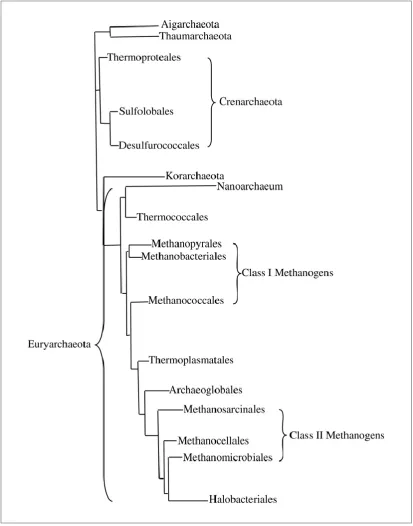
Figure 1.11: Phylogenetic tree showing the distinction between the 16SRNA sequences of the archaea.
[image:52.595.101.515.85.612.2]1.6.1. Archaeal SSB
SSBs from archaea show a large variation in structure and sequence, displaying
common features with both eukaryote and prokaryote SSBs, typically employing OB
folds. The euryarchaeon Methanosarcina acetivorans has three SSBs that all show
similarities to human RPA, and each has the capacity to form homomultimeric
complexes.102 These three proteins alone exhibit different binding modes and differ
in their molecular weight, multimeric state and the number of OB folds and zinc
fingers. Other methanogenic species that exhibit SSBs again show a range of
different ratios of OB folds and zinc fingers, including Methanopyrus kandleri,103
Methanocaldococcus jannaschii,104 and Methanothermobacter
thermautotrophicus.103 Proteins from non-methanogenic species of euryarchaea also
show similar zinc fingers and OB folds, including examples from Haloferax
volcanii,105 Ferroplasma acidarmanus,106Thermoplasma volcanium,107 and
Pyrococcus furiosus.108 Examples of SSBs from crenarchaea are not present or
unknown, the most well studied being from Sulfolobus solfataricus and a biophysical
analysis of this protein is presented in chapter 3 of this thesis.
1.6.1.1. Sulfolobus solfataricus SSB
The SSB from Sulfolobus solfataricus (SsoSSB) was identified by Wadsworth and
White, after purification from a cell extract.109 The mass of a monomer was
measured as 16.184 kDa, and in gel filtration of the recombinant protein was eluted
with an estimated size of 20 ± 4 kDa. The Kd for ssDNA-monomer complex was
strands with SSB monomers at a ratio of a single SSB monomer to approximately
4-5 nt of ssDNA, which was independent of the length of the ssDNA.109
Figure 1.12: The crystal structure of a SsoSSB monomer and the relative electrostatic charges on its surface.
(a) Cartoon of SsoSSB monomer with unstructured C terminal tail modelled on to the crystal structure. (b) Electrostatic surface generated on top of the structure shown in (a), clearly showing the acidic nature of the residues at the C terminus. (pdb: 1o7i)
The crystal structure of a truncated SsoSSB was solved as a monomer and is shown
in Figure 1.12(a). Crystals were produced from a construct where the highly flexible
C-terminal tail was removed by proteolytic digestion, at residue P115.27 This is
consistent with previous attempts to crystallise EcoSSB where the acidic terminal tail
had to be removed before the first structures were solved.110 A subsequent structure
of the full length E.coli protein could not provide any structure for the tail,
confirming its disordered nature.81 Previous work in the White lab has shown that
SsoSSB is expected to exist primarily as a monomer and multi-angle laser light
scattering experiments completed by Gamsjaeger et al. agree with SsoSSB adopting
a monomeric arrangement in solution.109 However Haseltine et al. also characterised
this protein and reported a mixture of dimeric, tetrameric and monomeric species
also suggested that this SSB is a tetramer, so clearly there is some debate in the
literature as to what multimeric form this protein takes.112
Figure 1.13: Part of the OB fold from SsoSSB.
An image of part of the crystal structure of SsoSSB showing the three aromatic residues (W56, W75 and F79) that can stack in between the bases of ssDNA. The loops that interact with the sugar phosphate backbone of ssDNA are also labelled as L12 and L45.
The crystal structure of SsoSSB shows the presence of a single OB fold, producing a
hydrophobic core for the binding of a single strand of DNA. Inside the core lie three
aromatic residues (W56, W75 and F79) which are involved in stacking between
bases, providing a major contribution to ssDNA binding, and are labelled in Figure
1.13. W75 appears to be both involved with base stacking and interacting with the
phosphate backbone.27, 113 SsoSSB has also been shown by Gamsjaeger et al. to bind
to ssDNA with a defined polarity identical to RPA.114 In the case of RPA, this plays
a crucial role in directing molecular traffic along ssDNA and SsoSSB could
potentially exploit this directionality in a similar vein.
The loop linking the β strands 1 and 2 (L12) and between 4 and 5 (L45) are expected
to close around ssDNA as it enters the OB fold, which is typical of other OB folds.27
protein-protein interactions are weaker and supports the idea SsoSSB is not as
capable of forming multimeric states which exploit the loops to strengthen the
interaction between monomers and guide the ssDNA around sets of monomers.27
The C terminal tail of SsoSSB has been shown to interact with RNA polymerase,
showing that SSB has the capacity to recruit RNA polymerase and/or initiate
transcription.86 SSB has been also shown to be able to substitute for the TATA
binding protein (TBP) and assist transcription factor II B (TFB) dependent
transcription.86 Proteolytic removal of the tail does not prevent SSB binding to
ssDNA but stops any interaction with other proteins and the deletion of the tail is
expected to cause severe repair-deficient phenotypes, similar to those seen in
EcoSSB.109
The level of SsoSSB expression in vivo only increases by a modest amount after
exposure to UV radiation, but has been shown to be able to selectively unwind
damaged dsDNA in vitro making SsoSSB a candidate for signalling DNA damage.
88, 115
It seems that SsoSSB is arguably one of the simplest SSBs structurally and yet it
contains all the necessary ingredients that an SSB requires to fulfil its role. A detailed
analysis of this stripped down model could provide a benchmark for answers to be
extrapolated into how other SSBs containing multiple OB folds bind to ssDNA and
facilitate essential cellular processes. It is clear that a nucleofilament must be a
capture the subtleties of how SsoSSB persists on ssDNA. Fluorescence microscopy
is capable of resolving single molecules and could provide the necessary level of
detail to be able to reach reliable conclusions about how SsoSSB and ssDNA
interact, and it is with these objectives in mind that single molecule techniques are
1.7. Single molecule assays
Achieving single molecule resolution allows the observer to simply look at the
structure and movement of individual molecules, which allows the observation of
temporal and spatial details that are otherwise lost in ensemble averaging. Difficult
ensemble experiments studying dynamic processes are made much easier as
synchronisation is not required when studying each molecule individually. Many
single molecule experiments can be completed quickly with ease producing large
amounts of data that can be used to construct frequency histograms, which are
effectively probability distribution functions. The shape and position of these
histograms not only give the ‘average’ result but can also reveal identify transient
states and heterogeneous behaviours that are difficult to observe with conventional
ensemble techniques.116
‘It is very easy to answer many of these fundamental biological questions; you just
look at the thing!’
Richard Feynman, ‘There’s plenty of room at the bottom,’1959117
Feynman was famously referring to electron microscopy in the quote above, but
there are a number of other single molecule techniques used to study biological
systems. A non-exhaustive list includes manipulation techniques (eg. atomic force
microscopy,118 scanning tunnelling microscopy,119 optical tweezers120 and magnetic
tweezers121), nanopore technology, and spectroscopic techniques such as surface
enhanced Raman spectroscopy122 and fluorescence microscopy, which is the primary