DOI 10.1007/s00792-016-0910-2
ORIGINAL PAPER
High-affinity RNA binding by a hyperthermophilic
single-stranded DNA-binding protein
Michael J. Morten1 · Roland Gamsjaeger2,3 · Liza Cubeddu2,3 ·
Ruvini Kariawasam2,3 · Jose Peregrina1 · J. Carlos Penedo1 · Malcolm F. White1
Received: 23 September 2016 / Accepted: 19 December 2016
© The Author(s) 2017. This article is published with open access at Springerlink.com
Introduction
Single-stranded DNA-binding proteins (SSBs) are essential for the genome maintenance of all known cellular organ-isms (Mushegian and Koonin 1996; Ashton et al. 2013) and are present in many viruses (Sun and Shamoo 2003). They play a vital role in DNA metabolism (Dickey et al. 2013), sequestering and protecting transiently formed ssDNA dur-ing DNA replication and recombination, meltdur-ing double-stranded DNA (dsDNA), and detecting DNA damage and recruiting repair proteins (Ashton et al. 2013; Sun and Shamoo 2003; Dickey et al. 2013; Theobald et al. 2003; Suck 1997). SSBs from the three domains of life share lit-tle sequence similarity and diverse subunit organisation (Dickey et al. 2013), but a common evolutionary feature of the SSB protein family is the oligonucleotide/oligosac-charide-binding (OB) fold (five-stranded beta-sheet coiled to form a closed beta-barrel), which can bind ssDNA with high affinity (Theobald et al. 2003). Although the persis-tence of the OB fold in all SSBs suggests a common ances-tor for these proteins (Suck 1997), the organisation of OB folds in SSBs varies considerably (Theobald et al. 2003). For example, Escherichia coli SSB (EcoSSB) is a homote-tramer, with each subunit consisting of a single OB domain for ssDNA binding, in conjunction with a flexible C-termi-nal extension involved in protein–protein interactions (Rag-hunathan et al. 2000). The Deinococcus/Thermus SSBs, whilst still utilising the tetrameric functional binding mode, arrive at this arrangement by combining two SSB homodi-mers: each SSB monomer encoding two OB folds linked by a conserved spacer sequence (Dabrowski et al. 2002). All eukaryotes utilise a heterotrimeric SSB known as rep-lication protein A (RPA) with six OB folds; two that medi-ate subunit interaction and four that are involved in ssDNA binding (Theobald et al. 2003; Bochkarev et al. 1999),
Abstract Single-stranded DNA-binding proteins (SSBs), including replication protein A (RPA) in eukaryotes, play a central role in DNA replication, recombination, and repair. SSBs utilise an oligonucleotide/oligosaccharide-binding (OB) fold domain to bind DNA, and typically oligomer-ise in solution to bring multiple OB fold domains together in the functional SSB. SSBs from hyperthermophilic cre-narchaea, such as Sulfolobus solfataricus, have an unusual structure with a single OB fold coupled to a flexible C-ter-minal tail. The OB fold resembles those in RPA, whilst the tail is reminiscent of bacterial SSBs and mediates interac-tion with other proteins. One paradigm in the field is that SSBs bind specifically to ssDNA and much less strongly to RNA, ensuring that their functions are restricted to DNA metabolism. Here, we use a combination of biochemical and biophysical approaches to demonstrate that the bind-ing properties of S. solfataricus SSB are essentially identi-cal for ssDNA and ssRNA. These features may represent an adaptation to a hyperthermophilic lifestyle, where DNA and RNA damage is a more frequent event.
Keywords RNA-binding proteins · OB fold · Single-molecule dynamics · Förster resonance energy transger · Nuclear magnetic resonance
Communicated by L. Huang.
* Malcolm F. White
[email protected]; [email protected]
1 Biomedical Sciences Research Complex, University of St
Andrews, St Andrews KY16 9ST, UK 2
School of Science and Health, Western Sydney University, Locked Bag 1797, Penrith, NSW 2751, Australia
3 School of Molecular Bioscience, University of Sydney,
whilst many also encode a second SSB (hSSB1/NABP2/ OBFC2B) with a single OB fold, which is involved in the maintenance of genome stability (Richard et al. 2008; Wu et al. 2016).
The arrangement of euryarchaeal SSBs is similar to eukaryotic RPA: a polypeptide or polypeptides with mul-tiple OB folds, including a characteristic OB fold inter-rupted by a zinc-binding domain (White 2003; Komori and Ishino 2001). This zinc-domain is also found in the large RPA70 subunit in eukaryotic RPA. It appears that some euryarchaeal SSBs form heterotrimers and others heter-odimers (Komori and Ishino 2001). In contrast, the cre-narchaeal SSB has a bacterial-like domain structure, with a single OB fold followed by a flexible C-terminal tail that is not involved in DNA binding and coats ssDNA with a stoichiometry of approximately 5 nucleotides (nt) DNA per SSB molecule (Wadsworth and White 2001). The crystal structure of the OB fold of Sulfolobus solfataricus SSB (SsoSSB) demonstrated its close structural relationship with the ssDNA-binding domains of human RPA70 (Kerr et al. 2003) and that of hSSB1 (Touma et al. 2016). The monomeric structure of SsoSSB in solution, both in the absence and presence of ssDNA, was recently confirmed by cross-linking experiments (Gamsjaeger et al. 2015) and by EPR and single-molecule molecule FRET (Morten et al.
2015).
Organisms inhabiting extreme environments where DNA damage is more frequent have a particular need to protect ssDNA, which is much more sensitive to damage than dsDNA (Ashton et al. 2013; Dickey et al. 2013). For example, the bacterium Deinococcus radiodurans main-tains a high level of SSB in the cell and increases that level nearly three-fold in response to ionising radiation (Bern-stein et al. 2004). Likewise, hyperthermophilic organ-isms, such as S. solfataricus, are also likely to experience elevated levels of DNA damage and it has been shown that SsoSSB expression is stimulated after UV irradiation (Wadsworth and White 2001; Gotz et al. 2007). Mutants of the archaeal halophile Halobacterium sp NRC1 with enhanced resistance to ionising radiation were shown to have enhanced expression of RPA (DeVeaux et al. 2007). E. coli, on the other hand, maintains a constant level of SSB that does not increase significantly in response to any DNA damage (Meyer and Laine 1990).
Materials and methods
Protein expression and purification
Recombinant SSB from S. solfataricus was prepared and purified as described previously (Wadsworth and White
2001). The A114C variant was constructed by site directed
mutagenesis using standard protocols (QuikChange, Strata-gene) and the sequences of oligonucleotides used for clon-ing and mutagenesis are available upon request. The variant was purified in the same manner as the wild-type SsoSSB. The A114C variant was then labelled with Alexa Fluor 647 using the manufacturers labelling buffer (Life Tech-nologies) and a ten times molar excess of fluorescent dye with the addition of urea to a final concentration of 8 M. The labelling reaction was left at room temperature for 3 h, and then overnight at 4 °C. The labelling mixture was then diluted with labelling buffer to half the concentration of urea. A pure sample of labelled proteins was obtained using an affinity column to remove the unlabelled pro-teins and any remaining free dye, as described previously (Wadsworth and White 2001). The labelling efficiency was checked by UV–vis spectroscopy and MALDI-TOF mass spectrometry and found to be >90%.
Oligonucleotides
Oligonucleotides were purchased from Eurofins MWG Operon and Qiagen. Sequences of oligonucleotides used in this study are shown in the table below. The positions of introduced biotin, fluorescein (FAM), and Cy3 and Cy5 dyes are indicated.
Name Sequences (5′–3′)
R21U UUUUUUUUUUUUUUUUUUUUU
R21A AAA AAA AAA AAA AAA AAA AAA
RNA-FAM FAM-UGAUAAUCUCUUAUAGAA
UUGAAAG
C12ssDNA Biotin-CCC CCC CCC CCC -Cy3
C12ssRNA Biotin-rCCC CCC CCC CCC -Cy3
RNA Hairpin Cy5-rUGAUAAUCUCUUAUAGAA
UUGAAAGU-Cy3
Isothermal titration calorimetry (ITC)
ITC buffer and/or ITC buffer to protein and were found to be similar to heats observed at the end of protein-DNA titrations. ITC-binding isotherms were analysed using a Single Set of Identical Sites model built-in to ITC Data Analysis in ORIGIN provided by the manufacturer. Non-linear least-squares fitting of the data to this model was performed using the ITC Data Analysis software. This fit does not consider any positive cooperativity and the KD values obtained are thus reported as “apparent KD’s”. This does not affect the main observation which is that RNA and DNA are bound similarly.
Ensemble-fluorescence experiments
Protein-induced fluorescence enhancement (PIFE) exper-iments were carried out in triplicate using a Varian Cary Eclipse fluorimeter, exciting the Cy3 dye at 550 nm. Oli-gonucleotides C12ssDNA Cy3 and C12ssRNA Cy3 (10 nM) were solubilized in 50 mM Tris–HCl pH 8.0, 10 mM KCl, and titrated with SsoSSB in the same buffer. Emis-sion intensity at each concentration of SsoSSB was cor-rected for dilution and the emission titration was fitted, as previously described (Morten et al. 2015), to a Hill model using Eq. 1.
where Bmax represents the maximum specific binding, KD is the concentration required for half-maximum binding, and n is the Hill coefficient.
Melting experiments were carried out using and intra-molecular FRET assay using Cy3 and Cy5 as FRET pair and the energy transfer efficiency was calculated using Eq. 2 and transformed into unwound fraction of hairpin. In Eq. 2, IDA and ID represent the intensity of the donor in the presence and absence of acceptor, respectively. Con-trol experiments to determine the variation in the emis-sion of Cy3 due to PIFE at each SsoSSB were also car-ried out.
Stoichiometric tryptophan quenching experiments were carried out as previously described (Ashton et al.
2013). We used an excitation wavelength of 300 nm and we titrated a 10 nM solution of unlabelled SsoSSB with increasing concentrations of unlabelled oligonucleotide. The area under the emission spectrum was taken at each data point. All ensemble data shown represent the average of three replicates.
(1)
FSSB
Fo =
BmaxXn
(
Kn
D+X
n)
(2)
EFRET=1−
IDA
ID
.
Single-molecule fluorescence
Single-molecule FRET data were taken using a home-built single-molecule prism-type total-internal reflec-tion microscope. Surface-immobilized oligonucleotides labelled with a donor Cy3 dye were exposed to SsoSSB labelled with the acceptor dye Alexa 647 as previously described (Morten et al. 2015). Quartz slides were pas-sivated using a PEG surface and biotin/neutravidin inter-actions head groups were exploited to immobilise C12 ssDNA Cy3 and C12 RNA Cy3 (Blouin et al. 2009). The sample was excited by a 532 nm laser (Crystalaser, USA) and the fluorescence from the donor and acceptor was collected using an electron-multiplying CCD cam-era (Ixon, Andor). Single-molecule intensity traces were analysed using laboratory-written MATLAB routines as previously described (McCluskey et al. 2013). Apparent FRET efficiencies after background corrections were cal-culated using (IA/(IA + ID)), where IA and ID represent the intensities of the acceptor and donor, respectively. Sin-gle-molecule FRET histograms were generated using the first 15 frames of each trajectory as previously reported (Morten et al. 2015; Bluoin et al. 2009; McCluskey et al.
2013). Single-molecule dwell-time histograms were cal-culated manually after filtering for blinking and pho-tobleaching effects and fitted to a monoexponential decay curve to extract the corresponding transition rate. Meas-urements were carried out at room temperature with inte-gration times of 50 ms per frame. The imaging buffer was identical to the ensemble binding buffer, with 200 μM Trolox, 6% (w/w) glucose and 0.1 mg/mL glucose oxi-dase, and 0.02 mg/mL glucose catalase added to reduce the rate of photobleaching and blinking of the fluorescent dyes.
NMR experiments and modelling
Exosome protection assay
Sulfolobus solfataricus exosome was purified as described previously (Witharana et al. 2012). 200 nM RNA labelled with a 5′-fluorescein (RNA-FAM) was incubated with wild-type SSB (concentrations from 0 to 480 µM) for 5 min at room temperature in 20 mM HEPES (pH 7.9), 0.1 mM EDTA, 60 mM KCl, 8 mM MgCl2, 2 mM DTT, and 10 mM K2HPO4. To each aliquot, 0.5 µl S. solfataricus Rrp41-Rrp42 hexameric ring and 0.4 µl Rrp4 protein were added. The total volume of each aliquot was 10 µl. The reaction was left to incubate at 60 °C for 1 h. 10 µl of each sample was added to acid phenol (Ambion) and mixed thoroughly, then spun at 13,000 rpm for 1 min. 5 µl from the resulting supernatant was added to 5 µl formamide (Promega) and loaded on to a denaturing gel (25% polyacrylamide, 7 M urea, 300 µl of ammonium persulfate (APS), and 30 µl of TEMED, 5 ml TBE, 5 ml water, total volume 50 ml) run at 85 W with a temperature threshold of 50 °C for 2.5 h. The gel was scanned using Fuji FLA5000 phosphorimager and analysed using the ImageJ software.
Results and discussion
The previous studies of eukaryotic and bacterial SSBs have suggested efficient discrimination between ssDNA and RNA (Mushegian and Koonin 1996; Ashton et al. 2013). In mesophilic organisms, this discrimination may serve to ensure that SSB is reserved for binding to ssDNA during replication and repair, not distributed over the much more abundant and omnipresent RNA in the cell. The ability of
these proteins to discriminate between ssDNA and ssRNA is not entirely understood, but it is thought to result from a combination of factors, including the lower plasticity of the RNA sugar pucker and the steric clash due to the pres-ence of the 2′ hydroxyl group that increases the energy barrier for binding and limits the conformational land-scape of ssRNA (Shamoo 2002). Usually, SSB proteins have only modest affinities for ssRNA (Meyer and Laine
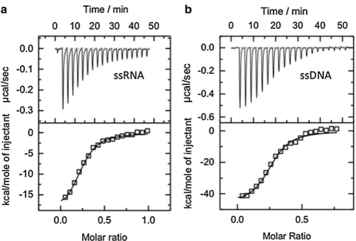
1990). For instance, human RPA binds to ssRNA with an affinity at least three orders of magnitude lower than that for binding ssDNA (Kim et al. 1992) and the early studies on the Escherichia coli SSB also indicated a much weaker affinity to ribopolymers than to their deoxy-counterparts (Ruyechan and Wetmur 1976; Molineux et al. 1975). Bac-terial cold shock proteins have been also reported to exhibit more than one order of magnitude decrease in binding affinity to ssRNA compared to ssDNA (Sachs et al. 2012). We were, therefore, surprised to observe using isothermal titration calorimetry that SsoSSB binds to a 21U RNA oli-gonucleotide (Fig. 1a) with a similar affinity (apparent KD = 93 ± 0.4 nM) as that seen for a 21T DNA oligonucleotide (apparent KD = 95 ± 0.6 nM) (Fig. 1b).
To investigate this unexpected property of SsoSSB fur-ther, we carried out ensemble-fluorescence experiments with 12 nucleotide ssRNA and ssDNA sequences func-tionalized with a Cy3 dye at the 3′ end. SsoSSB binding to these sequences was monitored using protein-induced fluorescence enhancement (PIFE). PIFE assays are based on the increase in the fluorescence emission of dyes due to the binding of proteins in close proximity and it has been extensively used as a molecular ruler to measure binding dynamics and distances shorter than those available by
Fig. 1 Representative iso-thermal titration calorimetry profiles for the interaction of
SsoSSB with a 21 nt poly-A DNA oligonucleotide (a) and a 21 nt poly-rA RNA oligo-nucleotide (b). The top panel
[image:4.595.184.544.470.714.2]Förster resonance energy transfer (FRET) (Morten et al.
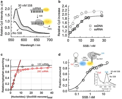
2015; Lerner et al. 2016). In the PIFE assay, we replaced the 21-mer employed for ITC by 12-mer ssDNA and ssRNA strands to ensure that monomer binding is within the distance range in which the PIFE mechanism can take place. The PIFE experiments with the ssRNA strand showed a >twofold increase in Cy3 emission (Fig. 2a), sim-ilar to the increase seen previously with ssDNA (Morten et al. 2015). The binding isotherms obtained when titrating
10 nM ssRNA and ssDNA were fitted to a Hill binding model and yielded similar apparent KD values of 4.2 ± 0.6 and 8.2 ± 0.9 nM, respectively (Fig. 2b). These values are very close to those reported previously for the binding of SsoSSB (Morten et al. 2015) to ssDNA under low ionic strength conditions where binding affinity is higher as dem-onstrated for other SSBs (Kernchen and Lipps 2006). The similarity between the affinity values also suggests that the presence of the dye at the 3′ end does not influence SsoSSB
Fig. 2 Ensemble-fluorescence characterization of the SsoSSB inter-action with single-stranded RNA oligonucleotides. a SsoSSB bind-ing to a 12-mer sbind-ingle-strand Cy3-labelled RNA monitored usbind-ing protein-induced fluorescence enhancement (PIFE). Fluorescence emission spectra of Cy3 as a function of SsoSSB concentration. The fluorescence spectrum in the absence of SsoSSB was normalized to unity at the wavelength of the maximum and taken as a reference to calculate the emission enhancement at each SsoSSB concentration. b
Relative variation in the emission intensity of a Cy3-labelled 12-mer ssDNA (black squares) and a Cy3-labelled 12-mer ssRNA (grey cir-cles) as a function of SsoSSB concentration obtained in a background of 10 mM KCl. Values represent the average of three experiments and are given as mean ± s.e.m. Solid lines represent the result from a non-linear squares fit to a Hill model as described by Eq. 1. c Stoichi-ometry of the SsoSSB-RNA interaction was determined using tryp-tophan emission quenching. A 460 nM concentration of SsoSSB was
[image:5.595.84.516.194.543.2]binding. From the fit, we obtained values for the Hill coef-ficients of 1.8 ± 0.3 for RNA and 1.6 ± 0.5 for DNA, imply-ing the interaction of more than one protein with a signifi-cant degree of positive cooperativity between them. Similar values for the apparent dissociation constant (6 ± 1 nM) and the Hill coefficient (1.7 ± 0.2) were obtained when the amount of titrated ssRNA was decreased to sub-nanomolar levels (~0.7 nM).
The number of ssRNA nucleotides occluded per SsoSSB monomer was further investigated using the intrinsic fluo-rescence of tryptophan as a reporter of binding (Fig. 2c). Structural studies of SsoSSB have confirmed that three tryptophan residues (W56, W75 and F79) are important for ssDNA binding (Wadsworth and White 2001; Kerr et al.
2003; Gamsjaeger et al. 2015). Stoichiometric titration of SsoSSB (460 nM) with increasing concentrations of a 12 C ssRNA sequence induced a 75% quenching of the trypto-phan emission and yielded a value of ~6–7 ribonucleotides interacting with each bound SsoSSB (Fig. 2c). Repeating the titration using a 20 C ssRNA yielded a similar number of nucleotides being protected by each SsoSSB monomer (Fig. 2c). This value is similar to that reported for the inter-action of SsoSSB with ssDNA using tryptophan quenching (~5–6 nt) (Wadsworth and White 2001) and gel electropho-resis-binding assays (~5 nt) (Cubeddu and White 2005) and in general agreement with the recent SsoSSB:ssDNA NMR structure where it was shown that 5 bases are sufficient for the recognition of ssDNA (Gamsjaeger et al. 2015).
It has been shown that SsoSSB can melt long stretches of duplex DNA in vitro at moderate temperatures (30–40 °C) and that this melting ability is enhanced when the duplex structure contains single mismatches and lesions, such as cyclobutane pyrimidine dimers (CPD) and extra-helical adducts (Cubeddu and White 2005). To explore whether this ability to disrupt secondary structure was also present for RNA sequences, we carried out FRET experiments using a RNA oligonucleotide capable of forming a hairpin structure containing a single-nucleotide bulge (Fig. 2d). FRET has extensively been used as a molecular ruler to monitor conformational changes within proteins and DNA–protein interactions (Blouin et al. 2009). The RNA hairpin was labelled with a Cy3–Cy5 FRET pair and the change in end-to-end distance was investigated as a func-tion of added protein (Fig. 2d). In the absence of SsoSSB, the fluorescence spectra obtained when exciting the Cy3 donor (λexc ~ 547 nm) showed a significant emission from the Cy5 acceptor dye (λem ~ 670 nm), indicative, as expected, of a high degree of energy transfer from the Cy3 to the Cy5 for the intact hairpin (Fig. 2d). However, in the presence of 20 nM SsoSSB, the spectrum was dominated by the emission from the Cy3, suggesting that the inter-dye distance had increased and, as a result, the FRET efficiency had decreased substantially. We interpreted this as evidence
that SsoSSB can efficiently disrupt the secondary structure of the hairpin RNA as previously observed for duplex DNA (Cubeddu and White 2005).
We have recently characterized the binding dynamics of SsoSSB monomers to surface-immobilized ssDNA using a single-molecule FRET approach (Morten et al. 2015). Single-molecule techniques are emerging as unique tools to unravel the dynamics of protein–DNA interactions (Morten et al. 2015; Blouin et al. 2009; Craggs et al. 2014) and they have been used extensively to investigate single-strand binding proteins, such as EcoSSB and RPA (Zhou and Ha
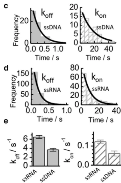
2012). To compared the dynamic properties of SsoSSB monomers binding to ssRNA and ssDNA, SsoSSB was labelled with an Alexa647 acceptor dye and a 12 C ssRNA or a 12 C ssDNA was doubly labelled with a biotin group at the 5′ end for surface immobilization to streptavidin coated microscope slides and with a Cy3 FRET donor at the 3′ end.
6 ± 1 s− 1 for ssRNA (Fig. 3e). The association rates were much slower than the dissociation rates, with values of 0.06 ± 0.01 s− 1 for ssDNA and 0.12 ± 0.08 s− 1 for ssRNA. Overall, the single-molecule data confirm that SsoSSB can bind ssDNA and ssRNA with similar efficiency and that individual SsoSSB monomers do not indefinitely persist on either of these oligonucleotides. Considering the harsh conditions to which thermophile organisms are exposed, a highly dynamic interaction between SsoSSB monomers and the nucleic acid sequence may provide the optimal bal-ance to ensure efficient protection whilst enabling access to nucleic acid processing proteins.
In the literature, there are examples of proteins that dis-criminate between ssDNA and ssRNA (Dickey et al. 2013). Schizosaccharomyces pombe Pot1 is the most extensively studied example of a protein that can selectively bind to ssDNA and it achieves this in a number of ways, including preferentially binding to thymine rather than uracil (Nanda-kumar et al. 2010). A strong hydrophobic interaction is
formed between the deoxythymine and a protein bind-ing site. In contrast, uracil lacks a methyl group, produc-ing an energetically unfavourable gap between the RNA and protein, weakening the strength of binding to RNA. Steric clashes between the 2′ hydroxyl group with Pot1 residue Ser123 and a phosphate group on the neighbour-ing nucleotide have also been identified as barriers to any strong affinity between RNA and the OB fold, and so facili-tate the selective binding of ssDNA (Nandakumar et al.
2010). The molecular basis for discrimination by RPA and EcoSSB between ssDNA and RNA is less well studied, but presumably arises from similar energetic penalties for the accommodation of the extra 2′ hydroxyl group in the bind-ing site of the protein, or from differences in the confor-mational flexibility of DNA and RNA (Chen et al. 2012). Having established that SsoSSB binds ssRNA with a simi-lar affinity and simisimi-lar kinetics as ssDNA, we next sought to determine whether there are any major structural differ-ences between DNA and RNA recognition. We carried out
Fig. 3 Single-molecule comparison of the interaction between Alexa647 labelled SsoSSB monomers and surface-immobilized 12-mer ssDNA (a) and 12 mer ssRNA (b) labelled with Cy3. Sin-gle-molecule donor (green) and acceptor (red) intensity trajectories (upper panel) are shown together with the corresponding FRET trace (black, bottom panel) obtained in the presence of 1 nM concentra-tion of SsoSSB. Anti-correlated fluctuations in the Cy3 and Alexa647 intensity signals result in FRET burst that indicate SsoSSB
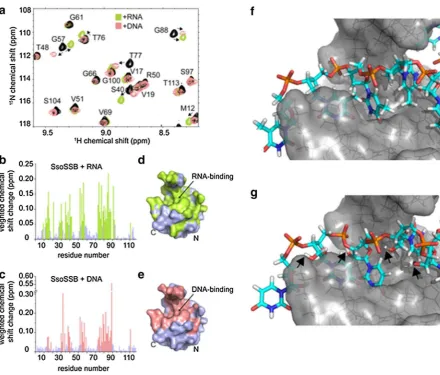
[image:7.595.306.508.53.359.2]NMR HSQC experiments of 15N-SsoSSB in the absence and presence of RNA revealing that the same residues that exhibit chemical shift changes upon binding of ssDNA are also significantly perturbed when RNA is added (Fig. 4a). These data suggest that the interaction surface is con-served between ssDNA and RNA. Indeed, mapping of the observed chemical shift changes onto the crystal structure of SsoSSB (PDB ID 1O7I) confirmed that ssDNA and RNA recognise essentially the same binding interface on the pro-tein (Fig. 4b–e). We have recently solved the structure of SsoSSB bound to ssDNA and have shown that the defin-ing feature of the complex structure is the base-stackdefin-ing of three aromatic residues (W56, W75 and F79) with three ssDNA bases (PDB ID 2MNA) (Gamsjaeger et al. 2015).
The NMR data suggest that this base-stacking mechanism is conserved between ssDNA and RNA. An in-silico model (Fig. 4f–g), calculated based on the NMR structure of the DNA-bound SsoSSB (Gamsjaeger et al. 2015) (assuming that replacing the ssDNA by RNA does not lead to a major change in the conformation of the nucleotide), provides further strong support for this notion. As seen from Fig. 4g, the model demonstrates that SsoSSB’s OB fold is capable of accommodating the 2′ hydroxyl group of the RNA and the effects of the resulting ring puckering without disrupt-ing the aromatic stackdisrupt-ing between the bases and aromatic residues in the OB fold.
In vivo, RNA in S. solfataricus is turned over by the exosome, which functions like the eukaryotic exosome
Fig. 4 NMR and molecular modelling characterization of SsoSSB binding to ssRNA and ssDNA. a Section of a 15 N HSQC spectrum of ~0.8–1 mM SsoSSB alone (black) and a 1:1 mixture of SsoSSB with 6U ssRNA (green) as well 6T ssDNA (salmon). Assignments and directions of movement are indicated. Weighted backbone chemi-cal shift changes of HN and N for SsoSSB upon binding to ssRNA (b) and ssDNA (c), respectively. Residues exhibiting changes larger than the average (binding residues) are coloured in green for RNA (b)
and salmon for DNA (c). Space-filling representation of the crystal structure of SsoSSB (PDB 1O7I) with binding residues coloured in
green for RNA (d) and salmon for DNA (e). Note the high similar-ity of the binding site for RNA compared to DNA. f Energy-lowest NMR structure (PDB ID 2MNA) of SsoSSB-DNA complex structure.
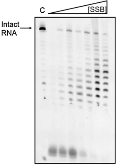
[image:8.595.57.498.249.624.2]by degrading RNA in a 3′–5′ direction (Evguenieva-Hackenberg et al. 2003). We, therefore, examined the effect of SsoSSB on the efficiency of RNA degradation by the exosome in vitro (Fig. 5). A 25 nt RNA oligonucleo-tide labelled with a 5′-fluorescein moiety was incubated with purified S. solfataricus exosome in the presence of increasing amounts of SsoSSB. At higher concentrations of SsoSSB, the activity of the exosome was progressively diminished, demonstrating that SsoSSB has the ability to bind and protect RNA against degradative enzymes in vitro. Partial protection of RNA by SsoSSB against digestion by benzonase was reported previously (Shi et al. 2013).
SsoSSB is clearly the major ssDNA-binding protein pre-sent in Sulfolobus cell extracts, and is estimated to consti-tute 0.1% of total soluble protein (Wadsworth and White
2001; Paytubi et al. 2012). Our data suggest that SsoSSB has the potential to associate with and stabilise unstruc-tured RNA molecules, such as mRNA, and thus increase its half-life at the elevated temperatures characteristic of hyperthermophilic organisms. In S. solfataricus, mRNA half-lives are longer than those seen in bacteria, which may reflect the increased stability and protection provided by
RNA-binding proteins (Bini et al. 2002). It is also possi-ble that SsoSSB plays a role in RNA remodelling in con-junction with RNA helicases, for example in ribosome bio-genesis, as SSB binding could protect unfolded rRNA and act as an RNA chaperone. We have shown previously that SsoSSB forms a tight physical interaction with RNA poly-merase via the C-terminal tail, and can stimulate transcrip-tion in vitro, consistent with a role as an mRNA chaperone (Richard et al. 2004).
There is a good reason to suppose that the OB fold evolved originally as an RNA-binding module, as RNA is thought to have predated DNA early in evolution (Orgel
1998), and several examples of OB fold domains spe-cialised for RNA binding have been reported. Examples include bacterial tRNA-binding proteins proposed to act as molecular chaperones to protect and stabilise tRNAs (Orgel 1998), N-terminal anti-codon binding domains of some class II tRNA synthetases (Swairjo et al. 2000), trans-lation initiation factors, and ribosomal proteins from bac-teria and archaea (Li and Hoffman 2001; Wu et al. 2003). The archaeal chromatin protein Alba, whose primary role is thought to require binding to dsDNA, has been shown to also interact quite strongly with RNA in vitro (Guo et al. 2003). SSBs from several hyperthermophilic species have been shown capable of binding RNA in vitro (Shi et al. 2013). The relaxed specificity of abundant nucleic acid binding proteins in hyperthermophiles may thus be a derived feature that has evolved to protect both ssDNA and RNA under extreme conditions, or alternatively reflect an ancestral state held over from the RNA world.
Acknowledgements Thanks to Elena Evguenieva-Hackenberg and Gabriele Klug, University of Giessen, Germany, for provision of the exosome sample.
Funding Wellcome Trust programme Grant [WT091825MA to M.F.W]. Royal Society Wolfson Merit Award (to M.F.W). Funding for open access charge: Wellcome Trust [WT091825MA].
Open Access This article is distributed under the terms of the Creative Commons Attribution 4.0 International License (http:// creativecommons.org/licenses/by/4.0/), which permits unrestricted use, distribution, and reproduction in any medium, provided you give appropriate credit to the original author(s) and the source, provide a link to the Creative Commons license, and indicate if changes were made.
References
Ashton NW, Bolderson E, Cubeddu L, O’Byrne KJ, Richard DJ (2013) Human single-stranded binding proteins are essential for maintaining genomic stability. BMC. Mol Biol 14:1–20
Bernstein DA, Eggington JM, Killoran MP, Misic AM, Cox MM, Keck JL (2004) Crystal structure of the Deinococcus radio-durans single-stranded DNA-binding protein suggests a
[image:9.595.70.270.351.632.2]mechanism for coping with DNA damage. Proc Natl Acad Sci USA 101:8575–8580
Bini E, Dikshit V, Dirksen K, Drozda M, Blum P (2002) Stability of mRNA in the hyperthermophilic archaeon Sulfolobus solfa-taricus. RNA 8:1129–1136
Blouin S, Craggs TD, Lafontaine DA, Penedo JC (2009) Functional studies of DNA–protein interactions using FRET techniques. Methods Mol Biol 543:475–502
Bochkarev A, Bochkareva E, Frappier L, Edwards AM (1999) The crystal structure of the complex of replication protein A subunits RPA32 and RPA14 reveals a mechanism for single-stranded DNA binding. EMBO J 18:4498–4504
Chen H, Meisbuger SP, Pabit SA, Sutton JL, Webb WW, Pol-lack L (2012) Ionic strength-dependent persistence lengths of single-stranded RNA and DNA. Proc Natl Acad Sci USA 109:799–804
Craggs TD, Hutton RD, Brenlla A, White MF, Penedo JC (2014) Single-molecule characterization of Fen1 and Fen1/PCNA complexes acting on flap substrates. Nucleic Acids Res 42:1857–1872
Cubeddu L, White MF (2005) DNA damage detection by an archaeal single-stranded DNA-binding protein. J Mol Biol 353:507–516
Dabrowski S, Olszewski M, Piatek R, Brillowska-Dabrowska A, Konopa G, Kur J (2002) Identification and characterization of single-stranded-DNA-binding proteins from Thermus thermo-philus and Thermus aquaticus—new arrangement of binding domains. Microbiology 148:3307–3315
de Vries R, van Dijk AD, Krzeminski M, van Dijk M, Thureau A, Hsu V, Wassenaar T, Bonvin AM (2007) HADDOCK versus HAD-DOCK: new features and performance of HADDOCK2.0 on the CAPRI targets. Proteins 69:726–733
DeVeaux LC, Muller JA, Smith J, Petrisko J, Wells DP, DasSarma S (2007) Extremely radiation-resistant mutants of a halophilic archaeon with increased single-stranded DNA-binding protein (RPA) gene expression. Radiat Res 168:507–514
Dickey TH, Altschuler SE, Wuttke DS (2013) Single-stranded DNA-binding proteins: multiple domains for multiple functions. Struc-ture 2:1074–1084
Dominguez C, Boelens R, Bonvin AM (2003) HADDOCK: a pro-tein–protein docking approach based on biochemical or biophys-ical information. J Am Chem Soc 125:1731–1737
Evguenieva-Hackenberg E, Walter P, Hochleitner E, Lottspeich F, Klug G (2003) An exosome-like complex in Sulfolobus solfatari-cus. EMBO Rep 4:889–893
Gamsjaeger R, Kariawasam R, Gimenez AX, Touma C, McIlwain E, Bernardo RE, Shepherd NE, Ataide SF, Dong Q, Richard DJ, White MF, Cubeddu L (2015) The structural basis of DNA bind-ing by the sbind-ingle stranded DNA bindbind-ing protein from Sulfolobus solfataricus. Biochem J 465:337–346
Götz D, Paytubi S, Munro S, Lundgren M, Bernander R, White MF (2007) Responses of hyperthermophilic crenarchaea to UV irra-diation. Genome Biol 8:R220
Guo R, Xue H, Huang L (2003) Ssh10b, a conserved thermo-philic archaeal protein, binds RNA in vivo. Mol Microbiol 50:1605–1615
Kernchen U, Lipps G (2006) Thermodynamic analysis of single-stranded binding activity of the archaeal replication protein A (RPA) from Sulfolobus solfataricus. BioChemistry 45:594–603 Kerr ID, Wadsworth RI, Cubeddu L, Blankenfeldt W, Naismith JH,
White MF (2003) Insights into ssDNA recognition by the OB fold from a structural and thermodynamic study of Sulfolobus SSB protein. EMBO J 22:2561–2570
Kim C, Snyder RO, Wold MS (1992) Binding properties of repli-cation protein A from human and yeast cells. Mol Cell Biol 12:3050–3059
Komori K, Ishino Y (2001) Replication protein A in Pyrococcus furiosus is involved in homologous DNA recombination. J Biol Chem 276:25654–25660
Lerner E, Ploetz E, Hohlbein J, Cordes T, Weiss S (2016) A quan-titative theoretical framework for protein-induced fluorescence enhancement-Forster-type resonance energy transfer (PIFE-FRET). J Phys Chem B 120:6401–6410
Li W, Hoffman DH (2001) Structure and dynamics of translation initi-ation factor aIF-1A from the archaeon Methanococcus jannaschii
determined by NMR spectroscopy. Protein Sci 10:2426–2438 McCluskey K, Shaw ES, Lafontaine DA, Penedo JC (2013)
Single-molecule fluorescence of nucleic acids. Methods Mol Biol 1076:759–791
Meyer RR, Laine LS (1990) The single-stranded DNA-binding pro-tein of Escherichia coli. Microbiol Rev 54:342–380
Molineaux IJ, Pauli N, Gefter ML (1975) Physical studies on the interaction between Escherichia coli DNA binding protein and nucleic acids. Nucleic Acids Res 2:1837–1921
Morten MJ, Peregrina JR, Figueira-Gonzalez M, Ackermann K, Bode BE, White MF, Penedo JC (2015) Binding dynamics of a mono-meric SSB protein to DNA: a single-molecule multi-process approach. Nucleic Acids Res 43:10907–10924
Mushegian AR, Koonin EV (1996) A minimal gene set for cellular life derived by comparison of complete bacterial genomes. Proc Natl Acad Sci USA 93:10268–10273
Nandakumar JE, Podell R, Cech TR (2010) How telomeric protein POT1 avoids RNA to achieve specificity for single-stranded DNA. Proc Natl Acad Sci USA 107:651–656
Orgel LE (1998) The origin of life—a review of facts and specula-tions. Trends Biochem Sci 23:491–495
Paytubi S, McMahon SA, Graham S, Liu H, Botting CH, Makarova KS, Koonin EV, Naismith JH, White MF (2012) Displacement of the canonical single-stranded DNA-binding protein in the Thermoproteales. Proc Natl Acad Sci USA 109:E398–E405 Raghunathan S, Kozlov AG, Lohman TM, Waksman G (2000)
Structure of the DNA binding domain of E. coli SSB bound to ssDNA. Nat Struct Biol 7:648–652
Richard DJ, Bell SD, White MF (2004) Physical and functional inter-action of the archaeal single-stranded DNA-binding protein SSB with RNA polymerase. Nucleic Acids Res 32:1065–1074 Richard DJ, Bolderson E, Cubeddu L, Wadsworth RI, Savage K,
Sharma GG, Nicolette ML, Tsvetanov S, McIlwraith MI, dita RK, Takeda S, Hay RT, Gautier J, West SC, Paull TT, Pan-dita TK, White MF, Khanna KK (2008) Single-stranded DNA-binding protein hSSB1 is critical for genomic stability. Nature 453:677–681
Ruyechan WT, Wetmur JG (1976) Studies on the noncooperative binding of the Escherichia coli DNA unwinding protein to sin-gle-stranded nucleic acids. BioChemistry 15:5057–5064 Sachs R, Max KEA, Heinemann U, Balcbach J (2012) RNA single
strands bind to a conserved surface of the major cold shock pro-tein in crystals and solution. RNA 18:65–76
Shamoo Y (2002) Single-stranded DNA-binding proteins. In: eLS. Wiley, Chichester. doi:10.1002/047001590X
Shi H, Zhang Y, Zhang G, Guo J, Zhang X, Song H, Lv J, Gao J, Wang Y, Chen L, Wang Y (2013) Systematic functional compar-ative analysis of four single-stranded DNA-binding proteins and their affection on viral RNA metabolism. PLoS One 8:e55076 Suck D (1997) Common fold, common function, common origin? Nat
Struct Biol 4:161–165
Sun S, Shamoo Y (2003) Biochemical characterization of interactions between DNA polymerase and single-stranded DNA-binding protein in bacteriophage RB69. J Biol Chem 278:3876–3881 Swairjo MA, Morales AJ, Wang CC, Ortiz AR, Schimmel P (2000)
Theobald DL, Mitton-Fry RM, Wuttke DS (2003) Nucleic acid rec-ognition by OB-fold proteins. Annu Rev Biomol Struct 32: 115–133.
Touma C, Kariawasam R, Gimenez AX, Barnardo RE, Ashton NW, Adams MN, Paquet N, Croll TI, O’Byrne KJ, Richards DJ, Cubeddu L, Gamsjaeger R (2016) A structural analysis of DNA binding by hSSB1 (NABP2/OBFC2B) in solution. Nucleic Acids Res. doi:10.1093/nar/gkw617
Wadsworth RI, White MF (2001) Identification and properties of the crenarchaeal single-stranded DNA binding protein from Sulfolo-bus solfataricus. Nucleic Acids Res 29:914–920
White MF (2003) Archaeal DNA repair: paradigms and puzzles. Bio-chem Soc Trans 31:690–693
Witharana C, Roppelt V, Lochnit G, Klug G, Evguenieva-Hackenberg E (2012) Heterogeneous complexes of the RNA exosome in Sul-folobus solfataricus. Biochimie 94:1578–1587
Wu B, Yee A, Pineda-Lucena A, Semesi A, Ramelot TA, Cort JR, Jung JW, Edwards A, Lee W, Kennedy M, Arrowsmith CH (2003) Solution structure of ribosomal protein S28E from Meth-anobacterium thermoautotrophicum. Protein Sci 12:2831–2837 Wu Y, Lu J, Kang T (2016) Human single-stranded DNA binding
pro-teins: guardians of genome stability. Acta Biochim Biophys Sin 48:671–677 (Shanghai)