The role of monocularly visible regions in depth and surface perception
Julie M. Harris1 and Laurie M. Wilcox2
1 School of Psychology, University of St. Andrews, South St., St. Andrews, KY16 9JP, Scotland
2 Department of Psychology Centre for Vision Research 4700 Keele St., North York Ontario, M3J 1P3 Canada
Abstract
The mainstream of binocular vision research has long been focused on understanding how binocular disparity is used for depth perception. In recent years, researchers have begun to explore how monocular regions in binocularly viewed scenes contribute to our perception of the three-dimensional world. Here we review the field as it currently stands, with a focus on
understanding the extent to which the role of monocular regions in depth perception can be understood using extant theories of binocular vision.
Key words: stereopsis, binocular vision, da Vinci Stereopsis, monocular regions, half-occlusion, monocular
(1) Introduction
(1.1) The problem: how do we get a 3-D view when the eyes see different things?
Because we have two forward facing eyes that are separated in the head, our visual system continuously receives two slightly different views of the world. For many decades scientists have been trying to understand how the visual system deals with the slight differences in the images that result from the lateral separation of the two eyes (since Wheatstone, 1838). Some have argued that the differences are so small that they are irrelevant. Instead, they suggest that the primary reason for having two eyes is for binocular concordance: that is, increasing visual
efficiency (reducing noise) by having effectively ‘a second go’ at viewing each scene (e.g. Jones & Lee, 1982). This approach makes sense when one views a distant scene, as the two views are very similar.
However, much of the time we function in environments in which there are multiple objects at close range, and for such scenes, there are substantial differences in the retinal images. Rather than emphasising concordance, a successful approach has been to consider the extent to which the visual system can exploit discordance by measuring the tiny differences between the two eyes’ views (binocular disparity), and by using disparity to represent the three-dimensional (3D)
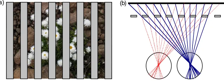
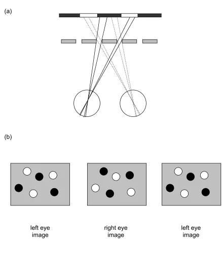
structure of the world. A rich literature has shown that binocular disparity is used by the visual system, and that we are exquisitely sensitive to it (e.g. Howard & Rogers, 2002). Measuring the differences between locations in the two eye’s views first requires a solution to the binocular correspondence problem. That is, which point on one retina matches with a given point on the other. This is a potentially complicated operation given the complex 3-D structure of the natural environment, but it is made even more difficult by the fact that not all image points in one eye have a partner in the other. For example, Figure 1a shows a natural scene viewed through an occluding foreground ‘fence’. We refer to this scene as the background because it is the furthest thing visible in the display. Figure 1b illustrates how there are features in one eye that are not present in the other, and vice versa. Notice that different portions of the background scene are occluded in each eye (in this extreme example none of the background is visible to both eyes).
For many years, researchers studying stereopsis treated these monocular regions as ‘noise’: a potential source of false matches and ambiguity, and therefore an obstacle to binocular disparity processing. Since the work of Gillam and Borsting (1988), and now many others, the prevailing view has changed. We have come to realize that these monocular regions or half-occlusions are useful, and play a potentially important role in binocular depth perception.
(1.2) History
The study of vision has a long history, stretching back to the ancient Greeks and Arabs. Howard (2002) gives an excellent introduction to the history of vision in general, and discusses binocular vision and occlusion in particular. He describes how Euclid, around 300 BC, first outlined the geometry of binocular vision and the fact that the two eyes see different parts of a sphere. Some 500 years later, Galen noted that when a foreground object is viewed, parts of objects lying behind that object are only seen by one eye. This point was later developed visually in a series of
drawings from Leonardo da Vinci (Wade, Ono & Lillakas, 2001; Richter, 1977) who was the first to note that depth perception can arise when each eye sees different parts of an object. Some of Leonardo’s drawings also illustrate that when looking through a hole, there are regions of the background scene that are only visible to one eye (see Strong, 1979).
Many of the beautiful and challenging examples of monocular regions that deliver depth
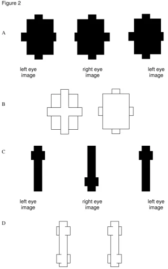
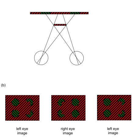
perception in binocular vision, that we will review below, were anticipated and demonstrated by von Szily (1921, translated by Ehrenstein & Gillam, 1998). Figure 2 shows two examples of his demonstrations. Lawson and Gulick (1967) first demonstrated experimentally that monocular regions of a scene can deliver a perception of depth akin to that from stereopsis. Many important issues in this field were discussed in a PhD thesis (otherwise unpublished) by Barrand (1979). Kaye (1978) was the first to show that a sensation of depth can be obtained from viewing an isolated point monocularly, and that its perceived depth depends on its location in on the retina.
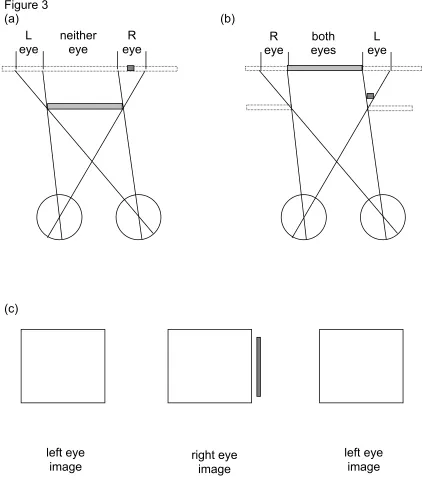
The first experimental evidence that monocular regions could specifically aid depth perception from binocular disparity came from Gillam’s lab in the 1980’s. She hypothesised that monocular regions could be used to identify the location of depth edges. This hypothesis was supported by evidence that perception of a depth edge occurs faster when explicitly textured monocular regions are present, than when they are left blank and the same colour as the surround (Gillam and Borsting, 1988). Perhaps the most well known study in this area is that which coined the term ‘da Vinci stereopsis’ to refer to the use of monocular regions in depth perception (Nakayama & Shimojo, 1990). In this extensive study of depth from monocular regions the authors used very simple stimuli, in which one eye’s view contained a bar that was not visible to the other eye (figure 3). When a stimulus is designed so that both eyes view the rectangle, and the right eye views an additional vertical bar to the right of the rectangle, the observer perceives the bar to lie behind the rectangle. This is consistent with viewing geometry as shown in figure 3a, where the bar is
occluded by the closer rectangle, in the left eye’s view.
Though known of for many years, monocular regions have been by-passed by much of the binocular vision community. Instead, there has been a focus on the significant computational problem of extracting the correct binocular disparity information from a pair of disparate images. This problem has perhaps most famously been set out by Marr, who used it as an exemplar of how a computational approach could yield a richer understanding of the mechanisms and algorithms underlying perception (Marr & Poggio, 1976, 1979). His approach, and that of many modellers of both human and machine vision since, was to initially break the problem into two parts: first corresponding image points must be identified between the two eyes views, and then the disparity can be extracted. Over the years, a variety of rules, or constraints, have been suggested to guide this process, including the assumption that each point has a single match (uniqueness constraint) and that the world is composed of piece-wise smooth objects
recent computational models that extract monocular regions. We discuss more recent, physiologically plausible models in section 5 below.
The exclusion of monocular regions from models of binocular disparity processing might be understandable if such phenomena were trivially rare. But in fact, monocular regions are
abundant in scenes containing a cluttered foreground. Figure 1a shows a foreground ‘picket-fence’ occluding a background scene, with figure 1b portraying how much of that scene is visible to each eye’s view. In this example, the background scene is only visible to one eye, or the other, never both. Whilst such an extreme example will occur rarely, monocular regions exist in real scenes at all vertical object boundaries where there is a significant depth discontinuity. In fact, such locations are arguably the regions of most interest in a scene, for they indicate where one object ends and the next begins (Gillam & Borsting, 1988; Anderson & Nakayama, 1994). Some recent modelling work has made this proposal explicit. For instance, Langer (2008) developed an artificial world in which square or spherical objects were randomly distributed through a volume (emulating natural cluttered scenes, like foliage or tree branches). He noted that occlusion in one eye’s view of a given point (resulting in a monocular region visible to the other eye) becomes increasingly more likely as its distance from the observer increases. In a world densely populated with objects, the further away a point is from the observer, the more likely it is that there will be an occluding object along any particular line of sight. Another very recent paper has taken this idea a step further. Changizi and Shimojo (2008) suggest that the main reason why forward-facing eyes have evolved is not for stereopsis, but rather to take advantage of the increased proportion of background objects that can be viewed in cluttered scenes with two eyes. As we describe below, this
hypothesis cannot account for the apparent utility of monocular regions in depth perception, so it surely cannot represent the whole story. However, the theoretical position adopted does illustrate the potential importance of monocularly visible regions for vision.
(1.3) Geometry and classification of monocular regions
In the real world objects are located at different depths. When viewed from certain locations, foreground objects result in partial occlusion of objects that are further away. These are different for each eye, because the eyes are laterally separated, which results in monocular regions of the scene; that is, regions that are visible to one eye only. There are several different ways in which this can occur. Here, we classify monocular regions into 3 types, that depend on the arrangement and features of the objects being viewed, as well as the relative location of the observer and her eyes. All the studies to be described in this review use stimuli that conform to one of these 3 types.
Type 1: monocular background
If foreground occluders are of a specific size and at a specific distance from a background scene, there will be regions of the images where the eyes are delivered completely different patterns and where there are no binocular regions that contain those patterns. Figure 1 is an example of this kind of stimulus, where the foreground occluders are just the right width, ensuring that no part of the background is simultaneously visible to both eyes (see also figures 4 and 10). This situation is rare in natural viewing, and although parts of these stimuli can appear rivalrous, scenes are perceived as stable and with a depth difference between foreground and background (Forte et al, 2002; Howard, 1995, Tsai & Victor, 2000). Phenomena linked to this configuration will be
discussed in section 3.2.
Type 2: binocularly visible foreground
real-world situations the foreground and background will both be visible, and differently textured or coloured. Under such conditions monocular regions are seen at the depth of the background.
Type 3: invisible foreground
Figure 3b shows a situation in which an observer views a background (featureless except for the binocularly visible rectangle) through a hole in a featureless foreground object. Such a scene is consistent with switching the two eye’s views in figure 3c, so that the left eye now views the stimulus containing the monocular bar. This has been called the camouflage configuration (Howard & Rogers, 2002), and can only occur when an object has the same texture and
luminance as the background (i.e. is camouflaged) in one eye, but not in the other (first described by Kaye, 1978). Figure 4a shows another example, where a small grey foreground line is
camouflaged to the left eye (it occludes a grey section of the background and is therefore invisible to that eye) but not to the right eye (from the right eye’s view the background region visible is striped). Stereo-pairs that simulate this configuration are shown in figure 4b. Although cases like this will be rare in the world, because they require the coincidence of identically patterned
foreground and background, the literature provides several recent examples that do appear to support depth perception from such images (see section 4.1, figure 10; section 4.3, figure 11).
A key issue to notice when considering these 3 types of occlusion is that Type 2 is much more common in the world than either Types 1 or 3, but that all are consistent with real 3-D scenes, as shown in the figures (this point runs contrary to the original ideas put forward in Nakayama & Shimojo, 1990, and will be taken up in more detail in section 2.3 below). The extent to which these three types of monocular stimulation may be processed via different mechanisms and whether depth perception mechanisms need to ‘know’ the geometrical constraints that underlie occlusion, is described and discussed in the sections below.
2 Monocular regions and depth perception
A number of different lines of research have explored depth from monocular regions, most of which use distinct stimuli. The challenge is to integrate the diverse effects that have been
discovered to form a coherent understanding. At the heart of our review is the question of whether the visual processing of monocular regions is distinctly different from classical stereopsis. Below we will examine some of the studies that have used stimuli containing monocular regions, where the monocular regions appear to be involved in depth perception. Some of these are directly aimed at understanding how depth is perceived from monocular regions, while others are related to this field by nature of the stimuli, but not by intent. We will consider these in separate sections reviewing what is widely accepted, what is controversial and what remains a puzzle.
2.1 Speed of depth perception
A monocular region is present whenever there is a significant depth discontinuity. Perhaps the simplest way that a monocular zone could provide information about depth is to signal the location of a depth discontinuity. Some work has demonstrated that monocular regions speed up depth processing. Saye and Frisby (1975) found that, for large disparities, monocular features did speed up depth detection in some configurations. Gillam & Borsting (1988) found that depth
discontinuities were more rapidly detected when the appropriate monocular zone was filled with the same pattern as the immediate surrounding background, than if it was left blank. Grove & Ono (1999) explored whether longer latencies occurred because a monocular region was missing, or because it was different from the background pattern (a possible, but unlikely, real configuration). They found that response latencies were longer when the monocular region was differently
A recent study used photographs of real objects in which half-occluded regions of boxes could be present, or absent (Wilcox & Lakra, 2007). Observers were asked to decide whether they were viewing scenes with correct depth configurations (where disparity information was congruent with depth from perspective, texture etc) or whether the disparity information had been reversed. For richly textured scenes reaction times were faster when monocular regions were present than when removed, but only for scenes where the disparity was congruent with other depth cues. This suggests that occlusion geometry must be consistent with other cues to depth for the rapid perception of depth ordering. One point to note about the stimuli used for this study is that the monocular regions were ‘self-occluded’ regions that were part of the object, rather than part of the flat background wall. Self-occlusions have been very rarely studied in detail but see work on contour stereopsis, e.g. Nefs, 2008). It is not known if monocular regions due to self-occlusion are processed differently from other forms of monocular region.
Overall, it appears that even though latency effects are subject to large individual differences and may be specific to particular stimulus characteristics, they suggest a facilitatory role for monocular regions in identifying depth discontinuities.
(2.2) Minimum requirements for depth from monocular regions
The simplest possible variant of a stimulus that delivers depth perception from stimulation of one eye is that of monoptic depth, where one eye views a point, or line, and the other a blank screen. Whether this phenomenon can be directly linked to depth from monocular regions is not yet fully understood.
In a systematic study of depth from monocular elements, Kaye (1978) showed that the perceived depth of a monocular element depends on its distance from the fovea. This issue has been studied in more detail recently, and experiments have shown that the phenomenon does not rest on the notion of a simple ‘local sign’. This concept, outlined by Hering (and discussed in detail in Howard, 2002) asserts that each location on the retina, in each eye, encodes a particular direction and relative distance. Wilcox and colleagues (Wilcox, Harris & McKee, 2007) ruled out the local sign account by showing that that no depth is perceived if the non-stimulated eye is patched, rather than viewing a blank screen. They also showed that the depth percept is lost at small disparities, and with eccentric fixation. Taken together their work suggests that the phenomenon is most likely due to a crude binocular mechanism that matches a point in one eye with the line of sight, or fovea, in the other eye.
Traditional stereoscopic mechanisms cannot account for monoptic depth phenomena. However, there is growing evidence for stereoscopic mechanisms that do not conform to our traditional understanding of binocular disparity processing. This topic is reviewed in more detail in a companion article (Wilcox & Allison, 2009). Whilst the conventional stereoscopic mechanism processes fine disparities present in luminance-defined stimuli such as bars and edges, there is at least one other type of disparity mechanism that is able to abstract over fine detail and provide a depth signal for the whole of an object regardless of the similarity of the interocular detail. This is commonly known as 2nd-order stereopsis (but is also referred to as coarse, or envelope,
stereopsis). Stereoacuity using the 2nd-order mechanism is much poorer than for 1st order stereopsis, but delivers depth perception for diplopic targets (Wilcox and Hess, 1995) and for patches of uncorrelated noise (Wilcox & Hess, 1996).
regions, or even monoptic elements, could account for some types of depth processing from monocular regions.
(2.3) Does occlusion geometry constrain depth perception for monocular regions?
Nakayama and Shimojo (1990) suggested that the brain’s knowledge, or experience, of 3-D occlusion geometry constrains our perceptions of depth from monocular regions. When a
monocular region is adjacent to an unambiguous background and foreground (unambiguous due to the presence of shading, texture or colour differences: a type 2 region), its depth interpretation is straightforward: the monocular region is assigned the same depth as the unambiguous
background (Julesz, 1971; Collett, 1985; Shimojo & Nakayama, 1990; Anderson & Nakayama, 1994). Further, monocular probe dots are located more reliably at a specific depth when they are located in a monocular region, than when they are placed in a binocularly visible part of a stimulus (Shimojo and Nakayama, 1994). When monocular regions are textured so that they are clearly not part of the background, less perceived depth results than if they have the same texture as the background (Grove et al, 2002).
A more ambiguous example, is one in which the scene is very sparse (figure 3a), and the
binocular surface is not specified by a pattern (also Type 2). In principle, the geometry (as shown in figure 3a) dictates that monocular regions should be perceived as lying somewhere behind a foreground occluder but precisely where it should be in depth is not specified. Nakayama and Shimojo (1990) defined the depth constraint zone, as shown in figure 5 (striped region). Any real points or objects lying within that zone will only be seen by one eye. An example monocular point in the right eye (indicated by the solid line in figure 5) could correspond to a real point with a depth anywhere along that eye’s line of sight, within the depth constraint zone. The zone extends back to an effectively infinite depth for a large foreground object, but could itself be constrained if the object were smaller, or if a textured background were present.
These geometric constraints raise the issue of whether the depth from monocular points is qualitative or quantitative in nature. That is, whether the perceived depth is somewhere within the constraint zone, but the location cannot be precisely identified, or at a specific location in depth, where the depth is matchable to depth from binocular disparity. This issue was dealt with in some detail by Nakayama & Shimojo (1990). They showed that, for a Type 2 monocular region, the perceived depth of a monocular point can be matched using a stereoscopic probe and that the depth matches are quantitative in nature: the perceived depth of the monocular point increased with increasing separation from the occluding edge. For points located close to the occluder (up to around 30 min arc in their hands, or around 10-15 min in a related study (Hakkinen & Nyman, 1996) the matched depth followed the forward edge of the depth constraint zone, suggesting that the depth assigned was the smallest possible that would be consistent with occlusion geometry. For larger separations the matched depth gradually fell to zero disparity. When the two eye’s views were inter-changed (resulting in a Type 3 monocular region, figure 3b), Nakayama and Shimojo found that the matched depth was zero disparity, whatever the separation between binocular rectangle and monocular bar. This suggested that there are fundamental differences between the ways in which Type 2 and Type 3 monocular regions are processed in visual perception. Nakayama and Shimojo discussed this difference in terms of viewing geometry, with configurations such as those in figure 3b dubbed ‘ecologically invalid’, suggesting that they did not correspond to a monocular region that would be present in a real scene. This assertion, and indeed the generality of the result itself, was subsequently challenged.
to do with whether the monocular region in the stimulus appears rivalrous (as can occur for these Type 3 configurations and sometimes for Type 1) than with the ecological validity of the stimulus itself.
Second, Hakkinen and Nyman (1996) found no differences in depth perception between configurations very similar to Nakayama & Shimojo’s ‘valid’ and ‘invalid’ cases. The main difference between the experiments was that stimuli in the Hakkinen & Nyman study also
contained an additional binocularly visible plane. These authors showed that the relative depths between that plane and the occluding plane (the one near which the monocular element was placed) affected the perceived location in depth of the monocular element. As suggested by Assee & Qian (2007), it may be that, when the stimulus is particularly ambiguous due to the sparseness of a scene (as in figure 3a and 3b), different observers place different interpretations on why a monocular bar is monocular. If the configuration in 3b was perceived as if behind a featureless occluder, then quantitative depth perception may result. If not, then no depth would be perceived. Adding additional binocularly visible objects to the scene, as Hakkinen & Nyman (1996) did, could result in rather different binocular interpretations. Further evidence that different observers may use different interpretations comes from a recent study in which the perceived depth location (near or far) of a monocular bar could be manipulated by adjusting the pictorial cues of bar size and contrast (Makino & Yano, 2006). Again, scenes were very sparse and observers were
idiosyncratic in their responses.
2.4 Can monocular regions be processed by standard disparity mechanisms?
Our understanding of Nakayama and Shimojo’s (1990) elegant result has recently been called into question in other ways. The issue is whether what they dubbed ‘da Vinci stereopsis’ (using a sparse Type 2 geometric arrangement) requires a specific novel brain mechanism, or whether known binocular processes (like those that may be involved in depth from Panum’s limiting case, or simply a coarse stereoscopic mechanism) can account for the depth perceived. A range of evidence supports the possibility that the depth perceived using their stimulus provides an
example of Panum’s limiting case, and thus could be detected using standard disparity processing mechanisms.
Panum’s case arises when one eye views a single line and the other a pair of lines. If the left eye views the single line, then the left line appears closer than the right. This configuration is
consistent with a pair of real lines, at different distances, that fall along the line of sight of the left eye (thus appearing as a single line in that eye, but two lines in the right eye). Ono et al (1992) pointed out that in a purely geometrical sense, this is an extreme example of occlusion in the da Vinci configuration, so the same mechanisms could govern the two phenomena.
Gillam et al (1995) showed that for small disparities, depth settings in Panum’s case are made as precisely as for normal stereopsis, and that disparity curvature effects can also be revealed when the monocular line is matched to a curved binocular line. One of the proposed explanations for Panum’s limiting case is that the lone target in one eye is matched to the already matched line in the other eye, a case of ‘double-duty matching’ (e.g. McKee et al, 1995). Gillam et al (2003) noted that the da Vinci configuration used by Nakayama and Shimojo (1990) was reminiscent of a
computational modelling efforts (Assee and Qian, 2007), which demonstrated that both Panum’s case, and the da Vinci configuration can be modelled using a variant of the disparity energy model (Chen and Qian, 2004).
So far, then it appears that standard stereoscopic mechanisms may be responsible for the perception of depth in the da Vinci configuration. As will become evident below, however, other effects evade such straightforward explanation.
2.5 Do monocular regions provide evidence for a separate, sophisticated depth mechanism?
When one eye views a black bar and the other a black bar with a central gap (figure 6b), the fused percept is of a pair of rectangles displaced in depth (another example of a Type 2 occlusion). Figure 6 (top) shows a possible occlusion situation, in which a pair of objects are located side by side at different depths. One eye sees through the gap between them to the featureless
background. In the other eye this gap is occluded by the near object. This effect has been dubbed monocular gap stereopsis (Gillam, Blackburn & Nakayama, 1999)1. This is a potentially important stimulus configuration because the depth settings are precise, and cannot easily be accounted for by mechanisms responsive to traditional binocular disparity. Depth in these stimuli appears to be mediated by mechanisms specifically sensitive to the width of the monocular gap. Gillam et al (1999) showed that the amount of depth perceived increases with the size of the gap, and can be matched to a stimulus containing depth from binocular disparity. This effect is
consistent with the visual system interpreting the central monocular gap as a gap between the objects that is occluded in the one eye’s view due to a depth difference between the objects.
Depth thresholds for monocular gap stereopsis have been found to be very similar to those for standard stereopsis, and, importantly, adaptation to stereopsis results in shifts in perceived depth from monocular gaps, and vice versa (Pianta & Gillam, 2003a). Adaptation techniques are
frequently used to explore whether different stimuli are processed by common sensory
mechanisms. The logic used is that if a stimulus adapts a particular mechanism, for example a depth mechanism, then perceived depth should be affected in other stimuli that are processed by the same mechanism. Pianta and Gillam’s powerful result, the first using adaptation to explore the perception of depth from monocular regions, suggests that the two forms of depth information may be processed by a common mechanism. However, an alternative interpretation cannot be ruled out: the depth could be processed by different mechanisms that converge on a later, common mechanism, which can be adapted. It has also been found that the perceived depth is closest to that provided by a disparity depth probe when the gap contains visual information consistent with it actually being a gap through to the background: if the ‘gap’ is a different colour or texture than the background region which surrounds it, less depth is perceived (Grove et al, 2002). Further, the stimulus configuration must be such that the gap and background can form a continuous surface (i.e. both are textured, or both featureless), or else perceived depth is attenuated (Grove, Sachtler & Gillam, 2006).
If monocular-gap stimuli and binocular disparity are processed by a common mechanism, do we need to rethink how disparity itself is processed, or could traditional disparity-processing
mechanisms account for both the depth threshold and adaptation results? Pianta & Gillam (2003b) suggested (and then tested) two forms of depth processing that might occur. In the original
monocular gap stimuli, the gap in one eye’s view is obtained by ‘pulling apart’ the rectangle that forms the other eye’s view: the resulting rectangle-with-gap is wider than the single rectangle in the other eye, wider by an amount equal to the gap width (see figure 6b). One way that depth could be perceived is if the visual system were to detect the disparity difference between the outer edges of the black rectangles and then to use the monocular region to simply label the location of the depth edge. Depth attributed to the rectangles would somehow need to propagate from the outer edges, a form of depth interpolation.
Monocular-gap stimuli can also be built without outer-edge disparity. This can be achieved by showing each eye a rectangle of the same width, then superimposing a ‘gap’ onto the centre of one of them. Now, the outer-edge disparity would be zero. Pianta and Gillam argued that depth could only be perceived at the gap if the visual system were able to infer an implicit depth signal, inferring that the lack of a gap in one eye can only be due to a particular geometric arrangement, involving occlusion.
By measuring depth thresholds for no-gap stimuli (with no gap the observer sees a narrower rectangle in one eye than the other, resulting in the perception of slant around a vertical axis), for same-width gap stimuli, and gap-with-outer-edge disparity stimuli, Pianta & Gillam (2003b)
demonstrated that the depth percept at the gap is robust when outer-edge disparity is present, and of the expected sign and magnitude. Importantly, depth is also perceived at the gap for same-width stimuli, when the outer-edge disparity is zero, and it still varies monotonically with gap size, but its magnitude is smaller than that found when outer-edge disparity is present. These results imply that some mechanism other than outer-edge disparity is at work when interpreting the depth in monocular gap stereopsis.
Another hint that there may be a separate mechanism for monocular gap stereopsis comes from work on how depth from monocular regions is scaled by changes in accommodation and/or viewing distance. Kuroki & Nakamizo (2006) showed that depth does not scale with distance for monocular gap stereopsis, as it does for other examples of monocular occlusion depth, and as it does for standard binocular disparity. Recently, models have been developed that use the output of disparity-detectors in ways that could make use of monocular gaps (see modelling section below, in particular Grossberg and Howe, 2003; Cao and Grossberg, 2005). These models rely on the use of outer-edge disparities and cannot explain the depth perceived when the outer-edge disparity is zero.
A further twist to this story is added by a stimulus containing even fewer clues to the presence of depth, the ‘stereoscopic sliver’ stimulus (Sachtler & Gillam, 2007). Here there is a monocular gap in one eye’s view, but no outer-edge disparity. The gap does not cover the full vertical extent of the stimulus but tapers in width from the centre until it disappears near the top and bottom. This is consistent with a torn piece of fabric, with a featureless background visible through the tear in one eye’s view, but the tear occluded in the other eye. Depth differences between the edges of the tear can be reliably discriminated.
One interesting point to note is that observers in these experiments did not see the gap itself in depth (Gillam et al, 1999). We have noticed that some observers perceive the monocular gap to be in depth with respect to the black surfaces, particularly with careful fixation on the gap itself. This percept is reminiscent of that found by Kumar (1995) who showed both eyes a light rectangle and one eye a superimposed dark bar at the centre, the other a lighter bar. For some
configurations the two halves of the rectangle appeared at different depths, for others the central bar appeared in depth. The percept may also be similar to that for the perception of monoptic depth (see section 2.2), where a single monocular element can appear in depth. It is also not clear whether the effect of instructions, of eye movements or some other stimulus property could
account for this alternative interpretation, and we currently do not know whether only a small proportion of the population achieve this alternative percept. Further studies with larger numbers of naïve observers, could help us to understand just how robust these percepts are.
3 Integration of binocular and half-occluded regions in 3D scenes
‘Leonardo’s constraint’ (Ono, Wade & Lillakas, 2002) is the constraint that two opaque objects cannot be seen in the same visual direction. An example of this is the situation where a
foreground objects occlude the background surface such that any one part of the background is only seen by a single eye. We defined this above as a Type 1 occlusion. In principal, the visual information is available to ‘see behind’ the foreground object. Can the visual system do this?
This question can be addressed in at least 3 ways. First, we could ask whether our visual impressions are stable under conditions where large regions of the scene are monocular. We explore this in section 3.1 below. Second, it is well known that when the eyes are shown different images, binocular rivalry results. Rivalry consists of the dominance of one eye’s view, which is periodically replaced with the other eye’s view (e.g. see Blake and Logothetis, 2002). There is a considerable literature on the spatial and temporal properties of binocular rivalry, though it rarely occurs under natural viewing conditions. We discuss literature in section 3.2 which demonstrates that depth can be perceived in occlusion situations, despite rivalry. As we will see, some
researchers believe that rivalry itself has an interpretation in terms of occlusion geometry. Third, when some parts of a scene are viewed by a single eye, and some by both eyes, it is not clear how our phenomenal perception of a single fully ‘stitched together’ world, can be obtained. Figure 7 illustrates the problem. Each eye views the world from a different direction, because the eyes are laterally separated. Yet we feel that we view the world as if from a single point, mid-way between the eyes. If some transformation occurred to deliver a representation from that point, the brain would have to squeeze the region defined by the separation between points a and d (the right eye view in figure 7), into a smaller region separating a’ and d’ (the view as if from a single central location). How, or even if, this is done is still a hotly debated topic and beyond the scope of this review, but the interested reader is directed to Erkelens & van Ee (2002) and Ono, Mapp & Howard (2002) and for reviews see Howard & Rogers (2002) and Ono et al (2009).
3.1 Scene stability despite large monocularly visible regions
Forte et al (2002) studied the stability of monocular regions when there was no binocularly visible background, but regions of the background surface were visible to one eye or the other (e.g. figure 1). In their stimuli both eyes viewed foreground ‘occluders’, which were arranged so that no part of the background surface was viewed by both eyes. In such displays qualitative measurements suggested that observers saw what was described as ‘stable diplopia’, perceiving the background surface as coherent and continuous. We do not know to what extent the information in such scenes is represented in the same way as for binocular regions, because the work of Forte et al (2002) has not been extended to quantitative predictive measures. In particular, they did not establish whether there was a processing advantage of stable diplopic viewing over a monocularly viewed scene. In other words, can the visual system use information from the stable diplopic scene as well as it can for a normal binocularly or monocularly viewed scene? Could this
information be used to help recognise objects, or to more accurately measure the number of items present behind a ‘screen’ of fence, or long grass? It does appear that all the items in monocularly viewed regions are visible (Erkelens, Muijs & van Ee, 1996), though the detectability of a
monocular point is poorer in a monocular region than when it is presented in a binocular location (Emoto & Mitsuhashi, 1998). It is even possible to read text behind a small foreground object when viewed binocularly (Ono, Lillakas, Grove & Suzuki, 2003), as can be demonstrated by holding a pencil between yourself and this text. However, it is not known whether the text is perceptually distorted or whether it is more difficult to read than normally viewed text.
3.2 Linking monocular regions to binocular rivalry
as rivalrous. However, as demonstrated by Forte et al (2002) some depth can be perceived even when the regions of an image seen at a different depth are totally different in the two eyes views.
The ‘sieve effect’ (a Type 1 occlusion arrangement) is another example of a viewing situation in which there is no consistent binocular disparity information, but where perceived depth is
attributed to mechanisms that may rely on knowledge of occlusion geometry. Figure 8 shows the viewing situation devised by Howard (1995). An observer fixates a near surface in which there are ‘holes’ through which a far surface can be seen. The hole size and background pattern are designed such that one eye sees a light patch in the background and the other eye a dark patch. This results in binocular rivalry within the patch, with a fluctuating percept between white and black, but the rivalrous region contained within the holes is perceived as lying behind a foreground occluder, into which the holes are punched. The effect requires some sort of surface percept, for instance it does not occur when there is only one hole. The real scene consistent with this situation is that of viewing a striped background surface through a number of holes (the ‘sieve’). Howard provides a number of examples of this stimulus, and finds that the depth effect is most robust when the holes are each smaller than 1 deg of visual angle, when there are many holes, when each is surrounded by a binocular rim that is clearly visible in both eyes. Rivalry occurs in addition to the perception of depth for many observers. It has been noted that not all observers perceive depth from sieve effect stimuli (Howard, 1995; Tsai and Victor, 2000), and it is not clear how commonly the effect delivers the full sieve effect without prompting.
A recent computational model, designed to account for how monocular regions could contribute to depth perception alongside disparity (Hayashi et al, 2004), delivers binocular rivalry as an
apparent ‘side-effect’ (see the modelling section 5 below). This is an interesting point given that others have suggested that almost all rivalry stimuli could be interpreted as examples of occlusion (Ooi & He, 2006). These authors noticed that when one scene is presented to the left eye and a different one to the right eye, the viewing arrangement is consistent with the geometrical
interpretation that the observer is viewing two different scenes, side by side, through a hole in a binocularly visible foreground (akin to the adjacent dark and light regions viewed through one of the holes in figure 8a). Ooi & He suggest that rivalry can be understood and explained via a 3-D interpretation, thus forming an intimate link between rivalry and binocular surface perception. For example, if one eye see a small patch of one texture, and the other eye a patch of another texture, as in a typical rivalry stimulus, this is consistent with viewing a background through a small
foreground hole, with the proviso that the background consists of regions containing both kinds of texture and the hole-eye arrangement is such that one eye sees one texture and the other eye the other texture. This is akin to viewing the sieve-effect stimulus through a single foreground hole (figure 8). The same group goes on to demonstrate that when rivalry configurations are consistent with this, or other, 3-D interpretations, scene stability is more likely to occur than rivalry. For
example, observers experience a stable percept when a monocular target is presented on a binocular background (Ooi & He, 2006) and when monocular regions are consistent with an invisible occluding foreground (a Type 3 occlusion, figure 9, Bogaert, Ooi & He, 2008). Rivalry is much more likely to occur when the two eyes images are switched. The group suggest that the visual system prefers selecting images that contain a monocular boundary contour, consistent with a 3-D occlusion, precisely because is it consistent with a real 3-D interpretation. This idea
resonates with Nakayama and Shimojo’s (1990) idea of ‘valid’ and ‘invalid’ scenes, though Assee & Qian’s (2007) interpretation suggests to us that rivalry occurs when the real 3-D scene formed by stimuli containing monocular regions is very unlikely to occur.
than for standard stereopsis, though the depth within the sieve elements was consistently seen as behind the occluder. This is similar precision to that found for judging depth from disparity for anti-correlated bars (Cogan et al, 1995), but notice that in the sieve-effect stimulus there is no disparity applied to the elements, so there must be a different mechanism at work.
In the same study, Tsai and Victor (2000) found other attributes of the sieve effect that were not consistent with an occlusion-based explanation. According to such an account, perceived depth should vary systematically with element width, but not height. Although the horizontal size of the holes did affect perceived depth a little, so did the vertical size. In a later study (Tsai and Victor, 2005), the relative locations and luminances of elements were varied to alter the minimum depth between the occluder and the background surface that would be consistent with occlusion geometry. Binocular viewing geometry dictates that if two elements are closer than the element width, their relative luminance polarity (same or different) will determine the perceived separation between occluder and background. The study found no evidence that the visual system could take account of these geometric constraints. These results cannot be explained by a standard stereoscopic mechanism, or by mechanisms that rely on appropriate occlusion geometry.
Some recent work corroborates this conclusion. Matsumiya et al (2007) measured depth from the sieve effect under a number of conditions and found it to be maximal when exclusive rivalry within the elements was also greatest (exclusive rivalry is defined as occurring when perception
correlates with the view from one eye, or the other, rather than some intermediate or partial effect, e.g. see Blake et al, 1992). These authors suggested that the same mechanisms might be at work in the processing of rivalry and depth from the sieve effect, although they did not speculate further. In sum, whilst there clearly are links between rivalry and depth from monocular regions, the available evidence is not wholeheartedly behind the idea that they arise as part of the same processing mechanism.
4 Monocular regions and surfaces
Monocular regions in a scene can generate the percept of an illusory surface, and associated illusory contours, consistent with an invisible occluder, that appears in the foreground in depth. We have defined monocular regions due to this type of occlusion as Type 3 and noted that they will occur very rarely in natural scenes. That an invisible occluder could be perceived from monocular regions was first shown using sparse dot patterns via depth magnitude estimation (Lawson & Mount, 1967; Lawson & Gulick, 1967). An extreme example of the phenomenon was
demonstrated by Nakayama & Shimojo (1990), who devised a stimulus where only 4 points in a sparse random dot stereogram are viewed monocularly, yet a clear illusory surface can be seen in depth (figure 10). Vertically oriented monocular regions have also been shown to generate a clear percept of an illusory surface (Anderson, 1994, figure 11d), and monocular regions presented at the edge of slanting binocular surfaces in a random dot stereogram can increase perceived slant (Gillam & Blackburn, 1998). In the sub-sections below we discuss depth perception from several surface-related instances of monocular regions like these.
4.1 Phantom stereopsis
stereoscopic matching. Liu et al (1997) tested a simple model of disparity processing, finding that, although disparity mechanisms would respond differently to phantom-stereopsis stimuli than to stimuli containing standard binocular disparity, there was a disparity signal that could be used to obtain depth sign information that is consistent with the psychophysical results. These depth signals are required for surface interpolation: the observer sees a plane in depth, rather than individual points. The authors noted that mechanisms that use some knowledge of occlusion geometry to guide disparity selection could achieve the depth sign consistent with perception, but also that much simpler mechanisms, perhaps using disparity averaging or other simple heuristics, might also work. Clearly, additional careful experimentation is needed to test these ideas more thoroughly.
A start has been made in this direction. Gillam and Nakayama (1999) designed an elegant stimulus composed only of vertical lines with gaps in them (Figure 11c). A central rectangle is perceived, standing out in depth, with strong illusory horizontal contours, dubbed the phantom occluder (type 3 monocular regions). Observers were able to match the perceived depth of the rectangle with a stereoscopic probe target, but performance was variable between observers and not as accurate as for standard stereoscopic stimuli. An interesting feature of phantom stereopsis is that the apparent depth between the phantom and the background (featureless except for the vertical lines) is greater than the depth constraint zone would predict (Gillam & Nakayma, 1999; Grove Gillam and Ono, 2002). By using visual search in noise defined by either disparity or half-occlusion elements, Mitsudo, Nakamizo and Ono (2005) were able to show that depth from phantom stereopsis appears to be processed at an early stage of visual perception.
In an earlier study Anderson (1994) showed that vertical offsets between the left and right eye views, consistent with an invisible occluder (see figure 11d), give rise to perception of a phantom occluding surface. A more general theory was later developed to account for the depth perceived from occlusion junctions (the places where objects overlap). Due to occlusion of one object by another, the occlusion junctions can have both a horizontal and vertical separation between the two eyes’ views. The separation between junctions was defined, by Malik, Anderson & Charowhas (1999), as ‘pseudodisparity’ and it was demonstrated that for such scenes there is a clear
quantitative relationship between perceived depth and image pseudodisparity. Remarkably, the orientation of the clear illusory contours can also be precisely judged: a neat demonstration of the clarity and crispness of such contours. While it seems clear that pseuododisparity can be exploited in some instances to support depth perception, this is not always the case. More recent studies by van Ee, Anderson & Farid (2001) have shown that depth detection near disparity threshold is not improved by the presence of pseudodisparities.
Depth perception has been demonstrated in dynamic versions of the phantom stimulus described above. This was first done by Shimojo, Silverman & Nakayama (1998). They showed that
sequentially stimulating each eye with a moving line that ‘disappears’ behind an occluder (so that the depth cue is the differing time and location of occlusion and reappearance) results in a clear percept of depth. This depth percept increases as the temporal gap becomes larger, consistent with the presence of an occluded object further away. Brooks & Gillam (2006b) used a similar stimulus and ruled out the possibility of depth being perceived via inter-ocular delay because the effect remains robust when the delay is eliminated. It is difficult to imagine a way to perceive depth in such spatial or temporal line stimuli that simply relies on conventional stereoscopic
mechanisms.
Hakkinen & Nyman (2001) have argued that phantom stereopsis must be closely linked to
background texture. Studies agree (Hakkinen & Nyman, 2001, Grove et al, 2002) that these effects occur most strongly when the monocular regions are consistent with there being a foreground invisible occluder. This is an important point because it emphasises that, whatever mechanisms are at work, both local processing (to account for monocular regions requiring locally consistent binocular regions) and larger-scale, or long-distance processing (to account for the global occlusion geometry) must be involved. This point has recently been demonstrated by Mitsudo, Nakamizo and Ono (2006). They measured contrast sensitivity for detecting stereopairs in noise and found greater sensitivity for a phantom stereopsis stimulus (like that in figure 11c) than for an equivalent stimulus with the two eye’s views switched round. In the latter case it is possible to contrive a ‘real’ scene arrangement that could deliver the left and right eye views, but they would be rarely encountered in the real world. Sensitivity to the foreground occluder
configuration was also greater than for a stimulus composed of only the left-most bar in each eye. This work suggests that, to obtain and use the phantom surface, information is combined by large-scale processing mechanisms which process information across the full extent of the stimulus, rather than relying on individual elements.
4.2 Monocular transparency
Howard & Duke (2003) presented a novel effect that they named monocular transparency, in which perceived depth is attributed to geometrical rules related to transparency, rather than to occlusion geometry. One of their stimuli is depicted in figure 12. One eye views a white rectangle, occluded by a slightly offset transparent square, and the other the same rectangle, with the square aligned. In the configuration shown in figure 12, the square is seen to float in front of the
rectangle. When the two eyes views are switched, it is seen behind. The depth of the square could be matched to a disparity-defined depth probe and delivered quantitative depth percepts. The key point to the design of this display is that occlusion cannot be required to explain the perception of depth because no part of the scene is occluded, and standard disparity processing cannot account for the depth because there are no vertical edges of the foreground object in one eye’s view.
Can other explanations account for these findings? Howard and Duke considered the possibility that the vertical contours from the eye containing the target could be matched to vertical contours above and below the gap in the other eye. They ruled this out because the contours are of opposite polarity. Grove, Brooks, Anderson & Gillam (2006) noted that the extent to which depth can be seen via opposite contrast edges is controversial. They performed experiments showing that such matches can result in perceived depth, and suggested that the depth in some
configurations could be obtained via disparity-processing mechanisms which are robust to local luminance contrast differences. This suggests that performance is instead mediated by a disparity mechanism that responds to the overall extent of the stimulus, perhaps akin to the 2nd-order mechanism proposed by Hess and Wilcox (1994) and referred to by Cogan et al (1995). In other configurations, where horizontal contours had the same polarity, Grove, Brooks et al (2006) found that disparity matches were more robust and consistent with standard stereoscopic matching of horizontal contours.
Grove, Brooks et al went on to study other versions of the transparent stimuli used by Howard and Duke and demonstrated that many effects do not require transparency. Instead, they appear to be examples akin to those described as monocular gap stereopsis (Gillam et al, 1999, section 2.4). In sum, it may not be necessary to invoke novel depth processing mechanisms to account for depth perceived in this class of visual stimuli.
4.3 Surface intrusion
right side. When the intrusion is presented on the nasal side of the stimulus (corresponding to the temporal retina)), some observers (4/7) see a white patch floating behind and partially occluded by the black figure-of-eight, as if viewed through a hole in the white foreground. The patch ‘intrudes’ into the black surrounding region, both of which are seen as behind the hole. When the left and right eye views are switched, so that the intrusion is now temporal, all observers see the intrusion as an object floating in front of a background that consists of the black figure-of-eight object and the white surround. This is a camouflage situation similar to that shown in figure 4 (type 3 region). Lateral motion of the intrusion has also been shown to result in the perception of motion in depth (Brooks & Gillam, 2007), just as standard disparity change allows the perception of motion in depth (e.g. see Harris, Nefs & Grafton, 2008).
For some observers, the depth perceived in both configurations varies as a function of the position of the vertical intrusion edge (for others this only works for the temporal stimulus configuration), suggesting a quantitative mechanism is at work. The key question, as ever, is whether traditional stereoscopic mechanisms can account for performance. One way to explore this issue would be to exploit the individual differences in precision, noted by Cook & Gillam. This has never been done, though is certainly tractable via forced choice psychophysical methods, where depth thresholds for monocular intrusion stimuli could be compared with those for binocular occlusion stimuli (where both eyes see an intrusion, but the intrusion is larger in one eye’s view, providing a traditional binocular disparity).
Cook and Gillam argued that an explanation based on traditional stereopsis is unlikely. First, because the three observers who could not see consistent depth in the nasal configuration could do so for the binocular occlusion equivalent. This is intriguing and requires further study. Second, they conducted a control experiment in which observers were asked to set a depth probe to the depth seen in a narrow bar, presented at the same location as the intruding edge in the intrusion displays. Depth did not vary consistently with bar position in these displays, leading the authors to conclude that intrusion is a necessary condition for quantitative depth perception. However, bars presented on the temporal side of the stimulus (nasal retina) were seen behind, and those on the nasal side, in front, reminiscent of the monoptic depth perceived from a monocular bar viewed alone (Kaye, 1978, Wilcox et al, 2007). The visual system may simply be matching the monocular bar to the luminance centroid of the figure-of-eight in the other eye. Thus, while this argument suggests that something different is occurring for the intrusion displays, it does not help clarify what this difference is.
Third, Cook and Gillam note that point-for-point stereo matching of each vertical location on the left eye’s edge with that on the right eye’s edge should result in perception of a complex 3-D shape at the edge, because the cusp-shape in one eye’s view must be matched to a vertical line in the other eye’s view. They demonstrated that this depth profile was perceived by three of their four observers in a control stimulus that delivered appropriate disparity information, but the complex profile was not perceived when the explicit disparity information was removed. The authors concluded that the depths reported in the intrusion stereogram can therefore not be explained using traditional stereoscopic mechanisms.
standard stereoscopic mechanisms could be employed to account for perceived depth in these stimuli. However, what our suggestion fails to account for are the crisp surface edges and contours that form part of the percept. Clearly, more research is needed to fully understand this intriguing stimulus.
4.4 Occlusion and slant
Monocular occlusion effects have an interesting effect on slant perception, suggesting there is a complex interaction between binocular stereopsis and occlusion. If the left and right eyes view images of objects with different horizontal extents, the observer perceives an object slanted around a vertical axis in 3-D. This interpretation is consistent with the real-world conditions that might generate such images in the two eyes (Rogers & Graham, 1983). But differences in
horizontal extent can also occur due to monocular occlusion. When a foreground object occludes an object further away, one eye’s view will be delivered an image of the background object that has a smaller horizontal extent than the image of the background object in the other eye. Yet in this case, observers do not observe slant, rather they perceive one object as lying front of the other. Figure 2a shows a nice example of this from von Szily’s work. This phenomenon has been quantified by Hakkinen & Nyman (1997), who showed that perceived slant of a rectangular region is much diminished when there is a consistent 3-D occlusion interpretation provided by a
binocularly visible plane. More slant is perceived when the relationship between the binocular and small test plane are consistent with the presence of occlusion, than when they are inconsistent. Thus slant processing appears to be linked to the processing of binocular disparity with
consideration of the presence of monocular occlusion.
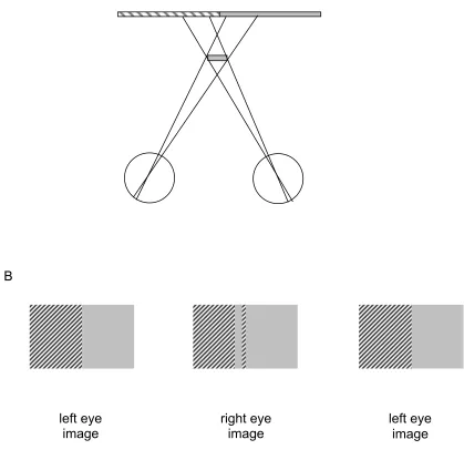
Gillam & Grove (2004) have addressed the related issue of what visual information might be used to distinguish between global occlusion and local slant interpretations, when scenes consist of sets of horizontal lines. Occlusion of a set of horizontal lines by an invisible vertical foreground occluder results in each line being shorter by a fixed amount in one eyes view (figure 14a). Yet this stimulus would also be consistent with the lines having different local slants in depth. Gillam and Grove showed that observers perceive the former, global, occlusion interpretation, rather than the local slant interpretation. However, a local slant interpretation is perceived when the two eyes views are switched, when the stimulus is no longer consistent with occlusion by an invisible foreground object. Grove, Byrne & Gillam (2005) have extended this work further, showing that for stimuli comprised of a set of oblique lines (each with differing orientations, figure 14b),
perception of the occluding contour is stronger than for a similar but horizontal line stimulus. The authors suggest that the vertical differences between left and right eye views of the lines (which we know deliver a strong perception of occlusion see Anderson, 1994, and figure 11d)
disambiguate the ambiguous information provided by horizontal binocular disparity.
5 Models of depth from monocular regions: revealing underlying mechanisms
When modelling the behavioural results obtained using the variety of half-occlusion stimuli
described in this review, the experimental approach used must be taken into account. Most of the experimental research has concentrated on designing stimuli that do not contain conventional binocular disparity information, yet which contain monocular regions consistent with 3-D occlusion geometry. Success in achieving this has been mixed, with some stimuli harbouring disparity information that is available to non-standard stereoscopic mechanisms. Those stimuli that have, so far, defied an explanation based on binocular disparity processing, have been discussed in terms of the geometrical constraints that they fulfil. Such accounts do not, of course, further our understanding of the neural mechanisms underlying the phenomena.
more work is required to try to understand how the brain’s basic binocular disparity processing mechanisms (whose understanding is fairly well advanced at the earliest levels, e.g. Ohzawa et al, 1990; Qian, 1994) might be used to incorporate monocular information in a relatively simple way. Some efforts in this direction are outlined below.
A recent suggestion has been directly inspired by the neurophysiology of stereopsis. Disparity-sensitive simple cells are known to code for disparity by both phase and position shifts (e.g. DeAngelis et al, 1991; Anzai et al, 1999, Prince et al, 2002, Tsao et al 2003). It has been suggested that some occlusion arrangements generate unusual combinations of phase- and position-shifts in simple cells and that an appropriate combination of phase- and position-coding could account for some half-occlusion phenomena (Tsao, Conway & Livingstone, 2003). This suggestion has yet to be formalised, but modelling along these lines could generate valuable, testable, predictions.
There have been several computationally implemented models of stereopsis that have attempted to deal with the presence of unpaired points, but not necessarily make use of them as a depth cue. We do not have space to review them all here (but see Egnal and Wildes for a comparison of several models, and Jones and Malik, 1992, for a model that takes account of monocular regions). Watanabe and Fukushima (1999) have developed a stereo algorithm that combines traditional binocular matching (based on Marr and Poggio’s 1979 cooperative stereoalgorithm, that
implements both a smoothness and uniqueness constraint) with monocular detectors that signal the presence of monocular regions. They use a cooperative stage to combine information from binocular disparity and neighbouring monocular regions, which relies on a constraint consistent with binocular occlusion geometry. In this sense the model is inspired by ‘purpose’: the known properties of the world are used to direct what depths are signalled. They demonstrate that their model can identify monocular regions that occur due to partial occlusion of one eye’s view, to aid in finding depth discontinuities (see also a similar model by Zitnick & Kanade, 2000).
Hyashi et al (2004) have extended Watanabe & Fukushima’s model. One of their aims was to use the known properties of early visual neurons to generate a physiologically plausible initial
matching stage, thus linking process with purpose. To do this they started with a disparity energy model (Ohzawa et al, 1990) and constructed a monocular-region detector by monitoring the output of a population of binocular disparity energy neurons, each looking at the same region of a scene. The signature of a monocular region is broad activation across the population of disparity
detectors, rather than specific activity over a narrow band of disparities, as would be delivered by strong disparity signals, signalling a particular depth. So these authors have ingeniously used the population response from a purely binocular mechanism to identify locations where there is no consistent binocular signal (and hence there must be a monocular region). Another key feature of the model describes what they call an additional occlusion constraint, that only one monocular representation can occur at one instant, in other words, that a monocular region is present in one eye, or the other, but not both. To achieve this, right and left eye representations for the
monocular regions are designed to inhibit one another. Not only does this model account nicely for some of the psychophysical results in particular, it also provides a ‘for-free’ model of binocular rivalry in general. When the two eye’s views are completely different, the interocular inhibition between the left- and right-eye monocular representations occurs across the whole scene, resulting in temporally alternating perception of one or the other views. This is the first model of stereopsis that can simultaneously accommodate conventional disparity processing, monocular regions as depth signals, and binocular rivalry.
eye-of-origin of monocular regions, then the geometric rules of occlusion are used to assign the same depth as the background.
An alternative model also uses simulated binocular neurons and includes input from monocular regions (Grossberg and McLoughlin, 1997, McLoughlin and Grossberg, 1998). Monocular regions are initially assigned all possible depths, then depth is determined based on higher-level
assumptions including filling in. The model has been developed to account for some results from the original da Vinci stereopsis study, monocular gap stereopsis (Grossberg & Howe, 2003) and can be elaborated to account for additional half-occlusion effects (Cao & Grossberg, 2005). The model does not explicitly require knowledge of occlusion geometry to deliver an output consistent with human perception, but does use a variety of other high-level rules, such as the propagation of depth from edges into a figure.
What we do not know is how these various models compare with one another. Egnal and Wildes (2002) compared a variety of computational models designed to detect monocular regions, but the models were not designed to be physiologically plausible, or to use monocular regions as a source of depth information. It would be fascinating to compare the current models directly on sets of the more challenging occlusion stimuli that have been described in this review.
6 Conclusions
In this article we have attempted to provide a comprehensive review of the literature related to depth from monocular regions. We have laid out in detail the experimental evidence showing how these regions can be used as part of a binocular visual representation. It is clear that monocular regions are important for forming surface representations and for depth perception. Research has shown that information from monocular regions is not simply thrown away by mechanisms
dedicated to forming a seamless representation of the world.
At the outset we classified monocular regions into 3 types, based on the form of the 3-D scene that would be delivered by the monocular region and its accompanying binocular information. Most of the examples described in this review fit into one of these categories. Our classification was not intended to delineate separate phenomena requiring distinct processing mechanisms, and we would not pretend to have done so, but some interesting points about each type have
emerged.
Type 1 occurs when the geometrical arrangements of foreground occluders is such that each portion of a textured or patterned background is seen by only one eye. This is interesting, because depth perception from these the arrangements is accompanied by binocular rivalry, but only sometimes. Stimuli exploring this type of monocular region have provided tantalising clues for how binocular vision, rivalry and depth from monocular regions, can be understood together. We are still a long way from this aim.
Type 2 represents the most commonly occurring monocular region, which is hidden by a binocularly visible foreground. As we described, some examples of this type can be explained using standard stereoscopic mechanisms, and modelling efforts are beginning to explain how binocular mechanisms can be adapted to obtain depth from the most common case, where the monocular region is accompanied by binocularly visible foreground and background. However, there remain a few stubborn effects that have not been amenable to straightforward explanation, in particular monocular gap stereopsis.
emerging from studying intrusion stimuli. These will provide challenges to future researchers engaged in forming theories and models of binocular vision.
We have shown that it is now possible to explain some phenomena that involve depth from monocular regions, using extensions of standard stereoscopic mechanisms. A parsimonious view would be that someday, all of these phenomena could be explained via elaborations of the
binocular mechanisms that we know underly standard disparity processing. But this has certainly not yet been demonstrated. The key question for the future is the extent to which each of these monocular region phenomena require explanation via theories and mechanisms that are distinct from normal stereopsis and that require inferences about the 3-D geometry of the external world. If this is necessary, then surely the mechanisms responsible for stereopsis are far more
sophisticated than we current know.
Acknowledgements
We thank the following individuals for providing comments and suggestions on early versions of this manuscript: Kevin Brooks, Barbara Gillam, Paul Hibbard, Ken Nakayama, Harold Nefs, Hiroshi Ono.
References
Anderson, B. L. (1994) The role of partial occlusion in stereopsis. Nature, 367, 365-368.
Anderson, B. L., & Nakayama, K. (1994). Towards a general theory of stereopsis: Binocular matching, occluding contours, and fusion. Psychology Review, 101, 414–445.
Anzai, A., Ohzawa, I., and Freeman, R.D. (1999a). Neural mechanisms for encoding binocular disparity: receptive field position versus phase. J. Neurophysiol. 82, 874–890.
Assee A., & Qian, N. (2007) Solving da Vinci stereopsis with depth-edge-selective V2 cells. Vis. Res., 47, 2585-2602.
Barrand, A. G. (1979) An ecological approach to binocular perception: the neglected facts of occlusion. Cornell University PhD, 1979. Published by University Microfilms International, Michigan.
Blake, R., & Logothetis, N. K. (2002). Visual competition. Nature Reviews Neuroscience, 3, 13–21.
van Bogaerts, E.A., Ooi, T.L. & He, Z. J. The monocular-boundary-contour mechanism in binocular surface representation and suppression (2008), Perception, 37, 1197-1215.
Brooks, K. R., & Gillam, B. J. (2006a). The swinging doors of perception: Stereomotion without binocular matching. Journal of Vision, 6(7):2, 685-695
Brooks, KR; Gillam, BJ (2006b) Quantitative Perceived Depth From Sequential Monocular Decamouflage, Vision Research, 46, 605-613.