Original Article
microRNA-21 is a potential indicator for
the pathological progression of cervical cancer
Fuying Xue1*, Huani Yi2*, Xiuli Ru3
1Department of Obstetrical, Taian City Central Hospital, Taian, Shandong, China; 2Department of Obstetrics and
Gynecology, Jinan Maternity and Child Care Hospital, Jinan, Shandong, China; 3Department of Reproductive
Medicine, The First Hospital of Hebei Medical University, Shijiazhuang, Hebei, China. *Co-first authors.
Received December 3, 2015; Accepted February 13, 2016; Epub April 1, 2016; Published April 15, 2016
Abstract: Cervical cancer is a disease of high incidence, affecting numerous female patients’ health and life. miR-21 is reported to play oncogenic roles in various cancers including cervical cancer, but its correlation with cervical cancer pathological progression is not fully understood. This study aims at revealing the expression pattern of miR-21 in the tissues of cervical intraepithelial neoplasia (CIN) and cervical cancer patients of different pathological grades. miR-21 levels in 35 CIN and 35 cervical cancer specimens were detected by qRT-PCR and compared to 40 normal specimens. Then in the 35 cervical cancer specimens, miR-21 levels were compared in different cancer variables. Its roles in cervical cancer cell migration and invasion were again verified by Transwell experiments after vector transfection to overexpress pre-miR-21 or miR-21 sponge in HeLa cells. miR-21 was promoted in CIN tissues compared to normal tissues (P < 0.05), and elevated in cervical cancer tissues compared to CIN tissues (P < 0.05). In cervical cancer tissues, miR-21 expression did not vary significantly with different patient ages or tumor sizes (P > 0.05), but it was significantly up-regulated in cancer tissues with higher pathology grades, clinical stages and lymphatic or distant metastasis (P < 0.05), which was supported by Transwell results that miR-21 promoted HeLa cell migration and invasion. These results indicated that the up-regulation of miR-21 might be associated with se-verer cervical cancer grades, implying the role of miR-21 as a possible indicator for the pathological progression of cervical cancer.
Keywords: Cervical cancer, miR-21, pathological progression, pathology grade, clinical stage
Introduction
Cervical cancer is a disease occurring in the cervix, thus being a female-attributed cancer that affects the normal life of tens of thousands of women [1]. Cervical cancer consists of two main types, squamous cell carcinoma and ade-nocarcinoma, with the former causing health problems in the majority of patients. Human papillomavirus (HPV) infection appears to be a leading risk factor of cervical cancer, and most patients are detected with HPV DNAs [2]. In addition, hereditary factors, as well as un- healthy habits like smoking and unhealthy sex life, are also potential causes of cervical can-cer. Invasive cervical cancer has become the third most common cancer for women world-wide, adding great burdens to low- and middle-income countries, especially. Though the intro-duction of screening programs has effectively
reduced the incidence and mortality of cervi- cal cancer in developed countries [3], greater efforts are still being paid to acquire more knowledge, diagnostic and therapeutic
meth-ods for fighting this disease.
Cervical cancer is developed from a series of precancerous lesions, cervical intraepithelial neoplasia (CIN) for instance, which is a patho-logical process from quantitative to qualitative changes with the accumulation of various ge- netic and phenotypic disorders. Continuous infection with high risk HPV may eventually lead grade III CIN to cervical cancer [4]. Modern genome and transcriptome sequencing
in human and is overexpressed in various can-cers [9]. Its association with cervical cancer,
including its influence on radiosensitivity [10]
and its oncogenic role of promoting cell prolif-eration [11], lymph node metastasis [12], is attracting increasing attention. Still, little is known about miR-21 and its relationship with cervical cancer malignancy. The aim of this study is to investigate miR-21 expression pat-tern in the pathological progression of cervical cancer. We collect tissue samples from nor- mal cervical and different degrees of CIN and cervical cancer, and detect miR-21 expression, which is compared in various cancer variables. miR-21 is found to be discriminately expressed in CIN and cervical cancer, and is related to dif-ferent pathology grades, clinical stages and
metastasis of cervical cancer. These findings
imply that miR-21 may be a valuable indicator for pathological progression and malignancy of cervical cancer.
Materials and methods
Tissue sampling
All the cervical tissues used in this study were sampled from patients of cervical cancer or CIN and patients with no cervical disease that we- re clinically and histopathologically diagnosed
and hospitalized from January to December in
2014. Patients with no cervical disease (n = 35) aged from 29 to 66 (48.5 ± 15.8). CIN patients (n = 35) aging from 30 to 65 (47.9 ± 16.1) were fell into three CIN degrees: degree I (11 individuals), degree II (12 individuals) and degree III (12 individuals). The pathological de-
grees and clinical stages were identified accord -ing to Diagnosis Criteria for Cervical Cancer proposed by Ministry of Health (December, 2011, China). The individual numbers of G1, G2 and G3 pathological degrees were 23, 7 and 5, respectively, and those of I, II, III and IV clinical
The tissue samples were ground in liquid nitro-gen and miRNAs were extracted using
miR-Neasy Mini Kit (Qiagen, Shenzhen, China)
according to the manufacturer’s instructions. Reverse transcription was performed using the
miRNAs samples, the specific primer for
has-miR-21 (5’-CTC AAC TGG TGT CGT GGA GTC GGC AAT TCA GTT GAG CTA CAA CT-3’) and Prime- Script Reverse Transcriptase (TaKaRa, Dalian, China). qRT-PCR was performed to detect the
expression level of miR-21 using the specific
primers (Fw: 5’-ACA CTC CAG CTG GGT AGC TTA TCA GAC TGA-3’ and Rv: 5’-TGG TGT CGT GGA GTC G-3’) on QuantStudio 6 Flex platform (Applied Biosystems, Carlsbad, CA). U6 (Fw: 5’-CTC GCT TCG GCA GCA CA-3’ and Rv: 5’-AAC GCT TCA CGA ATT TGC GT-3’) was used as an internal control. Experiments on each tissue sample were repeated for three times, and
data were analyzed with 2-ΔΔCt methods
com-pared to normal tissues.
HeLa cell culture and transfection
Human cervical adenocarcinoma HeLa cell line (ATCC, Manassas, VA) were cultured in
Dulbecco’s modified Eagle’s medium (DMEM,
high glucose) supplemented with 10% fetal bovine serum (FBS, Gibco, Carlsbad, CA), and
incubated in humidified atmosphere with 5%
CO2 at 37°C. The cells were passaged every two days. HeLa cells were transfected with vec-tors expressing pre-miR-21 or miR-21 sponge (QuantoBio, Beijing, China) and the correspond-ing blank vectors as controls uscorrespond-ing Lipofec- tamine 2000 (Invitrogen, Carlsbad, CA)
accord-ing to the manufacturer’s instructions. Briefly,
The cells were incubated in humidified atmo -sphere with 5% CO2 at 37°C. Medium was changed at 6 h post transfection. After incuba-tion for 48 h, cells were collected for further migration and invasion assays.
Cell migration and invasion assays by transwell
Cell migration and invasion were detected using Transwell (BD Biosciences, San Jose, CA). For cell migration assay, the cells were starved in serum-free medium for 24 h, and adjusted to a density of 5 × 10⁵/mL, after which the cells were added to the upper chamber of Transwell sets. The lower chamber contained DMEM with 20% FBS, and then the chambers were incu-bated for 24 h. After the incubation, the medi-um in the upper chamber was removed, and the cells on the lower side of the membrane were washed in phosphate buffered saline
(PBS), fixed in methanol for 30 min and stained
by crystal violet for 20 min. The cells left on the upper side of the membrane were wiped out with a cotton swab, and the membrane was washed in PBS and mounted. The migrated cells were observed using an optical micro-scope (Olympus, Tokyo, Japan) and counted in
five random visual fields.
For cell invasion assay, the upper chamber was pre-coated with Matrigel (BD Biosciences) and incubated at 37°C for 4 h for gel formation. Before the experiment, the gel was hydrated in serum-free medium for 2 h, after which the
same procedures as the cell migration assay were carried out.
Statistical analysis
Results of observed cell numbers and qRT-PCR were indicated as the mean ± standard
devia-tion. Data were analyzed by F test for homoge-neity of variance and then t test for significant
difference. P < 0.05 indicated the significant
difference between groups. For analysis of qRT-PCR results, in particular, the tissue samples
were first divided into different values accord -ing to the parameters of age, tumor diameter, pathology grade, clinical stage, lymphatic me- tastasis and distant metastasis, and then data
of the same parameter value were normalized
to the corresponding U6 data and compared to normal tissue. Also, data of all CIN samples
or all cervical cancer samples were analyzed
together compared to those of normal tissues for a general expression pattern.
Results
miR-21 level is associated with the pathologi-cal progression of cervipathologi-cal cancer
The expression level of miR-21 was analyzed in
all of the CIN tissues and cervical cancer tis-sues compared to normal tistis-sues for a general understanding of the expression pattern. As was shown in qRT-PCR results (Figure 1A),
miR-21 expression in CIN tissues was significantly
[image:3.629.101.531.80.213.2]increased than normal tissues (P < 0.05), and Figure 1. miR-21 level is associated with the pathological progression of cervical cancer. (A) miR-21 expression is detected in normal tissues, all cervical intraepithelial neoplasia (CIN) tissues and all cervical cancer tissues by qRT-PCR (triplicate detection). U6 is used as an internal control. The expression level of CIN and cancer tissue groups was compared to that of normal tissue group. Significant differences are found between CIN and normal tissues (P
significantly higher in cervical cancer tissues
than CIN tissues (P < 0.05). It seemed that miR-21 level was higher in the tissue with a severer pathological degree, which implied that the expression pattern of miR-21 was likely to be related to the pathological progression of cervi-cal cancer.
The tissue sections were then checked and
results confirmed the increasing severity of cer -vical pathology degrees from degree I of CIN to G3 of cervical cancer, as indicated by the den-sity of positive cells. For example, P16-positive cells were barely found in normal tis-sues, but were observed in degree II of CIN and G2 of cervical cancer tissues (Figure 1B). Moreover, the signal density of P16 was higher in cervical cancer tissues than CIN tissues. Together with the increased miR-21 level in cer-vical cancer tissues, miR-21 expression was thought to be associated with the progression of cervical cancer pathology.
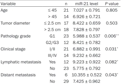
Then more detailed analyses were performed to reveal miR-21 expression patterns in various cancer variables including age of patients, tumor diameter, pathology grade, clinical stage, lymphatic metastasis, and distant metastasis (Table 1). No significant difference in miR-21
expression was found between the two age
groups (≤ 45 and > 45) or different tumor di-ameters (≤ 2.5 cm and > 2.5 cm) (P > 0.05). However, miR-21 expression was obviously
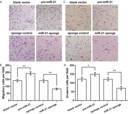
up-invasion by Transwell experiments. miR-21 was
promoted and inhibited by the specific vectors
expressing its precursor and sponge, respec-tively, and results were compared to the control groups transfected with corresponding blank vectors. Cell migration assay showed that the migrated HeLa cells were increased by miR-21 overexpression (P < 0.01, Figure 2A and 2B) and decreased by miR-21 inhibition (P < 0.01). Similarly, the invasive ability of HeLa cells were promoted by miR-21 overexpression (P < 0.05, Figure 2C and 2D) and inhibited by miR-21 inhi-bition (P < 0.01). These results indicated that miR-21 overexpression could promote HeLa cell migration and invasion, which might facili-tate the progression of cervical cancer. Discussion
miR-21 has been studied in cervical cancer before, being up-regulated in cervical cancer tissues and cells. In addition to its roles in cer-vical cancer cell radiosensitivity, proliferation and metastasis, its underlying relationship with the pathological progression of cervical cancer was investigated in this study. We detect miR-21 levels in CIN and cervical cancer tissues of altogether 75 patients to reveal its expression pattern in different pathological grades. miR-21 is expressed in lower level in normal cervical tissues and moderately expressed in CIN tis-sues. Its higher expression level is found in cervical cancer tissues with higher pathology
III/IV 14 9.232 ± 0.662
Lymphatic metastasis Yes 12 9.223 ± 0.922 0.082*
No 23 5.775 ± 0.792
Distant metastasis Yes 6 10.355 ± 0.522 0.043*
No 29 7.425 ± 0.962
*P < 0.05. **P < 0.01. amiR-21 level was indicated as the mean ±
stan-dard deviation compared to that of normal tissues.
ological progression of cervical cancer.
miR-21 promotes HeLa cell migration and invasion
[image:4.629.100.346.117.288.2]grade, clinical stage and lymphatic and distant metastasis.
Expression and functions of miR-21 have been reported in various diseases. Its expression is generally up-regulated in cancers and other diseases such as glioma, breast cancer, colo-
rectal cancer and lung cancer [13]. Specifically,
it is up-regulated in breast cancer, and anti-miR-21 oligonucleotides suppress cell growth and tumor growth [14]. It regulates key genes related to cell apoptosis, migration and inva-sion, and affects metalloproteinases, thus pro-moting invasiveness of glioma cells [15]. In colorectal cancer, miR-21 inhibits the
expres-sion of tumor suppressor PDCD4 and results in promoted cell invasion and metastasis [16]. Besides the up-regulated miR-21 level found in solid cancers like prostate and pancreatic tumors [17], it is also aberrantly expressed in
[image:5.629.102.529.81.459.2]HeLa cell proliferation, migration and invasion via inhibiting phosphatase and tensin homolog
[21]. On the basis of these findings, this study
further divided the cervical cancer tissues from 35 patients into different categories according to the cancer variables, which allowed us to dis-cover miR-21 expression patterns in different grades of cervical cancer.
The expression pattern of miR-21 sorted by dif-ferent cancer variables showed that miR-21 expression did not vary with patient age or tumor diameter. Instead, its expression
pos-sessed significant disparities between pathol -ogy grades, clinical stages, and cancer with lymphatic/distant metastasis or not, which implied the possible relationship between miR-21 level and these cancer variables. Similar functions of miR-21 have been reported in colorectal cancer, where miR-21 expression is close related to a prognostic value disease-free interval [22], lymph node positivity and devel-opment of distant metastases [23]. In breast
cancer, high level of miR-21 is significantly
associated with advanced clinical stage, lymph node metastasis and shortened survival of patients [24]. Besides, miR-21 may be a prog-nostic marker for squamous cell lung carcino-ma patients [25]. However, it seems that
miR-21 expression pattern cannot reflect clinical
prognosis in gastric cancer, albeit it promotes gastric cancer cell proliferation and invasion and is a biomarker for this disease [26, 27]. Taken together, the association between miR-21 and cancer pathological progression may
vary according to specific diseases, and in cer -vical cancer, miR-21 is capable of indicating pathological progression, as suggested in this study.
miR-21 has been proved to be a potential tis-sue biomarker, rather than a good serum mark-er, for diagnosing cervical cancer [28, 29]. High
miR-21 to be an indicator of cervical cancer
pathological progression. These findings uncov -er a new function of miR-21 for c-ervical canc-er diagnosis that may facilitate traditional diag-nostic methods.
Disclosure of conflict of interest
None.
Address correspondence to: Xiuli Ru, Department of Reproductive Medicine, The First Hospital of Hebei Medical University, 89 Donggang Road, Shijiazhuang 050031, Hebei, China. E-mail: ruxiuli7896@126. com
References
[1] Bray F, Ren JS, Masuyer E and Ferlay J. Global estimates of cancer prevalence for 27 sites in the adult population in 2008. Int J Cancer 2013; 132: 1133-1145.
[2] Dijkstra MG, van Niekerk D, Rijkaart DC, van Kemenade FJ, Heideman DA, Snijders PJ, Meijer CJ and Berkhof J. Primary hrHPV DNA testing in cervical cancer screening: how to manage screen-positive women? A POBASCAM trial substudy. Cancer Epidemiol Biomarkers Prev 2014; 23: 55-63.
[3] Vaccarella S, Lortet-Tieulent J, Plummer M, Franceschi S and Bray F. Worldwide trends in cervical cancer incidence: Impact of screening against changes in disease risk factors. Eur J Cancer 2013; 49: 3262-3273.
[4] Guan P, Howell-Jones R, Li N, Bruni L, de Sanjose S, Franceschi S and Clifford GM. Human papillomavirus types in 115,789 HPV-positive women: a meta-analysis from cervical infection to cancer. Int J Cancer 2012; 131: 2349-2359.
[6] Wan HY, Li QQ, Zhang Y, Tian W, Li YN, Liu M, Li X and Tang H. MiR-124 represses vasculogenic mimicry and cell motility by targeting amotL1 in cervical cancer cells. Cancer Lett 2014; 355: 148-158.
[7] Ribeiro J, Marinho-Dias J, Monteiro P, Loureiro J, Baldaque I, Medeiros R and Sousa H. miR-34a and miR-125b Expression in HPV Infection and Cervical Cancer Development. Biomed Res Int 2015; 2015: 304584.
[8] Shishodia G, Verma G, Srivastava Y, Mehrotra R, Das BC and Bharti AC. Deregulation of mi-croRNAs Let-7a and miR-21 mediate aberrant STAT3 signaling during human papillomavirus-induced cervical carcinogenesis: role of E6 on-coprotein. BMC Cancer 2014; 14: 996-1008. [9] Selcuklu SD, Donoghue MT and Spillane C.
miR-21 as a key regulator of oncogenic pro-cesses. Biochem Soc Trans 2009; 37: 918-925.
[10] Liu S, Song L, Zhang L, Zeng S and Gao F. miR-21 modulates resistance of HR-HPV positive cervical cancer cells to radiation through tar-geting LATS1. Biochem Biophys Res Commun 2015; 459: 679-685.
[11] Yao Q, Xu H, Zhang QQ, Zhou H and Qu LH. MicroRNA-21 promotes cell proliferation and down-regulates the expression of programmed cell death 4 (PDCD4) in HeLa cervical carcino-ma cells. Biochem Biophys Res Commun 2009; 388: 539-542.
[12] Chen J, Yao D, He C and Lu Y. Relationship be-tween serum miR-21 and lymph node metasta-sis in cervical cancer. Journal of Xi’an Jiaotong University (Medical Sciences) 2012; 33: 351-355.
[13] Krichevsky AM and Gabriely G. miR-21: a small multi-faceted RNA. J Cell Mol Med 2009; 13: 39-53.
[14] Si ML, Zhu S, Wu H, Lu Z, Wu F and Mo YY. miR-21-mediated tumor growth. Oncogene 2007; 26: 2799-2803.
[15] Gabriely G, Wurdinger T, Kesari S, Esau CC, Burchard J, Linsley PS and Krichevsky AM. MicroRNA 21 promotes glioma invasion by tar-geting matrix metalloproteinase regulators. Mol Cell Biol 2008; 28: 5369-5380.
[16] Asangani IA, Rasheed SA, Nikolova DA, Leupold JH, Colburn NH, Post S and Allgayer H. Micro- RNA-21 (miR-21) post-transcriptionally down-regulates tumor suppressor Pdcd4 and stimu-lates invasion, intravasation and metastasis in colorectal cancer. Oncogene 2008; 27: 2128-2136.
[17] Volinia S, Calin GA, Liu CG, Ambs S, Cimmino A, Petrocca F, Visone R, Iorio M, Roldo C, Ferracin M, Prueitt RL, Yanaihara N, Lanza G, Scarpa A, Vecchione A, Negrini M, Harris CC and Croce CM. A microRNA expression signature of
hu-man solid tumors defines cancer gene targets. Proc Natl Acad Sci U S A 2006; 103: 2257-2261.
[18] Jongen-Lavrencic M, Sun SM, Dijkstra MK, Valk PJ and Lowenberg B. MicroRNA expres-sion profiling in relation to the genetic hete-rogeneity of acute myeloid leukemia. Blood 2008; 111: 5078-5085.
[19] Deftereos G, Corrie SR, Feng Q, Morihara J, Stern J, Hawes SE and Kiviat NB. Expression of mir-21 and mir-143 in cervical specimens ranging from histologically normal through to invasive cervical cancer. PLoS One 2011; 6: e28423.
[20] Wang X, Tang S, Le SY, Lu R, Rader JS, Meyers C and Zheng ZM. Aberrant expression of onco-genic and tumor-suppressive microRNAs in cervical cancer is required for cancer cell gro- wth. PLoS One 2008; 3: e2557.
[21] Xu J, Zhang W, Lv Q and Zhu D. Overexpression of miR-21 promotes the proliferation and mi-gration of cervical cancer cells via the inhibi-tion of PTEN. Oncol Rep 2015; 33: 3108-3116. [22] Kulda V, Pesta M, Topolcan O, Liska V, Treska
V, Sutnar A, Rupert K, Ludvikova M, Babuska V, Holubec L Jr and Cerny R. Relevance of miR-21 and miR-143 expression in tissue samples of colorectal carcinoma and its liver metastases. Cancer Genet Cytogenet 2010; 200: 154-160. [23] Slaby O, Svoboda M, Fabian P, Smerdova T,
Knoflickova D, Bednarikova M, Nenutil R and Vyzula R. Altered expression of 21, miR-31, miR-143 and miR-145 is related to clini- copathologic features of colorectal cancer. Oncology 2007; 72: 397-402.
[24] Yan LX, Huang XF, Shao Q, Huang MY, Deng L, Wu QL, Zeng YX and Shao JY. MicroRNA miR-21 overexpression in human breast cancer is as-sociated with advanced clinical stage, lymph node metastasis and patient poor prognosis. RNA 2008; 14: 2348-2360.
[25] Gao W, Shen H, Liu L, Xu J, Xu J and Shu Y. MiR-21 overexpression in human primary squa-mous cell lung carcinoma is associated with poor patient prognosis. J Cancer Res Clin On- col 2011; 137: 557-566.
[26] Chan SH, Wu CW, Li AF, Chi CW and Lin WC. miR-21 microRNA expression in human gas- tric carcinomas and its clinical association. Anticancer Res 2008; 28: 907-911.
[27] Zhang Z, Li Z, Gao C, Chen P, Chen J, Liu W, Xiao S and Lu H. miR-21 plays a pivotal role in gastric cancer pathogenesis and progression. Lab Invest 2008; 88: 1358-1366.