Original Article
A sequence analysis of mitochondrial
DNA hypervariable segment I among Han
ethnic patients with HIV/AIDS in Guizhou, China
Jingsong Zhang1,3, Dexin Xiao3, Sha Li2, Hongyun Chen3, Jinqin Li3, Yidan Wang4, Juan Li1, Xinjiang Zhang2
1Department of Traditional Chinese Internal Medicine, School of Traditional Chinese Medicine, Southern Medical University, Guangzhou, Guangdong, P. R. China; 2Department of Dermatology, Affiliated Hospital of Zunyi Medical College, Zunyi, P. R. China; Departments of 3Dermatology, 4Stomatology, The First People’s Hospital of Zunyi, Zunyi, P. R. China
Received December 22, 2015; Accepted March 7, 2016; Epub June 1, 2016; Published June 15, 2016
Abstract: This study aims to investigate the gene polymorphisms in mitochondrial DNA (mtDNA) hypervariable seg-ment I (HVS I) among Han ethnic patients with HIV/AIDS in Guizhou, China, and to explore the effects of mitochon-drial gene polymorphisms on HIV infection. Blood samples were collected from 110 Han ethnic patients with HIV/ AIDS and 110 healthy Han ethnic newborn babies and anti-coagulated by EDTA. Then, mitochondrial DNA was
extracted, and the nucleic acid fragments of 643 bp containing non-encoding HVS I was amplified by PCR. After the purification of PCR products, sequencing was performed, and the sequencing results were compared with revised
Cambridge Reference Sequence to analyze mutant sites and mutation frequency in each site. Base mutation fre-quency distribution in single nucleotide site in mtDNA was compared between two groups. Results: In the nucleic acid fragment of 341 bp in the D-Loop HVS I (nucleotide sites, 16,024~16,365) among those patients with HIV/AIDS and the healthy controls, 380 base mutations occurred in the case group, with an average base mutation rate of 1.01%, and 241 base mutations in the control group, with an average base mutation rate of 0.64%, indicating that base mutation rate in the case group was obviously higher than that in the control group. These results suggest that mtDNA gene mutations, namely quality changes in mitochondria, exist in HIV/AIDS patients; however, whether HIV infection leads to mtDNA gene mutations in patients or the population with mtDNA gene mutations is susceptible to
HIV infection requires further study to confirm.
Keywords: Mitochondrial DNA hypervariable segment I, Han ethnic, HIV/AIDS, sequence analysis
Introduction
Acquired immune deficiency syndrome (AIDS) is a chronic infectious disease caused by human immunodeficiency virus (HIV) infection. Shortly after HIV entering the human body, it destroys the immune system, which results in patien- ts being susceptible to various opportunistic infections even secondary tumors, etc., leading to death. Clinical application of highly active antiretroviral therapy (HAART) extends survival time of patients and improves the life quality. However, HAART cannot completely eliminate HIV and patients need to receive long-term or lifelong medication, although the underlying treatment limits the virus replication in many patients. Currently, the disease is widely sp-
reading in Africa, Asia, Latin America, etc., resulting in a worldwide epidemic, which seri-ously affects the development of society and economy as well as threats the health of human being [1-4]. During the 30 years from 1985 when the first case of HIV/AIDS was detected in China, the epidemic situation has covered all provinces of the country [3, 4].
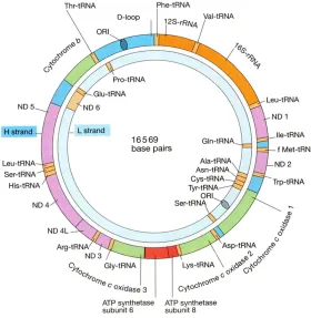
has finished the sequencing of human mtDNA, and grouped mtDNA into encoding region and non-encoding region (control region) [5]. mtDNA is a closed circular double-stranded molecule containing 16,569 base pairs (bp), with one heavy chain (H chain) located in outer ring and one light chain (L chain) in inner ring, both of which have encoding function (Figure 1) (http:// tupian.baike.com/a1_27_82_013005360713 09139023828680435_jpg.html&prd=so_ tupian). A total of 37 structure genes are encod-ed in the encoding region of mtDNA; among them, 13 structure genes encode polypepti- des which constitute oxidative phosphorylation enzymes and partial subunits of respiratory chain complex, while other 24 structure genes encode 2 ribosomal RNAs (rRNA: 12S rRNA and 16S rRNA)and 22 transfer RNAs (tRNA) [6, 7]. Non-encoding region, also called control reg- ion or hypervariable region, consists of base sequences with a size of 1,122 bp between 1-576 bp and 16,024-16,569 bp, forming a starting point of replication, a D-loop region and 2 starting points of transcription, and is a region with the rapidest evolutionary rate of mtDNA and the highest frequency of
polymor-and has an imperfect self-repair system after damage, leading to its susceptibility to muta-tions and obvious realization of polymorphisms [8, 9]. The present study, through sequencing in mtDNA HVS I among HIV/AIDS patients, at- tempts to investigate the gene polymorphisms in mtDNA HVS I, to explore the effects of mito-chondrial gene polymorphisms on HIV infec-tion, to make clear whether predisposing genes exist in AIDS, and to clarify the pathogenesis of AIDS from the aspect of molecular biology, and thereby providing novel evidence for the more effective prevention and treatment of AIDS.
Materials and methods
All experimental operations of the present study were conducted in the central laboratory of Zunyi Medical College and the Center for Disease Control and Prevention in Zunyi. Sub- jects were selected according to informed con-sent, ethical principles and security principles: 1. case group, blood samples of 110 Han eth-nic patients with HIV/AIDS were provided by the Center for Disease Control and Prevention in Zunyi, Guizhou; inclusion criteria were as fol-lows: HIV antibody-positive patients were con-Figure 1. Human mitochondrial gene structure.
[image:2.612.91.371.72.359.2]firmed by the Center for Disease Control and Prevention in Guizhou using western blot (WB) method, and age was over 18 years old; 2. con-trol group, blood samples of 110 healthy new-born babies in Guizhou were provided by the Affiliated Hospital of Zunyi Medical College and the Affiliated Hospital of Guiyang College of Traditional Chinese Medicine. Blood samples (5 ml) were collected from each subject in the case group and the control group, anti-coagu-lated by EDTA, and stored at -80°C. All above subjects were from the Chinese Han population in Guizhou. Whole blood genome was extracted by using the Ezup genomic DNA extraction col-umn extraction kit (Sangon Biotech Co., Ltd., Shanghai, China). Primers were designed acc- ording to revised Cambridge Reference Sequ- ence (rCRS) by Shanghai Sangon Biotech Co., Ltd. The sequence between 15,879~15,898 in L chain was selected as upstream primer (R15879 5’-AATGGGCCTGTCCTTGTAGT-3’), and
a PCR purification kit (Sangon Biotech Co., Ltd., Shanghai, China). The purified PCR products were sent to Shanghai Sangon Biotech Co., Ltd. for bidirectional sequencing, and the obtained DNA sequences were initially compared in http://www.ncbi.nlm.nih.gov/pubmed for sequ- ence source identification. Sequence diagrams were read with Chromas software and com-pared with derived sequences to revise nucleo-tide sequences. The forward and reverse sequences of the same nucleotide sequence were revised applying DNAStar software, and compared with rCRS after assembly. The obtained information on nucleotide variation was compared with the database in www.mito-map.org, and the new-found mutation sites were further confirmed. The average base mutation rate = the total number of mutation bases/the total number of detected bases × 100%.
Results
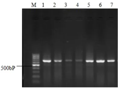
The integrity of the whole blood genome extracted with the Ezup genomic DNA extrac-tion column extracextrac-tion kit was verified by ran-domly selecting 7 mtDNA templates (Figure 2). Electrophoresis results for the PCR amplifica-tion products of the target fragments in mtDNA D. Loop HVS I demonstrated that all amplified target nucleic acid fragments presented clear bands with a size of 643 bp (Figure 3).
Sequencing results and bidirectional sequence diagrams for the PCR amplification products of the target fragments in mtDNA D. Loop HVS I showed that most PCR amplification products presented relatively clear base peaks by purifi-Figure 2. Verification of 7 mtDNA templates selected randomly by electro
[image:3.612.91.368.74.200.2]-phoresis, M is DNA Marker DL15000, band 1 is an mtDNA template selected randomly, with a size of 16,569 bp.
Figure 3. M is DNA Marker DL1000, bands 1, 2, 3, 4, 5, 6 and 7 are mtDNA bands selected randomly with a size of 643 bp.
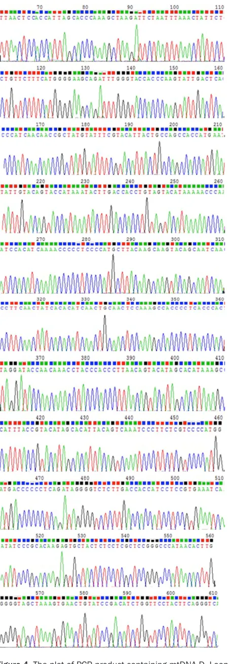
[image:3.612.91.287.260.405.2]cation and sequencing. Comparison in http://blast.ncbi.nlm.nih.gov/ revealed that the homology with human mtDNA reached more than 98% with a highest similarity, confirming that the amplified PCR products were in human mtDNA HVS I. The present study conducted bidirectional sequencing in the PCR products of HVS I of all samples, and the original peak chart for sequencing and the sequence map after splicing of one sample were selected randomly, as seen in the following figure (Figures 4, 5).
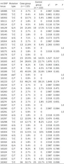
Sequencing in mtDNA D. Loop HVS I for vari-ation situvari-ation and site analysis was shown in Tables 1, 2. In the detected nucleic acid fragment of 341 bp in the D. Loop HVS I (nucleotide sites, 16,024~16,365), 380 base variations were detected, with an aver-age base variation rate of 1.01% (380/ [(16,365 - 16,024) × 110] × 100%); while 241 base variations were observed in the control group, with an average base varia-tion rate of 0.64% (241/[(16,365 - 16,024) × 110] × 100%). Comparison revealed an evidently higher base variation rate in the case group than the control group, showing significantly statistical significance (P<0.01). At the nucleotide site of 16,223, 53 cases with C-T variation were found in the case group, with an incidence of 48.2% (53/110), and 43 cases with C-T variation in the con-trol group, with an incidence of 39.1% (43/110), which implied that the variation frequency in the case group was higher than that in the control group, but showing no obviously statistical significance (P>0.05). At the nucleotide site of 16,362, there were 36 cases with T-C variation in the case group, with an incidence of 32.7% (36/110), and 25 cases with T-C variation in the con-trol group, with an incidence of 22.7% (25/110), demonstrating that the variation frequency in the case group was higher than that in the control group, without obviously difference (P>0.05). Statistics (the χ2 test
was conducted by using SPSS17.0 softwa- re) was performed in the base variation frequency of the nucleotide sites (16,0- 24~16,365) of the samples in both case group and control group, which revealed increases in the base variation frequency of all sites, but no apparent statistical differ-ence (P>0.05). The results were seen in
[image:4.612.93.318.69.723.2]Table 1. Figure 4. The plot of PCR product containing mtDNA D. Loop
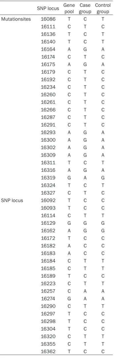
In the detected nucleic acid fragment of 341 bp in the D. Loop HVS I (nucleotide sites, 16,024~16,365) in the case group, single nucleotide polymorphism locus and mutation sites were seen in Table 2.
Discussion
Over the past 30 years, scholars have done a lot of researches on the etiology, pathogenesis, route of transmission, diagnosis, treatment and prevention of AIDS and the relationship between HIV and organism; moreover, some progress has been made. The current use of HAART facilitates the significant decrease of morbidity and mortality rates of AIDS, and rebuilds the immune functions of patients, whose survival time and life quality have been improved correspondingly. HIV infection in developed countries has been gradually becoming a controllable chronic disease, but HAART treatment is unable to restrict the late-phase viral replication while cannot entirely clear the virus in patients [10, 11]. Therefore, there is still a long hard way to go in the preven-tion and control of AIDS, and considerable in-depth researches are needed as well.
In recent years, studies have found that the pace of the progression from HIV-infection to AIDS varies possibly depending on individual DNA differences; in other words, the genotype of mtDNA may be associated with the pace of AIDS development. For instance, among the white population, the pace of AIDS progression in the patients with specific mutations of haplo-type groups J and U5a was two times faster than those without the two haplotype groups; while in those with a specific mutation of haplo-type group H3, the disease progression was 2 times slower than those without this haplotype group. Therefore, it is supposed that haplotype
mtDNA polymorphisms and the susceptibility and development of AIDS.
Apart from the nucleus, human mtDNA is the only genetic material and characterized by strict maternal inheritance, lack of recombina-tion, huge variation in groups, and high muta-tion rate, etc. [13, 14]. Mitochondria, free in the active oxygen environment of the matrix, are lacking to an effective damage/repair system and more prone to mutation damage than nuclear genes, and thereby influencing cellular functions and resulting in various diseases [15-17]. mtDNA is a closed circular double-strand-ed molecule with a full length of 16,569 bp, and grouped into encoding region and non-encoding region in which, D. loop region is a mutational hot spot, especially HVS I, with quite obvious polymorphisms; therefore, sequencing in HVS I can obtain a number of genetic infor-mation. The polymorphisms of mtDNA include heterogeneity, length polymorphisms, sequ- ence polymorphisms and single nucleotide polymorphisms (SNPs), among which, SNPs are a kind of polymorphism that currently receive much attention, and is caused by single-base mutation. Mitochondria are the main organelle of energy generation in human cells. Either HIV infection itself or anti retroviral drugs may affect the mitochondrial function [18, 19]. The polymorphism study of mitochondrial genes favors the understanding of the relationship between drug efficacy and AIDS disease pro-gression, which may offer support for the com-patibility of clinical medication and improve therapy regimen.
In this study, through the PCR amplification and sequencing in the HVS I of the D. loop region of the blood samples from patients with HIV/AIDS, and the comparison analysis with rCRS, it was revealed that among the patients with HIV/ Figure 5. The spliced sequence of PCR product containing mtDNA D. Loop
HVS I by bidirectional DNA sequencing.
Table 1. Mutation frequency of SNP locus in mt DNA D. Loop HVS I
mt-SNP
locus Mutation type Case group (n = 110)
Control group (n = 110) χ
2 Pa F
16086 T/C 3 2.7% 0 0 2.987 0.084 16092 T/C 7 6.4% 4 3.6% 0.861 0.353 16093 T/C 6 10.7% 3 5.4% 1.980 0.159 16111 C/T 2 1.8% 0 0 2.018 0.155 16114 C/T 5 4.5% 4 3.6% 0.116 0.734 16129 G/A 24 21.8% 19 17.3% 0.723 0.395 16136 T/C 3 2.7% 0 0 2.987 0.084 16140 T/C 2 1.8% 0 0 2.018 0.155 16162 A/G 4 7.1% 1 1.8% 3.173 0.075 16164 A/G 3 2.7% 0 0 2.987 0.084 16172 T/C 13 11.8% 6 5.4% 2.283 0.093 16174 C/T 1 0.9% 0 0 1.0 16175 A/G 2 1.8% 0 0 2.018 0.155 16179 C/T 1 0.9% 0 0 1.0 16182 A/C 23 20.9% 16 14.5% 1.527 0.217 16183 A/C 34 28.6% 25 22.7% 1.876 0.171 16184 C/T 9 8.2% 8 7.3% 0.064 0.801 16185 C/T 8 7.3% 5 4.5% 0.736 0.391 16189 T/C 45 40.9% 35 31.8% 1.964 0.161 16189 delT 1 0.9% 0 0 1.0 16192 C/T 1 0.9% 0 0 1.0 16223 C/T 53 48.2% 43 39.1% 1.848 0.174 16234 C/T 3 2.7% 0 0 2.987 0.084 16257 C/A 5 3.6% 3 2.7% 0.519 0.471 16260 C/T 3 2.7% 0 0 2.987 0.084 16261 C/T 3 2.7% 0 0 2.987 0.084 16266 C/T 2 1.8% 0 0 2.018 0.155 16274 G/A 3 2.7% 2 1.8% 0.205 0.651 16287 C/T 1 0.9% 0 0 1.0 16290 C/T 3 2.7% 0 0 2.987 0.084 16291 C/T 1 0.9% 0 0 1.0 16293 A/G 2 1.8% 0 0 2.018 0.155 16297 T/C 12 10.9% 9 8.2% 0.474 0.491 16298 T/C 14 12.7% 9 8.2% 1.214 0.27 16300 A/G 2 1.8% 0 0 2.018 0.155 16302 A/G 2 1.8% 0 0 2.018 0.155 16304 T/C 15 12.5% 11 10% 0.698 0.404 16309 A/G 2 1.8% 0 0 2.018 0.155 16311 T/C 3 5.4% 0 0 2.987 0.084 16316 A/G 2 1.8% 0 0 2.018 0.155 16319 G/A 3 5.4% 0 0 2.987 0.084 16320 C/T 9 8.2% 8 7.3% 0.074 0.786 16324 T/C 3 2.7% 0 0 2.987 0.084 16327 C/T 3 2.7% 0 0 2.987 0.084 16355 C/T 7 6.4% 5 4.5% 0.353 0.553 16362 T/C 36 32.7% 25 22.7% 2.745 0.098
AIDS, in the detected nucleic acid fragment of 341 bp in the D. Loop HVS I (nucleotide sites, 16,024~ 16,365), 380 base variations were detected, with an average base varia-tion rate of 1.01% (380/[(16,365 - 16,024) × 110] × 100%); while 241 base variations were observed in the control group, with an average base variation rate of 0.64% (241/[(16,365 - 16,024) × 110] × 100%), revealing an evidently higher base variation rate in the case group than the con-trol group, with significantly statisti-cal significance (P<0.01); at the nucleotide site of 16,223, 53 cases with C-T variation were found in the case group, with an incidence of 48.2% (53/110), and 43 cases with C-T variation in the control group, with an incidence of 39.1% (43/110), which implied that the variation fre-quency in the case group was higher than that in the control group, but showing no obviously statistical sig-nificance (P>0.05); at the nucleotide site of 16,362, there were 36 cases with T-C variation in the case group, with an incidence of 32.7% (36/110), and 25 cases with T-C variation in the control group, with an incidence of 22.7% (25/110), demonstrating that the variation frequency in the case group was higher than that in the control group, without obviously dif-ference (P>0.05).
dam-age of peripheral blood mononuclear cells and decreased mtDNA content, and HIV infection may lead to the reduction of mtDNA content in the organism, especially in immune-activated cells. After a 5-year follow-up research in 36 HIV-1-infected patients who underwent no anti-retroviral drug therapy, it has been demonstrat-ed that mtDNA content declindemonstrat-ed evidently after a 5-year seroconversion, contributing to the conclusion that HIV-1 infection influences mtDNA content, especially the mtDNA content in immune-activated cells [21]. These research results all implicated that HIV infection could affect cellular mtDNA content, which is the most significant in the immune-activated cells.
Our experimental findings demonstrate that: in the nucleic acid fragment of 341 bp that belongs to the D. Loop HVS I (nucleotide sites, 16,024~16,365) in the detected HIV/AIDS pa- tients, the occurrence of base mutation and its mutation frequency were significantly higher than those in the control group; gene mutations in mtDNA, namely quality changes in mitochon-dria, existed in patients with HIV/AIDS. Whether HIV infection leads to mtDNA gene mutations in patients or the population with mtDNA gene mutations is susceptible to HIV infection re- quires further study to confirm.
Acknowledgements
This study was supported by stadholder fund-ing of Guizhou Province (fundfund-ing number: 040- 109) and the funding title is “effect of mtDNA polymorphism on immune reconstitution of AIDS intervened by HAART therapy”.
Disclosure of conflict of interest
None.
Address correspondence to: Juan Li, Department of Traditional Chinese Internal Medicine, School of Traditional Chinese Medicine, Southern Medical University, Guangzhou, Guangdong, P. R. China. E-mail: [email protected]; Dr. Xinjiang Zhang, De-
partment of Dermatology, Affiliated Hospital of
Zunyi Medical College, Zunyi 563000, P. R. China. E-mail: [email protected]
References
[image:7.612.91.290.95.719.2][1] Chun TW, Carruth L, Finzi D, Shen X, DiGi-useppe JA, Taylor H, Hermankova M, Chadwick Table 2. Single nucleotide polymorphism
locus and mutation sites
SNP locus Gene pool groupCase Control group Mutationsites 16086 T C T
K, Margolick J, Quinn TC, Kuo YH, Brookmeyer R, Zeiger MA, Barditch-Crovo P and Siliciano
RF. Quantification of latent tissue reservoirs
and total body viral load in HIV-1 infection. Na-ture 1997; 387: 183-188.
[2] Castillo-Mancilla J, Allshouse A, Collins C, Hast-ings-Tolsma M, Campbell TB and Mawhinney S. Differences in sexual risk behavior and HIV/ AIDS risk factors among foreign-born and US-born Hispanic women. J Immigr Minor Health 2012; 14: 89-99.
[3] Liu K and Yuan J. The prevalence of AIDS and its impact on Chinses society and economy. J Xuahai 2003; 14: 89-99.
[4] Zeng Y. The prevalence of AIDS, research prog-ress and control strategy. China Public Health 2001; 17: 1062-1063.
[5] Anderson S, Bankier AT, Barrell BG, de Bruijn MH, Coulson AR, Drouin J, Eperon IC, Nierlich DP, Roe BA, Sanger F, Schreier PH, Smith AJ, Staden R and Young IG. Sequence and organi-zation of the human mitochondrial genome. Nature 1981; 290: 457-465.
[6] Wallace DC and Fan W. Energetics, epi-genetics, mitochondrial genetics. Mitochon-drion 2010; 10: 12-31.
[7] Wallace DC, Ye JH, Neckelmann SN, Singh G, Webster KA and Greenberg BD. Sequence analysis of cDNAs for the human and bovine ATP synthase beta subunit: mitochondrial DNA genes sustain seventeen times more muta-tions. Curr Genet 1987; 12: 81-90.
[8] DiMauro S and Schon EA. Mitochondrial DNA mutations in human disease. Am J Med Genet 2001; 106: 18-26.
[9] Paneto GG, Martins JA, Longo LV, Pereira GA, Freschi A, Alvarenga VL, Chen B, Oliveira RN, Hirata MH and Cicarelli RM. Heteroplasmy in hair: differences among hair and blood from the same individuals are still a matter of de-bate. Forensic Sci Int 2007; 173: 117-121. [10] Este JA and Cihlar T. Current status and
chal-lenges of antiretroviral research and therapy. Antiviral Res 2010; 85: 25-33.
[11] Quivy V, De Walque S and Van Lint C. Chroma-tin-associated regulation of HIV-1 transcrip-tion: implications for the development of thera-peutic strategies. Subcell Biochem 2007; 41: 371-396.
[12] Hendrickson SL, Hutcheson HB, Ruiz-Pesini E, Poole JC, Lautenberger J, Sezgin E, Kingsley
L, Goedert JJ, Vlahov D, Donfield S, Wallace DC
and O’Brien SJ. Mitochondrial DNA haplo-
groups influence AIDS progression. AIDS 2008;
22: 2429-2439.
[13] Yi YJ, Zimmerman SW, Manandhar G, Odhiam-bo JF, Kennedy C, Jonakova V, Manaskova-Pos-tlerova P, Sutovsky M, Park CS and Sutovsky P. Ubiquitin-activating enzyme (UBA1) is required for sperm capacitation, acrosomal exocytosis and sperm-egg coat penetration during por-cine fertilization. Int J Androl 2012; 35: 196-210.
[14] Jacobs HT. Disorders of mitochondrial protein synthesis. Hum Mol Genet 2003; 12 Spec No 2: R293-301.
[15] Martin AM, Hammond E, Nolan D, Pace C, Den Boer M, Taylor L, Moore H, Martinez OP, Chris-tiansen FT and Mallal S. Accumulation of mito-chondrial DNA mutations in human
immuno-deficiency virus-infected patients treated with
nucleoside-analogue reverse-transcriptase in-hibitors. Am J Hum Genet 2003; 72: 549-560. [16] Holt IJ, Harding AE and Morgan-Hughes JA. De-letions of muscle mitochondrial DNA in pa-tients with mitochondrial myopathies. Nature 1988; 331: 717-719.
[17] Wallace DC, Singh G, Lott MT, Hodge JA, Schurr TG, Lezza AM, Elsas LJ 2nd and Nikoskelainen EK. Mitochondrial DNA mutation associated with Leber’s hereditary optic neuropathy. Sci-ence 1988; 242: 1427-1430.
[18] Lehmann HC, Chen W, Borzan J, Mankowski JL and Hoke A. Mitochondrial dysfunction in
dis-tal axons contributes to human immunodefi -ciency virus sensory neuropathy. Ann Neurol 2011; 69: 100-110.
[19] Chiappini F, Teicher E, Saffroy R, Pham P, Falis-sard B, Barrier A, Chevalier S, Debuire B, Vitte-coq D and Lemoine A. Prospective evaluation of blood concentration of mitochondrial DNA as a marker of toxicity in 157 consecutively re-cruited untreated or HAART-treated HIV-posi-tive patients. Lab Invest 2004; 84: 908-914. [20] Bastard JP, Caron M, Vidal H, Jan V, Auclair M,
Vigouroux C, Luboinski J, Laville M, Maachi M, Girard PM, Rozenbaum W, Levan P and Ca-peau J. Association between altered expres-sion of adipogenic factor SREBP1 in li-poatrophic adipose tissue from HIV-1-infected patients and abnormal adipocyte differentia-tion and insulin resistance. Lancet 2002; 359: 1026-1031.