2,4-Diiodoaniline
Graham Smith* and Urs D. Wermuth
School of Physical and Chemical Sciences, Queensland University of Technology, GPO Box 2434, Brisbane, Qld 4001, Australia
Correspondence e-mail: [email protected]
Received 14 July 2009; accepted 31 July 2009
Key indicators: single-crystal X-ray study;T= 200 K; mean(C–C) = 0.005 A˚; Rfactor = 0.018;wRfactor = 0.038; data-to-parameter ratio = 20.8.
The structure of the title compound, C6H5I2N, shows a weak intermolecular amine–amine N—H N hydrogen-bonding interaction, giving a helical chain which extends along the a
axis. An intramolecular N—H I hydrogen bond is also observed.
Related literature
For related structures, see: Garden et al. (2002). For the synthesis, see: Dainset al.(1935); Hodgson & Marsden (1937); O’Neil (2001). For graph-set analysis of hydrogen bonding, see: Etteret al.(1990).
Experimental
Crystal data
C6H5I2N
Mr= 344.91
Orthorhombic,P212121
a= 4.3870 (1) A˚ b= 10.9626 (3) A˚ c= 16.9778 (4) A˚
V= 816.51 (3) A˚3
Z= 4
MoKradiation = 7.62 mm1 T= 200 K
0.300.180.18 mm
Data collection
Oxford Diffraction Gemini-S Ultra CCD-detector diffractometer Absorption correction: multi-scan
(SADABS; Sheldrick, 1996) Tmin= 0.146,Tmax= 0.250
6739 measured reflections 1873 independent reflections 1790 reflections withI> 2(I) Rint= 0.024
Refinement
R[F2> 2(F2)] = 0.018
wR(F2) = 0.038 S= 1.05 1873 reflections 90 parameters
H atoms treated by a mixture of independent and constrained refinement
max= 0.38 e A˚
3
min=0.47 e A˚
3
Absolute structure: Flack (1983), 737 Friedel pairs
Flack parameter:0.03 (4)
Table 1
Hydrogen-bond geometry (A˚ ,).
D—H A D—H H A D A D—H A
N1—H11 I2 0.77 (3) 2.81 (3) 3.283 (4) 122 (3)
N1—H12 N1i
0.80 (4) 2.30 (4) 3.106 (5) 180 (5)
Symmetry code: (i)x1 2;yþ
3 2;zþ2.
Data collection: CrysAlis CCD (Oxford Diffraction, 2008); cell refinement: CrysAlis RED (Oxford Diffraction, 2008); data reduc-tion:CrysAlis RED; program(s) used to solve structure:SHELXS97
(Sheldrick, 2008) withinWinGX(Farrugia, 1999); program(s) used to refine structure: SHELXL97 (Sheldrick, 2008) within WinGX
(Farrugia, 1999); molecular graphics:PLATON (Spek, 2009); soft-ware used to prepare material for publication:PLATON.
The authors acknowledge financial support from the Australian Research Council and the School of Physical and Chemical Sciences, Queensland University of Technology.
Supplementary data and figures for this paper are available from the IUCr electronic archives (Reference: IS2440).
References
Dains, F. B., Brewster, R. Q. & Davis, J. A. (1935).J. Am. Chem. Soc.57, 2326– 2327.
Etter, M. C., MacDonald, J. C. & Bernstein, J. (1990).Acta Cryst.B46, 256–262. Farrugia, L. J. (1999).J. Appl. Cryst.32, 837–838.
Flack, H. D. (1983).Acta Cryst.A39, 876–881.
Garden, S. J., Fontes, S. P., Wardell, J. L., Skakle, J. M. S., Low, J. N. & Glidewell, C. (2002).Acta Cryst.B58, 701–709.
Hodgson, H. H. & Marsden, E. (1937).J. Chem. Soc.pp. 1365–1366. O’Neil, M. J. (2001). Editor.The Merck Index13th ed., p. 560. Whitehouse
Station, NJ, USA: Merck & Co.
Oxford Diffraction (2008). CrysAlis CCD and CrysAlis RED. Oxford Diffraction Ltd, Abingdon, England.
Sheldrick, G. M. (1996).SADABS, University of Go¨ttingen, Germany. Sheldrick, G. M. (2008).Acta Cryst.A64, 112–122.
Spek, A. L. (2009).Acta Cryst.D65, 148–155.
Structure Reports
Online
supporting information
Acta Cryst. (2009). E65, o2108 [doi:10.1107/S1600536809030438]
2,4-Diiodoaniline
Graham Smith and Urs D. Wermuth
S1. Comment
Although the crystal structures of a number of nitro-substituted iodoanilines including 3-nitro-2,4-diodoaniline have been
reported (Garden et al., 2002), that of the title compound 2,4-diiodoaniline C6H6I2N (I) has not been determined and the
structure is reported here. The compound was isolated as the major crystalline product in the attempted synthesis of an
adduct of 4,5-dichlorophthalic acid with 4-iodoaniline in aqueous ethanol. This conversion of 4-iodoaniline to
2,4-diiodo-aniline has been reported previously (Dains et al., 1935), where solid 4-iodo2,4-diiodo-aniline was observed to undergo a ca 25%
conversion to the diiodo analogue in a sealed container over a period of three years. Hodgson & Marsden (1937) also
reported the ready formation of the diiodo derivative along with 4-iodoaniline from the reaction of aniline with iodine.
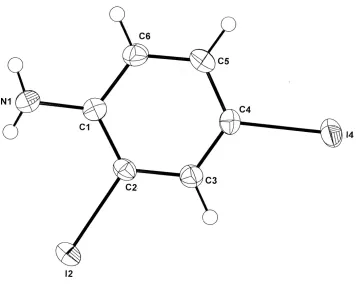
In the structure of (I) (Fig. 1), single weak intermolecular hydrogen bonds are found [N1—H1···N1i, 3.106 (5) Å;
symmetry code: (i) x - 1/2, -y + 3/2, -z + 2] [graph set S(4) (Etter et al., 1990)], linking the amine groups of 21
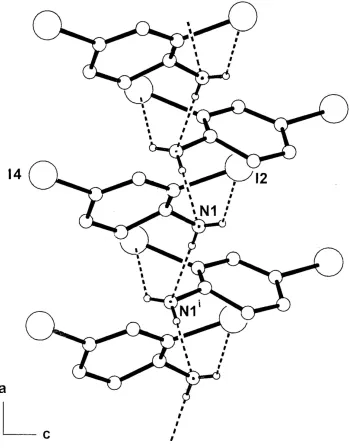
screw-related molecules. These form one-dimensional chains which extend down the a cell direction in the unit cell (Fig. 2).
In this structure there are, not unexpectedly, short intramoleculer N—H···I interactions [N1···I2, 3.283 (4) Å], which are
also present in the structure of 2,4-diiodo-3-nitroaniline [3.254 (7) Å (Garden et al., 2002)]. However, unlike the
nitro-derivative, no π–π stacking interactions are present in the structure of (I).
S2. Experimental
The title compound was formed in the attempted synthesis of a proton-transfer salt of 4,5-dichlorophthalic acid with
4-iodoaniline by heating together under reflux for 10 minutes 1 mmol quantities of the two reagents in 50 ml of 50%
ethanol-water. After concentration to ca 30 ml, partial room temperature evaporation of the hot-filtered solution gave
colourless needle prisms of 2,4-diiodoaniline [m.p. 368–389 K (O′Neil, 2001)] as the major product. This conversion of
4-iodoaniline to 2,4-diiodoaniline in the solid state has been reported previously (Dains et al., 1935).
S3. Refinement
The hydrogen atoms of the amino group were located in a difference Fourier map and their positional and isotropic
displacement parameters were refined freely. Other H-atoms were included in the refinement in calculated positions [C—
Figure 1
Figure 2
The one-dimensional hydrogen-bonded chain structure of (I) extending down the a axial direction of the unit cell,
showing hydrogen-bonding associations as dashed lines. Non-interactive H atoms are omitted. [Symmetry code (i): x -
1/2, -y + 3/2, -z + 2].
2,4-Diiodoaniline
Crystal data
C6H5I2N
Mr = 344.91
Orthorhombic, P212121
Hall symbol: P 2ac 2ab a = 4.3870 (1) Å b = 10.9626 (3) Å c = 16.9778 (4) Å
V = 816.51 (3) Å3
Z = 4 F(000) = 616 Dx = 2.806 Mg m−3
µ = 7.62 mm−1
T = 200 K
0.30 × 0.18 × 0.18 mm
Data collection
Oxford Diffraction Gemini-S Ultra CCD-detector
diffractometer
Radiation source: Enhance (Mo) X-ray tube Graphite monochromator
ω scans
Absorption correction: multi-scan (SADABS; Sheldrick, 1996) Tmin = 0.146, Tmax = 0.250
6739 measured reflections 1873 independent reflections 1790 reflections with I > 2σ(I) Rint = 0.024
θmax = 27.5°, θmin = 3.0°
h = −5→5 k = −13→14 l = −22→18
Refinement
Refinement on F2
Least-squares matrix: full R[F2 > 2σ(F2)] = 0.018
wR(F2) = 0.038
S = 1.05 1873 reflections 90 parameters 0 restraints
Primary atom site location: structure-invariant direct methods
Secondary atom site location: difference Fourier map
Hydrogen site location: inferred from neighbouring sites
H atoms treated by a mixture of independent and constrained refinement
w = 1/[σ2(F
o2) + (0.0207P)2]
where P = (Fo2 + 2Fc2)/3
(Δ/σ)max = 0.003
Δρmax = 0.38 e Å−3
Δρmin = −0.47 e Å−3
Absolute structure: Flack (1983), 737 Friedel pairs
Absolute structure parameter: −0.03 (4)
Special details
Geometry. Bond distances, angles etc. have been calculated using the rounded fractional coordinates. All su's are estimated from the variances of the (full) variance-covariance matrix. The cell e.s.d.'s are taken into account in the estimation of distances, angles and torsion angles
Refinement. Refinement of F2 against ALL reflections. The weighted R-factor wR and goodness of fit S are based on F2,
conventional R-factors R are based on F, with F set to zero for negative F2. The threshold expression of F2 > σ(F2) is used
only for calculating R-factors(gt) etc. and is not relevant to the choice of reflections for refinement. R-factors based on F2
are statistically about twice as large as those based on F, and R- factors based on ALL data will be even larger.
Fractional atomic coordinates and isotropic or equivalent isotropic displacement parameters (Å2)
x y z Uiso*/Ueq
I2 0.57888 (5) 0.42657 (2) 1.08868 (1) 0.0293 (1)
I4 0.48489 (5) 0.28546 (2) 0.75021 (1) 0.0330 (1)
N1 0.1721 (9) 0.6552 (3) 1.0212 (2) 0.0291 (11)
C1 0.2299 (7) 0.5690 (3) 0.9630 (2) 0.0218 (9)
C2 0.4096 (8) 0.4658 (3) 0.97570 (19) 0.0221 (9)
C3 0.4807 (8) 0.3859 (3) 0.9156 (2) 0.0247 (9)
C4 0.3689 (8) 0.4074 (3) 0.8407 (2) 0.0235 (10)
C5 0.1876 (8) 0.5075 (3) 0.8256 (2) 0.0260 (11)
C6 0.1216 (9) 0.5877 (3) 0.8865 (2) 0.0278 (11)
H3 0.60280 0.31820 0.92530 0.0300*
H5 0.11070 0.52100 0.77530 0.0310*
H11 0.190 (8) 0.626 (3) 1.062 (2) 0.038 (9)*
H12 0.043 (8) 0.704 (4) 1.010 (2) 0.040 (9)*
Atomic displacement parameters (Å2)
U11 U22 U33 U12 U13 U23
I2 0.0335 (1) 0.0341 (1) 0.0202 (1) 0.0009 (1) −0.0038 (1) 0.0021 (1)
I4 0.0383 (1) 0.0389 (1) 0.0219 (1) 0.0022 (1) 0.0017 (1) −0.0069 (1)
N1 0.037 (2) 0.0222 (16) 0.028 (2) 0.0055 (15) −0.0021 (16) 0.0013 (15)
C1 0.0214 (15) 0.0191 (16) 0.0248 (18) −0.0039 (15) 0.0014 (14) 0.0021 (14) C2 0.0251 (16) 0.0226 (16) 0.0186 (17) −0.0034 (15) −0.0009 (14) 0.0032 (11) C3 0.0289 (18) 0.0218 (14) 0.0234 (17) −0.0007 (11) −0.0010 (16) 0.0015 (12) C4 0.0252 (17) 0.0226 (18) 0.0228 (18) −0.0031 (13) 0.0028 (14) −0.0029 (13)
C5 0.027 (2) 0.0313 (19) 0.0197 (19) −0.0028 (14) −0.0018 (15) 0.0044 (14)
C6 0.0323 (19) 0.0236 (18) 0.0276 (19) 0.0028 (15) 0.0007 (15) 0.0051 (13)
Geometric parameters (Å, º)
I2—C2 2.101 (3) C2—C3 1.381 (5)
I4—C4 2.099 (3) C3—C4 1.383 (5)
N1—C1 1.391 (5) C4—C5 1.379 (5)
N1—H12 0.80 (4) C5—C6 1.388 (5)
N1—H11 0.77 (3) C3—H3 0.9300
C1—C6 1.398 (5) C5—H5 0.9300
C1—C2 1.396 (5) C6—H6 0.9300
H11—N1—H12 124 (4) I4—C4—C3 118.6 (2)
C1—N1—H11 110 (3) C3—C4—C5 120.7 (3)
C1—N1—H12 114 (3) C4—C5—C6 119.1 (3)
N1—C1—C6 119.9 (3) C1—C6—C5 121.9 (3)
N1—C1—C2 123.0 (3) C2—C3—H3 120.00
C2—C1—C6 117.0 (3) C4—C3—H3 120.00
I2—C2—C1 120.4 (2) C4—C5—H5 120.00
I2—C2—C3 117.7 (2) C6—C5—H5 121.00
C1—C2—C3 121.9 (3) C1—C6—H6 119.00
C2—C3—C4 119.4 (3) C5—C6—H6 119.00
I4—C4—C5 120.7 (2)
N1—C1—C2—I2 5.0 (5) C1—C2—C3—C4 −0.7 (5)
N1—C1—C2—C3 −175.5 (3) C2—C3—C4—I4 179.3 (3)
C6—C1—C2—I2 −178.9 (2) C2—C3—C4—C5 0.0 (5)
C6—C1—C2—C3 0.6 (5) I4—C4—C5—C6 −178.5 (3)
N1—C1—C6—C5 176.5 (3) C3—C4—C5—C6 0.9 (5)
C2—C1—C6—C5 0.3 (5) C4—C5—C6—C1 −1.0 (5)
D—H···A D—H H···A D···A D—H···A
N1—H11···I2 0.77 (3) 2.81 (3) 3.283 (4) 122 (3)
N1—H12···N1i 0.80 (4) 2.30 (4) 3.106 (5) 180 (5)