RESEARCH
PERSPECTIVES
Acute Stroke Imaging Research Roadmap
Max Wintermark Gregory W. Albers Andrei V. Alexandrov Jeffry R. Alger Roland Bammer Jean-Claude Baron Stephen Davis Bart M. Demaerschalk Colin P. Derdeyn Geoffrey A. Donnan James D. Eastwood Jochen B. Fiebach Marc Fisher Karen L. Furie Gregory V. Goldmakher Werner Hacke Chelsea S. Kidwell Stephan P. Kloska Martin Ko¨hrmann Walter Koroshetz Ting-Yim Lee Kennedy R. Lees Michael H. Lev David S. Liebeskind Leif Ostergaard William J. Powers James Provenzale Peter Schellinger Robert Silbergleit Alma Gregory Sorensen Joanna Wardlaw Ona Wu Steven Warach
ABSTRACT: The recent “Advanced Neuroimaging for Acute Stroke Treatment” meeting on September 7 and 8, 2007 in Washington DC, brought together stroke neurologists, neuroradiologists, emergency physicians, neuroimaging research scientists, members of the National Institute of Neurological Disorders and Stroke (NINDS), the National Institute of Biomedical Imaging and Bioengineering (NIBIB), industry representatives, and members of the US Food and Drug Administration (FDA) to discuss the role of advanced neuroimaging in acute stroke treatment. The goals of the meeting were to assess state-of-the-art practice in terms of acute stroke imaging research and to propose specific recommendations regarding: (1) the standardization of perfusion and penumbral imaging techniques, (2) the validation of the accuracy and clinical utility of imaging markers of the ischemic penumbra, (3) the validation of imaging biomarkers relevant to clinical outcomes, and (4) the creation of a central repository to achieve these goals. The present article summarizes these recommendations and examines practical steps to achieve them.
O
n September 7 and 8, 2007, the National Institute of Health, in conjunction with the American Society of Neu-roradiology and the NeuNeu-roradiology Education & Research Foundation, sponsored a research symposium entitled Ad-vanced NeuroImaging for Acute Stroke Treatment. The first dayof the symposium was devoted to presentations that provided an overview of technical and clinical aspects of acute stroke imaging, including perfusion imaging. These presentations fo-cused on topics that remain, to some extent, controversial and for which a higher degree of consensus is needed for research to proceed. For instance, the appropriate way to image the ischemic penumbra, ie, the region of hypoperfused— but not yet infracted—tissue at risk to proceed to infarction, and its exact role in triaging patients for therapy were debated. A number of issues with regard to study design and patient se-lection for clinical trials were also reviewed in detail. The sec-ond day consisted of 3 concurrent workshops, with 1 on each of the following major themes: (1) standardization of perfu-sion and penumbra imaging terminology and methodology, (2) trial design and patient selection for acute reperfusion therapy, and (3) development of multicenter collaborations and repositories to demonstrate that advanced stroke imaging improves acute stroke patients’ outcomes. This report pro-vides the salient points of the meeting, outlines the unresolved issues, and proposes the creation of a consortium that would greatly advance our efforts to overcome these issues. Specifi-cally, this report provides recommendations for stroke imag-ing research in terms timimag-ing of imagimag-ing studies for acute stroke patients, perfusion imaging protocols, and develop-ment of a central repository for images that will facilitate an-swering of major unresolved questions. There are important aspects of acute stroke imaging that were not addressed during Received December 13, 2007; final revision received February 4, 2008; accepted March 11,
2008.
From the University of California, San Francisco (M.W.); Stanford University (G.W.A., R.B.), Palo Alto, Calif; University of Alabama Comprehensive Stroke Center (A.V.A.), Birmingham, Ala; University of California, Los Angeles (J.R.A.); Addenbrooke’s Hospital Hills Road (J.-C.B.), Department of Neurology, Cambridge, UK; Department of Neurology (S.D.), Royal Melbourne Hospital, University of Melbourne, Australia; Mayo Clinic (B.M.D.), Phoenix, Arizona; Washington University (C.P.D.), St Louis, Mo; National Stroke Research Institute (G.A.D.), Austin Health, University of Melbourne; Department of Radiology (J.D.E., J.P.), Duke University Medical Center, Durham, NC; Charite´ University Hospital and Berlin NeuroImaging Center (BNIC) (J.B.F.), Berlin, Germany; University of Massachusetts Medical School (M.F.), Worchester, Mass; Massachusetts General Hospital (K.L.F., G.V.G., M.H.L., A.G.S., O.W.), Boston, Mass; Heidelberg University (W.H.), Germany; Georgetown Univer-sity, Washington Hospital Center (C.S.K.), Washington, DC; Department of Clinical Radi-ology (S.P.K.), University of Muenster, Germany; Department of NeurRadi-ology (M.K.), University Clinic at Erlangen, Germany; National Institute of Neurological Disorders and Stroke (W.K., S.W.), Bethesda, Md; Lawson Health Research Institute (T.-Y.L.), Canada; University Department of Medicine & Therapeutics (K.R.L.), Western Infirmary, University of Glasgow, UK; University of California, Los Angeles Stroke Center (D.S.L.); Center for Functionally Integrative Neuroscience (L.O.), Department of Neuroradiology, Aarhus, Denmark; Depart-ment of Neurology (W.J.P.), University of North Carolina at Chapel Hill; DepartDepart-ment of Neurology (P.S.), University Clinic at Erlangen, Germany; University of Michigan (R.S.), Ann Arbor; and Western General Hospital (J.W.), Edinburgh, UK.
Correspondence to Max Wintermark, MD, University of California, San Francisco, Depart-ment of Radiology, Neuroradiology Section, 505 Parnassus Avenue, Box 0628, San Fran-cisco, CA 94143-0628. E-mail [email protected]
Reprinted with permission fromStroke(DOI: 10.1161/STROKEAHA.107.512319).
RESEARCH
the meeting, including the impact of advanced imaging find-ings on the management of acute stroke patients beyond acute penumbral salvage. These include identification of stroke sub-type and mechanism (small vessel versus large vessel disease, atheroma versus dissection), large vessel patency status, and lesion volume, all of which have implications for acute and subacute management decisions. The specific issues pertinent to transient ischemic attacks and their imaging were not dis-cussed either.
Recommended Timing for Research Imaging Studies in Acute Stroke Patients
As a research model for evaluating the efficacy of reperfusion therapies or other interventions, acute stroke patients enrolled in clinical trials should ideally undergo imaging at 4 time points. The respective contraindications to CT and MRI, and to iodinated and gadolinium contrast material, should of course be taken into consideration when selecting the imaging modality and implementing the recommended time points described below. The rationale for this imaging protocol is detection of (1) the initial parenchymal and vascular state, (2) the biological effect of the intervention, (3) the occurrence of early hemorrhagic transformation, and (4) the final tissue outcome.
1. At baseline, acute stroke patients should undergo either a “baseline” MRI or CT study.
● Baseline MRI sequences should include: scout image, diffusion-weighted imaging (DW; Table 1), 3D time-of-flight MR-angiogram (MRA) of the intracranial arteries, gradient-recalled echo (GRE) imaging, perfusion-weighted imaging (PWI; Table 1), and T2-fluid attenu-ated inversion recovery (FLAIR). FLAIR images can be obtained before or after gadolinium administration. De-layed postgadolinium FLAIR images allow assessment for the presence of the Hyperintense Acute Reperfusion Marker (HARM) sign, possibly an indicator of early blood-brain barrier disruption. Time-of-flight or gado-linium-enhanced MRA to evaluate the cervical carotid and vertebral arteries should be obtained, either at base-line (if it does not delay treatment) or at any subsequent time point. Performing axial T1 fat-suppressed images of the neck is left to each site’s discretion.
● The baseline CT study should include: noncontrast CT, perfusion CT (PCT; Table 2), CT-angiography (CTA), and contrast-enhanced CT (PCT can be performed be-fore or after CTA). CTA must include the intracranial and cervical arteries.
[image:2.594.55.532.54.377.2]2. Typically 1 to 6 hours after treatment, patients enrolled in research protocols should undergo either an MRI or a CT study to assess for recanalization and reperfusion. Indeed, arterial occlusion is the first event in the chain of causality that leads to the stroke syndrome, perfusion and diffusion
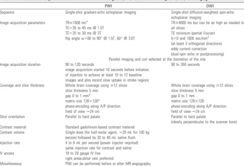
Table 1: Recommended Acquisition Protocols for Perfusion-Weighted (PWI) and Diffusion-Weighted (DWI) MR Imaging
PWI DWI
Sequence Single-shot gradient-echo echoplanar imaging Single-shot diffusion-weighted spin-echo echoplanar imaging
Image acquisition parameters TRⱕ1500 ms* TE⫽35 to 45 ms @ 1.5T TE⫽25 to 30 ms @ 3T
flip angle␣⫽60 to 90° @ 1.5T, 60° @ 3.0T
TRⱖ4000 ms but can be as high as needed to fit all slices
TE minimum (partial Fourier) b⫽0 and 1000 sec/mm2 (at least 3 orthogonal directions) eddy current correction (dual-spin echo or postprocessing) Parallel imaging and coil selected at the discretion of the site
Image acquisition duration 90 to 120 seconds
image acquisition started 10 seconds before initiation of injection to achieve at least 10 to 12 baseline images and also record slow uptake in stroke regions
90 to 260 seconds
Coverage and slice thickness Whole brain coverage usingⱖ12 slices slice thickness 5 mm
gap 0 to 1 mm* matrix size 128⫻128*
phase-encoding along A/P direction field of view⬇24 cm
Whole brain coverage usingⱖ12 slices slice thickness 5 mm
gap 0 to 1 mm matrix size 128⫻128
phase-encoding along A/P direction field of view⬇24 cm
Slice orientation Parallel to hard palate Parallel to hard palate
(ideally perpendicular to the scanner bore) Contrast material Standard gadolinium-based contrast material
Contrast volume Single dose (for half-molar agent,⬇20 mL for 100 kg person) followed by 20 to 40 mL saline flush Injection rate 4 to 6 mL per second (power injector required)
same injection rate for contrast and saline
IV access 18 to 20 gauge IV line
right antecubital vein preferred
Miscellaneous PWI can be performed before or after MR-angiography
MRI scanner: 1.5T or 3T (MRI scanners with 512 image limit per series should be excluded).
imaging abnormalities, and ultimately infarction. For treatments aiming at the recanalization of the occluded ar-tery, an appropriate assessment requires baseline and post-treatment assessment of arterial patency. The timing of the “reperfusion” scan should reflect a sufficient duration of the investigational therapy to demonstrate any effects. Ide-ally, the same modality (and MR field strength/CT param-eters) should be used for the baseline and this “reperfu-sion” scan.
● The reperfusion/recanalization MRI study should in-clude: scout image (no pregadolinium T1 required), DWI (Table 1), 3D time-of-flight MRA of the intracra-nial arteries, GRE, PWI (Table 1), and T2-FLAIR (FLAIR images can be obtained before or after gadolinium administration).
● The reperfusion/recanalization CT study should include: noncontrast CT, PCT (Table 2), and CTA, which can be limited to the intracranial arteries if the cervical arteries have been assessed at baseline (again, PCT can be per-formed before or after CTA).
If the patient has (1) undergone endovascular or intra-arterial (IA) therapy, or if the patient is (2) placed under continuous transcranial Doppler monitoring, and the recanalization (or persistent occlusion) status is known, then an MRA or CTA is not required, but may be obtained to assess for possible early reocclusion. PWI or PCT should be obtained in all cases to assess tissue reperfusion (or lack thereof, particularly consid-ering the possibility of distal embolization after intraarterial therapy).
For treatments other than reperfusion therapies, such as hyperoxia, induced hypertension, or collateral flow augmen-tation, an “on-treatment” scan should be considered instead of the “posttreatment”, “reperfusion” scan described above.
3. The third scan— either a noncontrast CT or GRE MRI of the brain—is a “safety scan” to assess the safety of investi-gational therapies, particularly with respect to the presence and degree of any hemorrhagic transformation. It may be obtained systematically or only in case of clinical worsen-ing, typically between 24 and 72 hours after symptom onset.
4. A follow-up imaging study should be obtained to deter-mine the final infarct volume. The appropriate timing for this follow-up scan is discussed below.
Recommended Perfusion Imaging Acquisition Protocols
Both PWI and PCT will be important components of the im-aging studies collected from acute stroke patients and contrib-uted to the central repository described below. The recom-mended imaging protocols for PWI and PCT are summarized in Tables 1 and 2. They are based on a consensus rather than solely evidence-based outcomes trials. The selected perfusion imaging parameters are based on first pass tracer kinetic mod-els and intended to provide the optimal balance between re-quirements for maximization of image quality and image analysis along with the need to minimize contrast material dose and CT radiation dose. Although these protocols are al-ready applied at the time points listed above at some institu-tions, their safety in terms of the total amount of contrast injected, the renal function, and the total radiation dose asso-ciated with the CT approach, requires further investigation.
Acute Stroke Imaging Central Repository
[image:3.594.53.283.62.544.2]The development of standardized, integrated, clinically useful imaging paradigms in acute stroke will require consolidation of existing data, prospective collection of new data, and the development of tools to analyze data in a standardized fashion at the time of image acquisition. This process will also require the systematic accumulation of evidence that specific imaging markers at determined time points accurately predict radio-graphic and clinical outcomes. An Acute Stroke Imaging Con-sortium could provide the framework for linking interna-tional resources. This organization will require leadership on the part of a small group of respected neuroimagers with a track record in collaborative endeavors. Criteria for inclusion
Table 2. Recommended Acquisition Protocol for Perfusion-CT (PCT)
Image acquisition rate 2 phases:
1st phase: 1 image per second, duration⫽30 to 45 seconds
2nd phase: 1 image per 2 to 3 seconds, duration⫽30 to 45 seconds
Total duration of the acquisition at least 70 to 90 seconds
Gantry rotation 1 second per gantry rotation
(up to every 3 seconds with “shuttle” or “toggle table” mode)
Image acquisition parameters 80 kVp, 100 mAs
Coverage and slice thickness Maximal coverage possible based on CT scanner configuration
(minimal coverage of 20 mm slab per contrast bolus injection preferable; two boluses is suggested to double coverage for all CT scanners with under 4 cm detector length unless precluded by contrast dose considerations) focus on supratentorial compartment/ anterior circulation
5- to 10-mm-thick slices field of view⬇24 cm Slice orientation Parallel to hard palate
lowest slice through the proximal middle/anterior cerebral artery (above the orbits)
Contrast material 350 to 370 mg/mL iodinated contrast material
high concentration, low/iso osmolar contrast preferred
follow local guidelines for contrast-induced nephropathy prevention Contrast volume 35 to 50 mL, followed by 20 to 40 mL
saline flush
Injection rate 4 to 6 mL per second (power injector required)
same injection rate for contrast and saline
IV access 18 to 20 gauge IV line right antecubital vein preferred (for anatomical reasons, reduces pooling of contrast, lowers the risk of extravasation and minimizes streak artifact at thoracic inlet in CTA portion) Miscellaneous PCT can be performed before or
in the consortium and definition of the structure for commit-tees and representation will need to be established. A charge to the leadership of such a consortium will be to secure funding from public and private sources and to foster collaboration with imaging equipment manufacturers and stroke pharma-ceutical/device companies.
An important initial step in effecting standardized anal-ysis will be the creation of a central repository. This ap-proach has been adopted by other organizations, as evidenced in acute stroke initiatives such as the American Heart Associ-ation’sStroke - Get With the Guidelinesprogram,1the Centers for Disease Control and Prevention’s (CDC)Paul Coverdell National Acute Stroke Registry,2 and the NINDSSpecialized Program in Translational Research in Acute Stroke (SPO-TRIAS).3 The Alzheimer Disease Neuroimaging Initiative (ADNI) group has successfully created an archive of imaging datasets publicly available for research images,4and there have also been nascent efforts to establish image repositories by SPOTRIAS,3the NIH Biomedical Informatics Research Net-work (BIRN),5the National Cancer Institute’scancer Biomed-ical Informatics Grid(caBIG),6and the International Consor-tium for Brain Mapping.7Investigators in Canada (Canadian Stroke Network and Canadian Stroke Consortium), Germany (Stroke Competence Network), United Kingdom and Scotland (NeuroGridandSINAPSE),8,9France (VIRAGE), Japan (Acute Stroke Imaging Standardization Group- ASIST),10Taiwan, and the international investigators from the MR Stroke Collabo-rative Group11and the I KNOW12and VISTA13projects have also established imaging repositories or are in the process of doing so. A coordinated centralized resource building on these individual efforts would significantly benefit the field of acute stroke imaging.
The central repository should include a statistically mean-ingful number of imaging studies obtained in acute stroke patients admitted within 12 hours of symptom onset. In addi-tion to these imaging studies, relevant metadata such as clini-cal information should be collected using standardized defini-tions, including (1) scores of clinical stroke severity, eg, NIH Stroke Scale, and other abstracted clinical parameters, (2) treatment records, (3) subsequent imaging studies, as well as information on (4) timing of symptom onset, admission, im-aging studies, interventions, and clinical evaluations, and (5) the results of these evaluations indicative of functional out-come, eg, modified Rankin scores, Barthel Index scores, and cognitive scales. In addition, whenever possible, blood should be banked from a subset of patients for the assessment of biomarkers.
The concepts underlying image-guided selection of stroke patients for therapy are that (1) only patients with reversible ischemia are going to benefit from treatment, and (2) imaging can identify these patients. To validate these concepts, it will be important for the set of patients included in the central repos-itory either (1) no treatment decision is based on imaging or (2) that matched control patients be identified in the case of image-guided treatment decisions, and that (3) all required imaging time points are obtained from all patients, including those deemed ineligible for treatment.
Documentation of early reperfusion (whether spontane-ous or following therapy) is important because it strongly in-fluences the appropriate predictive analysis and maximizes
ability to test acute imaging paradigms. Patients who achieve early reperfusion are informative with regard to distinguishing penumbra from core; nonrecanalizing patients are informa-tive with regard to distinguishing imaging benign oligemia from penumbra. Data would ideally be prospectively col-lected, but some retrospective data collected as part of existing networks and ongoing or completed trials, such as SPO-TRIAS,3Echoplanar Imaging Thrombolysis Evaluation Trial (EPITHET),11MR RESCUE,14Diffusion-weighted imaging Evaluation For Understanding Stroke Evolution (DE-FUSE),15etc, would also be included in the imaging database, as long as the datasets satisfy the minimal requirements listed below in terms of imaging acquisition protocols and time points for imaging studies. Contributors to the repository will need to confirm consent of their patients and approval from their institutional review board to allow inclusion and utiliza-tion of anonymized data. The collected informautiliza-tion (includ-ing the source or raw imag(includ-ing data) will be deidentified. Also, the potential for unblinding during the analysis of the scans collected in the imaging repository will be considered.
The data collected in the repository will be made accessible to qualified researchers worldwide, based on the recommen-dations of a scientific committee that will evaluate proposed research projects. The confidentiality of patients’ information will be rigorously protected. Contributors will be offered suit-able reassurance over the uses to which their data may be put, the acknowledgement that they as individuals and their insti-tutions will be granted for ensuing projects and developments, and an opportunity both to assist with the academic leadership of the consortium and to access the repository for projects of their own.
Adequate funding will be required to implement a data quality control program and to coordinate successful commu-nication among participating sites. The cost of local study co-ordination, data collection, and image transfer will need to be compensated. The consortium will require financial resources to reimburse centers for performance of additional images or tests that are not otherwise clinically indicated, facilitate com-munication with sites and data transfer, organize regular in-vestigator meetings, support centralized analysis, recruit ser-vices of dedicated stroke neuroimaging biostatisticians and technology assessment experts, and develop the technical in-frastructure for the repository. Several mechanisms are avail-able for potential funding through the NIH (U01), the Foun-dation for NIH, and the Institute of Medicine. Diverse partnerships will be explored with the NIH, private founda-tions, and industry.
Pilot Projects
As pilot studies for the proposed Acute Stroke Imaging Con-sortium, 3 “proof of concept” validation projects are proposed that would build on the optimized test dataset collected in the central repository.
Perfusion Imaging Processing
and of a venous output function (to correct for partial volume averaging in the AIF), are the most appropriate approach to process these datasets. However, a formal comparison with other analysis techniques (eg, nondeconvolution based or maximal slope methods) is required to demonstrate the supe-riority of this approach for predicting tissue fate and clinical outcome. This systematic comparison will also determine which parameters have, or do not have, a significant impact in terms of accurately representing acute perfusion status and predicting subsequent tissue outcome. Parameters studied will include cerebral blood flow, cerebral blood volume, and mean transit time, among others. The optimal method(s) should be most immune against slight raw image quality dif-ferences resulting from the use of different scanner hardware (ie, detector size configuration for multidetector CT scanners, magnetic field strengths, RF coils, scan parameters, injection protocols, and contrast agents used).
Imaging Prediction of Tissue Outcome
Still undetermined are the perfusion imaging parameters that indicate that tissue is at risk for infarction or that adequate reperfusion has taken place to prevent infarction. The “four scan” approach described above (baseline, 1 to 6 hours, 24 to 72 hours and final tissue outcome) will be used to develop, optimize, and validate imaging biomarkers of the infarct core and the ischemic penumbra. It will establish the value of base-line perfusion imaging in predicting final infarct size, using tissue fate as the outcome variable. Analysis will adjust for recanalization and reperfusion status, considered as a key de-terminant of tissue outcome and one that can be influenced by treatment. Different models of “operational” penumbra will be compared, and the optimal parameters (eg, cerebral blood flow, transit time, flow heterogeneity maps, etc) and optimal thresholds (eg, quantitative versus relative, gray matter versus white matter) to characterize the ischemic penumbra will be determined. Emphasis will be placed on quantitative ap-proaches. A consensus on the appropriate timing for deciding on the final infarct volume will be developed. Similarly, stan-dard definitions for recanalization (ie, changes in the degree of arterial patency) and reperfusion (ie, changes in the amount and spatial extent of perfusion changes) will be established before the final analysis. This analysis will incorporate patient characteristics at the time of scan acquisition, such as heart rate, blood pressure, glucose level, and hematocrit, which may have a significant impact on the distribution of contrast within collateral fields, and NIHSS which may reflect penumbral tis-sue shifting in and out of electric dysfunction. Imaging data in patients who have undergone reperfusion therapy and in those who have not will be analyzed separately to determine whether the results are the same for both groups.
Imaging Prediction of Clinical Outcome
One of the greatest challenges raised by pilot projects #1 and #2 is on the lack of consensus with respect to the optimal timing of outcome scans. Identification of key imaging biomarkers would facilitate the prediction of clinical outcome, define re-sponders/nonresponders to therapy, and permit monitoring of the efficacy of stroke treatment. This would represent a significant advance in the field of stroke imaging.
The third study will determine the optimal timing to
per-form imaging (48 hours, 1 week, 2 weeks, 1 month, 2 months, 3 months) to predict clinical outcomes at varying time points in the course of stroke recovery (eg, 30 days, 3 months, 6 months, 12 months). Analysis will be stratified according to management (eg, conservative care, IV/IA thrombolysis, me-chanical thrombectomy, collateral augmentation, or neuro-protective agents). The optimal imaging modality (MRI ver-sus CT) should be identified (many researchers believe that T2-FLAIR is the current best imaging modality for the identi-fication of final infarct, but this requires validation). Clinical outcomes will be documented using measures of global dis-ability (eg, the Modified Rankin Scale [mRS]), instrumental activities of daily living (eg, Barthel Index [BI]), neurological deficit (eg, NIHSS), cognitive function (neuropsychological testing), and quality of life. All clinical outcome assessments should be undertaken in a standardized manner and blinded to imaging and vice versa. Inclusion of generic and stroke-specific quality of life scores, and measures that identify values important to the patient (patient-derived recovery targets), are considered critical. This plan is in harmony with the Pa-tient-Reported Outcomes Measurement Information System (PROMIS), an NIH Roadmap initiative.16Cost-effectiveness analyses should be integrated into this and all future projects. For this third pilot project, follow-up imaging studies will be obtained at multiple time points. All datasets should be contributed to the central repository.
Deliverables
The goals of these 3 pilot projects, based on the clinical and imaging data from the central repository, will be to provide investigators with:
1. A standard set of imaging sequences to be performed at specific time points.
Overall, these deliverables will be accommodated in the clini-cal workflow of institutions using them and represent minimal impediment to enrollment of acute stroke patients in treat-ment protocols.
Next Steps
The deliverables outlined above, and the datasets stored in the central repository, will be available for further analyses. The initial focus will be on identifying the parameters that opti-mize the selection of acute stroke patients who benefit from reperfusion therapy. Other parameters of interest include as-pects that will improve our understanding of collateral perfu-sion, including determinants of tissue fate and clinical out-come, and predictors of hemorrhagic transformation. A consensus on the definition of clinically meaningful hemor-rhagic transformation will need to be developed.
At this stage, the efforts of the Acute Stroke Imaging Con-sortium will set the stage for 1 or more clinical trials. Indeed, the institutions contributing to the central repository will con-stitute a broad network of stroke care centers that could form the basis for an acute stroke trial/imaging network. They will all apply standardized imaging acquisition protocols, and use the same toolbox to process images and apply the same opti-mized criteria to interpret these processed images. This pro-cess will significantly minimize any source of variation other than the specific intervention (ie, drug or device) that will be tested in the clinical trial. The performance of the toolbox will be fully documented, facilitating sample size calculations for such trials. Initially, the identified imaging biomarkers will need to be validated in clinical trials with conventional clinical primary end points. Subsequently, it is anticipated that sample sizes will be reduced by the increased power afforded by the use of imaging biomarkers. In addition, if validated, the shorter follow-up periods that will be tested as part of the pilot projects will reduce loss to follow-up and minimize variation in clinical outcome due to unrelated events. This will greatly increase the feasibility and decrease the duration and cost of stroke treatment clinical trials.
Among the future stroke treatment clinical trials consid-ered, particular interest has focused on 2 that have the poten-tial to increase the proportion of acute stroke patients that are treated. The first trial is 1 of image-guided recanalization ther-apy in an extended time window (3 to 6 or 9 hours); the second one would assess image-guided recanalization therapy in wake-up stroke patients. Preliminary analysis (S.C. Johnston, personal communication, 2007) indicates that increasing the time window for acute reperfusion therapy from 3 hours to 6 hours could result in a 10-year societal benefit of $US 60 mil-lion. Neuroprotective agents and collateral enhancement could also be tested by the consortium, and future analyses should include attention to tissue repair, neurogenesis from stem cells, neurovascular remodeling, and stroke recovery.
Conclusion
Validation and widespread use of imaging for acute stroke patients’ management will be facilitated by the establishment of an Acute Stroke Imaging Consortium, consisting of an in-ternational, multi-institutional stroke neuroimaging network. This consortium would provide an expertise structure in which methodological issues in stroke imaging can be
ad-dressed and consensus reached among different groups of re-searchers and care providers. Initially, the consortium would create a central repository of imaging studies and clinical data obtained from acute stroke patients and develop a standard-ized image analysis toolbox. These could subsequently benefit clinical trials of acute stroke treatments, including, but not limited to, treatment of stroke patients in an extended time window, treatment of patients with wake-up stroke or those with long intervals between the time last seen well and time of symptom discovery, and neuroprotective, collateral enhance-ment, and neuroplasticity-stimulating therapies. Ultimately, these efforts, combined with strategies to change patient/pop-ulation behavior to promote earliest possible admission to hospital, should result in more acute stroke patients being ap-propriately treated and in an overall improvement of their outcome, as well as in reduced societal costs from economic disability. Collaboration between academia, the NIH, the FDA, and industry is integral to the successful realization of these aims.
Appendix
Contributors
Neuro-radiology Education and Research Foundation and American Society of Neuroradiology; Gregory V. Goldmakher, MD, PhD, setts General Hospital; R. Gilberto Gonzalez, MD, PhD, Massachu-setts General Hospital; Werner Hacke, MD, PhD, Heidelberg Univer-sity; Maxim D. Hammer, MD, University of Pittsburgh Medical School Stroke Center; Randall T. Higashida, MD, University of Cali-fornia, San Francisco; Michael D. Hill, MD, MSc, FRCPC, HSF Al-berta/NWT/NU Professorship Department of Clinical Neuro-sciences/Medicine/Community Health Sciences, Hotchkiss Brain Institute, University of Calgary, Canada; Ellen G. Hoeffner, MD, Uni-versity of Michigan; Amie W. Hsia, MD, Washington Hospital Center Stroke Center, Washington, DC; S. Claiborne Johnston, MD, PhD, University of California, San Francisco, Department of Neurology; Tudor G. Jovin, MD, University of Pittsburgh Medical School Stroke Center and Center for Neuroendovascular Therapy; Markku Kaste, MD, Department of Neurology, Helsinki University Central Hospi-tal, Helsinki, Finland; Chelsea S. Kidwell, MD, Goergetown Univer-sity, Washington Hospital Center; Stephan P. Kloska, MD, Depart-ment of Clinical Radiology, University of Muenster, Germany; Martin Ko¨hrmann, MD, Department of Neurology, University Clinic at Erlangen, Germany; Walter Koroshetz, MD, National Institute of Neurological Disorders and Stroke; Kohsuke Kudo, MD, PhD, Hok-kaido University, Japan; Paul Kwon, Genentech; Meng Law, MD MBBS FRACR, Mount Sinai Medical Center, New York; Ting-Yim Lee, PhD, Lawson Health Research Institute, Canada; Kennedy R. Lees, MD, University Department of Medicine & Therapeutics, West-ern. Infirmary, University of Glasgow, United Kingdom; Michael H. Lev, MD, Massachusetts General Hospital; David S. Liebeskind, MD, University of California, Los Angeles Stroke Center; Ke Lin, MD, New York University Medical Center; Weili Lin, PhD, Department of Ra-diology, University of North Carolina at Chapel Hill; Songling Liu, MD, University of California, San Francisco; Eng H. Lo, PhD, Depart-ments of Radiology and Neurology, Massachusetts General Hospital, Harvard Medical School; Bill McLaughlin, MS, CCRA, Mitsubishi Pharma America, Inc; Reto A. Meuli MD PhD, Centre Hospitalier Universitaire Vaudois and University of Lausanne, Switzerland; Patrik Michel, MD, Neurology Service, Centre Hospitalier Universi-taire Vaudois and University of Lausanne; David J. Mikulis, MD, Dept. of Medical Imaging, The University of Toronto, The University Health Network, The Toronto Western Hospital; Carlos A. Molina, MD, Neurovascular Unit. Hospital Vall d⬘Hebron. Barcelona, Spain; Kim Mouridsen, MD, Center for Functionally Integrative Neuro-science, Department of Neuroradiology, Aarhus University Hospital; Darius Nabavi, MD, Vivantes Klinikum Neuko¨lln, Berlin, Germany; Norbert Nighoghossian, MD, Cerebrovascular Unit, CHU de Lyon, Creatis CNRS 5520-INSERM U 630; Raul G. Nogueira, MD, Massa-chusetts General Hospital; Neil R.P. Ogden, MS, FDA*; Leif Oster-gaard, MD, MSc, PhD, DMSc, Center for Functionally Integrative Neuroscience, Department of Neuroradiology; Salvador A. Pedraza, MD, Servicio de Radiologia-IDI. IDIBGI. Hospital Universitario Dr Josep Trueta. Girona, Spain; Scott Pohlman, MSc, Philips Medical Systems; William J. Powers, MD, Department of Neurology, Univer-sity of North Carolina at Chapel Hill; James Provenzale, MD, Depart-ment of Radiology, Duke University Medical Center; Qin Qin, PhD, Johns Hopkins University; Philippe Raffy, PhD, Vital Images, Inc; Pat
Reilly, Genentech; Timothy Roberts, PhD, Department of Radiology, Children’s Hospital of Philadelphia; Joachim Ro¨ther, MD, Depart-ment of Neurology, Klinikum Minden, Academic Teaching; Hospital, Hannover Medical School, Minden, Germany; Howard A. Rowley, MD, University of Wisconsin; Eric J. Russell, MD, FACR, Department of Radiology, The Feinberg School of Medicine, Northwestern Uni-versity, Chicago; Pina C. Sanelli, MD, MPH, Weill Cornell Medical School, New York Presbyterian Hospital; Makoto Sasaki, MD, De-partment of Radiology, Iwate Medical University. Japan; Jeffrey L. Saver, MD, University of California, Los Angeles Stroke Center; Sean I. Savitz, MD, University of Texas Houston Medical School; Pamela W. Schaefer, MD, Massachusetts General Hospital; Peter Schellinger, MD, PhD, Department of Neurology, University Clinic at Erlangen, Germany; Gottfried Schlaug, MD, PhD, Neuroimaging Laboratory and Comprehensive Stroke Center, Beth Israel Deaconess Medical Center and Harvard Medical School; Uri Shreter, GE Healthcare; Lee H. Schwamm, MD, FAHA, Massachusetts General Hospital; Robert Silbergleit, MD, University of Michigan; Aneesh B. Singhal, MD, J. Philip Kistler Stroke Research Center, Massachusetts General Hospi-tal; Saad A. Sirohey, PhD, GE Healthcare; Wade S. Smith, MD, PhD, University of California, San Francisco, Department of Neurology; Jamal Smyej, PAION Deutschland GmbH; Bruno P. Soares, MD, Di-agnostic Imaging Department, Federal University of Sao Paulo, Bra-zil; Alma Gregory Sorensen, MD, Massachusetts General Hospital; Jeffrey L. Sunshine, MD, PhD, Interventional Neuroradiology Uni-versity Hospitals Health System, Case Western Reserve UniUni-versity; Turgut Tatlisumak, MD, PhD, Department of Neurology, Helsinki University Central Hospital, Helsinki, Finland; Wyatt M. Tellis, PhD, University of California, San Francisco; Thomas Tourdias, MD, CHU de Bordeaux - Universite´ Victor Segalen Bordeaux, Service de Neu-roradiologie, Hoˆpital Pellegrin, Bordeaux, France; Matthias van Osch, PhD, Leiden University Medical Center, Leiden, The Nether-lands; Peter C.M. van Zijl, MD, PhD, Johns Hopkins University School of Medicine, Dept. of Radiology and Kennedy Krieger Insti-tute, F.M. Kirby Research Center; J. Pablo Villablanca, MD, Univer-sity of California, Los Angeles; Sissel Vorstrup, MD, PhD, Depart-ment of Neurology, International Clinical Research, H. Lundbeck A/S; Steven Warach, MD, PhD, National Institute of Neurological Disorders and Stroke; Joyce A. Wahr, MD, CoAxia, Inc; Joanna Wardlaw, MD, Western General Hospital, Edinburgh, United King-dom; Lawrence R. Wechsler, MD, University of Pittsburgh Medical School Stroke Center; Andrew M. Weiss, CoAxia, Inc; Max Winter-mark, MD, University of California, San Francisco; Hans-Jo¨rg Witt-sack, PhD, Institute of Diagnostic Radiology, University of Duessel-dorf, DuesselDuessel-dorf, Germany; Ona Wu, PhD, Massachusetts General Hospital; Greg Zaharchuk, MD, PhD, Stanford University; Christian G. Zimmerman, MD, Institute of Medicine, Forum on Neuroscience and Nervous System Disease.
Acknowledgments
The participants of the “Advanced Neuroimaging for Acute Stroke Treatment” meeting on September 7 and 8, 2007 in Washington DC thank the National Institute of Neurological Disorders and Stroke (NINDS) and the Neuroradiology Education and Research Founda-tion (NERF) of the American Society of Neuroradiology (ASNR) for their support. Special acknowledgement and thanks for the members of the organizing committee of the meeting: Gregory W. Albers, MD, Stanford University; Colin P. Derdeyn, MD, Washington University; James D. Eastwood, MD, Duke University; Chelsea S. Kidwell, MD, Georgetown University, Washington Hospital Center; Michael H.
Lev, MD, Massachusetts General Hospital; Steven Warach, MD, PhD, National Institute of Neurological Disorders and Stroke; Max Win-termark, MD, University of California, San Francisco; Ona Wu, PhD, Massachusetts General Hospital.
Sources of Funding
The meeting was supported by a grant from the NINDS (1R13NS061371-01), as well as by contributions from the Neurora-diology Education and Research Foundation (NERF) of the American Society of Neuroradiology (ASNR), General Electric Healthcare, Sie-mens Medical Solutions, Mitsubishi-Pharma, Concentric, Lundbeck, Genentech, Paion, Toshiba Medical Systems, Vital Images, CoAxia, Philips Medical Systems, Sanofi-Aventis Pharmaceuticals, and Wyeth.
Disclosures None.
References
1. http://www.americanheart.org/presenter.jhtml?identifier⫽1165. Accessed September 2007.
2. http://www.cdc.gov/DHDSP/stroke_registry.htm. Accessed September 2007. 3. http://www.spotrias.com/. Accessed September 2007.
4. http://www.loni.ucla.edu/ADNI/. Accessed September 2007. 5. http://www.nbirn.net/. Accessed September 2007. 6. https://cabig.nci.nih.gov/. Accessed September 2007.
7. http://www.loni.ucla.edu/ICBM/About/. Accessed September 2007. 8. http://www.sbirc.ed.ac.uk/sinapse/sinapse.asp. Accessed September 2007. 9. http://www.neurogrid.ac.uk/. Accessed September 2007.
10. http://asist.umin.jp/index-e.htm. Accessed September 2007. 11. www.mrstroke.com. Accessed September 2007.
12. http://cordis.europa.eu/fetch?CALLER⫽PROJ_IST&ACTION⫽ D&DOC⫽3&CAT⫽PROJ&QUERY⫽1185280321622&RCN⫽78374. Accessed September 2007.
13. http://www.vista.gla.ac.uk/index.aspx. Accessed September 2007. 14. http://clinicaltrials.gov/ct/show/NCT00389467;jsessionid⫽76B7