STRUCTURAL ANALYSIS OF HIV
DOCKING WITH PHYTOCHEMICALS
1
Samriti Sharma,
21School of Biomolecular and Biomedical Science
2School of Biotechnology, Lovely Professional Univer
3Kaust Catalysis Center, King Abdullah University of Science and Technology, Thuwal, Saudi Arabia
ARTICLE INFO ABSTRACT
Human Immunodeficiency Virus (HIV) is a rapidly expanding global pandemic. Despite more than a decade of intense research to understand the HIV pathogenesis aimed at developing an effective therapy for AIDS,
significant progress has been made in the management of HIV
HIV-2 serotype of HIV was determined to be a cause of disease in the parts of the West African population, and there is evidence
reverse transcriptase (RT) demonstrates intrinsic resistance to non the two classes of anti
designing a potent drug that can block the activity of HIV
conducting a comparative analysis between the reverse transcriptase from the two strains of HIV viz. HIV-1 and HIV
molecular docking of the HIV
pertaining to mechanism of binding of the ligand to the receptor. Docking results revealed that Curcumin, Astralgin
to pocket1 (Pkt1).
Copyright ©2014 Samriti Sharma et al. This is an open access article distributed under the Creative Commons Attribution License, which permits unrestricted distribution, and reproduction in any medium, provided the original work is properly cited.
INTRODUCTION
Acquired Immunodeficiency Syndrome (AIDS) is a medical condition that results in weakening of the natural defense machinery, the immune system of the individual resulting in its susceptibility to a plethora of opportunistic infections and tumors to invade the body. This susceptibility gets worse as the disease continues (Sepkowitz 2001). Since the first cases of AIDS were identified in 1981, more than 30 million people have died from AIDS (as of 2013). An estimated 1.8 million people died as a result of AIDS in 2010 alone.
by Human Immunode ficiency Virus (HIV) be
genus lentivirus (Weiss 1993).The genus Lentivirus, itself is a subset of the Retroviridae family of RNA viruses
viruses which share acommon replicative cy caused by each of the lentiviruses including H 2,Simian ImmunodeficiencyVirus(SIV) cause c systemic events that initiatediseaseprocess.It
reported that Human Immunodeficiency Virus (HIV) enters human body through the transfer of blood, semen, vaginal fluid or breastmilk through four major routes of transmission that includes
*Corresponding author: 2,3
Mohit Chawla, 2
School of Biotechnology, Lovely Professional University, Punjab, India, 3
Kaust Catalysis Abdullah University of Science and Technology, Thuwal, Saudi Arabia
ISSN: 0975-833X
International Journal of Current Research
Vol.
Article History:
Received 20th
April, 2014 Received in revised form
18th
May, 2014
Accepted 08th
June, 2014
Published online 20th
July,2014
Key words:
HIV-2 Reverse Transcriptase, Comparative Analysis, Molecular Docking, Phytochemicals, Ligand, Receptor.
RESEARCHARTICLE
STRUCTURAL ANALYSIS OF HIV-2 REVERSE TRANSCRIPTASE AND ITS
DOCKING WITH PHYTOCHEMICALS
2
Ravneet KaurGrewal and *
2,3Mohit Chawla
Biomolecular and Biomedical Science, University College, Dublin- Biotechnology, Lovely Professional University, Punjab, India
Kaust Catalysis Center, King Abdullah University of Science and Technology, Thuwal, Saudi Arabia
ABSTRACT
Human Immunodeficiency Virus (HIV) is a rapidly expanding global pandemic. Despite more than a decade of intense research to understand the HIV pathogenesis aimed at developing an effective therapy for AIDS, achieving a true eradication of HIV remains a daunting challenge
significant progress has been made in the management of HIV-1 replication using potent inhibitors. 2 serotype of HIV was determined to be a cause of disease in the parts of the West African population, and there is evidence for its spread to Europe and Asia. It has also been found that HIV reverse transcriptase (RT) demonstrates intrinsic resistance to non-nucleoside RT inhibitors, one of the two classes of anti- AIDS drugs that target the viral RT. The given article is an
designing a potent drug that can block the activity of HIV-2 RT. This has been achieved by conducting a comparative analysis between the reverse transcriptase from the two strains of HIV viz. 1 and HIV-2. After the comparative analysis of two genomes and structure of the enzyme, molecular docking of the HIV-2 RT was performed using phytochemicals in order to gain insights pertaining to mechanism of binding of the ligand to the receptor. Docking results revealed that Curcumin, Astralgin and Tiliroside bind to pocket 2 (Pkt 2) with higher binding energies as compared to pocket1 (Pkt1).
is an open access article distributed under the Creative Commons Attribution License, which permits unrestricted distribution, and reproduction in any medium, provided the original work is properly cited.
yndrome (AIDS) is a medical condition that results in weakening of the natural defense machinery, the immune system of the individual resulting in its susceptibility to a plethora of opportunistic infections and ty gets worse as . Since the first cases of AIDS were identified in 1981, more than 30 million people have died from AIDS (as of 2013). An estimated 1.8 million AID Siscaused elong ingto the nus Lentivirus, itself is a viruses that includes ycle. Infection HIV-1andHIV-use cellular and
It is already irus (HIV) enters through the transfer of blood, semen, vaginal fluid or breastmilk through four major routes of transmission
School of Biotechnology, Lovely Kaust Catalysis Center, King Abdullah University of Science and Technology, Thuwal, Saudi Arabia.
unsafe sex, contaminated needles, breast milk, and from mother to fetus. Once HIV enters the human body, its target is the immune cells of the body i.e. T
(specifically, CD4+ T cells), macrophages, and dendritic cells (Cunningham et al.,2010). HIV infection leads to low levels of CD4+ T-cells through three main mechanisms: First, direct viral killing of infected cells; second, increased rates of apoptosis in infected cells; and third, killing of infected CD4+ T cells by CD8 cytotoxic lymphocytes that recognize infected cells. When CD4+ T cell numbers decline below a critical level, cell-mediated immunity is lost, and the body becomes progressively more susceptible to opportunistic infections. Reverse transcriptase converts the single stranded HIV RNA genome to double stranded DNA copy by catalyzing both DNA-dependent, RNA-dependent polymerization as well as RNase H cleavage activity to remove the RNA
the synthesis of its DNA. Due to these unique catalytic properties, Reverse transcriptase (RT) has been the target enzyme for many antiviral therapeutic agents used in treatment of AIDS, including nucleoside and non
analogues (De Clercq 1994). Reverse transcriptase inhibitors (RTIs) are a class of antiretroviral drug used to treat HIV infection by inhibiting the ability of the enzyme to form DNA from RNA. RTIs come from three main types: Nucleoside Analog Reverse- Transcriptase I
International Journal of Current Research
Vol. 6, Issue, 07, pp.7321-7325, July,2014
OF CURRENT RESEARCH
2 REVERSE TRANSCRIPTASE AND ITS
Mohit Chawla
Ireland India
Kaust Catalysis Center, King Abdullah University of Science and Technology, Thuwal, Saudi Arabia
Human Immunodeficiency Virus (HIV) is a rapidly expanding global pandemic. Despite more than a decade of intense research to understand the HIV pathogenesis aimed at developing an effective daunting challenge. However, 1 replication using potent inhibitors. 2 serotype of HIV was determined to be a cause of disease in the parts of the West African for its spread to Europe and Asia. It has also been found that HIV-2 nucleoside RT inhibitors, one of AIDS drugs that target the viral RT. The given article is an attempt towards 2 RT. This has been achieved by conducting a comparative analysis between the reverse transcriptase from the two strains of HIV viz. s of two genomes and structure of the enzyme, 2 RT was performed using phytochemicals in order to gain insights pertaining to mechanism of binding of the ligand to the receptor. Docking results revealed that and Tiliroside bind to pocket 2 (Pkt 2) with higher binding energies as compared
is an open access article distributed under the Creative Commons Attribution License, which permits unrestricted use,
or NRTIs); second, Nucleotide Analog Reverse
Inhibitors (NtARTIs or NtRTIs). The mode of action of NRTIs and NtRTIs is essentially the same; they are analogues of the naturally occurring deoxynucleotideneeded to synthesize t viral DNA and they compete with the natural deoxynucleotides for incorporation into the growing viral DNA chain
2006). Third, Non-Nucleoside Reverse
(NNRTIs), on the other hand, blocks reverse transcriptase by binding at a different site on the enzyme. NNRTIs are not incorporated into the viral DNA but instead inhibit the movement of protein domains of reverse transcriptase that areneeded to carry out the process of DNA synthesis. NNRTIs are therefore classified as non-competitive inhibitors of reverse
transcriptase.The crystal structure of HIV
transcriptase consists of two subunits p66 and p51 forming a heterodimer (Rodgerset al., 1995). The larger p66 subunit contains the fingers, palm, and connection subdomain as we as the RNaseH domain. The p51 is a product of the same gene as the p66 subunit, however the RNaseH domain is absent as a result of the proteolytic cleavage. HIV-2 RT on the other hand forms a more stable p68/p55 heterodimer compared with p66/p51 HIV-1 RT heterodimer (Ren et al., 2002
the two molecules possess high degree of sequence similarities, the difference lies in the kinetic parameters for RNaseH and the polymerase. In case of HIV-1 RT, the RNA/DNA binding pocket is blocked by thumb of p51 whereas it is open in case of
HIV-2 RT.Moleculardockingisamethodwhi
preferredorientationofonemoleculetoa secondwh
toeachothertoformastable
complex.Theaimofmoleculardockingisto evaluatethefeasiblebindinggeometriesofaputativel getwhosetargetsiteis
known.Thesebindinggeometriesareknownasbind cludesboththepositionof
ligandrelativetothereceptorandconformationalstateofthe ligandandthereceptor.The
dockingprocedureconsistsof(i)characterizationofbind positioningofligandtothe
bindingsite;and(iii)evaluatingthestrengthofintera icligandreceptorcomplex.
Theligandsusedtodocktheenzymewerefivephytoc vealreadybeenprovedto have anti-HIV activity
MATERIALS AND METHODS
Selection of binding pockets
The three dimensional structure of HIV-2 reverse transcriptase was retrieved from Protein Data Bank (PDB-id: 1MU2). The structure was then searched for possible binding pockets using LIGSITEcsc server (Hendlich et al., 1997
predicted two possible binding pockets which were named as Pkt-1 and Pkt-2 respectively.
Ligand selection
Five phytochemicals with a known anti-HIV activity such as
lycopene, curcumin, astralgin, tiliroside and 1
deoxynojirimycin were selected (Abhik Seal et al.,
2D structures of these phytochemicals were retrieved from PubChemdatabase (http://pubchem.ncbi.nlm.nih.gov/). These or NRTIs); second, Nucleotide Analog Reverse-Transcriptase nhibitors (NtARTIs or NtRTIs). The mode of action of NRTIs and NtRTIs is essentially the same; they are analogues of the naturally occurring deoxynucleotideneeded to synthesize the viral DNA and they compete with the natural deoxynucleotides for incorporation into the growing viral DNA chain (Paulet al., Reverse-Transcriptase (NNRTIs), on the other hand, blocks reverse transcriptase by ferent site on the enzyme. NNRTIs are not incorporated into the viral DNA but instead inhibit the movement of protein domains of reverse transcriptase that areneeded to carry out the process of DNA synthesis. NNRTIs ive inhibitors of reverse
transcriptase.The crystal structure of HIV-1 reverse
transcriptase consists of two subunits p66 and p51 forming a . The larger p66 subunit contains the fingers, palm, and connection subdomain as well as the RNaseH domain. The p51 is a product of the same gene as the p66 subunit, however the RNaseH domain is absent as a 2 RT on the other hand forms a more stable p68/p55 heterodimer compared with 2002). Although the two molecules possess high degree of sequence similarities, the difference lies in the kinetic parameters for RNaseH and the 1 RT, the RNA/DNA binding p51 whereas it is open in case of gisamethodwhichpredictsthe
ondwhen bound
iesofaputativeligandwithatar
sbindingposesthatin
lstateofthe
onofbindingsite;(ii)
actionforaspecif
chemicalsthatha
2 reverse transcriptase id: 1MU2). The structure was then searched for possible binding pockets using 1997).The server le binding pockets which were named as
HIV activity such as
lycopene, curcumin, astralgin, tiliroside and
1-et al., 2011). The
2D structures of these phytochemicals were retrieved from PubChemdatabase (http://pubchem.ncbi.nlm.nih.gov/). These
2D structures were then converted to 3D structures using openbabel (Figure1). These structures were evaluated for their drug likeliness based on Lipinski’s Rule of five. However, later on, lycopene molecule was excluded from the docking studies as it did not obey Lipinski’s Rule of five.
Figure 1.Structuresofthephytochemicalsusedasl
eptor (PDB id: 1MU2): a) Curcumin; b) Astralgin; c) Lycopene; d) Tiliroside; e) 1-Deoxynojirimycin
Protein-Ligand docking
The docking of Phytochemicals (ligand molecules) with HIV RT (receptor molecule) was then performed using Autodock 4.0 and Argus Lab (Thomson 2004).
Docking using Argus Lab
The coordinates of PDB id: 1MU2 were provided as an input for the Argus Lab and the residues of the binding pocket were selected as the possible binding site. Then, the ligand file was uploaded. After calculation of grid parameters
structures viz. ligand and receptor were docked. The results obtained were ranked by energy values and their binding poses. The best possible conformation was saved as a PDB interaction analysis.
Docking using Autodock 4.0
Docking was performed with both the binding pockets viz. Pkt 1 and Pkt 2. Prior to loading the molecule, all heteroatoms were removed and hydrogen atoms were added to the enzyme to generate a PDBQT file. Then ligand PDB files
(curcumin.pdb, astralgin.pdb, tiliroside.pdb, 1
deoxynojirimycin.pdb) were added each time to select bonds about which segments of the ligand will be rotated. Various docking parameters like torsion angles in the ligand were calculated and grid parameters were set to define the region for 2D structures were then converted to 3D structures using openbabel (Figure1). These structures were evaluated for their s based on Lipinski’s Rule of five. However, later on, lycopene molecule was excluded from the docking studies as it did not obey Lipinski’s Rule of five.
sligandsfordockingwithrec Curcumin; b) Astralgin; c) Lycopene;
Deoxynojirimycin
The docking of Phytochemicals (ligand molecules) with HIV-2 RT (receptor molecule) was then performed using Autodock
The coordinates of PDB id: 1MU2 were provided as an input for the Argus Lab and the residues of the binding pocket were selected as the possible binding site. Then, the ligand file was uploaded. After calculation of grid parameters, the two structures viz. ligand and receptor were docked. The results obtained were ranked by energy values and their binding poses. The best possible conformation was saved as a PDB
with both the binding pockets viz. Pkt 1 and Pkt 2. Prior to loading the molecule, all heteroatoms were removed and hydrogen atoms were added to the enzyme to generate a PDBQT file. Then ligand PDB files
(curcumin.pdb, astralgin.pdb, tiliroside.pdb, 1-
docking the ligand resulting in the generation of GPF file. Autodock runs in the directory where the macromolecule, ligand, grid parameter file (GPF), docking parameter file (DPF)
and maps are located. Upon completion of the program DLG files for several docked structures were obtained. The resulting files were then analyzed using Pymol (DeLano2008) to find possible hydrogen bonds present between the ligand and the interacting residues.
RESULTS AND DISCUSSION
Using the Pocketfinder server, two pockets namely, Pkt1 and Pkt2 were identified and docking study using the
phytochemicals was performed using the identified
pockets.Out of the two identified pockets, the residues present in Pkt2
showed better binding affinity as compared to the residues in
Pkt1 with phytochemicals. The structural and
interactionanalysis revealed the presence of hydrogen bonding with the residues His360, Arg365, Tyr404, and Trp405 present in Pkt2 with all the phytochemicals under study. The detailed analysis of the atoms that are involved in H-bonding of residues in receptor with that of ligands is shown (Figure 2 and Figure 3).
It was also observed that the phytochemicals viz. Curcumin,
Table1.Interaction energiesobtained using the dockingofdifferentphytochemicalswithtwo diferentpockets. Column2 provides the results of dockingwith ArgusLAband Autodock 4.0with phytochemicalsinPkt1 and column 3 gives the interaction energies
ofphytochemicalswithPkt2 respectively
Ligands Pkt1 Pkt2
CURCUMIN
1. Bindingenergy
2. Interactingresidues
3. Grid parameters
-3.11kcal/mol (Autodock) -9.189kcal/mol (ArgusLab)
Tyr183,Met184
17.381,10,10
-5.11kcal/mol (Autodock) -9.2423kcal/mol (ArgusLab)
His360, Arg365, Asn403,Tyr404,Trp405, Gln406Gly503 16.927,12.131, 11.355
ASTRALGIN
1. Bindingenergy
2. Interaction residues
3. Grid parameters
-2.19kcal/mol (Autodock) -7.158kcal/mol(ArgusLab)
Tyr183,Met184,
19.027,11.495, 13.247
-4.44kcal/mol (Autodock)-8.267kcal/mol(ArgusLab)
His360, Tyr404, Trp405, Gly503,Ala506,
Ser507
12.082,11.420, 10
TILIROSIDE
1. Bindingenergy
2. Interaction residues
3. Grid parameters
-2.19kcal/mol (Autodock) -8.577kcal/mol(ArgusLab)
Glu89, Gln91, Leu92,
14.311,14.081, 19,909
-3.73kcal/mol (Autodock) -8.176kcal/mol(ArgusLab)
His360, Arg365, Tyr404, Trp405,
Gly503,Ala506, Ser507
10,12.154,15.464
1-DEOXYNOJIRIMYCIN
1. Bindingenergy
2. Interactingresidues
3. Grid parameters
-2.29kcal/mol (Autodock) -6.141kcal/mol(ArgusLab)
Arg172, Asn175, Ile180
10,10, 10
-1.87kcal/mol (Autodock) -6.580kcal/mol(ArgusLab)
Trp405, Gly503, Ser507
Astralgin, and Tiliroside were interacting with Pkt2 with more number of H-bonds as compared to Pkt1.
Figure 2. Interaction between the amino acid residues
Figure 3.Interaction between the amino acid residues of Pkt1 and Pkt2 with the phytochemicals viz. Tiliroside and 1
Conclusion
The values of interaction energies of all the phytochemicals that were used as ligand molecules to dock with the receptor molecule were given in Table 1.Among all the phytochemicals under study, curcumin was found to bind with both the pockets of the receptor molecule with maximum interaction energy. (Table 1) The values of interaction energies obtained due to the binding of Curcumin with receptor was found to be maximum employing both the rigid and flexible docking methods. The rigid docking was performed using ArgusLab, and flexible using was performed using Autodock 4.0. The mo drawings were prepared using Pymol. The interaction analysis also revealed the presence of maximum number of H
Astralgin, and Tiliroside were interacting with Pkt2 with more
Interaction between the amino acid residues
Figure 3.Interaction between the amino acid residues of Pkt1 and Pkt2 with the phytochemicals viz. Tiliroside and 1-DNM.
The values of interaction energies of all the phytochemicals as ligand molecules to dock with the receptor molecule were given in Table 1.Among all the phytochemicals under study, curcumin was found to bind with both the pockets of the receptor molecule with maximum interaction energy. action energies obtained due to the binding of Curcumin with receptor was found to be maximum employing both the rigid and flexible docking methods. The rigid docking was performed using ArgusLab, and flexible using was performed using Autodock 4.0. The molecular drawings were prepared using Pymol. The interaction analysis also revealed the presence of maximum number of H-bonds in
case of curcumin interacting with residues of Pkt2. Docking of curcumin to Pkt2 shows best results with the highest binding energy of -9.24 kcal/mol using ArgusLab and
using Autodock followed by astralgin and tiliroside. However, the H-bonding pattern in case ofcurcumin, astralgin and tiliroside shows on an average five hydrogen bonds between the ligand and the binding pocket. The detailed information pertaining to possible H- bonds is listed in Table 2 of SI.
Conclusion
In the present study it has been concluded that despite of high amount of sequence similarities between the two enzymes, certain mutations in the amino acids result in different structure and altered activity towards most of the common inhibitors, making it a possible element of research. These variations call for the search of several different drug targets that can block the reverse transcriptase activity of the enzyme thereby restricting the viral replication inside the host cell. Phytochemicals offer a better alternative to chemically
synthesizedmoleculesasligands.Thisis b
increased biocompatibilityandless
Thedockingresults reveal
astralginandtilirosidetoPkt2withhighere Pkt2 servesas agood bindingsitetothesep
Acknowledgment
We thank School of Biotechnology, Lovely Professional University, Punjab and Department of Structural Biology, NCCS Complex, University of Pune Campus for providing us requisite infrastructure, cutting edgetechnology and excellent working environment to carry out this research work.
REFERENCES
Abhik Seal.; RijuAkkyal.; Rosana O.
Docking study of HIV-1 reverse transcriptasewith phytochemicals. Bioinformation. 2011, 5, 430
AZT Resistance?PLoSPathog. 2006, 2(2):e10.Rodgers, DW.; Gamblin, SJ.; Harris, BA.; Ray, S.; Culp,JS.; Hellmig, B.; Woolf, DJ.; Debouck, C.; Harrison, SC.; The structure of
unligated reverse transcriptase from the Human
Immunodeficiency Type-1virus.Proc.Natl.Acad.Sci.
USA.1995, 92, 1222-1226.
Cunningham, A.; Donaghy, H.; Harman, A.; Kim, M.; Turville, S. Manipulation of dendritic cell f byviruses. CurrOpinMicrobiol.2010, 13, 524
De Clercq, E. HIV resistance to reverse transcriptaseinhibitors. BiochemPharmacol. 1994, 47, 155
DeLano W.L., 2008.The PyMOL Molecular Graphics System‖. DeLano Scientific LLC, Palo Alto, CA, US Hendlich, M.; Rippmann, F.; Barnickel, G. LIGSITE:
automatic and efficient detection of potential
smallmolecule-binding sites in proteins.
Model.1997, 15,359-63, 389.
Paul L Boyer.; Stefan G Sarafianos.; Patrick K Clark.; Eddy Arnold.; Stephen H Hughes.; 2006.
HIV-2 Use Different Pathways to Develop
Ren, J.; Bird, LE.; Chamberlain, PP.; Stuart, DI.;Stammers, DK.; Structure of HIV-2 Reverse transcriptase at 2.35 Å and the mechanism of resistance to non
case of curcumin interacting with residues of Pkt2. Docking of curcumin to Pkt2 shows best results with the highest binding 9.24 kcal/mol using ArgusLab and -5.11kcal/mol using Autodock followed by astralgin and tiliroside. However, pattern in case ofcurcumin, astralgin and tiliroside shows on an average five hydrogen bonds between g pocket. The detailed information bonds is listed in Table 2 of SI.
In the present study it has been concluded that despite of high amount of sequence similarities between the two enzymes, ino acids result in different structure and altered activity towards most of the common inhibitors, making it a possible element of research. These variations call for the search of several different drug targets that can block tivity of the enzyme thereby restricting the viral replication inside the host cell. Phytochemicals offer a better alternative to chemically
hisis becauseoftheir
ndlesserchancesoftoxicity. ledthebindingofcurcumin, energies.This suggeststhat etothesephytochemicals.
We thank School of Biotechnology, Lovely Professional Department of Structural Biology, NCCS Complex, University of Pune Campus for providing us technology and excellent working environment to carry out this research work.
Abhik Seal.; RijuAkkyal.; Rosana O. Babu.; MrigankaGhosh.; 1 reverse transcriptasewith phytochemicals. Bioinformation. 2011, 5, 430-439. AZT Resistance?PLoSPathog. 2006, 2(2):e10.Rodgers, DW.;
Gamblin, SJ.; Harris, BA.; Ray, S.; Culp,JS.; Hellmig, B.; k, C.; Harrison, SC.; The structure of
unligated reverse transcriptase from the Human
1virus.Proc.Natl.Acad.Sci.
Cunningham, A.; Donaghy, H.; Harman, A.; Kim, M.; Turville, S. Manipulation of dendritic cell function
.2010, 13, 524-9.
De Clercq, E. HIV resistance to reverse transcriptaseinhibitors. BiochemPharmacol. 1994, 47, 155-69.
DeLano W.L., 2008.The PyMOL Molecular Graphics ‖. DeLano Scientific LLC, Palo Alto, CA, US dlich, M.; Rippmann, F.; Barnickel, G. LIGSITE:
automatic and efficient detection of potential
binding sites in proteins. J Mol Graph
Paul L Boyer.; Stefan G Sarafianos.; Patrick K Clark.; Eddy 2006. Why Do HIV-1 and 2 Use Different Pathways to Develop
[image:4.612.71.291.316.510.2]inhibitors.Proc.Natl.Acad.Sci.USA.2002, 99, 14410– 14415.
Sepkowitz, KA. AIDS—the first 20 years.N Engl J Med.2001, 344, 1764–72.
Thomson, M.A. Molecular docking using ArgusLa, an efficient shape based search algorithm and A Scorescoring function ACS meeting, Philadelphia, 172, CINF42, PA, 2004.
Weiss, RA. How does HIV cause AIDS? Science. 1993, 260, 1273–79.
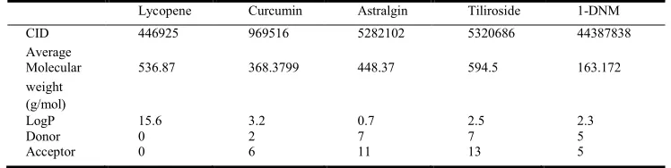
[image:5.612.121.495.157.251.2]SupplementaryInformation
Table 1.Phytochemicals thatwereselected based onLipinski’s Ruleof Five
Lycopene Curcumin Astralgin Tiliroside 1-DNM
CID 446925 969516 5282102 5320686 44387838
Average
Molecular 536.87 368.3799 448.37 594.5 163.172
weight (g/mol)
LogP 15.6 3.2 0.7 2.5 2.3
Donor 0 2 7 7 5
Acceptor 0 6 11 13 5
Table2.Possible Hydrogen bonds between theresidues inreceptormolecule with the Phytochemicals and their respectivebond lengths
Curcumin-Pkt1
O(MET184)---H(DEF)3.0Å N(MET184)---O(DEF)2.5Å
Curcumin-Pkt2
NE(ARG365)---O(DEF) 3.0Å NH2(ARG365)---O(DEF)2.3Å O(ASN403)---O(DEF)2.0Å OE1(GLN406)---O(DEF) 3.4Å O(GLY503)---O(DEF)2.6Å
Astralgin-Pkt1
OH(TYR183)---O(DEF)2.0Å Astralgin-Pk2
NE2(HIS360)---O(DEF)3.0Å N(HIS360)---O(DEF)2.7Å O(HIS360)---O(DEF)2.6Å OH(TYR404)---O(DEF)3.2Å NE1(TRP405)---O(DEF)2.9Å OG(SER507)---O(DEF)3.4Å
Tiliroside-Pkt1
O(GLN91)---O(DEF)3.5Å OE2(GLU89)---O(DEF)2.9Å
Tiliroside-Pkt2
NH1(ARG365)---O(DEF)3.1Å NH2(ARG365)--- O(DEF)3.3Å OH(TYR404)---O(DEF)3.4Å N(GLY503)--- O(DEF)2.9Å N(ILE504)--- O(DEF)2.7Å N(SER507)---O(DEF)3.3Å
DNM-Pkt1
O(ARG172)---O(DEF)2.1Å O(ASN175)---O(DEF)3.1Å N(ILE180)---O(DEF)3.1Å O(ILE180)---O(DEF)2.1Å
DNM-Pkt2
N(SER507)---N(DEF) 2.9Å O(GLY503) ---N(DEF) 2.7Å NE1(TRP405) --- O(DEF)3.1Å