0095-1137/95/$04.0010
Copyrightq1995, American Society for Microbiology
Comparison of Arbitrarily Primed PCR with Restriction
Endonuclease and Immunoblot Analyses for Typing
Clostridium difficile Isolates
YAJARAYMA J. TANG,
1SHERARD T. HOUSTON,
1PAUL H. GUMERLOCK,
1MAURY E. MULLIGAN,
2DALE N. GERDING,
3STUART JOHNSON,
3F. ROBERT FEKETY,
4ANDJOSEPH SILVA, JR.
1*
Cancer and Molecular Research Laboratory, Department of Internal Medicine, University of California, Davis
Medical Center, Sacramento,
1and Veterans Administration Medical Center, Long Beach,
2California;
VA Lakeside Medical Center and Northwestern University Medical School, Chicago, Illinois
3;
and University of Michigan, Ann Arbor, Michigan
4Received 20 April 1995/Returned for modification 8 June 1995/Accepted 15 September 1995
Arbitrarily primed PCR (AP-PCR) was used to genotype 26 clinical isolates of
Clostridium difficile
previously
analyzed by immunoblotting (IB) and 20 isolates typed by restriction endonuclease analysis (REA) with
Hin
dIII. Two levels of differentiation were achieved with the AP-PCR approach by use of two different arbitrary
primers. With the 19-mer arbitrary primer T-7 (first level of differentiation), a good correlation was found
between IB and AP-PCR typing. Twenty isolates grouped into six IB types were separated into seven major
AP-PCR types. These seven AP-PCR groups were further discriminated into 12 subtypes after genotyping with
the arbitrary primer PG-05 (second level of differentiation). The remaining six isolates, all of different IB types,
showed a unique and distinct DNA banding pattern with both of the arbitrary primers, T-7 and PG-05. Twenty
isolates representing 20 REA types from 15 REA groups were resolved into 13 AP-PCR DNA profiles with the
arbitrary primer T-7. A good correlation was found at this level of differentiation between the major REA
groups, Y and M, and AP-PCR typing. While AP-PCR with this primer failed to differentiate isolates in REA
groups J, G, R, and B, AP-PCR with PG-05 resolved these four isolates into four distinct AP-PCR types. In
addition, one of three M strains and one of four Y strains displayed a slightly different DNA banding pattern
by AP-PCR (with PG-05) from that of the other strains in the group. We conclude that AP-PCR is a rapid and
sensitive method which not only complements other typing schemes but also may be a substitute and prove to
be especially suited for immediate epidemiological tracking of nosocomial infections due to
C. difficile.
Toxigenic Clostridium difficile is the leading cause of
noso-comial diarrhea. Cross-infection between patients and
contam-ination from environmental surfaces and hospital personnel
are considered important factors in the nosocomial spread of
this enteropathogen (2, 6, 27). A number of typing schemes
have been developed to identify strains of C. difficile and to
better understand its transmission in hospital settings. Some of
these techniques are based on phenotypic characteristics,
in-cluding bacteriocin and bacteriophage susceptibility,
electro-phoretic protein patterns, and immunologic markers (8, 21, 26,
29). While these methods are useful, they are often more
labor-intensive and less discriminatory than methods targeting
genetic markers. Among the latter are restriction
endonucle-ase analysis (REA), ribotyping, pulse-field gel electrophoresis
(PFGE), and, recently, arbitrarily primed PCR (AP-PCR).
Ini-tial use of these techniques has proven useful in
epidemiolog-ical studies of C. difficile infections (5, 18, 20, 22, 28).
These methods have allowed the characterization of a
num-ber of strains of C. difficile, and in some cases, epidemiological
correlations to outbreaks have been made (1, 24). Recently,
Kristjansson et al. (20) found REA and PFGE to be
compa-rable in discriminatory power to and more sensitive than
ri-botyping for differentiating strains of C. difficile. Killgore and
Kato (19), comparing AP-PCR with immunoblot (IB) typing
for C. difficile isolates, found AP-PCR to be more sensitive
than IB typing. In a previous study, we reported the use of
AP-PCR to genotype serogrouped strains of C. difficile as well
as patient and environmental isolates from our institution (28).
In the latter study, four major AP-PCR groups, some of which
contained subgroups, were identified among 20 isolates with
the single 15-mer arbitrary primer PG-05. We have since
re-fined our typing system by using a two-level AP-PCR approach
with a 19-mer arbitrary primer, T-7, which groups strains into
major types (first level of differentiation). In addition, a second
arbitrary primer (PG-05) further discriminates strains within a
type into subtypes (second level of differentiation). A similar
system has been used by Barbut et al. to genotype a limited
number of C. difficile strains (1). This study described a
two-level AP-PCR approach to genotype isolates previously typed
by IB or analyzed by their restriction enzyme digestion pattern.
The discriminatory power and reproducibility of AP-PCR is
compared with that of IB and REA typing.
MATERIALS AND METHODS
C. difficilestrains.Forty-six isolates of C. difficile were studied. Twenty-six of these isolates had been typed previously by IB (21); the other 20 isolates had been analyzed by REA (5). We included in this analysis a diverse group of isolates, most of which were from hospital epidemiological studies or from hospital surveillance of C. difficile diarrhea cases at the Minneapolis Veterans Administration Medical Center. The isolates represented both asymptomatic carriers and diarrhea patients. Two of the strains in REA group B were outbreak strains. Table 1 lists a number of these isolates and their characteristics.
Oligonucleotides used as AP-PCR primers.The arbitrary primers used in this study were T7, a 19-mer (59GTAATACGACTCACTATAG39) which grouped strains into major types, and PG-05, a 15-mer (59AGCCCAGCTATGAAC39) used to discriminate isolates in major types into subtypes (15). These primers * Corresponding author. Mailing address: Department of Internal
Medicine, University of California, Davis Medical Center, 2315 Stock-ton Blvd., Sacramento, CA 95817. Phone: (916) 734-5356. Fax: (916) 734-7766.
3169
on May 15, 2020 by guest
http://jcm.asm.org/
were obtained from a commercial source (Integrated DNA Technologies, Coraville, Iowa).
IB analysis.IB was based on the method for enzyme-linked immunoelectro-transfer blotting described by Tsang et al. (31). Bacterial cell proteins from various strains and isolates were extracted with EDTA and used as the recogni-tion antigens following gel electrophoresis and blotting. Pooled human sera (from 100 patients) were used as the primary antibodies. Goat anti-human immunoglobulin G (gchain) conjugated with horseradish peroxidase was used as the secondary antibody. The blotting and detection procedure used was de-scribed previously (23).
Restriction endonuclease digestion.Whole-cell DNA was prepared as de-scribed previously (5). Briefly, a single C. difficile colony from a 48-h anaerobic blood agar plate was inoculated into 10 ml of reduced brain heart infusion broth (Difco Laboratories, Detroit, Mich.) and grown anaerobically for 18 h at 358C. A modification of the method of Cleary et al. was used for extracting DNA (7). Briefly, approximately 3 to 5mg of DNA obtained by the GES method was incubated with 20 U of HindIII restriction endonuclease and digested as de-scribed in the manufacturer’s instructions (Promega, Madison, Wis.) with the addition of 1ml of a 50-mg/ml concentration of bovine serum albumin (Gibco-BRL, Gaithersburg, Md.) and 3ml of a 100-mg/ml concentration of spermidine (Sigma Chemical Co., St. Louis, Mo.) to each reaction mixture. The restricted DNA fragments were separated in an 0.7% agarose gel (FMC Bioproducts) by electrophoresis in Tris-acetate buffer (40 mM Tris, 20 mM acetate, 2 mM EDTA [pH 7.9], with glacial acetic acid) at 50 V for 16 h. Gels were stained for 30 min in 2mg of ethidium bromide per ml, visualized by UV transillumination, and photographed. The restricted DNA pattern for each isolate was compared with that of all known types. Isolates were designated as distinct types if the DNA band patterns were unique (number designation) and grouped (letter designa-tion) if the band pattern displayed a$90% similarity (5). For example, types M1, M3, and M4 each display unique band patterns but are all designated as belong-ing to the M group because of$90% similarity of band patterns.
AP-PCR methodology.AP-PCR was performed as described previously (28). DNA was extracted from the cultures by scraping a single colony from each isolate with an inoculating loop into a 0.6-ml microcentrifuge tube containing 100 ml of sterile water. After boiling for 10 min, the sample was centrifuged at 5,000 3g for 3 min to remove cell debris, and the DNA in the supernatant was quantitated as described previously (28). PCRs were prepared in PCR buffer (50 mM KCl, 20 mM Tris-HCl, 2.5 mM MgCl2, 100mg of bovine serum albumin per ml [pH 8.4]) containing 40 pmol of the respective arbitrary primer, 1 mM each deoxynucleoside triphosphate, 2 U of recombinant thermostable DNA poly-merase (rTaq; Perkin-Elmer Cetus, Norwalk, Conn.), and 1mg of DNA. The reaction mixtures were covered with 150ml of light mineral oil (Sigma). The PCR profile was initiated with two cycles of low stringency, which included a dena-turing step at 958C for 1 min, followed by annealing of the primer at 268C for 1 min, with an extension for 2 min at 728C. After the initial 2 cycles, annealing of the arbitrary primer was done at 508C for the subsequent 55 cycles. To ensure reproducibility, all amplifications were done in duplicate and on DNA extracted
on different days. All strains were analyzed with both of the primers, T-7 and PG-05. Amplification products were visualized by running 12ml of the reaction mixture in a 1.5% agarose gel (Life Technologies, GIBCO BRL, Grand Island, N.Y.) in Tris-borate-EDTA buffer for 70 min at 110 V. A 123-bp DNA ladder was included as a size marker in all gels. Gels were stained in a 0.5-mg/ml ethidium bromide solution for 30 min, destained for 30 min, and photographed under UV light with a Polaroid Land camera.
Analysis of DNA banding patterns.The DNA profile of each isolate was compared with the DNA profile of each other previously typed isolate by running the amplified products simultaneously on the same gel. Scanning densitometry was then used, after visualization of the DNA banding patterns, to confirm the assignation of the strains to AP-PCR types. For this purpose, a negative of each gel was scanned through a laser densitometer (LKB, Pharmacia) with the com-puter program GSXL. The peaks produced by the DNA banding patterns of each isolate were compared with the scanning pattern of each other isolate. In addi-tion, for those isolates which differed by more than one band, the Dice similarity coefficient analysis was used to determine relatedness, arbitrarily setting 85% similarity as the cutoff point (9) for AP-PCR types. This similarity coefficient has frequently been used by others as the cutoff point for determining relatedness of isolates (17) and thus was selected in this study.
Amplification of toxin A and B genes.The primers and conditions for ampli-fication of the toxin A and B genes have been described elsewhere (16, 30).
RESULTS
AP-PCR and IB analysis.
AP-PCR with the arbitrary primer
[image:2.612.316.553.71.267.2]T-7 separated 26 isolates assigned to six different IB types into
seven major AP-PCR types as shown in Table 1. A
near-complete correlation in typing between IB and AP-PCR was
found at this level. In general, strains in the same IB type
showed the same DNA banding pattern by AP-PCR, with the
exception of isolates in IB type 1, which were separated by
AP-PCR into two groups. Strains genotyped with the primer
T-7 were assigned to major types (Table 1). Strains within
these types were discriminated at the second level with the
arbitrary primer PG-05. Twelve subtypes were identified with
this primer. The DNA banding patterns generated with this
primer showed some unique bands in addition to displaying the
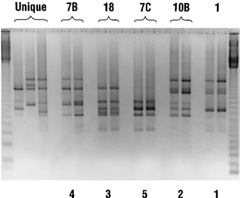
major common bands of the group. Figure 1 shows the
AP-PCR profiles of selected IB typed isolates with the primer T-7.
Strains in unique IB types were also unique by AP-PCR (Table
1).
TABLE 1. Molecular profiles of selected C. difficile isolates genotyped by AP-PCR with the arbitrary primer T-7
Isolate Toxigenicity IB type REA group AP-PCR (T-7) type
Y (1, 2, 4, 6) 1 ND Y 1
WA 197, KY 17 1 1 NDa 1
WA 182, WA 148 WA 171, WA 105
WA 40, WA 37 2 10B ND 2
WA 150
KY 28, WA 114 1 7B ND 4
WA 146, WA 50 2 7C ND 5
WA 84, WA 74 1 18 ND 6
PC 406, PC 997 1 10C ND 7
L1 1 ND L 8
M (1, 3, 4) 2 ND M 9
J1, G1 1 ND J, G 10
E1 1 ND E 11
N1 1 ND N 12
Z1 1 ND Z 13
BM1 1 ND BM 14
T1 2 ND T 15
WA 114 1 4 ND 16
B1, R1 1 ND R, B 17
AL1 1 ND AL 18
BD1 1 ND BD 19
a
[image:2.612.58.295.92.320.2]ND, not done.
FIG. 1. Genotyping of IB-typed C. difficile isolates by AP-PCR. A negative print of an ethidium bromide-stained 1.5% agarose gel of DNA patterns gener-ated by AP-PCR with the arbitrary primer T-7 is shown. Outer lanes, 123-bp DNA marker ladder. IB types are indicated by the numbers given at the top of the lanes. AP-PCR types are given by the numbers at the bottom of the lanes. Unique types had unique and different DNA banding patterns by AP-PCR (T-7) from those of all other isolates analyzed.
on May 15, 2020 by guest
http://jcm.asm.org/
AP-PCR and REA.
Genotyping C. difficile by AP-PCR with
the primer T-7 had a slightly less discriminatory power than
REA typing. Fifteen REA groups were resolved into 13
AP-PCR types. A good correlation was found between the major
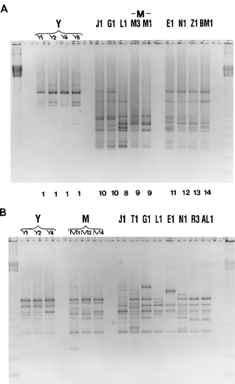
REA groups (Y and M) and our AP-PCR typing with T-7 (Fig.
2A); however, AP-PCR failed to discriminate the J and G
groups and the B and R REA groups and did not differentiate
M and Y types within these groups. A second level of typing
with PG-05 yielded a much higher level of discrimination.
Isolates G1, J1, R1, and B1 were all resolved into four different
AP-PCR types; in addition, one isolate in the Y group and one
isolate in the M group displayed a DNA banding pattern
slightly different from that of the other isolates in these groups
(Fig. 2B). However, by REA, each isolate within the Y and M
REA groups displayed minor but reproducible band
differ-ences representing unique REA types. All strains in unique
REA groups displayed unique DNA banding patterns by
AP-PCR with the PG-05 primer. In the analysis of these isolates,
we did not find any case in which the T-7 primer further
discriminated isolates grouped in the same AP-PCR type with
PG-05.
As indicated in Materials and Methods, the Dice coefficient
was used to group strains by percentage of similarity. In this
case, the isolates compared shared all the major bands but
differed in that they either lack or had one or more minor
(light) bands.
DISCUSSION
AP-PCR has been used to type a number of bacterial species
(3, 12, 32). McMillin and Muldrow first reported the use of
AP-PCR to generate DNA banding patterns from C. difficile
(22). This technique has also been used by Barbut et al. (1) to
study an outbreak of C. difficile diarrhea in AIDS patients. A
number of studies have correlated AP-PCR to other typing
systems. AP-PCR has been compared with ribotyping in typing
strains of Legionella pneumophila (14) and with PFGE in
typ-ing strains of Staphylococcus aureus (25) and
Stenotrophomo-nas maltophilia (33). With regards to C. difficile strain typing,
Killgore and Kato (19) applying AP-PCR analyzed 41 strains
typed by IB and which were isolated from patients during a C.
difficile colitis outbreak. These investigators found AP-PCR to
be comparable in discriminative power to IB, and in addition,
AP-PCR discriminated seven individual isolates which were
untypeable by IB.
In our study, a two-level AP-PCR technique was used to type
strains analyzed by IB and REA. This two-primer AP-PCR
approach provided a relative increase in discriminatory power
when compared with IB and AP-PCR typing with a single
arbitrary primer. At the first level of differentiation, AP-PCR
and IB typing correlated closely. These results are consistent
with the study of Killgore and Kato (19) comparing AP-PCR
with IB analysis. However, AP-PCR typing at this first level
was less discriminatory than REA. At the second level of
differentiation, the REA types that were grouped in the same
AP-PCR type with the first (T-7) arbitrary primer (J1, G1, R1,
and B1) resolved into separate types with the PG-05 primer. It
is interesting that REA types G1 and J1, which were identified
as the same type by AP-PCR with the primer T-7, have the
same ribotyping pattern (I) (5). At the second level of
differ-entiation, in some cases, strains in the same REA group were
further resolved into subtypes. Whether these AP-PCR
sub-type strains are different strains or represent strain
heteroge-neity within a single type is not known; however, it is known
that the REA type is stable and highly reproducible with
re-peated typing. Because of the good correlation between T-7
AP-PCR types and the major groups as defined by REA and
IB, this primer has value for typing by itself. However, the
clinical significance of typing with either oligonucleotide (T-7
or PG-05) remains unknown. It is possible that virulence may
correlate better to the major groups as identified with the T-7
primer. Typing with PG-05 leads to unique DNA fingerprinting
in most cases. These minor differences in DNA banding
pat-terns may not be significant enough to consider these strains as
different types; however, further clinical correlations may
re-veal their importance.
Not all of the IB-typed strains analyzed in this study have
been typed by REA. Although strains in IB type 1 and REA
group Y were grouped in AP-PCR type 1 with the T-7 primer,
the DNA banding pattern obtained with the arbitrary primer
PG-05 was different for IB type 1 and REA group Y.
[image:3.612.59.295.69.455.2]As was observed with REA typing, genotyping with both
oligonucleotides correlated with the toxigenicity of isolates. All
types within an REA group are either toxin positive or toxin
FIG. 2. Genotyping of REA-typed C. difficile isolates by AP-PCR with thearbitrary primers T-7 (A) and PG-05 (B). Negative prints of ethidium bromide-stained 1.5% agarose gels of DNA patterns are shown. Outer lanes, 123-bp DNA marker ladder. REA groups and types are indicated by the letters at the top of the lanes. (A) AP-PCR types are indicated by the numbers at bottom of lanes (see Table 1). (B) AP-PCR types were unique for all isolates of a particular REA group. Isolates Y4 and M4 displayed a DNA banding pattern slightly different from that of the other isolates in these groups.
on May 15, 2020 by guest
http://jcm.asm.org/
negative, and the same observation was found with AP-PCR
typing.
Kristjansson et al. (20) found the discriminatory power of
REA and PFGE to be comparable for typing strains of C.
difficile; however, REA was more sensitive than ribotyping in
differentiating strains. In a recent study, Chachaty et al. (4)
compared ribotyping with PFGE and AP-PCR in
differentiat-ing between 30 unrelated strains of C. difficile. The three
meth-ods showed a good correlation for clustering of strains;
how-ever, PFGE was slightly more discriminatory than AP-PCR,
with three different primers used in their study. PFGE,
how-ever, is labor-intensive and requires specialized equipment not
available to all clinical laboratories (20).
The selection of the two oligonucleotide primers used here
was based on the reproducibility of the DNA banding patterns,
the ability to type all of the strains, and the number of bands
obtained (in general, with the T-7 primer, the DNA profile
contained 6 to 10 bands, while with the primer PG-05, the
number of bands observed was usually 7 to 16). We also tested
three other arbitrary primers (10- to 12-mers), which resulted
in either no reproducible banding pattern, a small number of
bands (which we consider insufficient for an accurate typing
system), or no bands at all. Because of the inconclusive results
and poor reproducibility obtained with these three primers,
they were not used in this study. Although we made no attempt
to use primers with a GC content matching that of the C.
difficile genome, both primers used in this study (T-7 and
PG-05) provided a DNA banding pattern which was reproducible
and adequate for analysis. As has been reported by others (11),
some of the drawbacks of AP-PCR are its sensitivities to
changes in primer concentration, DNA concentration, brand
of thermal cycler used, and Taq polymerase. The DNA
band-ing pattern obtained from all 46 strains was reproducible from
day to day with both of the primers, T-7 and PG-05. The
assignation of isolates to AP-PCR types not only was based on
visual inspections of the DNA banding patterns in the gels but
was determined by laser scanning densitometry. This allowed
us to objectively discriminate true bands from background
bands. The number of AP-PCR types obtained by visualization
of the gels and by scanning densitometry showed 100%
corre-lation (data not shown). Scanning densitometry has been used
extensively to analyze protein patterns of polyacrylamide gel
electrophoresis analysis and has proved to be useful in
analyz-ing complex bandanalyz-ing patterns (10). The use of similarity
coef-ficient analysis for strains in which the DNA banding patterns
differed slightly allowed us to separate the strains into distinct
types when the isolates showed less than 85% similarity.
An important issue to be considered in the interpretation of
DNA banding patterns is the criterion used by the investigators
to score the DNA banding patterns. As has been the
experi-ence of others (1, 13, 19), we observed the presexperi-ence of faint
amplification products in some of the reactions when either
primer was used. Even though these bands were reproducible
in most cases, we chose not to include them when scoring the
DNA banding pattern. Inclusion of these bands would have
resulted in a larger number of subtypes being identified,
mak-ing the system unnecessarily complex.
In summary, AP-PCR typing is a reproducible, rapid, and
simple technique. Like the other typing methods (REA and
IB), it results in a sensitive and accurate identification of C.
difficile strains. The applicability of these typing methods to a
clinical laboratory is dependent on their economic feasibility.
AP-PCR can be performed in 36 to 48 h compared with the 3
to 4 days it takes to carry out some of the other typing
meth-ods; thus, it can be considered a cost-effective method for
determining the source of hospital outbreaks. (The
approxi-mate cost of duplicate reactions with both primers would likely
be in the range of $150 to $200 per specimen.) Thus, the
primarily practical advantage of AP-PCR is the greater
rapid-ity of this procedure compared with that of other molecular
typing methods.
ACKNOWLEDGMENT
We thank Gloria Marquez for excellent typing of the manuscript.
REFERENCES
1. Barbut, F., N. Mario, J. Frottier, and J. C. Petit. 1993. Use of the arbitrarily-primed polymerase chain reaction for investigating an outbreak of Clostrid-ium difficile-associated diarrhea in AIDS patients. Eur. J. Clin. Microbiol. Infect. Dis. 12:794–795.
2. Barlett, J. G. 1990. Clostridium difficile: clinical considerations. Rev. Infect. Dis. 12(Suppl. 2):S192–S199.
3. Bingen, E., C. Boissinot, P. Desjardins, H. Cave, N. Brahinai, N.
Lambert-Zechovsky, E. Denamur, P. Blot, and J. Elion.1993. Arbitrarily primed polymerase chain reaction provides rapid differentiation of Proteus mirabilis isolates from a pediatric hospital. J. Clin. Microbiol. 31:1055–1059. 4. Chachaty, E., P. Saulnier, A. Martin, N. Mario, and A. Andremont. 1994.
Comparison of ribotyping, pulsed field-gel electrophoresis and random am-plified polymorphic DNA for typing Clostridium difficile strains. FEMS Mi-crobiol. Lett. 122:61–68.
5. Clabots, C. R., S. Johnson, K. M. Bettin, P. A. Mathie, M. E. Mulligan, D. R.
Schaberg, L. R. Peterson, and D. N. Gerding.1993. Development of a rapid and efficient restriction endonuclease analysis typing system for Clostridium difficile and correlation with other typing systems. J. Clin. Microbiol. 31: 1870–1875.
6. Clabots, C. R., S. Johnson, M. M. Olson, L. R. Peterson, and D. N. Gerding. 1992. Acquisition of Clostridium difficile by hospitalized patients: evidence for colonized new admissions as a source of infection. J. Infect. Dis. 166: 561–567.
7. Cleary, P. P., E. L. Kaplan, C. Livdahl, and S. Skjold. 1988. DNA finger-prints of Streptococcus pyogenes are M type specific. J. Infect. Dis. 158:1317– 1323.
8. Delmee, M., Y. Laroche, B. Ayesani, and G. Cornelis. 1986. Comparison of serogrouping and polyacrylamide gel electrophoresis for typing Clostridium difficile. J. Clin. Microbiol. 24:991–994.
9. Dice, L. R. 1945. Measures of the amount of ecological association between species. Ecology 26:297–302.
10. Ehret, W., M. Turba, P. Pfaller, W. Heizmann, and G. Ruckdeschel. 1988. Computer-aided densitometric analysis of protein patterns of Clostridium difficile. Eur. J. Microbiol. Infect. Dis. 7:285–290.
11. Ellsworth, D. L., K. D. Rittenhouse, and R. L. Honeycutt. 1993. Artifactual variation in randomly amplified polymorphic DNA banding patterns. Bio-techniques 14:214–217.
12. Fang, F. C., M. McClelland, D. G. Guiney, et al. 1993. Values of molecular epidemiologic analysis in a nosocomial methicillin-resistant Staphylococcus aureus outbreak. JAMA 270:1323–1328.
13. Fekete, A., J. A. Bantle, S. M. Halling, and R. W. Stich. 1992. Amplification fragment length polymorphism in Brucella strains by use of polymerase chain reaction with arbitrary primers. J. Bacteriol. 174:7778–7783.
14. Gomez-Lus, P., B. S. Fields, R. F. Benson, W. T. Martin, S. P. O’Connor, and
C. M. Black.1993. Comparison of arbitrarily primed polymerase chain re-action, ribotyping, and monoclonal antibody analysis for subtyping Legionella pneumophila serogroup 1. J. Clin. Microbiol. 31:1940–1942.
15. Gumerlock, P. H., Y. J. Tang, M. E. Mulligan, and J. Silva, Jr. 1994. Clostridium difficile: correlation of genotyping and immunoblot analysis. (Fourth Annual Meeting for the Society of Hospital Epidemiology of Amer-ica.) Infect. Control Hosp. Epidemiol. 15(Suppl.):16.
16. Gumerlock, P. H., Y. J. Tang, J. B. Weiss, and J. Silva, Jr. 1993. Specific detection of toxigenic strains of Clostridium difficile in stool specimens. J. Clin. Microbiol. 31:507–511.
17. Herwaldt, L. A., J. Pottinger, S. Coffman, M. J. Jaqua-Stewart, and J.
Tjaden.1994. Molecular epidemiology of methicillin-resistant Staphylococ-cus aureus (MRSA) carriage. (Fourth Annual Meeting for the Society of Hospital Epidemiology of America.) Infect. Control Hosp. Epidemiol.
15(Suppl.):S20.
18. Kato, H., N. Kato, K. Watanabe, K. Ueno, H. Ushijima, S. Hashira, and T.
Abe.1994. Application of typing by pulsed-field gel electrophoresis to the study of Clostridium difficile in a neonatal intensive care unit. J. Clin. Micro-biol. 32:2067–2070.
19. Killgore, G. E., and H. Kato. 1994. Use of arbitrary primer PCR to type Clostridium difficile and comparison of results with those by immunoblot typing. J. Clin. Microbiol. 32:1591–1593.
20. Kristjansson, M., M. H. Samore, D. N. Gerding, P. C. DeGirolami, K. M.
Bettin, A. W. Karchmer, and R. D. Arbeit.1994. Comparison of restriction endonuclease analysis, ribotyping, and pulsed-field gel electrophoresis for
on May 15, 2020 by guest
http://jcm.asm.org/
molecular differentiation of Clostridium difficile strains. J. Clin. Microbiol.
32:1963–1969.
21. McFarland, L. V., G. W. Elmer, W. E. Stamm, and M. E. Mulligan. 1991. Correlation of immunoblot type, enterotoxin production, and cytotoxin pro-duction with clinical manifestations of Clostridium difficile infection in a cohort of hospitalized patients. Infect. Immun. 59:2456–2462.
22. McMillin, D. E., and L. L. Muldrow. 1992. Typing of toxigenic strains of Clostridium difficile using DNA fingerprints generated with arbitrary poly-merase chain reaction primers. FEMS Microbiol. Lett. 79:5–10.
23. Mulligan, M. E., L. R. Peterson, R. Y. Kwok, C. R. Clabots, and D. N.
Gerding.1988. Immunoblots and plasmid fingerprints compared with sero-typing and polyacrylamide gel electrophoresis for sero-typing Clostridium difficile. J. Clin. Microbiol. 26:41–46.
24. Samore, M. H., K. M. Bettin, P. C. DeGirolami, C. R. Clabots, D. N.
Gerding, and A. W. Karchmer.1994. Wide diversity of Clostridium difficile types at a tertiary referral hospital. J. Infect. Dis. 170:615–621.
25. Saulnier, P., C. Bourneik, G. Prevost, and A. Andremont. 1993. Random amplified polymorphic DNA assay is less discriminant than pulsed-field gel electrophoresis for typing strains of methicillin-resistant Staphylococcus au-reus. J. Clin. Microbiol. 31:982–985.
26. Sell, T. L., D. R. Schaberg, and R. Fekety. 1983. Bacteriophage and
bacte-riocin typing scheme for Clostridium difficile. J. Clin. Microbiol. 17:148–152. 27. Silva, J., Jr., and C. Iezzi. 1988. Clostridium difficile as a nosocomial
patho-gen. J. Hosp. Infect. 11(Suppl. A):378–385.
28. Silva, J., Jr., Y. J. Tang, and P. H. Gumerlock. 1994. Genotyping of Clos-tridium difficile isolates. J. Infect. Dis. 169:661–664.
29. Tabaqchali, S. 1990. Epidemiological markers of Clostridium difficile. Rev. Infect. Dis. 12(Suppl.) 2: S192–S199.
30. Tang, Y. J., P. H. Gumerlock, J. B. Weiss, and J. Silva, Jr. 1994. Specific detection of Clostridium difficile toxin A gene sequences in clinical isolates. Mol. Cell. Probes 8:463–467.
31. Tsang, V. C., J. M. Peratta, and A. R. Simons. 1983. Enzyme-linked immu-noelectrotransfer blot techniques (EITB) for studying the specificity of an-tigens and antibodies separated by gel electrophoresis. Methods Enzymol.
92:377–391.
32. vanBelkum, A., M. Struelens, and W. Quint. 1993. Typing of Legionella pneumophila strains by polymerase chain reaction-mediated DNA finger-printing. J. Clin. Microbiol. 31:2198–2200.
33. VanCouwenberghe, C. J., S. H. Cohen, Y. J. Tang, P. H. Gumerlock, and J.
Silva, Jr.1995. Genomic fingerprinting of epidemic and endemic strains of Stenotrophomonas maltophilia (formerly Xanthomonas maltophilia) by arbi-trarily primed PCR. J. Clin. Microbiol. 33:1289–1291.