paradoxically impairs the in vivo antitumor
efficacy of adoptively transferred CD8
+
T cells
Luca Gattinoni, … , Steven A. Rosenberg, Nicholas P.
Restifo
J Clin Invest.
2005;
115(6)
:1616-1626.
https://doi.org/10.1172/JCI24480
.
T cell differentiation is a progressive process characterized by phenotypic and functional
changes. By transferring tumor-specific CD8
+T cells into tumor-bearing mice at various
stages of differentiation, we evaluated their efficacy for adoptive immunotherapy. We found
that administration of naive and early effector T cells, in combination with active
immunization and IL-2, resulted in the eradication of large, established tumors. Despite
enhanced in vitro antitumor properties, more-differentiated effector T cells were less
effective for in vivo tumor treatment. Several events may underlie this paradoxical
phenomenon: (a) downregulation of lymphoid-homing and costimulatory molecules; (b)
inability to produce IL-2 and access homeostatic cytokines; and (c) entry into a proapoptotic
and replicative senescent state. While the progressive acquisition of terminal effector
properties is characterized by pronounced in vitro tumor killing, in vivo T cell activation,
proliferation, and survival are progressively impaired. These findings suggest that the
current methodology for selecting T cells for transfer is inadequate and provide new criteria
for the generation and the screening of optimal lymphocyte populations for adoptive
immunotherapy.
Research Article
Immunology
Find the latest version:
Acquisition of full effector function in vitro
paradoxically impairs the in vivo antitumor
efficacy of adoptively transferred CD8
+
T cells
Luca Gattinoni,1 Christopher A. Klebanoff,1,2 Douglas C. Palmer,1
Claudia Wrzesinski,1 Keith Kerstann,1 Zhiya Yu,1 Steven E. Finkelstein,1
Marc R. Theoret,1 Steven A. Rosenberg,1 and Nicholas P. Restifo1
1Center for Cancer Research, National Cancer Institute (NCI), NIH, Bethesda, Maryland, USA. 2Howard Hughes Medical Institute–NIH Research Scholars Program, Bethesda, Maryland, USA.
T cell differentiation is a progressive process characterized by phenotypic and functional changes. By
trans-ferring tumor-specific CD8
+T cells into tumor-bearing mice at various stages of differentiation, we evaluated
their efficacy for adoptive immunotherapy. We found that administration of naive and early effector T cells,
in combination with active immunization and IL-2, resulted in the eradication of large, established tumors.
Despite enhanced in vitro antitumor properties, more-differentiated effector T cells were less effective for in
vivo tumor treatment. Several events may underlie this paradoxical phenomenon: (a) downregulation of
lym-phoid-homing and costimulatory molecules; (b) inability to produce IL-2 and access homeostatic cytokines;
and (c) entry into a proapoptotic and replicative senescent state. While the progressive acquisition of terminal
effector properties is characterized by pronounced in vitro tumor killing, in vivo T cell activation,
prolifera-tion, and survival are progressively impaired. These findings suggest that the current methodology for
select-ing T cells for transfer is inadequate and provide new criteria for the generation and the screenselect-ing of optimal
lymphocyte populations for adoptive immunotherapy.
Introduction
Adoptive cell transfer therapy (ACT), the administration of ex vivo– activated and –expanded autologous tumor-reactive T cells, is cur-rently one of the few immunotherapies that can induce objective clinical responses in significant numbers of patients with metastatic solid tumors (1–3). In a previous study, ACT after lymphodeplet-ing conditionlymphodeplet-ing caused objective responses in 46% of patients with metastatic melanoma refractory to other therapeutic modalities (4). We have now tripled the size of the original study (4), and the objec-tive response rate exceeds 50%, and 11% of all patients treated are complete responders (5).
There are several theoretical advantages to the use of ACT in treatment of cancer. Tumor-specific T cells can be activated and expanded to large numbers ex vivo, independently of the immu-nogenic properties of the tumor. Perhaps most important, the functional and phenotypic qualities of T cells can be selected prior to their adoptive transfer.
Much progress has been made in the understanding of the proper-ties of T cell subpopulations associated with states of T cell differen-tiation in mice and humans, especially those states that are related to the generation of memory T cells capable of protecting against viral challenge (6–9). However, little progress has been made in identifying the characteristics of cell states that are associated with the successful treatment of large, established tumors in mice or in humans.
Currently, the only criterion applied to selecting cells for adop-tive transfer to patients with solid tumors is the ability of antitumor T cells to release IFN-γ and kill tumor cells upon coculture (2, 10). However, based on a retrospective analysis of ACT in melanoma patients, it is now clear that these criteria alone do not predict in vivo efficacy — despite enhanced in vitro antigen-specific IFN-γ release and cytolysis, tumor-specific CD8+ clones did not induce objective clinical responses upon adoptive transfer (10–12).
We sought to determine whether the functional and phenotypic qualities associated with CD8+ T cell differentiation affect the abil-ity of tumor-specific T cells to mediate tumor regression. We tested this hypothesis using T cells derived from pmel-1 TCR–Tg mice in adoptive transfer experiments (13). The pmel-1 system accurately models the human situation in many important respects. The tar-get antigen, pmel-17, is a highly homologous ortholog of the self/ tumor antigen gp100 that is often targeted in human clinical trials (14, 15). In addition, the exogenous administration of high doses of IL-2 and the lymphodepleting conditioning represent other ele-ments shared with our clinical protocols. In contrast to the human situation, in which cells are isolated from cancer patients, we used CD8+ T cells derived from naive non–tumor-bearing mice; however, it is important to note that in the presence of either B16 melanoma or a gp100-nonexpressing irrelevant tumor, pmel-1 T cells remain naive (16). After adoptive transfer, T cells from tumor-bearing pmel-1 mice are able to eradicate established tumors as well as T cells derived from naive pmel-1 mice (data not shown). Thus, there is neither activation nor tolerance of these tumor-reactive T cells in the presence of large established tumors — instead, these tumor-reactive T cells exist in a state of immunological ignorance (13).
In the current article, we elucidate the T cell differentiation state (i.e., the specific phenotypic and functional qualities) of cells associated with responses in the ACT setting and clarify the
Nonstandard abbreviations used: ACT, adoptive cell transfer therapy; CCR7, chemokine receptor 7; CM, culture media; hgp10025–33, human gp10025–33;
IL-7Rα, IL-7 receptor α; KLRG-1, killer cell lectin-like receptor G1; β2m–/–, β2
micro-globulin–/–; rFPhgp100, recombinant fowlpox virus expressing hgp100; rhIL-2,
recombinant human IL-2.
Conflict of interest: The authors have declared that no conflict of interest exists.
mechanisms that impair a T cell’s ability to cause tumor regres-sion. Finally, we suggest specific solutions to generate more effec-tive antitumor T cells for the development of experimental human adoptive cell transfer–based therapies.
Results
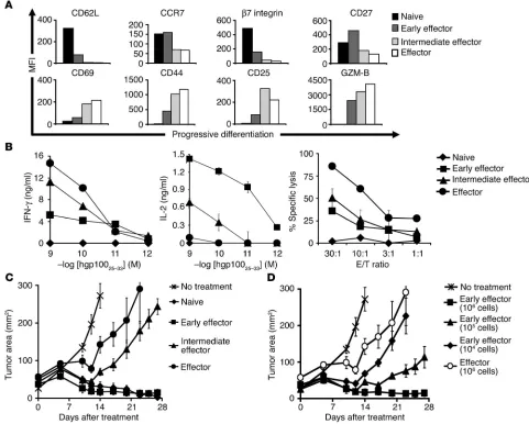
Acquisition of terminal effector functions in vitro impairs in vivo antitumor efficacy. To determine whether the differentiation state of CD8+ T cells may affect their ability to mediate tumor regression when adoptively transferred in vivo, we generated pmel-1 CD8+ T cells
[image:3.585.48.529.84.470.2]at progressive stages of differentiation (naive, early effector, inter-mediate effector, effector). We obtained these cells using multiple in vitro stimulations with antigen and IL-2 (unstimulated, stimu-lated once, stimulated twice, stimulated 3 times). We then assessed a variety of phenotypic and functional attributes to validate the stage of CD8+ T cell differentiation. After multiple in vitro stimu-lations, naive pmel-1 cells differentiated into fully mature effec-tor CD8+ T cells, as indicated by the progressive downregulation of CD62L, chemokine receptor 7 (CCR7), β7 integrin, and CD27 and the concurrent increase in the levels of CD44, CD69, CD25,
Figure 1
Acquisition of full effector function in vitro impairs antitumor efficacy. Pmel-1 CD8+ T cells at progressive stages of differentiation were
gener-ated using multiple in vitro stimulations with 1 μM hgp10025–33 and 30 IU/ml of rhIL-2. Seven days following the last stimulation, phenotypic and
functional assays were performed. (A) Phenotypic differences in naive and effector subgroups. Flow cytometry analysis for the expression of
lymphoid-homing molecules CD62L, CCR7, and β7 integrin; costimulatory molecule CD27; activation/effector markers CD69, CD44, CD25,
and granzyme-B (GZM-B) on CD8-enriched pmel-1 naive, early effector, intermediate effector, and effector T cells. Mean fluorescence intensity
(MFI) values after gating on CD8+ cells are shown. Data shown are representative of 3 independent experiments. (B) Functional differences in
naive and effector subgroups. IFN-γ and IL-2 release and cytotoxic assay against hgp10025–33–pulsed MCA-205 targets. MCA-205 cells plus the
irrelevant influenza nucleoprotein peptide were used as control. In the cytotoxic assay, values for control target cells (0–14%) were subtracted.
Data shown are representative of 3 independent experiments. (C and D) Differentiation of antitumor T cells is inversely associated with in vivo
effectiveness. WT mice bearing 10-day-old established s.c. B16 tumors were sublethally irradiated and left untreated as control or received
adoptive transfer of 1 × 10⁶(when not otherwise indicated) CD8-enriched pmel-1 naive, early effector, intermediate effector, and effector T cells
in conjunction with rFPhgp100 vaccination and exogenous rhIL-2 (36 μg per dose). Results for tumor area are the mean of measurements from
and granzyme-B (Figure 1A). The acquisition of an effector pheno-type was associated with an increase in cytolytic activity and IFN-γ release but a decrease in IL-2 production (Figure 1B).
We next adoptively transferred these cells into lymphodepleted C57BL/6 WT mice bearing subcutaneous B16 tumors established for 10 days to test their in vivo antitumor functions. Pmel-1 CD8+ T cells that were naive or at early effector stage, in combination with an altered ligand vaccine and IL-2, were able to effectively treat large, established B16 melanoma. Unexpectedly, despite enhanced antitumor properties in vitro, more-differentiated
T cells were less effective upon adoptive transfer (P = 0.009; Figure 1C). According to a titration experiment, effector cells were at least 100-fold less effective than early effector cells (Figure 1D). It was surprising that the least effective cells we used had the characteristics that we and others
cur-rently seek in T cells for use in current ACT-based human trials (2). Specifically IFN-γ release and cytotoxicity were
negatively correlated with in vivo antitumor efficacy.
Further characterization of in vivo effective and impaired CD8+
T cells. To investigate the mechanisms underlying the lack of antitumor effectiveness of highly differentiated T cells, we extended our characterization of early effector and intermediate effector T cells using microarray analysis. RNA was extracted from early effector and intermediate effector T cells and indirectly labeled after a single round of linear amplification. The labeled samples were combined and hybridized overnight on a murine 22,000-gene oligo-nucleotide array. All raw data sets have been deposited, and processed values and complete gene lists are available (GEO series id GSE2348; http://www.ncbi.nlm.nih.gov/geo/).
Microarray analysis confirmed that impaired cells dif-ferentially expressed genes associated with highly differ-entiated effector T cells. The expression of many genes involved in effector functions such as granzymes, perforin,
IFN-γ, and fas-ligand were strongly upregulated in impaired cells compared with in vivo effective cells (Table 1). Fur-thermore, intermediate effector cells overexpressed eomeso-dermin, a transcription factor recently described as a mas-ter regulator for CD8+ effector functions (17). Conversely, early effector cells showed a more naive profile, expressing higher levels of the lymphoid-homing molecules cd62l,
integrin-αE, and ccr7, the costimulatory molecule cd27 and
[image:4.585.52.304.115.388.2]IL-7 receptor α (il-7rα). However, highly differentiated T cells overexpressed a number of genes that may prejudice their survival capability, such as the proapoptotic mole-cules bid, bad, and fas-ligand (18–20). Intermediate effector cells also expressed higher levels of molecules associated with replicative senescence such as killer cell lectin-like recep-tor G1 (klrg1) (21, 22) and the mortality factor mrgx (23), which sug-gests that the proliferative potential of these cells may be decreased. Microarray data were validated by flow cytometry, Western blot, and functional analyses (Figure1, A and B; Figure 2, A and B). Interestingly, according to the Western blot analysis, it was the truncated, active, C-terminal products of caspase-3 and -8 cleavage of BID (tBID) that were present at higher levels in impaired cells (18, 20). It should be noted that it was the p13 product — usually a minor form exclusively associated with the mitochondria — that
Table 1
Microarray analysis of early effector and intermediate effector T cells
Genes highly expressed Genes highly expressed in early effector cells in intermediate effector cells
Lymphoid-homing E/I, Effector I/E,
fold change function fold
change
cd62I 5.6 granzyme A 5.1
ccr7 3.7 granzyme B 6.0
integrin αE 3.1 granzyme C 6.0
granzyme D 3.5
Costimulatory molecules granzyme E 2.9
cd27 2.9 granzyme F 3.9
granzyme G 3.1
granzyme K 5.0
T cell survival/memory generation perforin 2.4
fas-ligand 2.4
il-7rα 7.4 IFN-γ 3.4
cd27 2.9 eomesodermin 4.5
Apoptosis
bid 10.7
bad 4.4
fas-ligand 2.4
Replicative senescence
klrg-1 2.5
mrgx 2.3
Results shown are the geometric mean of 2 experiments using independently generated sets of cells. Reversing the flourochromes resulted in similar results. E, early effector cells; I, intermediate effector cells.
Figure 2
Cytofluorometry and Western blot analysis of
effec-tive and impaired CD8+ T cells. Pmel-1 CD8+ T cells
at progressive stages of differentiation were
gener-ated using multiple in vitro stimulations with 1 μM
hgp10025–33 and 30 IU/ml of rhIL-2. (A) Flow cytometry
for the surface expression of IL-7Rα and KLRG-1.
Results after gating for CD8+ cells are shown. (B)
[image:4.585.245.537.564.741.2]was strongly elevated (>3-fold by densitometry) in intermediate effector T cells (20). Thus, the Western blot confirmed and further refined the results of the analysis of global gene expression.
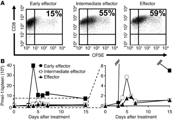
T cell differentiation is associated with progressive impairment of prolifer-ative capacity. The analysis of global gene expression suggested that fully differentiated effector T cells may be less “fit” (24) and more proapoptotic. We sought to determine the relative proliferative capacity of effector T cell subpopulations in vitro and in vivo. We evaluated the proliferative capability in vitro with a CFSE-based assay. Four days after antigen stimulation in IL-2, early effector cells had proliferated vigorously — only 15% of the starting popu-lation failed to dilute the CFSE label to levels comparable to those of unlabeled cells (data not shown). By sharp contrast, 55% of the intermediate effector and 59% of the effector T cells failed to fully dilute the label, which indicates that the differentiation state was inversely related to T cell proliferative capacity (Figure 3A).
To test whether the in vitro findings could predict the prolif-erative capacity of T effector subsets upon adoptive transfer, we directly quantified the absolute number of CD8+Vβ13+ pmel-1–Tg T cells at multiple time points in the blood and in the spleen. The differences observed in vivo were more pronounced than those observed in vitro. Each of the effector subsets was transferred (1.0 × 10⁶) with altered peptide ligand vaccine and IL-2. The prog-eny of all effector subgroups reached a peak number between days 4 and 6 after transfer. However, in the spleen alone, we found that 39.5 × 10⁶ progeny derived from early effectors but only 5.7 × 10⁶
and 1.8 × 10⁶ pmel-1 T cells derived from inter-mediate effector and effector cells, respectively (Figure 3B). A similarly pronounced difference was observed in the blood (data not shown). Thus, progressive acquisition of antitumor effector function led to deterioration of the numbers of surviving T cells in vivo.
Further subfractionation of early effector T cells by expression of CD62L identifies CD8+ T cells with
superior in vivo antitumor properties. In our experi-ments using the highly effective/early effector population, we observed significant homogene-ity with regard to most differentiation markers. However, in the early effector subpopulation there remained significant heterogeneity in the expression of CD62L, which was lost with fur-ther T cell differentiation and failure of antitu-mor function in vivo (Figure 1A).
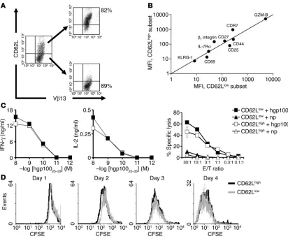
We sought to understand whether CD62L was functionally important or merely a marker of the state of differentiation. We further sub-fractionated the early effector T cell population by sorting cells into CD62Lhigh and CD62Llow subsets (Figure 4A). The CD62Llow population appeared slightly more differentiated on the basis of CD25 and CD69 expression, but all cells in the CD62Lhigh and CD62Llow subgroups demonstrated an “antigen-experienced” pheno-type. Most phenotypic markers were expressed at a similar level, including CD27, CD44, and IL-7Rα (Figure 4B). As KLRG-1 — a sensitive marker for replicative senescence — was virtually absent in both CD62Lhigh and CD62Llow subsets, neither of the 2 groups had entered into a senes-cence state (Figure 4B). Functionally, CD62Lhigh and CD62Llow subsets showed similar cytolytic activity and IFN-γ production. Importantly, no deficit in IL-2 release was seen in either popula-tion (Figure 4C). Furthermore, no differences in proliferapopula-tion were observed when CFSE-labeled CD62Lhigh and CD62Llow popula-tions were stimulated in vitro with antigen and IL-2 (Figure 4D).
[image:5.585.47.349.83.294.2]Despite these similarities, when transferred in conjunction with an altered ligand vaccine and IL-2, the CD62Lhigh subset had superior antitumor function (P = 0.009; Figure 5A). This was associated with pronounced in vivo expansion of the CD62Lhigh subset in the spleen (Figure 5B). Similar results were observed in tumor (Figure 5C) and blood (data not shown). Thus, these differences were not the result of selective accumulation in lymphoid tissues mediated by CD62L. However, no statistical difference was observed when high or low subsets were transferred in the absence of vaccination (P > 0.05; Figure 5A). To definitively assess whether CD62L was functionally important, we crossed pmel-1–Tg mice with CD62L–/– mice. Splenocytes from pmel-1-CD62L–/– and age-matched pmel-1-CD62L+/+ mice possessed similar phenotypic and functional attri-butes in vitro after activation (data not shown). By contrast, when adoptively transferred in WT mice bearing B16 tumors established for 10 days, pmel-1-CD62L–/– cells were less effective in treating tumors compared with pmel-1-CD62L+/+ (P = 0.014; Figure 5D). Thus, CD62L not only represents a marker of T cell differentiation state but also plays an important functional role in this adoptive immunotherapy model of large, established tumors.
Figure 3
The differentiation state of CD8+ T cells is inversely related to their proliferative capacity.
Pmel-1 CD8+ T cells at progressive stages of differentiation were generated using multiple
in vitro stimulations with 1 μM hgp10025–33 and 30 IU/ml of rhIL-2. (A) In vitro
prolifera-tion of effector subgroups. Early effector, intermediate effector, and effector T cells were
labeled with CFSE and were cultured with 1 μM hgp10025–33 peptide–pulsed on irradiated
splenocytes in the presence of 30 IU/ml rhIL-2. Cells were harvested after 4 days, and their
CFSE content was determined. Results after gating on Vβ13+ population are shown. This
experiment was performed twice with similar results. (B) In vivo proliferation of effector sub-groups. Absolute numbers of adoptively transferred pmel-1 cells (CD8+Vβ13+) in the spleens
of tumor-bearing, sublethally irradiated WT mice. Mice were treated with rFPhgp100, rhIL-2 (36 μg per dose), and 1 × 10⁶ pmel-1 early effector, intermediate effector, and
Tumor antigen presentation by BM-derived APCs is required for successful tumor treatment. Differences between CD62Lhigh and CD62Llow population were observed only in the presence of vac-cination. Consistent with findings of others (25, 26), we found that CD62Lhigh T cells preferentially homed to lymph nodes. This finding was obtained using confocal laser scanning microscopy (data not shown). Taken together, these data suggested that the lymphoid homing of CD62Lhigh T cells could result in a more effective interaction with APCs.
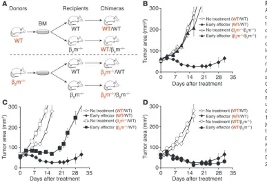
In order to evaluate the role of APCs in this model, we gen-erated a variety of chimeras by transferring BM cells from β2 microglobulin–/– (β2m–/–) and from WT mice into lethally irradi-ated β2m–/– and WT mice (Figure 6A). Chimeras were analyzed for reconstitution with the appropriate donor marrow after 8 weeks (data not shown) and subsequently challenged
[image:6.585.86.498.80.425.2]subcuta-neously with B16 melanoma. Adoptive transfer of early effector cells, in combination with an altered ligand vaccine and exog-enous IL-2, was unable to treat B16 tumors in β2m–/–/β2m–/– chi-meras (Figure 6B). As previously reported in studies using other models (27), self/tumor antigen presentation by B16 melanoma was insufficient to efficiently stimulate adoptively transferred T cells, and no significant differences in tumor growth were seen compared with untreated mice (P > 0.05). However, transfer of CD8+ T cells into β2m–/– mice that had been engrafted with BM cells derived from WT mice treated B16 melanoma as effi-ciently as did pmel-1 cells adoptively administered to WT/WT chimeras (P > 0.05; Figure 6D). Tumor antigen presentation by BM-derived APCs (presumably professional APCs) appear to be critical for successful treatment: adoptively transferred T cells in vivo activated by parenchymal cells, in β2m–/–/WT chimeras, were
Figure 4
Subfractionated CD62Lhigh and CD62Llow early effector cells possess similar properties in vitro. (A and B) Phenotypic characterization of CD62Lhigh
and CD62Llow subsets. Five days after the initial stimulation with antigen and IL-2, CD62Lhigh and CD62Llow populations were sorted by MACS
CD62L-positive selection column and analyzed by flow cytometry on day 6 for the expression of IL-7Rα; lymphoid-homing molecules β7 integrin
and CCR7; the costimulatory molecule CD27; activation/effector markers CD44, CD69, CD25, and granzyme-B; and the senescence marker
KLRG-1. MFI values after gating on CD8+V
β13+ cells are shown. The MFI was determined for all groups at the same time under the same
cyto-flourometric settings using the same reagents and isotype-matched controls. Data shown are representative of 2 independent experiments. (C)
CD62Lhigh and CD62Llow subsets have similar functional qualities. 51Cr, IFN-γ, and IL-2 release against hgp10025–33–pulsed MCA-205 targets.
MCA-205 cells plus influenza nucleoprotein peptide (np) were used as control. Where error bars are not visible, it is because they are obscured by
the symbols used. Data shown are representative of 2 independent experiments. (D) CD62Lhigh and CD62Llow subsets have similar proliferative
capacity. CD62Lhigh and CD62Llow cells were labeled with CFSE and were cultured with hgp10025–33 peptide–pulsed on irradiated splenocytes in
the presence of 30 IU/ml of rhIL-2. Cells were harvested from day 1 to day 4, and their CFSE content was determined. Results after gating on
less effective in controlling tumor growth (P = 0.016; Figure 6C). Thus, our findings suggest that in lymphoid tissues, adoptively transferred T cells utilize CD62L to target professional APCs expressing antigen as a result of the vaccination and that the downregulation of CD62L, due to progressive differentiation, impairs their in vivo activation, their proliferation, and conse-quently their antitumor activity.
IL-15 uncouples T cell proliferation from differentiation and preserves antitumor efficacy in vivo. Results here indicate that the optimal T cell populations for ACT are early effector cells that retain high levels of lymphoid-homing molecules. These findings raise a crucial clinical problem. Current successful ACT-based immu-notherapies inevitably start from low precursor frequencies of tumor-specific T cells. These T cells are currently activated and expanded to clinically therapeutic numbers using IL-2 (2, 10). During this expansion, cells inexorably differentiate and lose lymphoid-homing molecules (28). Although T cells that pos-sess such characteristics can cause objective clinical responses in a significant number of patients (4, 5), and can suboptimally treat established tumors in mice, there is an urgent need to improve these results.
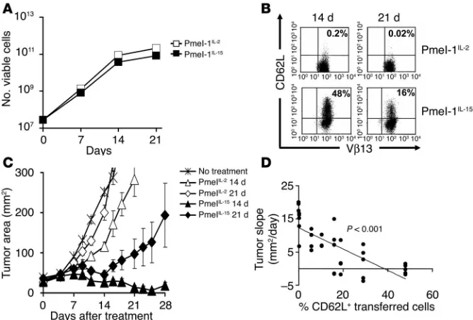
We and others have previously reported that splenocytes primed in the presence of IL-15 for 7 days polarized to a less-differenti-ated CD8+ T cell population that retained high levels of CD62L and CCR7 (26, 29, 30). Here we have extended these findings by test-ing the ability of IL-15 to sustain the growth of large numbers of T cells through multiple restimula-tions. After 14 days in culture, cells grown in either IL-2 or IL-15 pro-liferated extensively, undergoing a 1,000-fold expansion (Figure 7A). While the CD62L marker was vir-tually absent on T cells grown in IL-2 (0.2%), significant numbers of T cells cultured in IL-15 (48%) retained the expression of CD62L (Figure 7B). This resulted in the generation of a T cell population that was significantly more effec-tive even when used at limiting doses of 1 × 10⁶ cells (P = 0.009, IL-15 vs. IL-2, day 14; P = 0.02, IL-15 vs. IL-2, day 21) (Figure 7C). Enhanced tumor responses were associated with increased prolif-eration of IL-15–cultured cells upon adoptive transfer (data not shown). Interestingly, there was a statistically significant inverse cor-relation between the state of differ-entiation, as a measured by CD62L expression of transferred T cells, and tumor growth (P < 0.001; Fig-ure 7D). These data suggest that the use of IL-15 instead of IL-2 could result in significant expansion of T cells while delaying the acquisition of a detrimental effector state.
Discussion
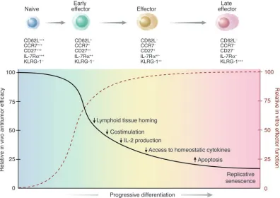
T cells used for current adoptive immunotherapy trials are selected for their capacity to produce high levels of IFN-γ and for their ability to efficiently and specifically lyse relevant tar-get cells (2, 10). However, we have found that CD8+ T cells that acquire complete effector properties and exhibit increased anti-tumor reactivity in vitro are less effective at triggering anti-tumor regressions and cures in vivo. In addition, as discussed in detail below, emerging data from our human clinical trials support the hypothesis that less-differentiated T cells are more thera-peutically effective upon adoptive transfer. These findings may appear paradoxical but can be resolved by a summary of our results represented diagrammatically in Figure 8.
[image:7.585.49.392.80.304.2]After adoptive transfer, several events must occur for T cells to cause the regression of established tumors. (a) T cells must be acti-vated in vivo through antigen-specific vaccination. (b) They must then vigorously expand to levels capable of causing the destruction of significant tumor burdens. (c) Finally, antitumor T cells must
Figure 5
CD62L-selectin identifies CD8+ T cells with superior in vivo antitumor properties. (A)CD62Lhigh subset
possesses superior in vivo antitumor properties. WT mice bearing 10-day-old established subcutane-ous B16 tumors were sublethally irradiated and left untreated as control or received adoptive transfer of 1 × 10⁶ of pmel-1 CD62Lhigh or CD62Llow cells with exogenous rhIL-2 (36 μg per dose) with or
with-out rFPhgp100 vaccination. Results for tumor area are the mean of measurements from 5 mice per
group (± SEM). Data shown are representative of 3 independent experiments. (B and C) CD62Lhigh
subset expands vigorously in vivo. (B) Absolute numbers of adoptively transferred CD8+ pmel-1cells in
the spleen of tumor-bearing, sublethally irradiated WT mice. Mice were treated with FPhgp100, rhIL-2 (36 μg per dose), and 1 × 10⁶ of pmel-1 CD62Lhigh or CD62Llow cells. Data shown are the mean of 2 mice
per group. This experiment was performed twice, with similar results. (C) Percentage of CD8+
pmel-1cells in the tumor. Mice were treated with FPhgp100, rhIL-2 (36 μg per dose), and 1 × 10⁶ of pmel-1
CD62LhighLy5.1 or CD62LlowThy1.1 cells. Data shown are the mean of 2 mice per group. This experiment
was performed twice with similar results. (D) CD62L expression is required to maximize the antitumor
efficacy of adoptively transferred T cells. WT mice bearing 10-day-old established s.c. B16 tumors were
sublethally irradiated and left untreated as control or received adoptive transfer of 1 × 10⁶
one-week-cultured pmel-1 CD62L+/+ cells or pmel-1 CD62L–/– cells in conjunction with rFPhgp100 vaccination and
exogenous rhIL-2 (36 μg per dose). Tumor area results are the mean of measurements from 5 mice per
survive long enough to complete the eradication of all tumor cells (13). In the present work, we found that the progressive differen-tiation of T cells to a terminal differentiated effector stage results in a series of phenotypic and functional changes that make them less “fit” to perform these functions.
We previously reported that antigen-specific vaccination using an altered peptide ligand is required to eradicate large, established B16 melanoma (13). Here, we extend these findings by determin-ing that the deprivation of a productive interaction of T cells with BM-derived APCs (presumably professional APCs) diminishes the therapeutic impact of T cells. Interestingly, priming T cells ex vivo does not abrogate the requirement for stimulation by “profes-sional” APCs. In addition, the expression of lymphoid-homing and costimulatory molecules on the surfaces of highly effective T cells provides further evidence that, to be fully therapeutic, adoptively transferred CD8+ T cells must home to secondary lymphoid tissue where they interact effectively with professional APCs. We cannot exclude the possibility that homing to lymph nodes would not be required in the presence of more-immunogenic tumors. Terminally differentiated T cells may have potent antitumor activity directly on tumor cells after infusion, as activation in lymph nodes may be unnecessary. Our results are consistent with the report that in pre-ventive infectious models, the adoptive transfer of “central memo-ry” CD8+ T cells that possess enhanced lymphoid-homing proper-ties and higher levels of the costimulatory molecule CD27 provides superior immunity compared with the transfer of CD62L–CCR7– CD27– “effector memory” T cell populations (31). Some authors have reported that the adoptive transfer of CD62Llow T cells pro-vides enhanced tumor treatment (32). However, these polyclonal cells derived from tumor-draining lymph node were selected from an open T cell repertoire only on the basis of CD62L expression. Thus, it is likely that a CD62Llow population is relatively enriched for tumor-reactive T cells compared with the naive, CD62Lhigh pop-ulation that has not been stimulated by exposure to tumor.
Although CD62L homing to lymphoid tissues, and the conse-quent ability of T cells to be properly activated by professional APCs, is important, it is clearly not the only mechanism underly-ing the differences in T cell efficacy after adoptive transfer. Neither intermediate effector cells nor effector cells expressed CD62L, but the intermediate effector populations were reproducibly more efficient than effector cells. Enhanced efficacy was associated with greater proliferation and survival in vivo.
Recent findings indicate that costimulatory interaction of CD27 and CD70 can result in prolonged expansion, survival, and memory differentiation of activated CD8+ T cells (7, 33–36). These findings suggest that the downregulation of CD27 that accompanies progressive T cell differentiation might contribute to the observed impairment of CD8+ T cell expansion and sur-vival after adoptive transfer.
Because recombinant IL-2 is provided both in the ex vivo expan-sion of antitumor CD8+ T cells and immediately after their adop-tive transfer, T cells may undergo apoptosis upon IL-2 withdrawal (37). Thus, the capacity of T cells to produce their own IL-2 may allow them to survive and expand. In our model, only early effector CD8+ T cells could produce significant levels of IL-2. Naive cells acquire this capability upon antigen stimulation. Cotransfer of CD4+CD25– helper cells, as a natural source of IL-2, may be helpful for the persistence and effectiveness of adoptive T cells (38).
[image:8.585.42.429.83.348.2]Homeostatic cytokines are important for the survival of T cells. For example, IL-7 has been shown to be critical for the generation and survival of memory CD8+ T cells, and IL-7Rα may identify effector CD8+ T cells that become long-lived memory cells (39–42). Thus, the progressive downregulation of IL-7Rα associated with T cell differentiation may be detrimental to their survival. Similarly, IL-15 plays a supportive role in survival and proliferation of adop-tively transferred T cells (40, 43–45). We have recently reported that endogenous IL-15 enhances the antitumor efficacy of adoptively transferred CD8+T cells (29). IL-15 appeared to be presented to
Figure 6
Antigen presentation by BM-derived APCs is critical for tumor
treatment. (A) Generation of BM
chimeras. We generated chime-ras by transferring 5 × 10⁶ BM
cells from β2m–/– and WT mice into
lethally irradiated WT or β2m–/–
mice. (B–D) Results from BM chi-meras indicate that BM-derived APCs expressing MHC class I are required for tumor treatment. Chimeras bearing 10-day-old established s.c. B16 tumors were sublethally irradiated and left untreated as controls or received adoptive transfer of 1 × 10⁶
pmel-1 cells cultured for pmel-1 week in con-junction with rFPhgp100
vaccina-tion and exogenous rhIL-2 (36 μg
CD8+ T cells in trans by APCs (46, 47). Thus, CD8+ T cell trafficking to lymphoid tissues and subsequent interaction with professional APCs may be pivotal not only for in vivo antigen vaccination but also for better access to this homeostatic cytokine.
Induction of apoptosis of activated effector CD8+ T cells is required to maintain lymphoid homeostasis and to prevent the development of autoimmune manifestations and lymphoid neo-plasia, but apoptosis after adoptive transfer is clearly undesirable. We consistently found that less therapeutically effective/highly differentiated T cells overexpressed molecules critically involved in activation-induced cell death, such as Fas ligand. We also found that the molecules involved in activated T cell–autonomous death, such as the BH3-only proteins BID and BAD (18, 19), were overexpressed in impaired effector T cells.
Replicative senescence is the ultimate fate of all somatic cells, including T cells. In both mice and humans, KLRG-1 identifies antigen-experienced T cells with an effector/effector memory phe-notype that are severely impaired in their proliferative capacity but can perform full effector functions (21, 22). Similarly, terminally differentiated pmel-1 cells expressed high levels of KLRG-1 and consistently had reduced replicative capacity.
Although it is possible that these results are specific to self/tumor antigens as well to this particular model, the finding that more-differentiated cells, with superior in vitro antitumor properties, are less capable of mediating tumor regression upon adop-tive transfer may be highly relevant to the immunotherapy of cancer patients. In cur-rent techniques (2, 10), tumor-specific CD8+ T cells generated ex vivo for ACT are gener-ally CD62L–CCR7– effector/effector mem-ory T cells (28). However, within this popu-lation, the degree of differentiation can be discriminated on the basis of the expres-sion of the costimulatory molecules CD28 and CD27 and T cell senescence markers KLRG-1 and CD57 and telomere length. The in vivo persistence of T cells following adoptive transfer significantly correlated with clinical responses in a study carried out on a panel of 25 patients (48). Interestingly, phenotypic and functional analyses on a single responding patient indicated that the fractions of transferred cells that persisted were phenotypically less-differentiated (CD45RO+CD27+CD28+CD57–KLRG-1–) clonotypes (unpublished observations). However, the most consistent finding that is emerging from the study of T cell clono-types after adoptive transfer regards the length of telomeres — T cells with longer telomeres persisted and mediated tumor regression, while cells possessing very short telomeres rapidly disappeared after transfer (unpublished observations). These observa-tions are now being extended using large cohorts of patients.
The idea that highly differentiated T cells are suboptimal to confer immunity is also supported by recent data from HIV patients. The incidence of IFN-γ+perforinhigh HIV-specific CD8+ T cells has been reported to correlate with HIV-1 disease progression (49, 50). In addition, HIV long-term nonprogressors were associated with HIV-spe-cific CD8+ T cells characterized by less-differentiated phenotype (CD45RO+/CD27+/CD28+) and high proliferative capacity (51).
We and others have found that highly tumor-reactive T cell clones did not cause objective regressions of cancer (10–12). Based on the data presented here, it seems clear that clones represent terminally differentiated CD8+ T cells with strong effector func-tions. These T cells, however, have exhausted their proliferative and survival capabilities — T cell clones rapidly disappeared after adoptive transfer (10–12). In contrast, adoptively transferred T cell clones can prevent complications from cytomegalovirus infec-tions in BM-transplanted patients (52). This could be related to the preventive nature of the treatment, which undoubtedly requires fewer cells; the repeated administration of T cell clones; or the strong immunogenicity of the antigen.
[image:9.585.43.377.82.308.2]Our work suggests that the current practice of using IFN-γ release or cytolysis (2, 10) alone to select T cells for transfer could be mislead-ing because it results in the selection of highly differentiated cells
Figure 7
IL-15 uncouples T cell proliferation from differentiation and preserves antitumor efficacy in vivo.
Pmel-1 splenocytes were cultured and restimulated weekly in the presence of 1 μM hgp10025-33
and CM containing 30 UI/ml of rhIL-2 (Pmel-1IL-2) or 10 ng/mL rhIL-15 (Pmel-1IL-15). (A) T cells
expand similarly in media containing IL-2 and IL-15. Pmel-1IL-2 and pmel-1IL-15 cells were
enu-merated using trypan blue exclusion at indicated time points. Data shown are representative of
2 independent experiments. (B) IL-15 preserves the expression of lymphoid-homing molecules
after multiple in vitro stimulations. Flow cytometry analysis for the expression of the
lymphoid-homing molecule CD62L on pmel-1IL-2 and pmel-1IL-15 after 14 and 21 days of culture. Results
after gating for CD8+ cells are shown. Data are representative of 2 independent experiments. (C)
IL-15 preserves in vivo antitumor efficacy of T cells after multiple in vitro stimulations. WT mice bearing 10-day-old established subcutaneous B16 tumors were sublethally irradiated and left untreated as control or received adoptive transfer of 1 × 10⁶ pmel-1IL-2 or pmel-1IL-15 cells cultured
for 14 and 21 days in conjunction with rFPhgp100 vaccination and exogenous rhIL-2 (36 μg per
dose). Tumor area results are the mean of measurements from 5 mice per group (± SEM). Data
shown are representative of 2 independent experiments. (D) Correlation between CD62L
with decreased proliferative and survival capabilities. We propose that in addition to the usual functional assays, phenotypic analyses and the evaluation of the length of telomeres can be used to identify less-differentiated/more effective T cells for adoptive transfer.
Our results also raise questions about the optimal strategy for the in vitro generation of T lymphocytes for adoptive transfer — the mere generation of large numbers of highly differentiated T cells for adoptive transfer is insufficient to trigger tumor regression. Cytokines, acting in concert with signals through the TCR, can have differential effects on T cell differentiation and proliferation (24). High doses of IL-2 have been used to obtain large numbers (up to 1011 cells) of tumor-reactive T cells for adoptive transfer, but T cells rapidly differentiate under this condition. In contrast, IL-15 uncouples differentiation from proliferation, enabling the genera-tion of large numbers of less-differentiated/more effective T cells. These findings are now being applied to current clinical efforts in the treatment of established tumors and may be useful in the treatment of established infectious diseases, including HIV and chronic hepatitis.
Methods
Mice and tumor lines. Pmel-1 TCR–Tg mice (13) were crossed with C57BL/6 Ly-5.1–Tg, C57BL/6 Thy1.1-Tg, and C57BL/6 Sell–/– mice (Jackson
Labo-ratory) to derive pmel-Ly5.1–double-Tg mice, pmel-Thy1.1–double-Tg mice, and pmel-1 CD62L–/– mice, respectively. B16 (H-2b), a spontaneous
gp100+ murine melanoma, was maintained in culture media (CM) (13).
The gp100– MCA-205 (National Cancer Institute Tumor Repository) was
maintained in CM and used as irrelevant H-2b target.
Generation of BM chimeras. BM cells were obtained from femurs and tibias of β2m–/– and C57BL/6 mice (Taconic). Recipients were irradiated
with 10 Gy and injected i.v. with 5 × 10⁶ BM cells. PBMC chimeras were
analyzed at 8 weeks.
In vitro activation, cytokine release, and cytolytic assays. Pmel-1 splenocytes were isolated as described previously (13) and cultured in the presence of 1 μM human gp10025–33 (hgp10025–33) and CM containing 30 IU/ml of
recombinant human IL-2 (rhIL-2) (Chiron Corp.) or 10 ng/ml rhIL-15 (PeproTech Inc.). Cells were restimulated weekly with 1 μM hgp10025–33–
pulsed, irradiated splenocytes. Pmel-1 cells were frozen after each restimulation. Prior to adoptivetransfer, pmel-1 lymphocytes were thawed and kept overnight in IL-2 culture medium at 37°C. Cells were concurrently used for flow cytometry analysis, recognition, or cytolytic assays. When naive cells were used, pmel-1 splenocytes were depleted of non-CD8+ T cells using a MACS negative selection column (Miltenyi
Biotec). CD62Lhigh and CD62Llow populations were sorted the day before
adoptive transfer using a MACS CD62L-positive selection column (Milt-enyi Biotec). Cytokine release assays and cytolytic assays were performed as previously described (29).
[image:10.585.95.491.83.363.2]Adoptive cell transfer, vaccination, and cytokine administration. Female C57BL/6 mice (Jackson Laboratory) at 6–12 weeks of age were injected s.c. with 2 × 10⁵ to 5 × 10⁵ B16 melanoma cells. Mice (n = 5 for all groups)
Figure 8
A schematic illustration summarizing results indicating the inverse relationship of in vitro and in vivo effector functions of adoptively transferred naive and effector T cell subsets.After primary antigen stimulation, naive CD8+ T cells proliferate and progressively differentiate into terminally differentiated
were treated 10–14 days later with i.v. adoptive transfer of pmel-1 CD8+ T
enriched splenocytes or in vitro activated splenocytes, as indicated. Lym-phopenia was induced by sublethal irradiation (500 cGy) of tumor-bearing mice on the day of treatment. Mice were vaccinated with 2 × 10⁷ PFU of a
previously described recombinant fowlpox virus expressing human gp100 (rFPhgp100; Therion Biologics) (53) and rhIL-2 at 600,000 IU (36 μg per dose) were administered by i.p. injection twice daily for a total of 6 doses. Calipers were used to measure tumors, and the products of the per-pendicular diameters were recorded. All experiments were performed in a blinded, randomized fashion (the investigator who performed the mea-surements had no knowledge of the experimental group) and performed independently at least twice with similar results. All animal experiments were approved by the NCI Animal Ethics Committee of the NCI.
Enumeration of adoptively transferred cells. On the days indicated, mice were bled by tail vein into heparin-containing microcentrifuge tubes (Marsh Bio Products) and sacrificed. We harvested spleen and tumor and homogenized them into a single-cell suspension using the rubber end of a 3-cc syringe and a 40-μM filter cup. Samples were enumerated in our laboratory using trypan blue exclusion for splenocyte count, and in the NIH Clinical Labo-ratory for peripheral blood analysis, and were analyzed by flow cytometry for CD8 and Vβ13, Ly5.1, or Thy.1 expression by cells. We calculated the absolute number of pmel-1 cells/μl by multiplying the splenocyte count or ALC by the ratio CD8+ Vβ13+/lymphocyte gate, CD8+ Ly5.1+/lymphocyte
gate, or CD8+ Thy1.1+/lymphocyte gate.
In vitro CFSE proliferation assay. Pmel-1 cells were labeled with 1 μM CFSE (Invitrogen Corp.) and restimulated with 1 μM hgp10025–33–pulsed,
irradi-ated splenocytes in CM containing 30 UI/ml of rhIL-2. From day 1 to day 4 after restimulation, cells were harvested and analyzed by flow cytometry for CFSE, CD8, and Vβ13 expression.
Flow cytometry and antibodies. All antibodies were purchased from BD Biosciences except goat polyclonal antibody against CCR7 (Abcam Inc.), donkey anti-goat–PE (Jackson ImmunoResearch Laboratories Inc.), KLRG-1–PE (2F1) (SouthernBiotech), and anti-human granzyme B–PE (GB11) (CALTAG Laboratories). Samples were analyzed using a FACSCali-bur flow cytometer and CellQuest software (Becton Dickinson).
Detection of BID and BAD protein expression by Western blot analysis. Cells were lysed in RIPA buffer (Boston BioProducts Inc.) with 1 mM of phenylmethylsulfonyl fluoride (Sigma-Aldrich). Then 10 μg protein
from each lysate was loaded to a well of a 12% PAGE gel (PAGEgel Inc.) and run in reducing conditions for an hour. After transfer, the mem-brane was blocked with 10% nonfat milk in wash buffer containing 10 mM Tris (pH 7.5), 100 mM NaCl (Quality Biological Inc.), and 0.1% Tween-20 (Fisher Scientific) overnight. The membrane was incubat-ed with 1 μg/ml rat anti-mouse BID (R&D Systems), 0.5 μg/ml mouse anti-mouse BAD (BD Biosciences), and 0.3 μg/ml mouse anti-mouse GAPDH (Chemicon International) for 1 hour and subsequently washed 3 times. We used 1 μg/ml of HRP-conjugated anti-mouse Ig (Amersham Biosciences) or anti-rat IgG (SouthernBiotech) as secondary antibodies. The membrane was developed in SuperSignal West Pico Chemilumi-nescent Substrate (Pierce) and photographed by LAS-1000 lumiChemilumi-nescent image analyzer (FUJIFILM Medical Systems).
Microarray analysis. RNA was isolated from CD8+-enriched pmel-1 cells
after 1 and 2 stimulations using RNeasy columns (QIAGEN) and indi-rectly labeled via a single round of linear amplification with Amino Allyl MessageAmp reagents (Ambion). The labeled samples were combined and hybridized overnight to 22,000-gene oligonucleotide arrays supplied by the Laboratory of Molecular Technology (NCI). We obtained data image files using a GenePix 4000B scanner (Axon Instruments) and imported them into GeneSpring version 6.2 (Silicon Genetics).
Statistical analysis. Tumor graphs were compared using Wilcoxon rank sum test.
Acknowledgments
The authors would like to thank A. Mixon and S. Farid of the Flow Cytometry Unit (Surgery Branch of the NCI) for flow cytometry analysis and P.A. Antony, C.M. Paulos, C.S. Hinrichs, and A. Ran-ganathan for critical review of the manuscript.
Received for publication January 14, 2005, and accepted in revised form April 12, 2005.
Address correspondence to: Nicholas P. Restifo, National Cancer Institute, National Institutes of Health, Mark O. Hatfield Clinical Research Center, Room 3-5762, 10 Center Drive, Bethesda, Mary-land 20892-1502, USA. Phone: (301) 496-4904; Fax: (301) 496-0011; E-mail: [email protected].
1. Rosenberg, S.A., Yang, J.C., and Restifo, N.P. 2004. Cancer immunotherapy: moving beyond current vaccines. Nat. Med.10:909–915.
2. Dudley, M.E., and Rosenberg, S.A. 2003. Adoptive-cell-transfer therapy for the treatment of patients with cancer. Nat. Rev. Cancer.3:666–675. 3. Childs, R.W., and Barrett, J. 2004.
Nonmyeloabla-tive allogeneic immunotherapy for solid tumors.
Annu. Rev. Med.55:459–475.
4. Dudley, M.E., et al. 2002. Cancer regression and autoimmunity in patients after clonal repopulation with antitumor lymphocytes. Science.298:850–854. 5. Rosenberg, S.A., and Dudley, M.E. 2004. Cancer regres-sion in patients with metastatic melanoma after the transfer of autologous antitumor lymphocytes. Proc. Natl. Acad. Sci. U. S. A. 101(Suppl. 2):14639–14645. 6. Sallusto, F., Geginat, J., and Lanzavecchia, A. 2004.
Central memory and effector memory T cell sub-sets: function, generation, and maintenance. Annu. Rev. Immunol.22:745–763.
7. Masopust, D., Kaech, S.M., Wherry, E.J., and Ahmed, R. 2004. The role of programming in memory T-cell development. Curr. Opin. Immunol.
16:217–225.
8. Appay, V., and Rowland-Jones, S.L. 2004. Lessons from the study of T-cell differentiation in per-sistent human virus infection. Semin. Immunol.
16:205–212.
9. Maus, M.V., et al. 2004. Extensive replicative capac-ity of human central memory T cells. J. Immunol.
172:6675–6683.
10. Yee, C., et al. 2002. Adoptive T cell therapy using antigen-specific CD8+ T cell clones for the treat-ment of patients with metastatic melanoma: in vivo persistence, migration, and antitumor effect of transferred T cells. Proc. Natl. Acad. Sci. U. S. A.
99:16168–16173.
11. Dudley, M.E., et al. 2001. Adoptive transfer of cloned melanoma-reactive T lymphocytes for the treatment of patients with metastatic melanoma.
J. Immunother.24:363–373.
12. Dudley, M.E., et al. 2002. A phase I study of nonmy-eloablative chemotherapy and adoptive transfer of autologous tumor antigen-specific T lymphocytes in patients with metastatic melanoma. J. Immunother.
25:243–251.
13. Overwijk, W.W., et al. 2003. Tumor regression and autoimmunity after reversal of a functionally toler-ant state of self-reactive CD8+ T cells. J. Exp. Med.
198:569–580.
14. Gold, J.S., et al. 2003. A single heteroclitic epitope determines cancer immunity after xenogeneic DNA immunization against a tumor differentia-tion antigen. J. Immunol.170:5188–5194. 15. Turk, M.J., Guevara-Patino, J.A., Rizzuto, G.A.,
Engelhorn, M.E., and Houghton, A.N. 2004.
Con-comitant tumor immunity to a poorly immuno-genic melanoma is prevented by regulatory T cells.
J. Exp. Med.200:771–782.
16. Palmer, D.C., et al. 2004. Vaccine-stimulated, adop-tively transferred CD8+ T cells traffic indiscrimi-nately and ubiquitously while mediating specific tumor destruction. J. Immunol.173:7209–7216. 17. Pearce, E.L., et al. 2003. Control of effector CD8+
T cell function by the transcription factor Eomeso-dermin. Science.302:1041–1043.
18. Danial, N.N., and Korsmeyer, S.J. 2004. Cell death: critical control points. Cell.116:205–219. 19. Hildeman, D.A., Mitchell, T., Kappler, J., and
Mar-rack, P. 2003. T cell apoptosis and reactive oxygen species. J. Clin. Invest.111:575–581. doi:10.1172/ JCI200318007.
20. Degli, E.M., et al. 2003. Post-translational modifi-cation of Bid has differential effects on its suscep-tibility to cleavage by caspase 8 or caspase 3. J. Biol. Chem.278:15749–15757.
21. Voehringer, D., et al. 2001. Viral infections induce abundant numbers of senescent CD8 T cells.
J. Immunol.167:4838–4843.
that reverses the immortal phenotype of a subset of cells and is a member of a novel family of transcrip-tion factor-like genes. Mol. Cell. Biol.19:1479–1485. 24. Lanzavecchia, A., and Sallusto, F. 2002. Progressive
differentiation and selection of the fittest in the immune response. Nat. Rev. Immunol.2:982–987. 25. Von Andrian, U.H., and Mempel, T.R. 2003.
Hom-ing and cellular traffic in lymph nodes. Nat. Rev. Immunol.3:867–878.
26. Weninger, W., Crowley, M.A., Manjunath, N., and Von Andrian, U.H. 2001. Migratory properties of naive, effector, and memory CD8(+) T cells. J. Exp. Med.194:953–966.
27. Speiser, D.E., et al. 1997. Self antigens expressed by solid tumors do not efficiently stimulate naive or activated T cells: implications for immunotherapy.
J. Exp. Med.186:645–653.
28. Powell, D.J., Jr., Dudley, M.E., Robbins, P.F., and Rosenberg, S.A. 2005. Transition of late-stage effector T cells to CD27+ CD28+ tumor-reactive effector memory T cells in humans after adoptive cell transfer therapy. Blood.105:241–250. 29. Klebanoff, C.A., et al. 2004. IL-15 enhances the in
vivo antitumor activity of tumor-reactive CD8+ T cells. Proc. Natl. Acad. Sci. U. S. A.101:1969–1974. 30. Manjunath, N., et al. 2001. Effector
differentia-tion is not prerequisite for generadifferentia-tion of memory cytotoxic T lymphocytes. J. Clin. Invest.108:871–878. doi:10.1172/JCI200113296.
31. Wherry, E.J., et al. 2003. Lineage relationship and protective immunity of memory CD8 T cell sub-sets. Nat. Immunol.4:225–234.
32. Kagamu, H., Touhalisky, J.E., Plautz, G.E., Krauss, J.C., and Shu, S. 1996. Isolation based on L-selec-tin expression of immune effector T cells derived from tumor-draining lymph nodes. Cancer Res.
56:4338–4342.
33. Hendriks, J., et al. 2000. CD27 is required for gener-ation and long-term maintenance of T cell immu-nity. Nat. Immunol.1:433–440.
34. Hendriks, J., Xiao, Y., and Borst, J. 2003. CD27
pro-motes survival of activated T cells and complements CD28 in generation and establishment of the effec-tor T cell pool. J. Exp. Med.198:1369–1380. 35. Arens, R., et al. 2004. Tumor rejection induced
by CD70-mediated quantitative and qualitative effects on effector CD8+ T cell formation. J. Exp. Med.199:1595–1605.
36. Rowley, T.F., and Al Shamkhani, A. 2004. Stimu-lation by soluble CD70 promotes strong primary and secondary CD8+ cytotoxic T cell responses in vivo. J. Immunol.172:6039–6046.
37. Liu, K., and Rosenberg, S.A. 2001. Transduction of an IL-2 gene into human melanoma-reactive lymphocytes results in their continued growth in the absence of exogenous IL-2 and mainte-nance of specific antitumor activity. J. Immunol.
167:6356–6365.
38. Antony, P.A., et al. 2005. CD8+ T cell immunity against a tumor/self-antigen is augmented by CD4+ T helper cells and hindered by naturally occurring T regulatory cells. J. Immunol.174:2591–2601. 39. Schluns, K.S., Kieper, W.C., Jameson, S.C., and
Lefrancois, L. 2000. Interleukin-7 mediates the homeostasis of naive and memory CD8 T cells in vivo. Nat. Immunol.1:426–432.
40. Goldrath, A.W., et al. 2002. Cytokine requirements for acute and basal homeostatic proliferation of naive and memory CD8+ T cells. J. Exp. Med.
195:1515–1522.
41. Kaech, S.M., et al. 2003. Selective expression of the interleukin 7 receptor identifies effector CD8 T cells that give rise to long-lived memory cells. Nat. Immunol.4:1191–1198.
42. Huster, K.M., et al. 2004. Selective expression of IL-7 receptor on memory T cells identifies early CD40L-dependent generation of distinct CD8+ memory T cell subsets. Proc. Natl. Acad. Sci. U. S. A.
101:5610–5615.
43. Tan, J.T., et al. 2002. Interleukin (IL)-15 and IL-7 jointly regulate homeostatic proliferation of mem-ory phenotype CD8+ cells but are not required
for memory phenotype CD4+ cells. J. Exp. Med.
195:1523–1532.
44. Judge, A.D., Zhang, X., Fujii, H., Surh, C.D., and Sprent, J. 2002. Interleukin 15 controls both pro-liferation and survival of a subset of memory-phe-notype CD8(+) T cells. J. Exp. Med.196:935–946. 45. Tuma, R.A., and Pamer, E.G. 2002. Homeostasis of
naive, effector and memory CD8 T cells. Curr. Opin. Immunol.14:348–353.
46. Dubois, S., Mariner, J., Waldmann, T.A., and Tagaya, Y. 2002. IL-15Ralpha recycles and presents IL-15 in trans to neighboring cells. Immunity.17:537–547. 47. Schluns, K.S., Klonowski, K.D., and Lefrancois, L.
2004. Transregulation of memory CD8 T-cell pro-liferation by IL-15R{alpha}+ bone marrow-derived cells. Blood.103:988–994.
48. Robbins, P.F., et al. 2004. Cutting edge: persistence of transferred lymphocyte clonotypes correlates with cancer regression in patients receiving cell transfer therapy. J. Immunol.173:7125–7130. 49. Papagno, L., et al. 2004. Immune activation and
CD8(+) T-cell differentiation towards senescence in HIV-1 infection. PLoS Biol.2:E20. doi:10.1371/ journal.pbio.0020020.
50. Heintel, T., et al. 2002. The fraction of perforin-expressing HIV-specific CD8 T cells is a marker for disease progression in HIV infection. AIDS.
16:1497–1501.
51. Migueles, S.A., et al. 2002. HIV-specific CD8+ T cell proliferation is coupled to perforin expression and is maintained in nonprogressors. Nat. Immunol.
3:1061–1068.
52. Walter, E.A., et al. 1995. Reconstitution of cellular immunity against cytomegalovirus in recipients of allogeneic bone marrow by transfer of T-cell clones from the donor. N. Engl. J. Med.333:1038–1044. 53. Overwijk, W.W., et al. 1998. gp100/pmel 17 is a
murine tumor rejection antigen: induction of “self ”-reactive, tumoricidal T cells using high-affinity, altered peptide ligand. J. Exp. Med.