The development of RNA-based control systems to
regulate signaling and dictate cell fate in a model MAPK
pathway
Thesis by
Kate Elizabeth Galloway
In Partial Fulfillment of the Requirements for the Degree of
Doctor of Philosophy
California Institute of Technology Pasadena, California
2012
© 2012
Acknowledgments
This thesis has been seven years in the making. It represents not only my efforts, but also the efforts of many people who have provided valuable academic, professional, and technical assistance and guidance, not to mention personal support. While this list is not exhaustive, I would like to particularly thank the people below for their much appreciated help.
given me great flexibility to be around for my family. I am so grateful to have had Christina as my advisor and for her compassion and guidance throughout the past six years.
I would also like to thank my committee: Prof. Michael Elowitz, Prof. Richard Murray, and Prof. Dave Tirrell. I am particularly thankful to Michael for allowing me to use his lab’s Quanta flow cytometer, which was critical for characterizing my system. When the Quanta needed to be moved, Michael willingly released it to its new home in the Murray lab. I am so thankful to Richard for coordinating and financing the move of the Quanta as well as providing space in his lab for it. Thanks to Dave for his willingness to help when I needed equipment in his lab and for serving as the chair of my thesis committee.
The Smolke lab move to Stanford in 2009 gave me the privilege of being adopted by a number of labs over the last several years. I am so thankful to Dr. Anand Asthagiri and the Asthagiri lab for providing me a lab home for two years. I am especially thankful to Dr. Steve Chapman for teaching me his Western blot protocol and sharing his stocks with me. I have been more recently adopted by Dr. Frances Arnold’s lab and the German contingent of postdocs. Thanks to Dr. Sabine Bastian for her thoughtfulness in helping me maintain a bench through all the remodeling chaos on the second floor and overcrowding upstairs. I will also remember the reliable Ernie’s lunch bunch, Dr. Kersten Rabe, Dr. Ryan Lauchli, Dr. Sebastian Schoof, Dr. Martin Engqvist, and Chris Farwell for our varied discussions ranging from new cloning methods to global politics. The last few years have found me increasingly borrowing time, space, and equipment across the campus. Thanks to the Tirrell, Elowitz, and Murray labs for being welcoming and friendly when I came to borrow things.
There is a lot of work that goes into administering the grad program at Caltech. I want to say thank you to the people who work behind the scenes on these critical tasks. I am particularly grateful to Kathy Bubash, Laura Lutz-King, and Martha Hepworth. In addition to their work supporting students, staff, and faculty, these three ladies made my transition back to work easier following the births of my children. For IT support, I am thankful to Suresh Gupta for the many times he rescued my computer and even saved the computer for the aforementioned flow cytometer.
I am very thankful for the timely emergence of the unofficial Caltech grad- student-mom’s club, whose members include Dr. Katie Brenner, Dr. Melissa Pope, and Dr. Kristin Gleitzman. I am especially glad that I witnessed Katie’s pioneering efforts as a grad-student–mom. Her example and encouragement gave me the inspiration and the hope that I needed to complete this journey and without which I am not sure that I would have had the courage to attempt it. Also, I am grateful for Melissa and her friendship as we journeyed together figuring out which doctor to see, how to manage infant feedings, and how to finally get some work done in lab. Sometimes it felt like a wild ride. I am glad I had someone else who was there to share in the unique joys and trials of being a mom in grad school.
taught me how to use an X-Acto knife and rubber cement. He instilled his love of design within me. My mom dared to dream big dreams for me and believed I could achieve them long before I could. I hope I have made them proud. I am also grateful for the support of my siblings Anna Johansen and Michael Gropp. Thanks for letting me wear the lab coat and goggles when we were kids and for lots of fun with our “secret chemistry labs”, which I am sure horrified Mom. Also, I am so glad for my mother-in-law, Rosemary Galloway, who has helped me finish my thesis by coming and taking care of our family, especially when the children were very little. Additionally, I am thankful to SaraJean Wright, who began as Allie’s nanny over two years ago and has since become like family. Thank you for taking excellent care of the babies so I could go to lab and get work done.
through their eyes. Allie recently told me, “Mommy, if you have a dream in your heart, you should get it out.” Over these last seven years, I have realized many of the dreams in my heart, including their births, and now a thesis.
Abstract
Cells integrate extracellular information via native signaling pathways to spatially and temporally coordinate complex tasks such as development and the immune response. Cellular programming holds the potential of harnessing the sophisticated and complex biological processes of living cells for diverse applications. In the last decade, cellular reprogramming has emerged as a viable therapeutic strategy. In large, reprogramming strategies have relied on statically programmed levels of gene expression to alter cellular behaviors. To construct more sophisticated programs requires dynamic control of expression and strategies for the facile construction of complex control architectures. Additionally, the application of synthetic programs to the control of native regulatory pathways requires the development of tools for interfacing with these pathways, as well as the construction of stringent controllers. Further, control systems composed of modular and tunable elements will facilitate the expansion of synthetic circuitry to a wide array of natural networks with varying system properties.
Table of Contents
Acknowledgments……….………...…...
Abstract………...
Table of Contents………...
List of Tables……….…..
List of Figures………..
Chapter 1: Introduction………...
Synthetic gene regulatory networks to control biological systems…………. Building a modular interface from non-coding RNAs………
Selecting for RNA-based sensors….……….………... Natural RNA switches as gene expression control systems…..………
Synthetic RNA switches that act through ribozyme-based cleavage
mechanism...
MAPK cascades as universal signaling modules in eukaryotes……… Combining synthetic biology and systems biology to build gene regulatory
networks that control cellular fate……….…….………. Thesis organization………... References………...…….
Chapter 2: Identifying pathway regulators and constructing RNA-based control systems to control decision-making in the yeast mating pathway………
Abstract………. Introduction……….……. Results……….……. Discussion………. Materials and methods………. Acknowledgments………...……. References………...……. Supplementary figures………. Supplementary tables….……….………
Chapter 3: Constructing synthetic gene networks to control decision-making in the yeast mating pathway………...
Abstract………. Introduction……….……. Results……….……. Discussion………. Materials and methods……….
Acknowledgments………...……. References………...……. Supplementary figures………. Supplementary tables….……….………
Chapter 4: Development of trans-ribozymes as actuators controlling gene expression………
Abstract………. Introduction……….……. Results……….……. Discussion………. Materials and Methods………. Acknowledgments………...……. References………...……. Supplementary figures………. Supplementary tables...……….………
Chapter 5: Conclusions………..
Immediate challenges..………...……….………. Future directions..………...………….…………. References………...
III-43 III-44 III-48 III-52
IV-1
IV-2 IV-4
List of Tables
Supplementary Table 2.1 GFP plasmids Supplementary Table 2.2 Primer sequences
Supplementary Table 2.3 Galactose-titration plasmids Supplementary Table 2.4 Primers for mutagenesis Supplementary Table 2.5 Switch sequence information Supplementary Table 2.6 Msg5 plasmids
Supplementary Table 2.7 Ste4 plasmids
Supplementary Table 2.8 Plasmids for strain construction Supplementary Table 2.9 Yeast strains
Supplementary Table 2.10 Primers for integration
Supplementary Table 3.1 pCS2094-based dual-expression cassette plasmids Supplementary Table 3.2 pCS1128-based dual-expression cassette plasmids Supplementary Table 3.3 pCS1128-based single-module booster plasmids Supplementary Table 4.1 Plasmids
Supplementary Table 4.2 Ribozyme sequences Supplementary Table 4.3 Yeast strains
Supplementary Table 4.4 Cloning primers Supplementary Table 4.5 In vitro assay primers
List of Figures
Figure 1.1 Building synthetic circuitry that interfaces with the environment and native regulatory networks to control cellular behavior
Figure 1.2 Components for interfacing with the environment and native regulatory networks
Figure 1.3 RNA-based switches regulate gene expression in response to small-molecule concentration.
Figure 1.4 Converting cis-acting actuators to trans-acting requires engineering an intramolecular reaction into an intermolecular reaction.
Figure 1.5 MAPK cascades as universal signaling modules in eukaryotes Figure 1.6 Yeast mating pathway and phenotypic response
Figure 1.7 Induced network topology shapes the dynamics in a natural regulatory pathway dictating cellular fate
Figure 1.8. Potential application of molecular network diverters to tissue engineering via small-molecule regulated patterning of cell fate.
Figure 2.1 Regulator expression modulates pathway activity and over a narrow range of expression transitions to an alternative fate
Figure 2.2 Identifying titratable regulators of pathway activity in the yeast mating pathway
Figure 2.3 Overexpression of Msg5 and Ste4 modulates pathway activity and routes cells to an alternative fate
Figure 2.4 Identifying titratable regulators in the yeast osmolarity pathway
Figure 2.5 Reshaping the native molecular network with molecular network diverters Figure 2.6 Implementation of a molecular network diverter from various genetic parts Figure 2.7 Composition of molecular network diverter from well-defined parts
Figure 2.9 Tetracycline-inducible positive network diverters of different architectures conditionally route cells to the promiscuous phenotype.
Figure 2.10 Tracing the pathway response curve of Ste4 expression to the promiscuous fate
Figure 2.11 Theophylline-inducible molecular network diverters constructed with constitutive Msg5 expression conditionally route cells to the chaste phenotype.
Figure 2.12 Theophylline-inducible molecular network diverters constructed with feedback expression of Msg5 route cells to the chaste phenotype. Figure 2.13 Tracing the pathway response curve of Msg5 to the chaste fate Figure 3.1 Composition of an expression module from well-defined parts Figure 3.2 Molecular network diverters are composed from single- and
double-expression modules that allow fate-routing dependent on small-molecule input.
Figure 3.3 Integration of positive and negative single-module diverters fails to achieve dual-fate routing.
Figure 3.4 Addition of booster module to positive feedback diverter enhances fate switching in the presence of the resistance diverter.
Figure 3.5 Pathway activation ratio is enhanced for positive feedback diverters in network configurations including a low-strength resistance module. Figure 3.6 Structuring networks to amplify switching by layering positive feedback
with a resistance module and a booster module
Figure 3.7 Networks configured with an amplifying diverter and various attenuating diverters show that pathway attenuation is a weak function of the strength of the negative feedback module.
Figure 3.8 A dual-module positive diverter shows that pathway activation is sensitive to the activity of the resistance module.
Figure 3.9 A dual-module negative diverter shows that pathway attenuation is a strong function of the activity of the resistance module.
Figure 3.11 Benchmarking dual diverters against various positive MAAAs indicates strong performance by Diverter A.
Figure 3.12 Benchmarking dual diverters against various negative MAAAs indicates strong performance from Diverter B and D.
Figure 3.13 Higher small-molecule inputs improve dual-fate routing.
Figure 3.14. Metabolic modulation restores wild-type halo in the absence of either trigger and enhances promiscuous fate routing from dual-diverters.
Figure 3.15. Routing genetically identical cells to divergent fates in response to small-molecule triggers supported by metabolic cues
Figure 3.16 Reducing expression from the positive feedback and resistance modules in Diverter B may optimize the dual diverter network.
Figure 4.1 The hammerhead ribozyme
Figure 4.2 The anatomy of a cis-acting hammerhead ribozyme and trans-acting hammerhead ribozyme with target transcript
Figure 4.3 Various thRz designs
Figure 4.4 Initial ribozyme designs with pCS933
Figure 4.5 Initial ribozyme designs with extended targeting arms show improved in vitro efficiency but fail to knockdown expression in vivo.
Figure 4.6 Redesigned expression system
Figure 4.7 Improved ribozymes with chRz-processing expression cassette show only modest in vivo knockdown despite significant improvement in vitro. Figure 4.8 Controlling target expression via the galactose-inducible promoter
demonstrates that knockdown increases at higher expression levels of target transcript.
Supplementary Figure 2.1 Engineered strain linearly increases the mean level of
expression from the galactose-inducible promoter in response to increasing galactose.
Supplementary Figure 2.2 Promoter characterization
Supplementary Figure 2.4 Constitutive expression with low-strength promoter shows high pFUS1-GFP levels, yet switches fail to cross the phenotypic transitory range.
Supplementary Figure 2.5 Theophylline-inducible positive network diverters mirror response of tetracycline-responsive diverters.
Supplementary Figure 2.6 Selection of mating-resistant cells occurs over time at super-threshold levels of positive feedback.
Supplementary Figure 2.7 Constitutive expression of Msg5 routes cells to chaste for entire range of promoter strengths.
Supplementary Figure 2.8 Histograms show population distribution differences for constitutive and feedback network diverters.
Supplementary Figure 2.9 Neglecting the low pFUS1-GFP population, both feedback architectures show similar mean levels of pathway activity are required for diverting pathway response to the chaste phenotype.
Supplementary Figure 2.10 Tracing the pathway response curve of constitutive Msg5 expression including the low GFP population shows similar transitory range for fate divergence
Supplementary Figure 2.11 Original plasmids for construction of molecular network diverter and reporters
Supplementary Figure 2.12 Original plasmids for construction of yeast strains Supplementary Figure 3.1 Range of switch expression strengths
Supplementary Figure 3.2 Single-module diverters fail to achieve dual-fate routing. Supplementary Figure 3.3 Reducing the strength of the feedback module from S3tc to
S2tc yields weak promiscuous routing while modestly improving chaste routing.
Supplementary Figure 3.4 Reducing the strength of the booster module from S4tc to S3tc yields weak promiscuous routing and does not significantly improve chaste routing.
Supplementary Figure 4.1 Comparison of minimal ribozyme folded with truncated and extended target sequence in RNAstructure
Supplementary Figure 4.2 Control ribozyme sequences from pCS933 folded with extended 137 nucleotide yEGFP target sequence
Supplementary Figure 4.3 Canonical ribozyme sequences from pCS933 folded with extended 137 nucleotide yEGFP target sequence
Supplementary Figure 4.4 Initial ribozyme designs with chRZ processing expression cassette (pCS975)
Supplementary Figure 4.5 Control ribozyme sequences from pCS975 folded with extended 137 nucleotide yEGFP target sequence
Supplementary Figure 4.6 Canonical ribozyme sequences from pCS975 folded with extended 137 nucleotide yEGFP target sequence
Supplementary Figure 4.7 Canonical ribozyme sequences from pCS975 folded with extended 137 nucleotide yEGFP target sequence
Supplementary Figure 4.8 Promoter characterization
Supplementary Figure 4.9 Ribozyme designs with extended arms in pCS933 Supplementary Figure 4.10 Plasmid maps for ribozyme expression vectors
Chapter 1
Cellular differentiation, organism development, and tissue homeostasis require precise temporal and spatial regulation of cellular responses to external inputs. Improper regulation and coordination of these processes can lead to deformities, disease, and death. Controlling cell fate offers the potential to coordinate and redirect cellular trajectories. Efforts to control cell fate have primarily focused on regulating the chemical, biochemical, and mechanical environments in the extracellular space. Yet for a wide range of applications, environmental cues alone are insufficient to alter cellular behaviors. Further, many in vivo applications preclude precise control of the extracellular environment. In these instances, effective control strategies must often be performed in antagonistic environments. Synthetic gene regulatory networks that control the internal decision-making process offer an alternative approach to directing cellular fate.
Synthetic gene regulatory networks to control biological systems
modeled and experimentally realized networks has informed our fundamental understanding of the importance of various process in biological systems, including degradation, cooperativity, and noise [15-17]. Despite remarkable advances in the realization of synthetic circuits, translation of these systems to real-world applications has been limited by the availability of methods to connect them to the requisite information in living cells [18]. There exists a need for modular interfaces that can extract specific information from biological systems and route this information to synthetic gene networks capable of programming rational responses via their interaction with native regulatory networks (Figure 1.1).
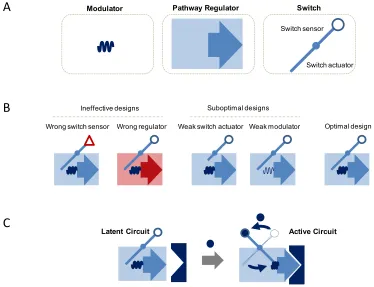
Figure 1.1. Building synthetic circuitry that interfaces with the environment and native regulatory networks to control cellular behavior. Environmental signals are transduced into changes in the native regulatory network via synthetic circuitry. The composition of the synthetic circuitry dictates the environmental interface, how environmental information is input into the synthetic circuit, and the network interface, how the circuitry implements regulation and extracts information from the native network.
Building a modular interface requires that circuitry be composed of modular parts such sensors, actuators, and other components that may be swapped in and out to connect to different environmental cues and various native networks. A synthetic circuit’s ability to actuate changes in a native network depends on the properties of the circuit’s parts, as well as, their interaction with each other (Figure 1.2A, B). An optimally placed circuit
Synthetic circuitry
+
Network interface Environmental
interface
runs quiescently until activated by the proper environmental cue to trigger a change in the native network (Figure 1.2C). The development of modular parts with tunable properties will enhance design flexibility, facilitating the optimal positioning of synthetic circuits for a broad range of applications.
Figure 1.2. Components for interfacing with the environment and native regulatory networks. A. Parts used to interface with the environment and network. Function of the circuit relies on the properties of the elements and how they are integrated into the larger device. B. Composing circuits for optimal performance requires selection of the proper parts to efficiently transduce the environmental input into changes in the regulatory machinery. C. The latent circuit is activated by increasing environmental signal transduced by a sensor to imbedded synthetic circuitry. The synthetic circuit processes increasing levels of input by raising the profile of regulatory machinery that interfaces with the native network, mediating changes in the native network behavior.
Building a modular interface from ncRNAs
The modular, tunable, and programmable nature of RNA makes it an ideal candidate to perform sensing, actuation, and regulatory functions within a synthetic circuit [19]. Over the last twenty years the scientific community has become increasingly
Latent Circuit Active Circuit
Pathway Regulator
Modulator Switch
A
B
Optimal design Weak modulator
Wrong regulator Weak switch actuator Wrong switch sensor
C
Suboptimal designs Ineffective designs
[image:22.612.133.507.189.476.2]aware of the role of non-coding RNAs (ncRNAs) in cellular control over gene expression. ncRNAs were thought to provide rather generic functions as ribosomal RNAs (rRNAs) and transfer RNAs (tRNAs) in translation and as small nuclear RNAs (snRNAs) in splicing. Studies of prokaryote genomes and their regulation forged the central dogma of biology, dictating that RNA had a relatively passive role in the transfer of genetic information from genes to proteins. In addition to specifying cellular state, proteins were thought to direct the trajectory of cellular fate by controlling gene expression networks. The vast tracks of ncRNA found in eukaryotes were hypothesized to be the evolutionary accumulation of inert sequences. However, large sets of these sequences are transcribed [20]. In fact, the majority of transcribed sequences in higher eukaryotes are never translated [21]. Additionally, the percentage of non-coding transcripts scales with organism complexity, while the number of coding genes does not [22]. With increasing discoveries of small ncRNAs that modulate gene expression [23-25] as well as with the discovery of the RNA interference (RNAi) pathway [26-28], ncRNAs appear to be the defining layer of sophisticated biological control that differentiates species with remarkably similar genomes [29].
implementation of RNA based-control systems that provide post-transcriptional control. To date, synthetic network engineering has largely focused on protein-based regulatory elements to build control systems [30]. However, there is a fundamental limitation to the complexity and specificity of protein-based regulatory schemes, which have a higher energetic cost. The burden of carrying additional genes encoding single protein regulators increases exponentially as complexity is introduced. Ultimately, in these accelerating networks, the cost of producing another protein regulator exceeds the benefit to the organism [31]. Regulation through alternative mechanisms such as ncRNA offers organisms an alternative that allows for specificity and diversity at a lower energetic cost [32].
[37]. In composing synthetic circuits, ncRNAs provide an additional degree of freedom for tuning circuit performance within the desired application.
Selecting for RNA-based sensors
To construct a modular environmental interface, requires modular sensors that can transduce the environmental input into changes in the synthetic circuit. We propose that RNA aptamers demonstrate the requisite modularity as environmental sensors that can be wired to actuation and regulatory elements. Aptamers are a class of small nucleic acids, including some ncRNAs, that bind to a wide range of ligands, such as small molecules and peptides, with sensitivity and selectivity that can rival that of proteins [38]. Aptamers are thought to bind ligands through a process called adaptive recognition, in which ligand binding occurs as the RNA molecule transitions through relatively unstructured conformations until the appropriate binding pocket is formed. Upon formation of the binding pocket, the aptamer associates with the ligand which stabilizes the ligand-bound structure. Due to evolutionary pressure during selection, the three-dimensional structure of aptamer complexes reflects highly optimized scaffolds for ligand recognition [39].
The development of new aptamer sequences to cellular molecules of interest offers the potential to connect to endogenous networks in a rational way that can direct information into exogenous control systems. Synthetic RNA aptamers have been generated de novo to various small-molecule and protein targets through in vitro selection or SELEX strategies [40, 41]. Briefly, a large library of RNA molecules (~ 1014–1015) is incubated with the target of interest.
Collected sequences are then reverse transcribed and amplified to generate an enriched library that will serve as the input pool for the next round of selection. The Smolke laboratory has generated RNA aptamers that exhibit varying specificities to benzylisoquinoline alkaloids [42] and folinic acid derivates, and is developing high-throughput strategies for the direct selection and characterization of new protein- and small-molecule-responsive aptamers. Developing modular interfaces that facilitate information exchange between natural and engineered systems is critical for constructing biological control systems that program cell behavior.
Natural RNA switches as gene expression control systems
adopts its active conformation, gene regulation occurs through an array of diverse mechanisms such as transcription termination, mRNA cleavage, or translation initiation [19].
Riboswitches are implemented by the cell as autonomous biological control systems. These RNA elements provide feedback control by sensing metabolites that are substrates and products of the riboswitch-regulated enzymes and modulating the levels of these enzymes in response to cellular metabolite concentrations. One such example is the glutamine-fructose-6-phosphate (GlcN6P) amidotransferase ribozyme-based riboswitch that is located within the 5’ untranslated region (UTR) of the glmS gene. This enzyme metabolizes GlcN6P, which is the small-molecule effector of the riboswitch located upstream of glmS [46]. While some riboswitches have been discovered to promote gene expression [47], the majority repress the expression of their target gene. However, these natural RNA switches generally have evolved nonmodular architectures, in which the sequences of the sensor and actuator components interact to allow the switch to adopt different functional conformations, making the adaptation of these ncRNA controllers to new molecular inputs and regulatory mechanisms through direct component swapping unfeasible [19, 45, 47].
Synthetic RNA switches that act through ribozyme-based cleavage mechanisms
regulate diverse genetic targets are of interest for integration into these synthetic controllers. A number of design strategies have been developed to functionally couple aptamers to small molecules and proteins to diverse ncRNA regulatory elements, from miRNAs [48] to ribozymes [50], such that binding of the ligand to the aptamer domain results in a change in the activity of the ncRNA regulatory element.
Ribozymes are RNA molecules that catalyze a variety of reactions such as self-cleavage or ligation [51]. Thus, ribozyme activity is independent of cell-specific machinery, and these RNA elements may provide a regulatory strategy that can be used across diverse organisms, including bacteria and eukaryotic cells. The hammerhead ribozyme is one of the most extensively studied ribozymes [51-54]. Previous work coupled aptamers to the stem-loop regions in hammerhead ribozymes, allowing for in vitro allosteric ribozymes [55-57]. However, the coupling strategies used in these early allosteric ribozyme designs inactivated the ribozyme activity at physiological salt conditions, not allowing these switches to be implemented as controllers inside cells. Through elucidation of the design rules for in vivo catalytic activity [58, 59], the stage was set for the design of RNA switch platforms that functionally integrate hammerhead ribozymes as in vivo regulatory elements.
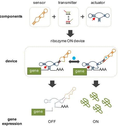
state, the ribozyme favors the active conformation, promoting cleavage of the transcript. Addition of ligand biases the ribozyme switch to the ligand-bound inactive conformation, resulting in increased gene expression. Small-molecule-dependent regulation of gene expression has been demonstrated on various heterologous genes and enabled the construction of RNA switches exhibiting up- and down-regulation of target expression levels. The design of the transmitter domain is a critical design feature that supports the insulation and modularity of the sensor and actuator components and allows the forward design and tuning of synthetic RNA switches. The resulting ability to mix-and-match sensing and actuation domains makes the modular RNA switch platforms powerful tools for developing tailored gene expression control systems.
Figure 1.3. RNA-based switches regulate gene expression in response to small-molecule concentration. The switch device is constructed from three primary components, a sensor, a transmitter, and an actuator. These devices regulate the expression of a gene when placed in the 3’ untranslated region (3’ UTR). Shown above in the OFF state, the switch reduces gene expression in the absence of the small molecule by cleaving and destabilizing the transcript. Presence of the ligand alters the structure of the sensor which is transduced to the changes in the actuator structure via the transmitter. In the presence of ligand, the switch is ON and the actuator structure allows for increased gene expression. Adapted from Liang, J, et al. (Submittedn).
components
device
AAA
gene gene AAA
gene expression
sensor transmitter actuator
ribozyme ON device
ON OFF
AAA
[image:29.612.214.442.351.595.2]Figure 1.4. Converting cis-acting actuators to trans-acting requires engineering an intramolecular reaction into an intermolecular reaction. The cis-acting ribozyme (at left) can be converted to a trans-acting ribozyme by opening up stem I of the ribozyme to allow binding of a target transcript (shown in red).
MAPK cascades as universal signaling modules in eukaryotes
Mitogen-activated protein kinase (MAPK) cascades are highly conserved signaling pathways that control such processes as differentiation, mitosis, and apoptosis (Figure 1.5) [70]. Signaling through this pathway begins when extracellular signals are transduced across the cell membrane through receptor binding events that activate G-proteins. G-proteins relay these signals by facilitating phosphorylation of MAPKKKKs that continue phosphorylation through a three-tiered cascade until reaching the MAPK [71]. MAPKs regulate cellular behavior through interaction with repressors and transcription factors that determine entry into various cellular programs [72]. Many human cancers and other diseases are known to result from aberrant activation of cellular programs connected to MAPK signaling [73, 74]. Thus, controlling improperly activated signals has important implications in the development of therapeutics. Control systems that modulate MAPK pathways will provide an opportunity to interface with a large class of endogenous regulatory networks by which cellular fate can be programmed.
Cis-acting actuator Rotated Cis-acting actuator Trans-acting actuator
Stem I
Figure 1.5. MAPK cascades as universal signaling modules in eukaryotes. From yeast to mammals MAPK pathways preserve the transmembrane receptor and downstream three-tiered MAPK cascade that ultimately generates a phenotypic response to the stimuli. Adapted from [75].
Despite an increase in our understanding of MAPK cascades, the development of therapeutics to intervene and redirect cellular fate through targeting components of this pathway has been primarily limited to kinase inhibitors [76-78]. These inhibitors act competitively to limit signal transduction; however, in pathways where control loops provide redundant verification of signaling, these inhibitors may be overwhelmed. Additionally, the delivery of these inhibitors is not restricted to diseased cells, which can result in unintended toxic side effects in healthy cells [79]. Effective therapeutics that redirect aberrant signaling through these pathways may need to compete with transcriptional feedback and discriminate between healthy and diseased cells. Implementation of control systems that regulate protein
Transmembrane receptor
MAPKKK
MAPKK
MAPK Extracellular signal
Pathway
response Mating DifferentiationProliferation Development
Inflammation Apoptosis Development
Yeast Mammalian
Pheromone Growth factors, cytokines, cell stress
Figure 1.6. Yeast mating pathway and phenotypic response. A. Pheromone (α-factor) binding the transmembrane receptor initiates signaling in the internal G-proteins which is relayed to the canonical three-tiered MAPK cascade (Ste11, Ste7, Fus3). Phosphorylation is relayed down the cascade and culminates with phosphorylated Fus3 translocating to the nucleus to activate a range of transcription factors, transcription at mating genes, and ultimately the canonical mating response. Signaling is antagonized by Msg5, a phosphatase specific to Fus3. B. Phenotypic evaluation of the mating pathway can be performed via halo assay and by observing cell morphology. At bottom, a typical halo assay with a filter paper (center dark circle) saturated with pheromone establishing a gradient of pheromone. Cells within a particular radius corresponding to a particular concentration undergo pheromone-induced cell cycle arrest generating a halo in which cell density is significantly reduced relative to the plate outside of this radius. Above, cells stimulated with pheromone form “shmoos” by undergoing polarized cell growth.
Combining synthetic biology and systems biology to build gene regulatory networks
that control cellular fate
Several modeling efforts have focused on evaluating the dynamics of MAPK cascades and posited that levels of signaling molecules are responsible for divergent cell fates [83-85]. Experimental results in the yeast pheromone-responsive MAPK pathway have demonstrated that particular profiles of signaling molecules are associated with
5 µm
1 cm α-factor
Nucleus
S
te5
Ste11
Fus3 Ste7
pp
pp
pp Cell Membrane
Mating Response
Ste4
Ste50
Msg5
pFUS1
[image:34.612.132.508.79.371.2]entry into specific fates [86-88]. Further, it has been suggested that network topology and the associated positive and negative feedback loops are ultimately responsible for these profiles and thus phenotype [87, 89]. Recent work in mammalian PC-12 cells indicates that the induced ERK MAPK network topology and resulting dynamics direct cell fate (Figure 1.7) [90]. Altering network topology routes cells to alternative fates. Questions still remain as to whether varying the induced network topology represents a conserved strategy across multiple MAPK cascades and eukaryotic organisms. Nevertheless, these results bode well for employing synthetic layers of positive and negative feedback loops as an engineering strategy to regulate cellular behavior
Figure 1.7. Induced network topology shapes the dynamics in a natural regulatory pathway dictating cellular fate. A. Transient stimulation with NGF and EGF leads to different Erk1/2 profiles that correspond to divergent cell fates. Adapted from [90]. B. Graph of network topology induced by different growth factors establishes feedback loops of different signs leading to divergent cell fates. Adapted from [91].
Construction of synthetic biological control systems that interact with natural circuits has seen success in regulating pathway activity by incorporating endogenous promoters in feedback control schemes [81]. Additionally, construction of protein
ProliferationProliferation Differentiation
Proliferation
EGF NGF
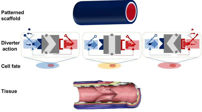
approaches (Figure 1.8). Such systems may be realized in the near future as researchers continue to unravel the systems biology governing cell-fate decisions [94, 95]. Advances in gene therapy delivery may allow molecular network diverters to be translated in vivo as cancer therapeutics targeting hyperactive MAPK pathways. Finally, the selection of new sensors responsive to pathway components may allow these diverters to perform autonomous corrective control of cell fate.
Figure 1.8. Potential application of molecular network diverters to tissue engineering via small-molecule regulated patterning of cell fate. A scaffold with the appropriate geometry is patterned with two small-molecules to trigger diverter action at particular regions within the scaffold. Activated diverters route cells to two alternative fates. All three fates are properly distributed to compose the constructed tissue.
Thesis organization
This thesis is organized into two primary sections. The first section focuses on constructing RNA-based control systems that regulate signaling in the yeast mating pathway. Chapter 2 focuses on using a synthetic titration system to identify regulators of
Patterned scaffold
Diverter action
Cell fate
[image:37.612.112.541.246.484.2]pathway activity and tracing the pathway response curve that routes cells to alternative fates via varying regulator expression. Using this knowledge, we construct synthetic circuits called “molecular network diverters” composed with engineered RNA controllers and feedback modules that conditionally route cellular fate. Chapter 3 examines the construction of diverters with more complex network architectures composed of multiple modules with different expression modes and RNA controllers that amplify ligand-induced phenotypic switching. We demonstrate an integrated network diverter capable of routing genetically identical cells to one of three fates dependent on environmental signals received. In the second section, chapter 4 discusses RNA-based controllers and efforts to develop a ligand-responsive trans-ribozyme platform that may be used to target both heterologous and synthetic transcript enhancing the design flexibility of synthetic control systems.
References
1. Purnick PE, Weiss R: The second wave of synthetic biology: from modules to systems. Nat Rev Mol Cell Biol 2009, 10:410-422.
2. Endy D: Foundations for engineering biology. Nature 2005, 438:449-453. 3. Gibson DG, Glass JI, Lartigue C, Noskov VN, Chuang RY, Algire MA, Benders
GA, Montague MG, Ma L, Moodie MM, et al: Creation of a bacterial cell controlled by a chemically synthesized genome. Science 2010, 329:52-56. 4. Gibson DG, Young L, Chuang RY, Venter JC, Hutchison CA, 3rd, Smith HO:
Enzymatic assembly of DNA molecules up to several hundred kilobases. Nat Methods 2009, 6:343-345.
5. Canton B, Labno A, Endy D: Refinement and standardization of synthetic biological parts and devices. Nat Biotechnol 2008, 26:787-793.
6. Elowitz MB, Leibler S: A synthetic oscillatory network of transcriptional regulators. Nature 2000, 403:335-338.
7. Stricker J, Cookson S, Bennett MR, Mather WH, Tsimring LS, Hasty J: A fast, robust and tunable synthetic gene oscillator. Nature 2008, 456:516-519.
8. Tigges M, Marquez-Lago TT, Stelling J, Fussenegger M: A tunable synthetic mammalian oscillator. Nature 2009, 457:309-312.
10. Tamsir A, Tabor JJ, Voigt CA: Robust multicellular computing using genetically encoded NOR gates and chemical 'wires'. Nature 2011, 469: 212-215.
11. Win MN, Smolke CD: Higher-order cellular information processing with synthetic RNA devices. Science 2008, 322:456-460.
12. Danino T, Mondragon-Palomino O, Tsimring L, Hasty J: A synchronized quorum of genetic clocks. Nature 2010, 463:326-330.
13. Tabor JJ, Salis HM, Simpson ZB, Chevalier AA, Levskaya A, Marcotte EM, Voigt CA, Ellington AD: A synthetic genetic edge detection program. Cell 2009, 137:1272-1281.
14. Elowitz M, Lim WA: Build life to understand it. Nature 2010, 468:889-890. 15. Eldar A, Elowitz MB: Functional roles for noise in genetic circuits. Nature
2010, 467:167-173.
16. Hasty J, McMillen D, Collins JJ: Engineered gene circuits. Nature 2002,
420:224-230.
17. Rosenfeld N, Elowitz MB, Alon U: Negative autoregulation speeds the response times of transcription networks. J Mol Biol 2002, 323:785-793.
18. Kobayashi H, Kaern M, Araki M, Chung K, Gardner TS, Cantor CR, Collins JJ:
Programmable cells: interfacing natural and engineered gene networks. Proc Natl Acad Sci U S A 2004, 101:8414-8419.
19. Isaacs FJ, Dwyer DJ, Collins JJ: RNA synthetic biology. Nat Biotechnol 2006,
20. Mattick JS: Non-coding RNAs: the architects of eukaryotic complexity. EMBO Rep 2001, 2:986-991.
21. Carninci P, Kasukawa T, Katayama S, Gough J, Frith MC, Maeda N, Oyama R, Ravasi T, Lenhard B, Wells C, et al: The transcriptional landscape of the mammalian genome. Science 2005, 309:1559-1563.
22. Taft RJ, Pheasant M, Mattick JS: The relationship between non-protein-coding DNA and eukaryotic complexity. Bioessays 2007, 29:288-299.
23. Moss EG: Non-coding RNA's: lightning strikes twice. Curr Biol 2000,
10:R436-439.
24. Meister G, Landthaler M, Dorsett Y, Tuschl T: Sequence-specific inhibition of microRNA- and siRNA-induced RNA silencing. Rna 2004, 10:544-550.
25. Meister G, Tuschl T: Mechanisms of gene silencing by double-stranded RNA.
Nature 2004, 431:343-349.
26. Fire A, Xu S, Montgomery MK, Kostas SA, Driver SE, Mello CC: Potent and specific genetic interference by double-stranded RNA in Caenorhabditis
elegans. Nature 1998, 391:806-811.
27. Dykxhoorn DM, Lierberman J: The Silent Revolution: RNA Interference a Basic Biology, Research Tool, and Therapeutic. Annual Review of Medicine 2005, 56:401-423.
28. Hannon GJ, Rossi JJ: Unlocking the potential of the human genome with RNA interference. Nature 2004, 431:371-378.
30. van Nimwegen E: Scaling laws in the functional content of genomes. Trends Genet 2003, 19:479-484.
31. Mattick JS, Gagen MJ: Mathematics/computation. Accelerating networks.
Science 2005, 307:856-858.
32. Mattick JS, Gagen MJ: The evolution of controlled multitasked gene networks: the role of introns and other noncoding RNAs in the development
of complex organisms. Mol Biol Evol 2001, 18:1611-1630.
33. Chen YY, Jensen MC, Smolke CD: Genetic control of mammalian T-cell proliferation with synthetic RNA regulatory systems. Proc Natl Acad Sci U S A 2010, 107:8531-8536.
34. Culler SJ, Hoff KG, Smolke CD: Reprogramming cellular behavior with RNA controllers responsive to endogenous proteins. Science 2010, 330:1251-1255. 35. Pfleger BF, Pitera DJ, Smolke CD, Keasling JD: Combinatorial engineering of
intergenic regions in operons tunes expression of multiple genes. Nat Biotechnol 2006, 24:1027-1032.
36. Osella M, Bosia C, Cora D, Caselle M: The role of incoherent microRNA-mediated feedforward loops in noise buffering. PLoS Comput Biol 2011,
7:e1001101.
37. Xie Z, Wroblewska L, Prochazka L, Weiss R, Benenson Y: Multi-input RNAi-based logic circuit for identification of specific cancer cells. Science 2011,
333:1307-1311.
39. Hermann T, Patel DJ: Adaptive recognition by nucleic acid aptamers. Science 2000, 287:820-825.
40. Tuerk C, Gold L: Systematic evolution of ligands by exponential enrichment: RNA ligands to bacteriophage T4 DNA polymerase. Science 1990, 249: 505-510.
41. Bowser MT: SELEX: just another separation? Analyst 2005, 130:128-130. 42. Win MN, Klein JS, Smolke CD: Codeine-binding RNA aptamers and rapid
determination of their binding constants using a direct coupling surface
plasmon resonance assay. Nucleic Acids Res 2006, 34:5670-5682.
43. Tucker BJ, Breaker RR: Riboswitches as versatile gene control elements. Curr Opin Struct Biol 2005, 15:342-348.
44. Bauer G, Suess B: Engineered riboswitches as novel tools in molecular biology. J Biotechnol 2006, 124:4-11.
45. Mandal M, Breaker RR: Gene regulation by riboswitches. Nat Rev Mol Cell Biol 2004, 5:451-463.
46. Winkler WC, Nahvi A, Roth A, Collins JA, Breaker RR: Control of gene expression by a natural metabolite-responsive ribozyme. Nature 2004,
428:281-286.
47. Winkler WC, Breaker RR: Genetic control by metabolite-binding riboswitches.
Chembiochem 2003, 4:1024-1032.
48. Beisel CL, Chen YY, Culler SJ, Hoff KG, Smolke CD: Design of small molecule-responsive microRNAs based on structural requirements for
49. Babiskin AH, Smolke CD: Engineering ligand-responsive RNA controllers in yeast through the assembly of RNase III tuning modules. Nucleic Acids Res 2011, 39:5299-5311.
50. Win MN, Smolke CD: A modular and extensible RNA-based gene-regulatory platform for engineering cellular function. Proc Natl Acad Sci U S A 2007. 51. Long DM, Uhlenbeck OC: Self-cleaving catalytic RNA. Faseb J 1993, 7:25-30. 52. Pley HW, Flaherty KM, McKay DB: Three-dimensional structure of a
hammerhead ribozyme. Nature 1994, 372:68-74.
53. Hammann C, Norman DG, Lilley DM: Dissection of the ion-induced folding of the hammerhead ribozyme using 19F NMR. Proc Natl Acad Sci U S A 2001,
98:5503-5508.
54. Blount KF, Uhlenbeck OC: The structure-function dilemma of the hammerhead ribozyme. Annu Rev Biophys Biomol Struct 2005, 34:415-440. 55. Soukup GA, Breaker RR: Nucleic acid molecular switches. Trends Biotechnol
1999, 17:469-476.
56. Soukup GA, Breaker RR: Engineering precision RNA molecular switches.
Proc Natl Acad Sci U S A 1999, 96:3584-3589.
57. Soukup GA, Breaker RR: Allosteric nucleic acid catalysts. Curr Opin Struct Biol 2000, 10:318-325.
59. Khvorova A, Lescoute A, Westhof E, Jayasena SD: Sequence elements outside the hammerhead ribozyme catalytic core enable intracellular activity. Nat Struct Biol 2003, 10:708-712.
60. Win M, Smolke C: Universal riboswitch platforms for engineering cellular function. In Submission.
61. Beisel CL, Bayer TS, Hoff KG, Smolke CD: Model-guided design of ligand-regulated RNAi for programmable control of gene expression. Mol Syst Biol 2008, 4:224.
62. Bayer TS, Smolke CD: Programmable ligand-controlled riboregulators of eukaryotic gene expression. Nat Biotechnol 2005, 23:337-343.
63. Aagaard L, Rossi JJ: RNAi therapeutics: principles, prospects and challenges.
Adv Drug Deliv Rev 2007, 59:75-86.
64. Grimm D, Streetz KL, Jopling CL, Storm TA, Pandey K, Davis CR, Marion P, Salazar F, Kay MA: Fatality in mice due to oversaturation of cellular microRNA/short hairpin RNA pathways. Nature 2006, 441:537-541.
65. Li MJ, Kim J, Li S, Zaia J, Yee JK, Anderson J, Akkina R, Rossi JJ: Long-term inhibition of HIV-1 infection in primary hematopoietic cells by lentiviral
vector delivery of a triple combination of anti-HIV shRNA, anti-CCR5
ribozyme, and a nucleolar-localizing TAR decoy. Mol Ther 2005, 12:900-909. 66. Weinberg MS, Rossi JJ: Comparative single-turnover kinetic analyses of
trans-cleaving hammerhead ribozymes with naturally derived non-conserved
67. Weinberg MS, Ely A, Passman M, Mufamadi SM, Arbuthnot P: Effective anti-hepatitis B virus hammerhead ribozymes derived from multimeric
precursors. Oligonucleotides 2007, 17:104-112.
68. Burke DH, Greathouse ST: Low-magnesium, trans-cleavage activity by type III, tertiary stabilized hammerhead ribozymes with stem 1 discontinuities.
BMC Biochem 2005, 6:14.
69. Saksmerprome V, Roychowdhury-Saha M, Jayasena S, Khvorova A, Burke DH:
Artificial tertiary motifs stabilize trans-cleaving hammerhead ribozymes
under conditions of submillimolar divalent ions and high temperatures. Rna 2004, 10:1916-1924.
70. Seger R, Krebs EG: The MAPK signaling cascade. Faseb J 1995, 9:726-735. 71. Qi M, Elion EA: MAP kinase pathways. J Cell Sci 2005, 118:3569-3572.
72. Dohlman HG, Thorner JW: Regulation of G protein-initiated signal transduction in yeast: paradigms and principles. Annu Rev Biochem 2001,
70:703-754.
73. McCormick F: Signalling networks that cause cancer. Trends Cell Biol 1999,
9:M53-56.
74. Hanahan D, Weinberg RA: The hallmarks of cancer. Cell 2000, 100:57-70. 75. Pierce KL, Premont RT, Lefkowitz RJ: Seven-transmembrane receptors. Nat
Rev Mol Cell Biol 2002, 3:639-650.
angiogenesis, and induces tumor cell apoptosis in hepatocellular carcinoma
model PLC/PRF/5. Cancer Res 2006, 66:11851-11858.
77. Shapiro P: Discovering new MAP kinase inhibitors. Chem Biol 2006, 13: 807-809.
78. Dominguez C, Powers DA, Tamayo N: p38 MAP kinase inhibitors: many are made, but few are chosen. Curr Opin Drug Discov Devel 2005, 8:421-430.
79. Dambach DM: Potential adverse effects associated with inhibition of p38alpha/beta MAP kinases. Curr Top Med Chem 2005, 5:929-939.
80. Elion EA: The Ste5p scaffold. J Cell Sci 2001, 114:3967-3978.
81. Park SH, Zarrinpar A, Lim WA: Rewiring MAP kinase pathways using alternative scaffold assembly mechanisms. Science 2003, 299:1061-1064.
82. Elion EA: Pheromone response, mating and cell biology. Curr Opin Microbiol 2000, 3:573-581.
83. Chapman S, Asthagiri AR: Resistance to signal activation governs design features of the MAP kinase signaling module. Biotechnol Bioeng 2004, 85: 311-322.
84. Levchenko A, Bruck J, Sternberg PW: Scaffold proteins may biphasically affect the levels of mitogen-activated protein kinase signaling and reduce its
threshold properties. Proc Natl Acad Sci U S A 2000, 97:5818-5823.
85. Wang X, Hao N, Dohlman HG, Elston TC: Bistability, stochasticity, and oscillations in the mitogen-activated protein kinase cascade. Biophys J 2006,
86. Esch RK, Errede B: Pheromone induction promotes Ste11 degradation through a MAPK feedback and ubiquitin-dependent mechanism. Proc Natl Acad Sci U S A 2002, 99:9160-9165.
87. Esch RK, Wang Y, Errede B: Pheromone-induced degradation of Ste12 contributes to signal attenuation and the specificity of developmental fate.
Eukaryot Cell 2006, 5:2147-2160.
88. Maleri S, Ge Q, Hackett EA, Wang Y, Dohlman HG, Errede B: Persistent activation by constitutive Ste7 promotes Kss1-mediated invasive growth but
fails to support Fus3-dependent mating in yeast. Mol Cell Biol 2004, 24: 9221-9238.
89. Ingolia NT, Murray AW: Positive-feedback loops as a flexible biological module. Curr Biol 2007, 17:668-677.
90. Santos SD, Verveer PJ, Bastiaens PI: Growth factor-induced MAPK network topology shapes Erk response determining PC-12 cell fate. Nat Cell Biol 2007,
9:324-330.
91. Kholodenko BN: Untangling the signalling wires. Nat Cell Biol 2007, 9: 247-249.
92. Bashor CJ, Helman NC, Yan S, Lim WA: Using engineered scaffold interactions to reshape MAP kinase pathway signaling dynamics. Science 2008, 319:1539-1543.
94. Kueh HY, Rothenberg EV: Regulatory gene network circuits underlying T cell development from multipotent progenitors. Wiley Interdiscip Rev Syst Biol Med 2011.
Chapter 2
Identifying pathway regulators and constructing RNA-based regulatory
Abstract
Introduction
To orchestrate complex, coordinated, multicellular tasks, organisms dynamically program their extracellular space with distributed molecular signals that are processed by individual cells into concerted responses. These extracellular signals activate signaling cascades that induce specific network topologies, leading cells to divergent cellular fates. MAPK cascades are a class of highly conserved signaling pathways that control such key cellular processes as differentiation, mitosis, and apoptosis [1]. Many diseases, including one third of human cancers, result from aberrant signaling through MAPK pathways [2, 3]. In the model eukaryotic organism Saccharomyces cerevisiae, multiple MAPK cascades direct cellular fate via divergent regulatory programs. Decisions to upregulate gene expression, halt cell cycle, and change cell morphology are programmed through these pathways as the rational response to changing environmental signals.
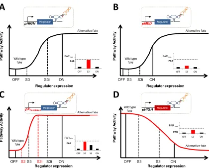
within the continuum of regulator expression levels between wild type and high levels of ectopic overexpression there exists a transitory region over which cell fate diverges (Figure 2.1). In such a model, at subtransition levels of regulator expression, cells mirror the wild-type response. At expression levels above the requisite transitory range, cells adopt an alternative cell fate. By constructing synthetic circuits that can toggle between subtransition and supertransition levels of regulator expression, cellular behavior can be synthetically switched from wild type to programmed alternative fates.
Figure 2.1. Regulator expression modulates pathway activity and over a narrow range of expression transitions to an alternative fate
We present a method for identifying and interfacing with control points in the S. cerevisiae mating and osmolarity response pathways. Using a galactose-responsive titration system, we successfully identified both a positive and a negative regulator in the mating pathway and two coexpressed positive regulators in the osmolarity pathway. The regulators of the mating pathway were shown to route cells to alternative fates above a particular threshold of expression. By incorporating these regulators into various network architectures with RNA-based controllers [12], we successfully constructed several types
Regulator expression
P
at
hw
ay
ac
tiv
ity
Wildtype fate
Transitory region
Alternative fate
of molecular network diverters in the mating pathway which conditionally route cells to one of two alternative cell fates based on the presence of distinct environmental signals. Our results demonstrate the utility of conditionally activated control systems for applications modulating cell grow and/or viability as well as the rational tuning of these synthetic circuits via the exchange of well-characterized parts. Further, we show that positive feedback reshapes the pathway activity response curve, shifting the threshold at which cell fate diverges to lower levels of the positive regulator. Finally, we have elucidated design principles and methodology for constructing molecular network diverters that can be readily extended to new pathways and applications.
Results
Identifying key pathway regulators that allow routing of cell fate decisions to
alternative phenotypes in the mating pathway
measured by monitoring expression from a transcriptional fusion construct (pFUS1-GFP, GFP fused to a mating responsive promoter), where GFP levels represent a measure of pathway activation, and by observing mating-associated cell cycle arrest via halo assays (see Materials and methods).
Figure 2.2. Identifying titratable regulators of pathway activity in the yeast mating pathway. A. A molecular view of signaling in the yeast mating pathway. Pheromone (α-factor) binds to a transmembrane receptor to initiate signaling. That binding event is transduced across the cell membrane and relayed down to the G-proteins including, Ste4 and Ste50, an adaptor protein, that in turn relay signaling to the Ste5 scaffold-bound three-tiered MAPK cascade. Phosphorylation of the MAPK Fus3 results in its translocation to the nucleus and is antagonized by the phosphatase Msg5. Fus3 translocation activates a host of transcription factors which ultimately upregulate expression at various mating gene including FUS1. Pathway activity can be monitored via an integrated pFUS1-GFP promoter fusion. Additionally, the mating response is characterized by pheromone-induced cell cycle arrest and polarized cell growth called shmoo formation. B. To find pathway activators, cells were monitored in the absence of stimulating pheromone. Ste4 increased pathway activity in a galactose-dependent manner indicating it is a titratable regulator. C. To find pathway inhibitors, the pathway was stimulated with pheromone. Msg5 overexpression reduced stimulated pathway activity. Cells were cultured in 0, 0.25 and 1% galactose for 6 hours. Pheromone was added 3 hours post-dilution to stimulate the pathway. Pathway activity was monitored via flow cytometry measurement of cellular fluorescence of GFP from the promoter fusion pFUS1-GFP.
0.0 0.5 1.0 1.5 2.0
Blank Ste4 Ste50 Ste11 Ste7 Fus3 Msg5
A C X pGAL1 X pGAL1 a-factor Nucleus S te5 Ste11 Fus3 Ste7 pp pp pp Cell Membrane Mating Response Ste4 Ste50 Msg5 GFP pFUS1 B No rm al iz ed P at hw ay Act ivi ty No rm al iz ed P at hw ay Act ivi ty 0.0 0.1 0.2 0.3
Blank Ste4 Ste50 Ste11 Ste7 Fus3 Msg5
Figure 2.3. Overexpression of Msg5 and Ste4 modulates pathway activity and routes cells to an alternative fate. A. Increasing Msg5 overexpression results in reduced pathway activity over the galactose range as measure by pFUS1-GFP levels B. Halo assays at various concentrations of galactose indicate that as galactose concentration increases Msg5 overexpression inhibits pheromone-induced cell cycle arrest leading to a “chaste” phenotype. C. Phosphorylated levels of Fus3 correspondingly drop across the galactose range while the Cdc28 control levels remain constant. D. For Ste4 overexpression, pathway activity increases as galactose levels increase. E. Halo assays at various concentrations of galactose indicate that as galactose concentration increases Ste4 overexpression generates pheromone-independent cell cycle arrest or “promiscuous” fate. F. Phosphorylated levels of Fus3 correspondingly drop across the galactose range while the Cdc28 control levels remain constant.
Identifying pathway regulators in the osmolarity pathway
To examine the flexibility of the synthetic titration system for identifying pathway regulators, we applied it to a second yeast MAPK pathway, the osmolarity response pathway. The osmolarity response is triggered by high osmotic pressure that initiates signaling at two different receptors Sho1 and Sln1, which converge on a three-tiered
0 0.25 0.5 1.0 2.0 3.0 % Galactose
% Galactose
Fus3pp
Cdc28
0 0.25 0.5 1.0 2.0 3.0 % Galactose 0.0 0.2 0.4 0.6 0.8 1.0 1.2
0 0.25 0.5 1 2 3
Ctrl Msg5
0 0.25 0.5 1.0 2.0 3.0
% Galactose
No rm al iz ed P at hw ay Act ivi ty
0 0.25 0.5 1.0 2.0 3.0 % Galactose
Fus3pp
Cdc28
% Galactose
0 0.25 0.5 1.0 2.0 3.0 % Galactose 0.0 0.1 0.2 0.3 0.4 0.5
0 0.25 0.5 1 2 3
Ctrl Ste4
0 0.25 0.5 1.0 2.0 3.0 % Galactose A B C D E F Msg5
pGAL1 pGAL1 Ste4
MAPK cascade (Figure 2.4A). Signaling results in phosphorylation and translocation of the dedicated MAPK Hog1 to the nucleus, and ultimately upregulation of osmo-genes and production of glycerol. Activation of the osmolarity response is also known to repress the mating pathway. We used the engineered galactose-inducible strain to evaluate the potential of several signaling proteins as titratable positive pathway regulators. Negative regulators were not explored with this method as inhibiting the osmolarity pathway in yeast challenged with high osmotic pressure results in cell death [17].
Figure 2.4. Identifying titratable regulators in the yeast osmolarity pathway. A. A molecular view of signaling in the yeast osmolarity pathway. High osmotic pressure initiates signaling via two transmembrane sensor proteins. Signaling is relayed to the Pbs2 scaffold-bound three-tiered MAPK cascade. Phosphorylation of the MAPK Hog1 results in its translocation to the nucleus and is antagonized by the phosphatase Ptc1. Hog1 translocation activates a host of transcription factors which ultimately upregulate expression at various osmo genes including STL1. Pathway activity can be monitored via an integrated pSTL1-GFP promoter fusion. Additionally, the osmolarity response is characterized by glycerol production. B. Increasing pathway activity in the osmolarity pathway requires overexpression of both Hog1 and Pbs2. To find pathway activators, cells were monitored in the absence of sorbitol. Cells were cultured in 0, 1, and 3% galactose for 6 hours. Pathway activity was monitored via flow cytometry measurement of cellular fluorescence of GFP from the promoter fusion pSTL1-GFP.
The impact of overexpression of three pathway proteins on osmolarity pathway activation was examined using the galactose-titratable expression system. Pathway activation was measured via a transcriptional fusion between a promoter that is activated through osmolarity pathway stimulation and GFP (pSTL1-GFP). Even at high induction
Osmolyte Nucleus pp pp pp Cell Membrane Osmolarity Response Ptc1 Pb s2 Ste7 Ste11 Hog1 GFP pSTL1 N or m al iz ed P at hw ay A ct ivi ty 0.0 0.3 0.5 0.8 1.0
Blank Pbs2 Ptc1 Hog1 Hog1 and
Pbs2
1 M Soribitol 0% 1% 3%
X
pGAL1
levels, the overexpression of single pathway proteins resulted in very small increases in pathway activation (Figure 2.4B). We hypothesized that co-overexpressing both the scaffold-kinase, Pbs2, and the MAPK Hog1 would lead to more substantial increases in pathway activity. We observed that co-overexpression of Pbs2 and Hog1 significantly increased pathway activation over single component expression.
osmolarity pathway. Under such time scale constraints, transcriptional control systems may modulate the initial conditions that guide the response, but dynamic modulation of signaling via feedback would be necessarily precluded.
Building molecular network diverters for programming cell fate decisions
Figure 2.5. Reshaping the native molecular network with molecular network diverters. A. The latent diverter is activated by increasing trigger concentrations with flip the switch which raises the profile of the pathway regulator via the modulator. Above a certain threshold, the pathway regulator docks to the regulatory node to reshape the native network. B. Trigger binding to its cognate sensor flips the switch. Binding is transduced via the switch actuator to the modulator and finally to the imbedded pathway regulator, raising the profile of the regulator which plugs into its cognate regulatory node, reshaping the molecular network and diverting fate. The diverter function can be tuned by the properties of the switch, the modulator, and addition of feedback to the modulator.
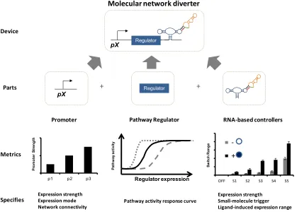
Figure 2.6. Implementation of a molecular network diverter from various genetic parts. The molecular network diverter is composed with a modulator, pathway regulator, and a switch into the larger device. Mapping these parts to their genetic representation, the modulator is a promoter, the pathway regulator is a gene that regulates pathway activity, and the switch is an RNA-based controller. Assembling the genetic parts in the proper configuration allows the realized molecular network diverter to conditionally regulate pathway activity and divert cell fate.
Construction of a molecular network diverter requires the selection of regulatory elements with the appropriate strength and range for modulating the pathway regulator levels to conditionally route cellular fate. The selection of the promoter driving the expression of the regulator is a key component in setting the activity of the molecular network diverter. Constitutive promoters of varying strength achieve different levels of regulator expression (Figure 2.7). The pathway activity response curve indicates where a promoter-regulator pair falls on this curve, which dictates whether cells route to the wild-type or alternative fate. Using a promoter responsive to pathway activation results in the construction of a feedback molecular network diverter. Feedback in molecular networks
Regulator
pX
Regulator
pX
Pathway Regulator
Promoter RNA-based controller
Pathway Regulator
Modulator Switch
Abstract molecular network diverter
Realization of molecular network diverter
Parts Device
Parts
Device Abstract
representation