D
E
V
E
LO
P
M
E
N
T
INTRODUCTION
Vertebrate limb development occurs along three cardinal axes: proximodistal (PD), anteroposterior (AP) and dorsoventral (DV) (reviewed by Niswander, 2003; Tickle, 2003). These axes are established through molecular networks, beginning when the limbs bud out of the flank and continuing with the organization of signaling centers, such as the zone of polarizing activity (ZPA) in the posterior limb mesenchyme (reviewed by Pearse and Tabin, 1998). Continued outgrowth and patterning of the distal limb requires a molecular feedback loop between the ZPA and a strip of specialized epithelium at the apex of the limb, known as the apical ectodermal ridge (AER). Crucial to this interaction is the expression of sonic hedgehog (Shh) in the ZPA (Riddle et al., 1993) and of fibroblast growth factors (Fgfs) in the AER (Sun et al., 2002). Shh signaling establishes the limb AP axis (Chiang et al., 2001) and leads to the formation of distal elements of the posterior zeugopod (i.e. ulna and fibula) and autopod (Harfe et al., 2004).
As of yet, the molecular triggers that activate Shhexpression are mostly unknown, although a few candidate genes have been proposed. For example, Hand2(previously known as dHand) has been shown to act upstream of Shhas Hand2-deficient (hereafter,
Hand2–/–) embryos lack Shhexpression (Charite et al., 2000). Hox genes (reviewed by Krumlauf, 1994; Capecchi, 1997; Deschamps and van Nes, 2005) have been more recently proposed as upstream regulators of Shh(Zakany et al., 2004) and of AP and PD axis formation (Kmita et al., 2005). For example, prior to Shhonset, early Hox colinear expression in limb mesenchyme leads to their transcript accumulation in the ZPA (Zakany et al., 2004; Tarchini and Duboule, 2006), while their ectopic expression leads to Shh transcription (Charite et al., 1994; Stratford et al., 1997; Knezevic et al., 1997). Finally, in vertebrates, functional ablation of multiple 5⬘ Hox leads to distal limb truncations partially mediated by Shh absence (Kmita et al., 2005).
Although early Hox colinearity appears essential for Shhonset, their dynamic expression throughout limb development aids in patterning elements along the PD axis. Indeed, patterning alterations occur in limb skeletal elements of specific developmental modules in mice where multiple paralogous Hox genes have been genetically ablated, suggesting a functional redundancy within paralogous groups (Condie and Capecchi, 1994; Davis et al., 1995; Davis and Capecchi, 1996; Ramain et al., 1996a; Fromental-Ramain et al., 1996b; Wellik and Capecchi, 2003). These findings highlight Hox role as global regulators of patterning throughout limb development.
The present understanding of Hox function is that they act partially through the aid of co-factors, such as Pbx TALE homeoproteins (Burglin, 1997; Burglin, 1998), that increase Hox DNA-binding specificity and selectivity (reviewed by Mann and Chan, 1996; Mann and Affolter, 1998; Moens and Selleri, 2006). Pbx proteins, when forming complexes with Hox, are further known to transcriptionally regulate Hox genes themselves (Popperl et al., 1995; Maconochie et al., 1997; Jacobs et al., 1999; Ferretti et al.,
Pbx1/Pbx2
requirement for distal limb patterning is
mediated by the hierarchical control of Hox gene spatial
distribution and
Shh
expression
Terence D. Capellini1,2,*, Giuseppina Di Giacomo1,*, Valentina Salsi3, Andrea Brendolan1, Elisabetta Ferretti1, Deepak Srivastava4, Vincenzo Zappavigna1,3 and Licia Selleri1,†
Vertebrate limb development occurs along three cardinal axes – proximodistal, anteroposterior and dorsoventral – that are established via the organization of signaling centers, such as the zone of polarizing activity (ZPA). Distal limb development, in turn, requires a molecular feedback loop between the ZPA expression of sonic hedgehog (Shh) and the apical ectodermal ridge. The TALE homeoprotein Pbx1 has been shown to be essential for proximal limb development. In this study, we first uncover that Pbx1 and Pbx2are co-expressed in the lateral plate and early limb field mesoderm. Later, Pbx2 is expressed throughout the limb, unlike Pbx1, which is expressed only in the proximal bud. By exploiting a Pbx1/Pbx2loss-of-function mouse model, we demonstrate that, despite the lack of limb abnormalities in Pbx2-deficient (Pbx2–/–) embryos, compound Pbx1–/–; Pbx2+/–mutants, in addition to their
exacerbated proximal limb defects, exhibit novel and severe distal abnormalities. Additionally, we reveal that Pbx1–/–; Pbx2–/– embryos lack limbs altogether. Furthermore, we establish that, unlike in flies, where the leg develops independently of Hox and where the Pbx ortholog Exdis required for specification of proximal (but not distal) limbs, in vertebrates, distal limb patterning is Pbx1/Pbx2dependent. Indeed, we demonstrate that Pbx genetic requirement is mediated, at least in part, through their
hierarchical control of Hox spatial distribution and Shhexpression. Overall, we establish that, by controlling the spatial expression of Hox genes in the posterior limb and regulating ZPA function, Pbx1/Pbx2exert a primary hierarchical function on Hox genes, rather than behaving merely as Hox ancillary factors.
KEY WORDS: Pbx1/Pbx2, Hox, Shh, Limb development, Distal limb patterning, Mouse
Development 133, 2263-2273 (2006) doi:10.1242/dev.02395
1Department of Cell and Developmental Biology, Cornell University Weill Medical
School, New York, NY 10021, USA. 2New York Consortium in Evolutionary
Primatology, The Graduate School and University Center, The City University of New York, NY 10016, USA. 3Department of Animal Biology, University of Modena-Reggio
Emilia, Modena 41100, Italy. 4Gladstone Institute of Cardiovascular Disease,
Department of Pediatrics, University of California, San Francisco, CA 94158, USA.
*These authors contributed equally to this work
†Author for correspondence (e-mail: [email protected])
D
E
V
E
LO
P
M
E
N
T
2000). However, recent findings strongly suggest that Pbx can also function more broadly in Hox-independent manners (Knoepfler et al., 1999; Berkes et al., 2004) (reviewed by Mann and Morata, 2000; Moens and Selleri, 2006).
Recently, we elucidated roles of Pbx genes in skeletal development and found that although Pbx1is required for proximal limb patterning (Selleri et al., 2001), Pbx2or Pbx3loss does not determine skeletal or limb phenotypes (Selleri et al., 2004; Rhee et al., 2004). Specifically, in Pbx1–/–embryos, skeletal structures of girdles (i.e. scapula and pelvis) and proximal limb stylopod (i.e. humerus and femur) that normally express nuclear Pbx1at early developmental stages, are malformed, while their distal elements and joints appear normal. These findings parallel the role of Exd(the DrosophilaPbx ortholog) (Peifer and Weischaus, 1990; Rauskolb et al., 1993) in governing proximal domains of the fly appendage, where its expression is restricted (Mercader et al., 1999).
In this study, we first uncover that Pbx1and Pbx2are co-expressed in the early vertebrate limb field and that, later, Pbx2 is expressed throughout the limb mesenchyme, while Pbx1 is expressed only proximally. Next, by exploiting a Pbx1/Pbx2loss-of-function mouse model, we determine that, despite the lack of skeletal and/or limb abnormalities in Pbx2–/–embryos (Selleri et al., 2004), decreasing Pbx2dose in the absence of Pbx1does affect limb development more severely than the loss of Pbx1 alone (Selleri et al., 2001). We demonstrate that compound Pbx1/Pbx2embryos, in addition to their proximal limb defects, exhibit novel and severe distal limb abnormalities; Pbx1–/–;Pbx2+/– embryos display loss of distal hindlimb elements, whereas Pbx1–/–;Pbx2–/–embryos lack hindlimbs altogether. We establish that in vertebrates distal limb patterning is genetically regulated by Pbx1/Pbx2, at least in part, through their hierarchical control of Hox spatial distribution and Shhexpression.
MATERIALS AND METHODS
Mice
Intercrosses of Pbx1+/–(Selleri et al., 2001) and Pbx2+/–(Selleri et al., 2004) were conducted to obtain Pbx1+/–;Pbx2+/–mice. On a C57BL/6 background, the number of Pbx1+/–;Pbx2+/–obtained was below the expected Mendelian ratio. To increase their number, we crossed C57BL/6 Pbx1+/–;Pbx2+/–males to an outbred strain, Black Swiss [NIH-BL(S)], and observed a slight amelioration of the limb phenotype that remained fully penetrant. We then intercrossed these C57BL/6 females and mixed C57BL/6-Black Swiss males and analyzed the progeny for lethality and soft/hard tissue morphologies.
Skeletal preparations
Differential staining of cartilage and bone in whole mouse embryos (E12.5 and E13.5) was visualized using Alcian Blue and Alizarin Red (Selleri et al., 2001).
Whole-mount in situ hybridization
Whole-mount in situ hybridization was performed on somite-matched embryos at different gestational days using digoxigenin or fluorescein-labeled antisense RNA probes as described (Di Giacomo et al., 2006). These analyses were performed on all compound genotypes, including key controls (Pbx1+/–;Pbx2–/–and Pbx1–/–;Pbx2+/+), but only shown for those genotypes
that displayed alterations in gene expression.
Whole mount immunohistochemistry
Mouse embryos were dissected at E10.5, fixed with 4% paraformaldehyde in phosphate-buffered saline (PBS) and processed for immunohistochemistry to detect CD44 localization (Sherman et al., 1998). Embryos were permeabilized in 0.1%TritonX-100/PBS, blocked in 3%BSA/0.1%TritonX-100/PBS and then incubated in anti-CD44 (antibody IM7 rat monoclonal, Pharmingen) overnight. The embryos were washed in 0.1% TritonX-100/PBS and incubated with anti-rat Alexa 568 (Molecular Probes-Invitrogen) overnight. After rinsing, embryos were visualized with a fluorescence microscope.
Chromatin immunoprecipitation (ChIP)
Formaldehyde cross-linking and ChIP (Aparicio et al., 1999) from E10.5 mouse limbs were performed according to described protocols (Orlando et al., 1997), with the following modifications: to disaggregate tissues, samples were forced through 18G needles, cells were fixed for 15 minutes with 1% formaldehyde at room temperature and reactions were quenched with 0.125 M glycine in PBS for 5 minutes. Crosslinked samples were sonicated for 15⫻25 seconds to obtain average fragment lengths of 500-1000 bp. Immunoprecipitation was performed with 10 l of protein G-Agarose (KPL) and blocked twice with 1 g/ml salmon sperm DNA (Sigma) and 1 g/ml bovine serum albumin, for 2 hours, then overnight. Chromatin was precleared by adding 20 l of protein G-Agarose for two hours, and incubated with 5 g of the respective specific antisera, or with 5 g of anti-Flag (F3165, Sigma) antibody as a control. Incubations were performed overnight at 4°C. These primers were used for PCR amplifications (42 cycles): mouse ShhE1, 5⬘-CTTTGATTTGAAGTCCTGGC-3⬘; mouse ShhE2, 5⬘-ACTGAGGGGAAAAGTCATC-3⬘; mouse ShhC1, 5⬘ -TCAA-GAGAGATCAACAAAAG-3⬘; mouse ShhC2, 5⬘ -TTGGACTCAAGT-CCAGAC-3⬘.
Cell culture and transfection
P19 mouse embryonal carcinoma cells (McBurney and Rogers, 1982) were cultured in media (MEMA, GIBCO, Life Technologies) supplemented with 10% fetal calf serum, 2 mM L-glutamine, 100 U/ml penicillin and 100 g/ml streptomycin. Transfections were performed by CaPO4precipitation.
In a typical experiment, reporter plasmid (5 mg), expression construct (2.5-5.0 g) and CMV-gal (Clontech) (0.1 g) as an internal control, were used per 6 cm dish. Forty-eight hours after transfection, cells were washed, lysed and assayed for luciferase and -galactosidase expression (Zappavigna et al., 1994).
RESULTS
Loss of Pbx1/Pbx2causes not only proximal but
also distal limb defects
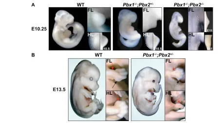
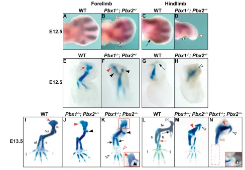
Pbx1+/–;Pbx2+/–mice were intercrossed to generate progeny with different compound genotypes. Axial and limb skeletal abnormalities were evident only in Pbx1–/–;Pbx2+/+, Pbx1–/–;Pbx2+/– and Pbx1–/–;Pbx2–/– embryos. Pbx1–/–;Pbx2–/– embryos died at E9.5/10.5 during the period crucial for limb bud initiation, owing to multiple organogenesis defects, and therefore lacked limbs. Thus, our analyses focused on limb development in Pbx1–/–;Pbx2+/– (hereafter, Pbx1/2 mutant) embryos (Fig. 1), which survived until E13.5/14.5. Pbx1/2 mutant embryos exhibited drastic exacerbations of Pbx1–/–axial and proximal limb defects (Selleri et al., 2001) and novel distal phenotypes (Fig. 1A,B). Fore- and hindlimb development in Pbx1/2 mutant embryos were differently affected. For example, although E9.5 Pbx1/2 mutant forelimbs already appeared malformed, E10.5 hindlimbs still exhibited relatively normal morphology (Fig. 1A), despite the typical half-day delay in development. However, by E13.5, gross morphology of both limbs was severely affected; specifically, forelimbs displayed shortened zeugopodia and hypoplastic autopodia, whereas hindlimbs appeared rudimentary and lacked true autopodia (Fig. 1B).
D
E
V
E
LO
P
M
E
N
T
striking abnormalities, including loss of distal elements (Fig. 2I-N). Pbx1/2 mutant forelimbs exhibited mis-shapened scapulae that were fused to skeletal elements, probably representing duplicated proximal humeral heads. In addition, humeri had shafts that were thickened and contorted, with cartilaginous anlagen mimicking hypertrophic deltoid tuberosities, more severely than in Pbx1–/–;Pbx2+/+ (Fig. 2I-K). Although both forelimb zeugopod elements were present, albeit malformed, digits one and five were missing or rudimentary (Fig. 2K). In Pbx1/2 mutant embryos, pelvic girdles were missing most elements except isolated anlagen reminiscent of ischia (Fig. 2L-N). These residual pelvic elements were fused to truncated thickened femurs more severely than in Pbx1–/–;Pbx2+/+ (Fig. 2L-N). The only remaining hindlimb zeugopodial element appeared as a dysmorphic tibia, whereas its fibula was absent, as were most autopodial elements (except for single rudimentary digital rays) (Fig. 2N).
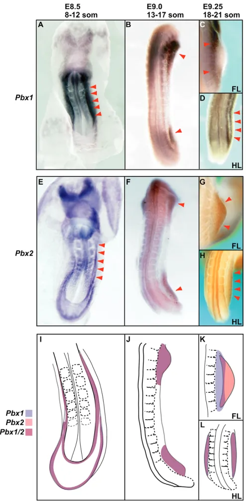
Pbx1/Pbx2early mesenchymal expression is
overlapping and during later limb development is dynamic and, in part, complementary
To determine how specific spatiotemporal differences in Pbx limb expression underlie such abnormalities, we performed in situ hybridization on wild-type embryos with Pbx1- and Pbx2-specific probes. From E8.5-9.0, we detected both Pbx1and Pbx2 in lateral plate-intermediate mesoderm (Fig. 3A,E,I) and early limb fields (Fig. 3B,F,J). From E9.25-E11, Pbx1was then present in the flank and forelimb proximal mesenchyme (Fig. 3C,K and Fig. 4A,C), while at E9.25 in the hindlimb field it still colocalized with Pbx2
(Fig. 3D,H,L). By contrast, Pbx2remained throughout forelimb (E9.25-11; Fig. 3G,K and Fig. 4B,C) and hindlimb mesenchyme (E9.5-11; Fig. 4B,C), but was particularly intense distally. From E11 to E12, Pbx1and Pbx2expression then changed dynamically and became restricted to the anterior and posterior mesenchyme proximal to the autopod (Fig. 4A-C). We also analyzed Pbx1 expression in Pbx2–/–limbs, and Pbx2expression in Pbx1–/–and Pbx1/2 mutant limbs. Pbx1 displayed a slightly broader expression domain but remained proximal (data not shown), while Pbx2 (data not shown) expression remained unaltered in these experiments, thus indicating that Pbx1 and Pbx2do not markedly control each other’s expression.
In Pbx1/2 mutant limbs, early anterior and
posterior mesenchymal patterning remains preserved
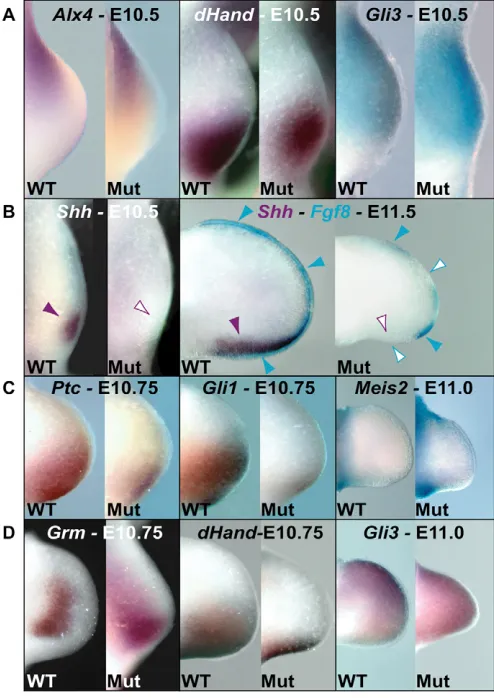
[image:3.612.52.504.58.311.2]Starting at early gestational days prior to Shhonset, a mutual genetic antagonism has been shown to exist between Gli3in the anterior limb mesenchyme and Hand2 in the posterior mesenchyme. In addition, Hand2has been shown to restrict Alx4 anteriorly (te Welscher et al., 2002a; te Welscher et al., 2002b). These genetic interactions subdivide the early limb into anterior and posterior domains. At E10.5, we found normal Alx4and Gli3 expression in Pbx1/2 mutant hindlimbs (Fig. 5A), indicating that the anterior and posterior domains of the hindlimb mesenchyme were initially preserved. Similarly, Hand2expression resembled that of Alx4and Gli3, although it was slightly reduced proximally. Finally, although marked morphological alterations were observed
Fig. 1. Abnormal morphology of Pbx1–/–;Pbx2+/–and Pbx1–/–;Pbx2–/–embryos. (A) At E10.25 (and E10.5), Pbx1–/–;Pbx2+/–embryos exhibit
abnormal morphology and appear delayed. Their forelimbs are PD shortened and AP dysmorphic, while their hindlimbs appear relatively normal compared with wild type. Pbx1–/–;Pbx2–/– embryos are severely delayed compared with wild type, exhibiting striking morphological abnormalities,
including the lack of limbs. Enlargements of all wild-type and mutant limbs are shown on the right-hand side. In addition, both wild-type and
Pbx1–/–;Pbx2+/–hindlimbs are shown at two gestational days in enlargements on the far right, whereas Pbx1–/–;Pbx2–/– limbs are shown at E11 to
demonstrate rudimentary forelimb bud formation (lower magnification). (B) At E13.5, Pbx1–/–;Pbx2+/–embryos are smaller than wild type with
D
E
V
E
LO
P
M
E
N
T
in Pbx1/2 mutant forelimbs prior to E10.5 (Fig. 1A), no gross perturbations of Alx4, Hand2and Gli3expression were detected (data not shown).
Pbx1/Pbx2 are required for normal Shhexpression
in the limb bud ZPA
Given the similarities between the distal hindlimb phenotypes of Pbx1/2 mutant and Shh–/–embryos (Chiang et al., 2001; Kraus et
al., 2001), we examined Shh expression in Pbx1/Pbx2 limbs. Strikingly, Shhwas never detected in Pbx1/2 mutant hindlimbs from E9.5 to E13.5, as represented at E10.5 and 11.5 (Fig. 5B). This absence could not be attributed to either a global Shh downregulation or to a delay in posterior embryonic development, as both Pbx1/2 mutant cloaca and notochord developed normally and displayed normal Shh expression (see Fig. S1A in the supplementary material). Interestingly, we observed low levels of
[image:4.612.52.540.252.591.2]Ptch1and Gli1(readouts of hedgehog signaling) (reviewed by Niswander, 2003) at E10.75 (Fig. 5C), suggesting that a minimal or transient wave of Shhactivity probably occurred. In contrast to Pbx1/2 mutant hindlimbs, in forelimbs Shhexpression was present, albeit at reduced levels, and Ptch1 and Gli1 were expressed (see Fig. S1B in the supplementary material), indicating that Shh absence was hindlimb specific.
We next found a relatively preserved CD44 distribution (Sherman et al., 1998) in Pbx1/2 mutant hindlimb AER, despite slight anterior reductions within smaller limbs (data not shown). This finding indicates that Pbx1/2 mutant hindlimb AER development proceeded normally, at least until E10.5, and thus was unlikely to be responsible for Shhabsence. This result was corroborated by our findings that AER-specific markers, such as Dlx6 (Robledo et al., 2002), remained unperturbed in Pbx1/2 mutant posterior hindlimbs (data not shown). Overall, these data demonstrate that the posterior AER
Fig. 2. Limb skeletal phenotypes of Pbx1–/–;Pbx2+/–(mutant) embryos. (A-D) At E12.5, Sox9expression indicates that mutant forelimb (B)
mesenchymal condensations for digits 1 (empty red arrowhead) and 5 (empty black arrowhead) are rudimentary compared with wild type (A). In mutant hindlimbs (D), digit (red empty arrowhead) and fibula condensations (empty black arrowhead) are absent compared with wild type (C) (black arrow). (E-H) At E12.5, unlike wild type (E) (red arrow), mutant scapular (F) anlagen is reduced (red arrowhead) and the humerus is dysmorphic with a hypertrophic anlagen reminiscent of a deltoid tuberosity (F) (black arrowheads). In mutant (H), the pelvis is virtually absent, except for one element (empty black arrowhead) that may be a rudimentary ischium (G) (compare with wild type, long black arrow). (I-N) At E13.5, unlike wild type (I, red arrow), mutant scapula (K; inset) is dysmorphic (red arrowhead) and the humeral head is fused to it and duplicated medially (double black arrowhead), more severely than in Pbx1–/–;Pbx2+/+mutants (J) (arrowheads). In mutant hindlimbs (N), only a rudimentary anlagen reminiscent of an ischium (open black arrowhead) remains fused to the dysmorphic femur (red arrow). This phenotype is more severe than in
D
E
V
E
LO
P
M
E
N
T
was preserved before Shhonset. Conversely at E11.5, after Shh initiation, Fgf8expression was patchy in Pbx1/2 mutant hindlimbs (Fig. 5B) and resembled that reported for Shh–/–embryos (Chiang et al., 2001; Kraus et al., 2001).
Intriguingly, normal proximal limb identity and outgrowth were observed (at E11) in Pbx1/2 mutant hindlimbs, as Meis2, a proximal mesenchymal marker (Capdevila et al., 1999), remained unchanged (Fig. 5C). Last, analyses of gremlin (Scherz et al., 2004), Hand2and Gli3(te Welscher et al., 2002a; te Welscher et al., 2002b) at later days (Fig. 5D) further confirmed Shhabsence in Pbx1/2 mutant hindlimbs, with the latter two expression patterns resembling those observed in Shh–/–(Chiang et al., 2001; Kraus et al., 2001) and
Hoxa/Hoxd-deficient (Kmita et al., 2005) embryos. Overall, these findings establish that Pbx1/Pbx2 are required in the limb mesenchyme for Shhexpression.
Pbxand Hand2probably control Shhthrough
parallel pathways; Shh and Pbx1/Pbx2do not form a cross regulatory loop
Shhabsence is intriguing, as Hand2, which has been shown to act upstream of Shh(Charite et al., 2000), remained unaltered in Pbx1/2 mutant limbs (Fig. 5A). However, we found that Pbx expression remained mostly unperturbed in Hand2–/–forelimbs, although Pbx1 was slightly broadened posteriorly (see Fig. S2A in the supplementary material). Accurate hindlimb analyses were hampered by the poor growth and posterior developmental delay in Hand2–/–embryos, as well as their lethality at E10.5 (Charite et al., 2000). Given that Hand2, Shhand Hox appear to form a regulatory loop in limb development, wherein Hand2lies up- and downstream of Shh(Zakany et al., 2004), we also examined if Pbx1/Pbx2and Shhform a similar regulatory loop. Analyses in Shh–/–embryos revealed relatively unperturbed Pbx1/Pbx2expression (see Fig. S2B in the supplementary material), despite their abnormal limb morphology and marked apoptosis in distal domains (Chiang et al., 2001).
Hox gene expression, prior to and after, onset of Shh expression is spatially perturbed or absent in
Pbx1/2 mutant limbs
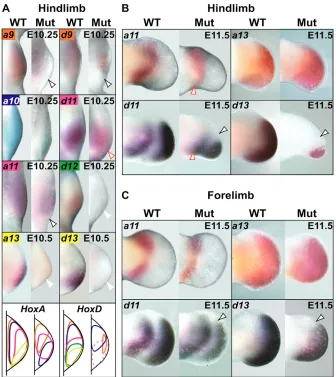
Recent findings have indicated that Hox expression in limb mesenchyme is crucial for Shhactivation (Zakany et al., 2004). Given the lack of Shhin Pbx1/2 mutant hindlimbs and its marked downregulation in forelimbs, we examined Hoxa/Hoxdexpression in compound mutant limbs. Intriguingly, all analyzed Hox genes were severely reduced or absent in future Shh-expressing domains of early Pbx1/2 mutant hindlimbs at E10.25-10.5, despite their relatively normal morphology (Fig. 6A; Hoxd10data not shown). By contrast, despite their abnormal expression, most Hox genes remained expressed in Pbx1/2 mutant forelimb ZPA (data not shown). In addition, in Pbx1/2 mutant forelimbs, multiple Hox genes were either up-regulated (e.g. Hoxa9 and Hoxd9), severely downregulated, or absent (e.g. Hoxa13and Hoxd13), suggesting a multifaceted, hierarchical control of Hox by Pbx. Finally, Hox expression was unaltered in limbs of all other compound mutant genotypes (data not shown).
[image:5.612.53.300.124.626.2]Given the suggested regulatory loop, wherein Shhregulates digit patterning via its control of autopod-specific 5⬘Hox gene reverse colinearity, Hox gene expression was also examined at E11.5. Interestingly, 5⬘ Hoxd gene expression was reduced in Pbx1/2 mutant hindlimb anterior domains, while 5⬘Hoxa gene expression remained normal (Fig. 6B; Hoxa10, Hoxd10 and Hoxd12, data not shown). This altered Hoxd gene expression pattern is similar to that reported in Shh-deficient limbs (Chiang et al., 2001). Additionally, Pbx1/2 mutant forelimbs displayed only slight anterior alterations of 5⬘ Hoxd gene, but not 5⬘ Hoxa gene, expression (Fig. 6C; Hoxa10, Hoxd10and Hoxd12, data not shown), coincident with the
Fig. 3. Pbx1and Pbx2colocalize in lateral plate-intermediate mesoderm and early limb fields. (A,E,I) At E8.5, Pbx1(A) and Pbx2
D
E
V
E
LO
P
M
E
N
[image:6.612.54.496.58.297.2]T
Fig. 4. Pbx1and Pbx2mesenchymal expression in later limb development is dynamic and, in part, complementary.(A,B) At E9.5-10.75,
Pbx1is expressed in proximal forelimbs and hindlimbs, mostly anteriorly, whereas Pbx2is expressed distally and throughout the limb. At E11-11.75,
Pbx1is still expressed proximally in limbs, but localized anteriorly and posteriorly in the mesenchyme proximal to the autopod. Similarly, Pbx2
expression becomes restricted to this domain, albeit more broadly. (C) Diagrams depicting dynamic expression of Pbx1, Pbx2and overlapping domains of Pbx1/Pbx2at different gestational days. These schemes represent summaries of the expression patterns present at the gestational time-points indicated by the somite range at the top of each panel. Proximal is leftwards, anterior towards the top. som, somites.
Fig. 5. Mesenchymal gene expression in Pbx1–/–;Pbx2+/–
(mutant) hindlimbs. (A) Expression of Alx4, Hand2and Gli3is relatively unperturbed in mutant hindlimbs, before and/or at Shh
onset, indicating that until E10.5 the mesenchyme is patterned similar to wild type. (B) Shhexpression is absent throughout mutant hindlimb development at E10.5 and E11.5. At E11.5, Shhabsence (empty purple arrowhead) in mutant hindlimbs is associated with a similar and expected alteration in Fgf8expression (empty blue arrowheads), compared with wild-type hindlimbs (normal Shh
expression indicated by solid purple arrowhead and Fgf8 by blue arrowheads). (C) Although reduced, expression of Ptch1and Gli1is present (E10.75), suggesting minimal and transient Shhactivity. Meis2
is unperturbed in mutant hindlimbs (E11.0), suggesting that proximal limbs remain relatively intact. (D) Grmis extended posterodistally in mutant hindlimbs (E10.75), indicating Shhabsence in the ZPA (Scherz et al., 2004). Hand2expression is reduced (E10.75) and Gli3
expression is diffused throughout mutant hindlimbs (E11), resembling expression patterns in mice that lack Hoxa/Hoxd (Kmita et al., 2005) and suggesting an AP patterning defect in the absence of sustained
[image:6.612.50.297.391.740.2]D
E
V
E
LO
P
M
E
N
T
observed reduction in Shhexpression. Therefore, in the context of reduced Pbx1/Pbx2dose, the mechanisms underlying the spatial regulation of Hoxa versus Hoxd expression differed during autopod development.
Hox proteins bind in vivo to, and activate transcription through, the Shhlong-range limb enhancer
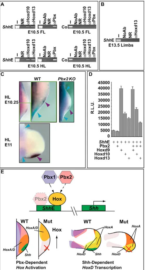
To ascertain whether Shhexpression was directly regulated by Hox and/or Pbx proteins in developing limbs, we tested for their in vivo binding to an evolutionarily conserved Shhregulatory region (ShhE), which was shown to drive the correct spatiotemporal expression of Shh within developing mouse limbs (Lettice et al., 2003). We performed chromatin immunoprecipitation (ChIP) analyses (Orlando et al., 1997) on E10.5 mouse limb cells, using specific antisera against Hoxd10, Hoxd13 and Pbx (Fig. 7A). The enrichment for genomic DNA fragments containing the ShhE sequence was verified by PCR amplification. As shown (Fig. 7A), significant enrichment was observed for Hoxd10 and anti-Hoxd13 antisera in forelimb and hindlimb immunoprecipitated chromatin, indicating that these Hoxd proteins are indeed bound to the ShhE in vivo. No enrichment was observed using a non-specific control antibody (Fig. 7A). Using an antiserum against all Pbx isoforms, Pbx protein was also found to bind in vivo to the ShhE in limb cells (Fig. 7A). The specificity of the Hoxd10, Hoxd13 and Pbx binding to the ShhE was further verified by the lack of enrichment
on a genomic region located upstream to this enhancer (Fig. 7A). Finally, as an additional control, no binding was detected for Hoxd13 to the ShhE at E13.5 when Shhexpression had ceased in limbs (Fig. 7B).
To determine which Pbx family member colocalized with Shhin limbs we performed in situ hybridization and immunohistochemistry. Notably, Pbx2 was the only Pbx that colocalized with Shhin the ZPA, as it was present throughout the limb (Fig. 4B). By contrast, two-color in situ hybridization revealed that Pbx1did not colocalize with Shh, even in the absence of Pbx2 (Fig. 7C). These results were corroborated by immunohistochemistry, whereby Pbx1 and Pbx2 proteins were detected on adjacent limb sections after Shh expression had been visualized by in situ hybridization (data not shown). Finally, we recently uncovered that Pbx3 is not expressed in hindlimbs (Di Giacomo et al., 2006) and reported findings demonstrated that Pbx4 is present only in testes (Wagner et al., 2001). In conclusion, Pbx2 is the only Pbxfamily member that colocalizes with Shh, and thus is responsible for the observed binding to the ShhE.
[image:7.612.50.386.59.436.2]The functional significance of the Hoxd10, Hoxd13 and Pbx2 binding to the ShhE was next tested in transient transfection assays using P19 embryonal carcinoma cells. A luciferase reporter construct (pT81ShhE) was generated, which contained a 745 bp fragment representing the complete ShhE (Lettice et al., 2003). Co-transfection of the ShhE reporter with Hoxd9, Hoxd10 and Hoxd13 expression constructs resulted in significant transcriptional
Fig. 6. Hox gene expression is altered in
Pbx1–/–;Pbx2+/–(mutant) limbs. (A) Hox
gene expression (E10.25-10.5) is absent or reduced in somite-matched mutant hindlimb posterior domains before, or at, Shhonset, despite their relatively normal morphology. Open black arrowheads indicate lack of posterior expression (Hoxa9, Hoxa11,
Hoxd9). Open red arrowhead indicates reduced posterior expression (Hoxd11). White arrowheads indicate absence of expression (Hoxa13, Hoxd12, Hoxd13). Diagrams depict summaries of expression data across two time-points (E10.25 and E10.5): Hox gene expression domains are represented by the same colors displayed in their respective in situ hybridization panels. Dashed domains in mutant indicate reduced
Hoxd11(pink) expression, whereas missing domains reflect complete absence of expression (Hoxa13, Hoxd12, Hoxd13). Blue domain in Hoxd diagram represents Hoxd10
expression. (B) Distal 5⬘Hoxa (Hoxa11 and
Hoxa13) expression (E11.5) remains relatively unperturbed in mutant hindlimbs, while Hoxd (Hoxd11andHoxd13) expression is severely reduced anteriorly (open black arrowheads). Open red arrowheads indicate perturbed proximal posterior expression (Hoxa11 and Hoxd11). (C) 5⬘Hoxa (Hoxa11
D
E
V
E
LO
P
M
E
N
T
activation above the reporter’s basal activity in this system (Fig. 7D). Conversely, Pbx2 co-transfection had virtually no effect on the reporter’s activity (Fig. 7D) and its co-transfection with Hoxd9, Hoxd10 or Hoxd13 did not significantly alter their activation of the reporter (Fig. 7D). These findings are consistent with the non-essential function of Pbx2 throughout embryonic development (Selleri et al., 2004). Taken together, our results show that Hoxd proteins can bind in vivo to the limb ShhE and can activate transcription through this regulatory element in cultured cells, suggesting that they directly control Shhexpression.
DISCUSSION
Pbx1/Pbx2are required for proximal and distal
limb patterning
Our results establish that, although Pbx1is required for proximal limb patterning (Selleri et al., 2001), Pbx1/Pbx2share overlapping functions in proximal and distal limb development. In this respect, the fundamental roles of Pbx1/Pbx2are underscored by our finding
that Pbx1–/–;Pbx2–/–mutants do not form limbs. Nevertheless, by decreasing Pbx2dose to one allele on a Pbx1-null background we were able to generate a mouse model where limb development could be studied in depth. Accordingly, Pbx1/2 mutant embryos died earlier in utero than Pbx1–/–embryos and had limbs with more drastic skeletal defects. Furthermore, compared with those present in Pbx1–/–embryos, axial skeletal defects were also worsened in Pbx1/2 mutant embryos (data not shown). Overall, these results establish that Pbx1/Pbx2 genetically interacts in axial and appendicular skeletal development.
[image:8.612.52.299.61.522.2]We observed that the distal Pbx1/2 mutant forelimb and hindlimb morphologies varied. In the forelimb, the ulna and most digits were present, albeit malformed, whereas in the hindlimb, the fibula and most digits were lost. Although limb-specific expression levels of Pbx1/Pbx2could explain this variability, we never detected gross differences in their expression by in situ hybridization or immunohistochemistry. Instead, the less severe distal Pbx1/2 mutant forelimb phenotype is more consistent with our finding that Pbx3is expressed only in forelimbs up to E11.5 (Di Giacomo et al., 2006). Indeed, in Pbx1–/–;Pbx2–/–mutants that survive to E11, rudimentary forelimbs, but not hindlimbs, develop (Fig. 1A), coincident with their Pbx3expression.
Fig. 7. 5⬘⬘HoxD proteins are bound to and directly activate transcription from the Shhlimb enhancer (ShhE). (A) ChIP analyses on E10.5 mouse fore- and hindlimbs, using specific anti-Hoxd10 (␣Hoxd10), anti-Hoxd13 (␣Hoxd13) and anti-Pbx (␣Pbx) antisera, demonstrate their direct in vivo binding to the mouse ShhE. A 424 bp fragment of the ShhE was amplified by PCR. No PCR amplification of a 373 bp negative-control region (Co) located 1400 bp upstream of this enhancer was detected using E10.5 limb chromatin. Representative reactions of all PCR amplifications, carried out in triplicate, are shown. (B) ChIP on pooled E13.5 limbs using the ␣Hoxd13 antiserum revealed no amplification of the ShhE. (C) At E10.25 and E11, Pbx1(blue) and
Shh(purple) mRNA transcripts do not colocalize in wild-type and
Pbx2–/–(KO) hindlimbs. (D) HoxD proteins can activate transcription from the ShhE (eightfold for Hoxd9, fourfold for Hoxd10 and threefold for Hoxd13) in P19 embryonal carcinoma cells. Luciferase activity, in arbitrary units (R.L.U.), was assayed from extracts of transiently transfected P19 cells. Co-transfection assays were performed in the presence (+) of the indicated expression vectors encoding Pbx2, Hoxd9, Hoxd10 and Hoxd13, and with a luciferase reporter construct
(pT81mShhE) containing the ShhE. Bars represent the mean±s.e.m. of at least four independent experiments. (E) Simplified model (top) depicts the overlapping genetic hierarchical roles of Pbx1and Pbx2in controlling Hox gene spatial distribution and Hox recruitment to the limb ShhE. In addition, Pbx2, albeit not functionally essential within this network, is also recruited to this enhancer in limb cells. Intensity of the hue of Pbx2 correlates with its functional relevance. Lower left diagram illustrates the requirement of Pbx1(violet), Pbx2(rose) and Pbx1/Pbx2
(purple) for controlling the positioning of 5⬘Hox genes (i.e. Hoxa/Hoxd, orange) to future Shh-expressing domain (green). In Pbx1–/–;Pbx2+/–
(mutant) limbs, Hox gene expression remains external to the ZPA, and
D
E
V
E
LO
P
M
E
N
T
Of importance, except for Pbx1–/– (Selleri et al., 2001), Pbx1–/–;Pbx2–/–and Pbx1/2mutant embryos, all other compound genotypes lacked limb defects and alterations in gene expression. These findings highlight the primary role of Pbx1and the crucial impact that Pbx1/Pbx2spatiotemporal expression patterns have on limb development. Indeed, the early colocalization of Pbx1 and Pbx2 in lateral plate and limb field mesoderm (Fig. 3I,J,L), appears crucial for distal limb development. Concomitantly, the later proximal versus distal restrictions of Pbx1and Pbx2, respectively, suggest a scenario where both genes provide a dynamic spatiotemporal ‘code’ along the entire PD limb axis.
Pbxrequirement for distal limb patterning is, at least in part, mediated by Shhcontrol
The Pbx1/2 mutant distal limb phenotype markedly resembles those observed in embryos lacking Shh(Chiang et al., 2001; Krause et al., 2001) and Hoxa/Hoxd (Kmita et al., 2005). These similarities are particularly evident in Pbx1/2 mutant hindlimbs, which initially exhibit relatively normal morphology and AP patterning (until E10.25-10.5), but subsequently lack Shh expression. Shhabsence is further supported by Gli3 posterior expansion and the severe reduction of 5⬘Hoxd expression, as also documented in Shh–/–
embryos (Chiang et al., 2001; Kraus et al., 2001). Recently, studies on the interaction of 5⬘Hoxd and Gli3 proteins (Chen et al., 2004) suggest that their ratio across the limb regulates digit pattern and identity. Therefore, digital loss in Pbx1/2 mutant hindlimbs appears mediated by Shhabsence (Fig. 7E; right panel). This exact scenario does not hold for Pbx1/2 mutant forelimbs, which already appear malformed early (E9.5-E10.5), but bear residual Shhexpression throughout development. Thus, it is not surprising that forelimbs display less severe digit malformations and/or losses. In fact, digit five absence in Pbx1/2 mutant forelimbs conforms to the expectation that the most posterior digits form under the influence of maximum and sustained Shh signaling (Ahn and Joyner, 2004; Harfe et al., 2004). Interestingly, digit one absence in Pbx1/2 mutant forelimbs probably occurs via the disruption of Shh-independent pathways, as early forelimbs display morphological abnormalities prior to Shh onset.
Our findings additionally establish that Pbx1/Pbx2hierarchically control Shhexpression and that this process may be independent of regulation of Hand2(i.e. Pbxand Hand2may control Shhthrough parallel pathways) (Charite et al., 2000). We cannot, however, rule out that Hand2 controls Pbx function at the post-transcriptional level and that, therefore, they may act on the same regulatory pathway. In light of the known regulatory interactions among Hand2and Shhin limb development, wherein Hand2lies both up- and downstream of Shh(see Zakany et al., 2004), we demonstrate that Pbx1/Pbx2are not downstream of Shhand thus that Pbx/Shhdo not crossregulate each other in limb development.
Pbx1/Pbx2hierarchical control of Hox gene spatial
distribution mediates Shhexpression in the limb As it had been reported that Shhexpression is triggered by Hox genes (Charite et al., 1994; Stratford et al., 1997; Knezevic et al., 1997; Zakany et al., 2004), we analyzed Hox gene expression prior to Shh onset in Pbx1/2 mutant limbs. In Pbx1/2 mutant hindlimbs, expression was severely perturbed for all 3⬘and 5⬘Hoxa/Hoxd genes analyzed, especially in posterior domains, while in forelimbs, the alterations varied depending on the Hoxa/Hoxd paralog. These alterations occurred in mutant limbs that exhibited relatively normal early AP patterning. Therefore, Pbx1/Pbx2act upstream of Hox genes in a manner that appears to occur independently from known
molecular regulators required for the establishment of early AP asymmetry. Furthermore, this Hox gene control occurs early, perhaps in the limb field, because both 3⬘and 5⬘Hoxa/Hoxd genes are already altered at the onset of limb development.
The mechanisms by which Pbx hierarchically control and maintain Hox genes spatial distribution in early limb mesenchyme remain elusive. Pbxrequirements in auto- and/or cross-regulatory interactions (Popperl et al., 1995; Popperl et al., 2000; Jacobs et al., 1999; Ferretti et al., 2000; Waskiewicz et al., 2002), or their cooperative roles with other transcription factors upstream of Hox genes (Berkes et al., 2004) (reviewed by Sagerstrom, 2004; Moens and Selleri, 2006) may have bearings. Indeed, with respect to the former, we cannot exclude the possibility that Pbx might directly regulate early Hox gene colinearity, given their co-expression with Hox genes in the early limb.
The absence of Shhexpression in Pbx1/2 mutant hindlimb ZPA is thus in keeping with the severe spatial perturbations of multiple Hox genes in posterior mutant hindlimbs (Fig. 7E; left panel). Conversely, in forelimbs, Shhregulation by Hox requires Pbx1/Pbx2 only in part, as the reduction in Shhexpression coincided with a less severe alteration in posterior Hox gene localization. Therefore, Hox gene localization to forelimb ZPA appears to require additional factors, with one potential candidate being Pbx3, the expression of which only in forelimbs partially overlaps with Pbx1/Pbx2in the mesenchyme (Di Giacomo et al., 2006).
We also found that several 5⬘HoxD proteins bind in vivo to the Shhlimb enhancer (Lettice et al., 2003) at a gestational stage when Shhis expressed, but not at a later stage when Shhexpression has waned. Furthermore, we found that several HoxD proteins can activate transcription through this regulatory element in cell culture. These results provide evidence in favor of the proposed direct involvement of HoxD proteins in the control of Shh expression (Zakany et al., 2004; Kmita et al., 2005) and that such control may be exerted by Hox in an overlapping or synergistic manner (Fig. 7E; top schematic).
In agreement with Pbx2 non-essential function throughout embryonic development (Selleri et al., 2004), we found that despite its colocalization with Shh and its binding in vivo to the ShhE, Pbx2 does not activate transcription or cooperate with Hox proteins in transcriptional activation through this regulatory element in our cellular readout. Therefore, Pbx2 in vivo binding to the ShhE is functionally unessential within the Pbx-Hox-Shhregulatory network (Fig. 7E; top schematic). It may, however, represent a ‘mark’ for the possible recruitment of other yet uncharacterized transcription factors, as reported in the case of the myogenin promoter (Berkes et al., 2004) (reviewed by Sagerstrom, 2004). Thus, our data indicate that the primary role of Pbx2is exerted in concert with Pbx1in the early limb field via their genetic control of Hox gene spatial expression (Fig. 7E; top schematic).
D
E
V
E
LO
P
M
E
N
T
experimental settings. Overall, we establish that Pbx1/Pbx2-dependent Hox gene spatial perturbation in the posterior limb has an equally drastic effect on Shhonset as does the disruption of early temporal Hox gene colinearity and the complete ablation of Hoxa/Hoxd gene function (Kmita et al., 2005).
Early mesenchymal interactions of Pbx1/Pbx2may
drive mouse limb outgrowth
As Hoxa/Hoxd mutants bear limb truncations (Kmita et al., 2005) and Hox genes are known to synergistically pattern all limb elements (Davis et al., 1995; Davis and Capecchi, 1996; Fromental-Ramain et al., 1996a; Fromental-Ramain et al., 1996b; Wellik and Capecchi, 2003), they must govern limb outgrowth. Similarly, as Pbx1/Pbx2 genetic interactions probably occur in early limb field mesenchyme, where they colocalize, they appear to have crucial bearings on overall limb outgrowth, especially in view of their control of Hox gene spatial distribution. Accordingly, stylopod, zeugopod and autopod elements are grossly mispatterned even at the onset of mesenchymal condensation, suggesting that early Hox-dependent patterning alterations underlie the Pbx1/2 mutant defects. Furthermore, Pbx1/Pbx2homozygous loss results in the absence of hindlimb buds, suggesting that a complete disruption of the Hox gene code occurs in early limb development. In light of the early specification model for limb patterning (Dudley et al., 2002), we suggest that interactions of Pbx genes early in outgrowth may drive specification and patterning of distal limb elements. Limb mesenchyme-specific Pbx inactivation will test this scenario.
Despite similarities to Hoxa/Hoxd and Shhmutant phenotypes, the Pbx1/2 mutant limb defects are indeed more severe. Therefore, our results do not completely negate other concomitant scenarios involving disruptions of different networks that govern limb patterning. For example, as suggested (Kmita et al., 2005), Hox genes may sustain distal limb outgrowth by ensuring proper AER function. Similarly, Pbx1/Pbx2, via control of Hox genes, may allow for the proper AER function required for Shhmaintenance. Three factors, however, argue in favor of a primary role for Pbx in Shh control that is independent of AER function: (1) the relatively normal Pbx1/2 mutant hindlimb AER morphology and observed gene expression prior to Shh onset; (2) the finding that ectopic Hox gene expression triggers Shhtranscription (Charite et al., 1994; Stratford et al., 1997; Knezevic et al., 1997; Zakany et al., 2004); and (3) the strong resemblance of the Pbx1/2 mutant hindlimb phenotype to those reported in Shh(Chiang et al., 2001; Kraus et al., 2001), as well as Hoxa/Hoxd absence (Kmita et al., 2005). However, Pbxroles in limb development may resemble those in spleen genesis, where Pbx1 hierarchically controls all known pathways required for spleen development (Brendolan et al., 2005).
Pbxroles in proximal-distal limb patterning are poorly conserved from flies to vertebrates
Evolutionarily, our findings indicate that the mechanisms of PD limb patterning are poorly conserved from insects to vertebrates. Unlike in vertebrates (Capecchi, 1997), the fly leg PD axis develops independently of Hox genes (Mann and Morata, 2000). In addition, in flies, Exdand Hth(the homolog of vertebrate Meis) are restricted to proximal limbs (Rauskolb et al., 1995; Capdevila et al., 1999; Mercader et al., 1999) and Exdis required for patterning of proximal identities (Gonzales-Crespo et al., 1998). By contrast, we demonstrate that in vertebrates distal limb patterning is also Pbx1/Pbx2 dependent. Part of this vertebrate innovation may lie in Pbx1/Pbx2interaction in early limb field mesenchyme and possibly also in later Pbx2localization to distal limb domains. In addition, in
flies, mutations in Exdresult in homeotic transformations without altering Hox gene expression, thereby indicating that Exd can act as an ancillary co-factor to Hox proteins (Peifer and Weischaus, 1990; Rauskolb et al., 1993; Rauskolb et al., 1995). Conversely, loss of Pbx1/Pbx2 causes drastic alterations in Hox gene expression, pointing to their primary role as upstream regulators of Hox genes in vertebrate limbs. This vertebrate novelty indeed has crucial bearings on Shh regulation, as well as on the origins of morphological complexity of distal appendages in tetrapods. Finally, we propose that Pbx1/Pbx2 proteins began to exert their functions differentially in hindlimbs versus forelimbs, because in vertebrates only hindlimb patterning is strictly dependent upon Pbx1/Pbx2-Hox-Shhregulation.
We thank Drs Anderson, Behringer, Boulet, Capecchi, de Crombrugghe, Duboule, Harland, Izpisua-Belmonte, Joyner, Lefebvre, Manley, McMahon, Meijlink, Milenkovic, Niswander, Popperl, Scott and Tabin for probes; Dr Mackem for anti-Hoxd13 antisera; Dr Beachy for Shh–/–mice; Dr Hadjantonakis for E8.5 embryos; Drs Barna, Mann, Martin, Niswander and Tabin for discussions and protocols; Dr R. Hogg and T. Peburn for editing; and Dr L. Lacy for constant support. T.D.C. is a graduate student in the New York Consortium in Evolutionary Primatology and a recipient of the Carole and Morton Olshen Fellowship. The work was supported by grants from the Association for the Study of Malformations and the Italian Association for Cancer Research to V.Z. and by grants from the National Institutes of Health (HD43997) and from the March of Dimes and Birth Defects Foundation (6-FY03-071) to L.S. L.S. is an Irma T. Hirschl Scholar and a recipient of an Alice Bohmfalk Charitable Trust grant.
Supplementary material
Supplementary material for this article is available at http://dev.biologists.org/cgi/content/full/133/11/2263/DC1
References
Ahn, S. and Joyner, A. L.(2004). Dynamic changes in the response of cells to positive hedgehog signaling during mouse limb patterning. Cell118, 505-516. Aparicio, O. M.(1999). Characterization of proteins bound to chromatin by
immunoprecipitation from whole-cell extracts. In Current Protocols in Molecular Biology (ed. F. M. Ausubel, R. Brent, R. E. Kingston, D. M. Moore, J. G. Seidman, J. A. Smith and K. Struhl), pp. 21.3.1-21.3.12. New York: John Wiley.
Berkes, C. A., Bergstrom, D. A., Penn, B. H., Seaver, K. J., Knoepfler, P. S. and Tapscott, S. J.(2004). Pbx marks genes for activation by MyoD indicating a role for a homeodomain protein in establishing myogenic potential. Mol. Cell14, 465-477.
Brendolan, A., Ferretti, E., Salsi, V., Moses, K., Quaggin, S., Blasi, F., Cleary, M. L. and Selleri, L.(2005). A Pbx1-dependent genetic and transcriptional network regulates spleen ontogeny. Development132, 3113-3126. Burglin, T. R.(1997). Analysis of TALE superclass homeobox genes (MEIS, PBC,
KNOX, Iroquois, TGIF) reveals a novel domain conserved between plants and animals. Nucleic Acids Res. 25, 4173-4180.
Burglin, T. R.(1998). The PBC domain contains a MEINOX domain: coevolution of Hoxand TALE homeobox genes? Dev. Genes Evol. 208, 113-116.
Capdevila, J., Tsukui, T., Rodriquez Esteban, C., Zappavigna, V. and Izpisua Belmonte, J. C.(1999). Control of vertebrate limb outgrowth by the proximal factor Meis2 and distal antagonism of BMPs by Gremlin. Mol. Cell 4, 839-849. Capecchi, M. R.(1997). Hoxgenes and mammalian development. Cold Spring
Harb. Symp. Quant. Biol. 62, 273-281.
Charite, J., de Graaff, W., Shen, S. and Deschamps, J.(1994). Ectopic expression of Hoxb-8causes duplication of the ZPA in the forelimb and homeotic transformation of axial structures. Cell78, 589-601.
Charite, J., McFadden, D. G. and Olson, E. N.(2000). The bHLH transcription factor dHand controls Sonic hedgehogexpression and establishment of the zone of polarizing activity during limb development. Development127, 2461-2470. Chen, Y., Knezevic, V., Ervin, V., Hutson, R., Ward, Y. and Mackem, S.(2004).
Direct interaction with Hoxd proteins reverses Gli3-repressor function to promote digit formation downstream of Shh. Development131, 2339-2347. Chiang, C., Litingtung, Y., Harris, M. P., Simandl, B. K., Li, Y., Beachy, P. A.
and Fallon, J. F.(2001). Manifestation of the limb prepattern: limb development in the absence of Sonic hedgehog function. Dev. Biol. 15, 421-435.
Condie, B. G. and Capecchi, M. R.(1994). Mice with targeted disruptions in the paralogous genes Hoxa-3and Hoxd-3reveal synergistic interactions. Nature 370, 304-307.
D
E
V
E
LO
P
M
E
N
T
Davis, A. P., Witte, D. P., Hsieh-Li, H. M., Potter, S. S. and Capecchi, M. R. (1995). Absence of radius and ulna in mice lacking Hoxa-11and Hoxd-11. Nature375, 791-795.
Deschamps, J. and van Nes, J.(2005). Developmental regulation of the Hox genes during axial morphogenesis in the mouse. Development132, 2931-2942. Di Giacomo, G., Koss, M., Capellini, T. D., Brendolan, A., Popperl, H. and
Selleri, L. (2006). Spatio-temporal expression of Pbx3during mouse organogenesis. Mech. Dev. Gene Exp. Patterns (in press).
Dudley, A. T., Ros, M. A. and Tabin, C. J.(2002). A re-examination of proximodistal patterning during vertebrate limb development. Nature418, 539-544.
Ferretti, E., Marshall, H., Popperl, H., Maconochie, M., Krumlauf, R. and Blasi, F.(2000). Segmental expression of Hoxb2in r4 requires two separate sites that integrate cooperative interactions between Prep1, Pbx and Hox proteins. Development127, 155-166.
Fromental-Ramain, C., Warot, X., Messadecq, N., LeMeur, M., Dolle, P. and Chambon, P.(1996a). Hoxa-13and Hoxd-13play a crucial role in the patterning of the limb autopod. Development122, 2997-3011.
Fromental-Ramain, C., Warot, X., Lakkaraju, S., Favier, B., Haack, H., Birling, C., Dierich, A., Dolle, P. and Chambon, P.(1996b). Specific and redundant functions of the paralogous Hoxa-9and Hoxd-9genes in forelimb and axial skeleton patterning. Development122, 461-472.
Gonzalez-Crespo, S., Abu-Shaar, M., Torres, M., Martinez-A, C., Mann, R. S. and Morata, G.(1998). Antagonism between extradenticlefunction and Hedgehog signaling in the developing limb. Nature394, 196-200.
Harfe, B. D., Scherz, P. J., Nissim, S., Tian, H., McMahon, A. P. and Tabin, C. J. (2004). Evidence for an expansion-based temporal Shh gradient in specifying vertebrate digit identities. Cell20, 517-528.
Jacobs, Y., Schnabel, C. A. and Cleary, M. L.(1999). Trimeric association of Hox and TALE homeodomain proteins mediates Hoxb2hindbrain enhancer activity. Mol. Cell. Biol. 19, 5134-5142.
Kmita, M., Tarchini, B., Zakany, J., Logan, M., Tabin, C. J. and Duboule, D. (2005). Early developmental arrest of mammalian limbs lacking HoxA/HoxDgene function. Nature435, 1113-1116.
Knezevic, V., De Santo, R., Schughart, K., Huffstadt, U., Chiang, C., Mahon, K. A. and Mackem, S.(1997). Hoxd-12 differentially affects preaxial and postaxial chondrogenic branches in the limb and regulates Sonic hedgehog in a positive feedback loop. Development124, 4523-4536.
Knoepfler, P. S., Bergstrom, D. A., Uetsuki, T., Dac-Korytko, I., Sun, Y. H., Wright, W. E., Tapscott, S. J. and Kamps, M. P.(1999). A conserved motif N-terminal to the DNA-binding domains of myogenic bHLH transcription factors mediates cooperative DNA binding with pbx-Meis1/Prep1. Nucleic Acids Res. 27, 3752-3761.
Kraus, P., Fraidenraich, D. and Loomis, C. A.(2001). Some distal limb structures develop in mice lacking Sonic hedgehog signaling. Mech. Dev. 100, 45-58. Krumlauf, R.(1994). Hoxgenes in vertebrate development. Cell78, 191-201. Lettice, L. A., Heaney, S. J., Purdie, L. A., Li, L., de Beer, P., Oostra, B. A.,
Goode, D., Elgar, G., Hill, R. E. and de Graaff, E.(2003). A long-range Shh enhancer regulates expression in the developing limb and fin and is associated with preaxial polydactyly. Hum. Mol. Genet. 12, 1725-1735.
Maconochie, M. K., Nonchev, S., Studer, M., Chan, S. K., Popperl, H., Sham, M. H., Mann, R. S. and Krumlauf, R.(1997). Cross-regulation in the mouse HoxB complex: the expression of Hoxb2 in rhombomere 4 is regulated by Hoxb1. Genes Dev. 11, 1885-1895.
Mann, R. S. and Chan, S. K.(1996). Extra specificity from extradenticle: the partnership between HOX and PBX/EXD homeodomain proteins. Trends Genet. 12, 258-262.
Mann, R. S. and Affolter, M.(1998). Hox proteins meet more partners. Curr. Opin. Genet. Dev. 8, 423-429.
Mann, R. S. and Morata, G.(2000). The developmental and molecular biology of genes that subdivide the body of Drosophila. Annu. Rev. Cell Dev. Biol. 16, 243-271.
McBurney, M. W. and Rogers, B. J.(1982). Isolation of male embryonal carcinoma cells and their chromosome replication patterns.Dev. Biol. 89, 503-508.
Mercader, N., Leonardo, E., Azpiazu, N., Serrano, A., Morata, G., Martinez, C. and Torres, M.(1999). Conserved regulation of proximodistal limb axis development by Meis1/Hth. Nature402, 425-429.
Moens, C. B. and Selleri, L. ( 2006). Hox cofactors in vertebrate development. Dev. Biol.291, 193-206.
Niswander, L.(2003). Pattern formation: old models out on a limb. Nat. Rev. Genet. 4, 133-143.
Orlando, V., Strutt, H. and Paro, R.(1997). Analysis of chromatin structure by in vivo formaldehyde cross-linking. Methods11, 205-214.
Pearse, R. V., 2nd and Tabin, C. J.(1998). The molecular ZPA. J. Exp. Zool. 282, 677-690.
Peifer, M. and Wieschaus, E.(1990). Mutations in the Drosophila gene extradenticleaffect the way specific homeodomain proteins regulate segmental identity. Genes Dev. 4, 1209-1223.
Popperl, H., Bienz, M., Studer, M., Chan, S. K., Aparicio, S., Brenner, S., Mann, R. S. and Krumlauf, R.(1995). Segmental expression of Hoxb-1is controlled by a highly conserved autoregulatory loop dependent upon exd/Pbx. Cell81, 1031-1042.
Popperl, H., Rikhof, H., Chang, H., Haffter, P., Kimmel, C. B. and Moens, C. B. (2000). Lazarusis a novel pbxgene that globally mediates hoxgene function in zebrafish. Mol. Cell6, 255-267.
Rauskolb, C., Peifer, M. and Weischaus, E.(1993). Extradenticle, a regulator of homeotic gene activity, is a homolog of the homoebox-containing human proto-oncogene Pbx1. Cell74, 1101-1112.
Rauskolb, C., Smith, K. M., Peifer, M. and Weischaus, E.(1995). extradenticle determines segmental identities throughout Drosophiladevelopment. Development121, 3663-3673.
Rhee, J. W., Arata, A., Selleri, L., Jacobs, Y., Arata, S., Onimaru, H. and Cleary, M. L.(2004). Pbx3 deficiency results in central hypoventilation. Am. J. Pathol. 165, 1343-1350.
Riddle, R. D., Johnson, R. L., Laufer, E. and Tabin, C. J.(1993). Sonic hedgehog mediates the polarizing activity of the ZPA. Cell75, 1401-1416.
Robledo, R. F., Rajan, L., Li, X. and Lufkin, T.(2002). The Dlx5and Dlx6 homeobox genes are essential for craniofacial, axial and appendicular skeletal development. Genes Dev. 16, 1089-1101.
Sagerstrom, C. G.(2004). Pbx marks the spot. Dev. Cell6, 737-748.
Scherz, P. J., Harfe, B. D., McMahon, A. P. and Tabin, C. J.(2004). The limb bud Shh-Fgf feedback loop is terminated by expansion of former ZPA cells. Science 305, 396-399.
Selleri, L., Depew, M. J., Jacobs, Y., Chanda, S. K., Tsang, K. Y., Cheah, K. S., Rubenstein, J. L., O’Gorman, S. and Cleary, M. L.(2001). Requirement for Pbx1in skeletal patterning and programming chondrocyte proliferation and differentiation. Development128, 3543-3557.
Selleri, L., DiMartino, J., van Deursen, J., Brendolan, A., Sanyal, M., Boon, E., Capellini, T., Smith, K. S., Rhee, J., Popperl, H. et al.(2004). The TALE homeodomain protein Pbx2 is not essential for development and long-term survival. Mol. Cell. Biol. 24, 5324-5331.
Sherman, L., Wainwright, D., Ponta, H. and Herrlich, P.(1998). A splice variant of CD44 expressed in the apical ectodermal ridge presents fibroblast growth factors to limb mesenchyme and is required for limb outgrowth. Genes Dev. 12, 1058-1071.
Stratford, T. H., Kostakopoulou, K. and Maden, M.(1997). Hoxb-8has a role in establishing early anterior-posterior polarity in chick forelimb but not hindlimb. Development 124, 4225-4234.
Sun, X., Mariani, F. V. and Martin, G. R.(2002). Functions of FGF signaling from the apical ectodermal ridge in limb development. Nature418, 501-508. Tarchini, B. and Duboule, D.(2006). Control of Hoxdgenes’ collinearity during
early limb development. Dev. Cell10, 93-103.
te Welscher, P., Zuniga, A., Kuijper, S., Drenth, T., Goedemans, H. J., Meijlink, F. and Zeller, R.(2002a). Progression of vertebrate limb development through Shh-mediated counteraction of Gli3. Science298, 827-830. te Welscher, P., Fernandez-Teran, M., Ros, M. A. and Zeller, R.(2002b).
Mutual genetic antagonism involving GLI3 and dHAND prepatterns the vertebrate limb bud mesenchyme prior to SHH signaling. Genes Dev. 16, 421-426.
Tickle, C.(2003). Patterning systems – from one end of the limb to the other. Dev. Cell4, 449-458.
Wagner, K., Mincheva, A., Korn, B., Lichter, P. and Popperl, H.(2001). Pbx4, a new Pbx family member on mouse chromosome 8, is expressed during spermatogenesis. Mech. Dev. 103, 127-131.
Waskiewicz, A. J., Rikhof, H. A. and Moens, C. B.(2002). Eliminating zebrafish pbx proteins reveals a hindbrain ground state. Dev. Cell3, 723-733.
Wellik, D. M. and Capecchi, M. R.(2003). Hox10and Hox11genes are required to globally pattern the mammalian skeleton. Science301, 363-367.
Wright, E., Hargrave, M. R., Christiansen, J., Cooper, L., Kun, J., Evans, T., Gangadharan, U., Greenfield, A. and Koopman, P.(1995). The Sry-related gene Sox9is expressed during chondrogenesis in mouse embryos. Nat. Genet. 9, 15-20.
Zakany, J., Kmita, M. and Duboule, D.(2004). A dual role for Hoxgenes in limb anterior-posterior asymmetry. Science304, 1669-1672.