Woods, DR (2017) Copeptin reflects physiological strain during thermal stress. European Journal of Applied Physiology, 118 (1). pp. 75-84. ISSN 1439-6319 DOI: https://doi.org/10.1007/s00421-017-3740-8
Link to Leeds Beckett Repository record:
http://eprints.leedsbeckett.ac.uk/4357/
Document Version: Article
Creative Commons: Attribution 4.0
The aim of the Leeds Beckett Repository is to provide open access to our research, as required by funder policies and permitted by publishers and copyright law.
The Leeds Beckett repository holds a wide range of publications, each of which has been checked for copyright and the relevant embargo period has been applied by the Research Services team.
We operate on a standard take-down policy. If you are the author or publisher of an output and you would like it removed from the repository, please contact us and we will investigate on a case-by-case basis.
Vol.:(0123456789)
1 3
DOI 10.1007/s00421-017-3740-8ORIGINAL ARTICLE
Copeptin reflects physiological strain during thermal stress
Michael John Stacey1,2 · Simon K. Delves3 · Sophie E. Britland3 ·Adrian J. Allsopp3 · Stephen J. Brett1 · Joanne L. Fallowfield3 · David R. Woods2,4
Received: 19 April 2017 / Accepted: 8 October 2017
© The Author(s) 2017. This article is an open access publication
correlated strongly with osmolality after the exposure
(r = 0.70, P = 0.004). In volunteers with maximum
Tc > 38 °C (n = 8) vs ≤ 38 °C (n = 7) there were significantly greater elevations in copeptin (10.4 vs. 2.4 pmol L−1) and
creatinine (10 vs. 2 μmol L−1), but no differences in cortisol,
free normetanephrine or osmolality.
Conclusions Changes in copeptin reflected Tc response more closely than sympathoadrenal markers or osmolal-ity. Dynamic relationships with tonicity and kidney func-tion may help to explain this finding. As a surrogate for integrated physiological strain during work in a field envi-ronment, copeptin assay could inform future measures to prevent heat-related illnesses.
Keywords Dehydration · Heat illness · Acute kidney injury · Arginine vasopressin · Normetanephrine · Cortisol
Abbreviations
ANOVA Analysis of variance
AUC-Tc37 Area under the core body temperature–time curve (baseline Tc 37 °C)
AVP Arginine vasopressin
LT38 Volunteers with maximum Tc
observed ≤ 38.0 °C
GFR Glomerular filtration rate
GPS Global positioning system
GT38 Volunteers with maximum observed
Tc > 38.0 °C
PRE Before assault exposure
POST After assault exposure
sCr Serum creatinine
SD Standard deviation
Tc Core body temperature
TcMax Maximum observed Tc
WBGT Wet bulb globe temperature
Abstract
Purpose To prevent heat-related illnesses, guidelines rec-ommend limiting core body temperature (Tc) ≤ 38 °C during thermal stress. Copeptin, a surrogate for arginine vasopres-sin secretion, could provide useful information about fluid balance, thermal strain and health risks. It was hypothesised that plasma copeptin would rise with dehydration from occupational heat stress, concurrent with sympathoadrenal activation and reduced glomerular filtration, and that these changes would reflect Tc responses.
Methods Volunteers (n = 15) were recruited from a British Army unit deployed to East Africa. During a simulated com-bat assault (3.5 h, final ambient temperature 27 °C), Tc was recorded by radiotelemetry to differentiate volunteers with maximum Tc > 38 °C versus ≤ 38 °C. Blood was sampled beforehand and afterwards, for measurement of copeptin, cortisol, free normetanephrine, osmolality and creatinine.
Results There was a significant (P < 0.05) rise in copeptin from pre- to post-assault (10.0 ± 6.3 vs. 16.7 ± 9.6 pmol L−1,
P < 0.001). Although osmolality did not increase, copeptin
Communicated by Narihiko Kondo.
* Michael John Stacey
1 Department of Surgery and Cancer, Imperial College London, Care of General Intensive Care Unit, Hammersmith Hospital Campus, Du Cane Road, London W12 0HS, UK 2 Department of Military Medicine, Royal Centre for Defence
Medicine, ICT Building, Birmingham Research Park, Vincent Drive, Edgbaston, Birmingham B15 2SQ, UK 3 Institute of Naval Medicine, Alverstoke,
Hampshire PO12 2DL, UK
Introduction
Individuals who perform strenuous work are at risk of adverse effects associated with excessive body heat (‘heat illness’), varying from physical incapacity to severe injury and death (Armstrong et al. 2007). A widely accepted defini-tion of heat stroke is as ‘a form of hyperthermia associated with a systemic inflammatory response, leading to a syn-drome of multiorgan dysfunction in which encephalopathy predominates’ (Leon and Bouchama 2015).
To minimise the incidence of heat illness, occupational guidelines recommend that core body temperature (Tc) is maintained at or below 38.0 °C. Despite these guidelines,
Tc exceeds 38.0 °C during a number of occupational activi-ties (Meade et al. 2016). There are considerable practical limitations to rectal or intra-gastric Tc monitoring and sam-pling at the tympanic membrane may be unhelpful, due to lack of uniformity of the temperature response (Yeoh et al. 2017) and the potential for hearing and thus safe working to be affected. Furthermore, the inter-individual response to thermal stress is diverse; the exact significance of core temperature in the pathophysiology of heat stroke remains debated (Noakes 2008); and evidence from civilian occu-pational settings (Garzon-Villalba et al. 2016) and military training (Wallace et al. 2005) indicates a cumulative effect of prior-day thermal stress on risk of heat-related illnesses. Thus instantaneous Tc values may incompletely reflect the risk of heat illness. A marker that integrates the physiologi-cal strain incurred during thermal stress, including but not limited to Tc response, could prove useful in prospectively identifying which individuals exposed to the same working conditions will become casualties.
The posterior pituitary hormone arginine vasopressin (AVP) classically rises in response to hypovolaemia and increasing osmolality, and is secreted as a large peptide pre-cursor ProAVP. ProAVP consists of AVP, copeptin, neuro-physin II and a signal peptide. With exertion in a warm envi-ronment, AVP has been shown to relate closely to plasma osmolality (Montain et al. 1997), which is a preferred blood marker of dehydration from thermal sweating (Cheuvront and Kenefick 2014). Thus an index of AVP secretion could usefully reflect physiological strain under thermal stress.
The assay of AVP itself is highly problematic (it is labour intensive, requires multiple pre-analytical steps, has a half-life < 30 min and is unstable even in isolated plasma), therefore recently copeptin (which is stable and can be measured on an automated analyser) has found favour as a valid and practical surrogate for AVP secre-tion (Morgenthaler 2012; Christ-Crain and Fenske 2016). Copeptin secretion occurs in response to various stressors (Christ-Crain and Fenske 2016; Katan et al. 2008a, b) and its concentration in peripheral blood has been proposed as a sensitive marker of the individual stress level (Katan
et al. 2008a). In healthy subjects performing a high alti-tude trek, plasma concentrations of copeptin and AVP associated closely and the correlation was strengthened (r = 0.834, p < 0.001) at the highest and most stressful measurement point (Mellor et al. 2015). In disease, copep-tin has been found to be of prognostic value in diverse conditions such as sepsis (Seligman et al. 2008), septic and haemorrhagic shock (Morgenthaler et al. 2007), lower respiratory tract infections (Müller et al. 2007), cardio- and cerebrovascular disease (Sun et al. 2015; Stoiser et al. 2006; Marston et al. 2016; Greisenegger et al. 2015) and for end stage renal failure, coronary heart disease and all-cause mortality in patients with type 1 diabetes (Velho et al. 2016).
One important factor that may contribute to AVP/ copeptin release is body temperature. Enhanced secretion of AVP with increasing tissue temperature has been dem-onstrated both in resting human subjects (Takamata et al. 1995a) and in hypothalamic-pituitary explants warmed from 37.0 to 39.5 °C (Sladek and Johnson 2013), whereby the rise in AVP was additive with that from increasing osmolality of plasma/perfusate. The effect of Tc on copep-tin concentration is yet to be investigated, however, and relationships of copeptin with other parameters of physi-ological strain under thermal stress have not been charac-terised. We hypothesised that plasma copeptin would rise with dehydration from occupational heat stress, concur-rent with serum osmolality and creatinine (sCr), and that changes in copeptin would reflect Tc response. As a Tc of 38.0 °C marks an important thermoregulatory threshold during exertional heat stress (Commission for Thermal Physiology 1987) above which vasomotor compensation plateaus (Gonzalez-Alonso et al. 1999) and sensitive con-trol of Tc is lost (Wyndham et al. 1965) we chose a Tc
of 38.0 °C as a threshold for the initial investigation of copeptin in response to thermal stress. As sympathoadre-nal activation is augmented by Tc rise (Rhind et al. 1999), we additionally investigated changes in cortisol and free normetanephrine levels as alternative markers of physi-ological strain under heat stress.
Methods
Ethical approval
1 3
Study design
Preliminary measures
Volunteers were recruited to the study from a British Army infantry battalion who took part in an overseas military exercise in northern Kenya (East Africa). A total of fifteen volunteers completed study measures in the deployed field environment.
Prior to departure, volunteers underwent baseline meas-urements at their home duty station in the UK. A digital scale and stadiometer (SECA Ltd, Leicester, UK) were used to determine body mass, to the nearest 0.01 kg, and height, to the nearest 0.1 cm, while wearing the clothing ensemble to be worn in Kenya, minus boots. Body fat as a percentage of body composition was determined by segmental multi-frequency bioimpedance (Tanita UK Ltd., Yiewsley, Mid-dlesex, UK).
Deployed measures
The volunteers deployed to Kenya 3 weeks after baseline measurements, where they undertook low-level activi-ties in cooler upland environs. Environmental conditions were monitored locally using a wet bulb globe temperature (WBGT) monitor (Grant, Cambridge, UK) throughout. Average WBGT was 16 °C during this initial phase, which lasted for 3 weeks. They then re-located to a warm lowland environment to take part in simulated combat assaults con-ducted over varying terrain while on foot, with live firing of munitions. Volunteers wore full combat dress and carried an equipment load of 38.9 ± 5.0 kg. The WBGT during this phase was 26 ± 5 °C.
Deployed study measures were made in relation to a simulated combat assault on the morning of the sixth day of training in the warm environment, 6 weeks following the UK measures. Volunteers rose from overnight rest-ing at 2.00 a.m. local time and underwent study measures in a 1 h sampling window (2.30 a.m.–3.30 a.m.). They were weighed by digital scale, to the nearest 0.01 kg (SECA Ltd, Leicester, UK), provided pre-assault (PRE) blood samples and had a plastic sweat-collection pouch applied to the left upper arm. Volunteers were then trans-ported by vehicle to the training area, where they rested (5.00 a.m.–7.00 a.m.) until moving into position for the start of the assault (7.00 a.m.–7.30 a.m.). The field assault lasted for 3.5 h, from 8.00 a.m. to 11.30 a.m. It consisted of a staggered advance on foot into a series of ‘enemy’ defensive positions in a mock-up village, with intermittent static periods of co-ordinated weapon firing, re-grouping and re-supply (including drinking water). Post-assault (POST) blood samples were drawn after the conclusion of the assault, between 30 and 60 min following cessation
of strenuous exertion (12.00 p.m.–12.30 p.m.). Where still intact, sweat was sampled from the collection pouch. Vol-unteers were allowed to drink water only, from rising until the completion of POST measures (2.00 a.m.–12.30 p.m.).
For Tc monitoring, each volunteer was provided with two telemetric pills (VitalSense, Mini Mitter Company Inc, Oregan, USA) to ingest 12 and 2 h before the field assault (6.00 p.m. and 6.00 a.m.). This was with the inten-tion of using measurements from the pill that had been in the gastrointestinal tract for the longest period, in order to reduce confounding from the ingestion of water dur-ing the assault, while allowdur-ing for the possibility that the first ingested pill could be excreted before the end of the monitoring period. These transmitted wirelessly to a receiver (VitalSense, Mini Mitter Company Inc, Oregan, USA) carried on the volunteer’s person, which logged Tc
locally every 60 s during the field assault. A member of each sub-unit within the group (n = 4) carried a Global Positioning System (GPS) (Sportslog, UK), which also logged locally every 60 s.
Biochemical sampling and processing
Blood was obtained by venesection in rested volunteers in a seated position from an antecubital fossa vein. Blood was drawn into in a serum separator tube (SST) (cortisol, osmolality and creatinine) and EDTA tube (plasma copep-tin and free normetanephrine). Samples were stored in ice and centrifuged within 1 h. With sweat samples, they were then immediately frozen to −20 °C and maintained at this temperature during transportation back to the UK, where they were stored at −80 °C until analysis. All measurements were performed in duplicate. Copeptin was assayed using an automated sandwich immunofluorescent assay based on TRACE technology (Brahms CT-proAVP Kryptor Compact Plus, Hennigsdorf, Germany). This assay has a coefficient of variation (CV) of 2.5–3.7% and a lower limit of detection of 0.9 pmol L−1. Cortisol was measured using the Roche
Elecsys Cortisol I assay, an automated competitive chemilu-minescence immunoassay (CLIA), using the Roche modular
E unit (Roche Diagnostics, Burgess Hill, UK). The analytical range of the assay is 0.5–1750 nmol L−1 and the intra-assay
CV is 9.3–11.7%. Free normetanephrine was measured using an in-house liquid chromatography/tandem mass spectrom-etry method (CV 4–12%). Osmolality was measured using a depression of freezing point method (Advanced® Model
Sample size
A power calculation was not performed prospectively as the sample size was dictated by the availability of equipment and personnel during a busy infantry exercise in a remote environment, in the face of significant logistic challenges. It was noted, however, that Morgenthaler et al. (2006) showed a significant, near two-fold increase in copeptin concentra-tions in a sample of 12 volunteers performing bicycle exer-cise (ambient conditions and Tc response not specified) and that significant elevations in AVP and osmolality were dem-onstrated in ten dehydrated subjects performing treadmill-walking while deprived of fluid intake over 90 min in the heat (Maresh et al. 2004). No rise in osmolality was shown when same ten subjects performed the protocol in the euhy-drated state while taking water ad libitum. Thus a minimum sample size of 12 volunteers was considered adequate to mitigate the occurrence of type 2 statistical errors in respect of changes in copeptin and osmolality.
Statistical methods
Statistical calculations were performed using the software package GraphPad Prism (GraphPad Prism version 5.01 for Windows, GraphPad Software, San Diego California USA). Parametric or non-parametric statistical tests were applied after exploring the data for normality using the Shapiro–Wilk test. Changes in two-group paired continu-ous data before and after the assault were analysed by Stu-dent’s paired t test or Wilcoxon signed ranks test for para-metric and non-parapara-metric data, respectively. For unpaired two-group comparisons of parametric and non-parametric data an unpaired t test and a Mann–Whitney test were used, respectively. Relationships between copeptin, osmolality and normetanephrine responses were assessed using Pearson’s (parametric data) and Spearman’s (non-parametric data) coefficients. For the reasons outlined above, Tc of 38.0 °C is a thermal threshold with physiological and occupational relevance, so an exploratory analysis was performed for the effects of the field assault (PRE to POST) against Tc
group using two-way repeated measures analysis of variance (ANOVA). Sidak’s multiple comparisons test was applied where appropriate. Individuals were assigned to Tc group according to whether the maximum temperature attained during the assault was > 38.0 °C (GT38) or ≤ 38.0 °C (LT38). The effect of Tc group on each biochemical param-eter was expressed as Cohen’s d = (mean changeGT38 −mean
changeLT38)/pooled standard deviation (SD), where pooled
SD = √((SDGT382 + SD
LT382)/2). Thermal
(tempera-ture–time) curves were also plotted, taking a normal resting
Tc of 37 °C as the baseline above which the area under the curve (AUC-Tc37) was calculated. Data are presented as mean ± SD, except where specified. Data on Tc response are
provided for the telemetric pill that had been in situ for the longest period.
Results
Volunteer characteristics
Pre-deployment (UK) measures were as follows: age 25 ± 5 years; height 1.79 ± 0.07 m; weight 77.5 ± 8.9 kg; body mass index 25.3 ± 2.1 kg m2. body fat 16.2 ± 3.3%.
Assault exposure
The ambient (dry bulb) temperature measured local to the training area increased from 20 to 27 °C during the hours of the simulated combat assault. The median distance covered from GPS measurements was 1781 (range 585–2265) m. The median moving speed was 1.3 km h−1 (range 0–5.8 km h−1).
Core temperature
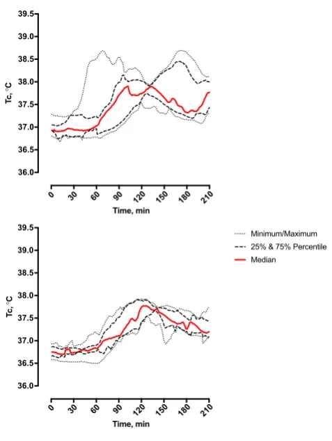
Figure 1 presents the group Tc response before, during and after the field assault. Group Tc ranged from a mini-mum value (TcMin) of 36.8 ± 0.2 °C to a maximum value (TcMax) of 38.1 ± 0.4 °C, a rise of 1.3 °C (95% CI 1.2–1.5 °C,
P < 0.0001). The highest individual Tc recorded was 38.7 °C and the greatest individual rise in Tc was 1.9 °C. Eight vol-unteers attained TcMax ≥ 38.0 °C, spending 48 ± 20 min at or above this threshold, whereas Tc remained ≤ 38.0 °C in seven volunteers. The rise in Tc for GT38 (1.5 ± 0.3 °C) was higher compared with the LT38 group (1.2 ± 0.1 °C, 95% CI 0.1–0.6 °C for the difference in maximal Tc change between GT38 and LT38).
Body mass
Clothed body mass decreased from 77.1 ± 8.2 kg PRE to 75.9 ± 8.1 kg POST (95% CI −1.69 to −0.66 kg for the difference in clothed body mass). In GT38 vs. LT38, the change in clothed body mass did not differ in either abso-lute (−1.4 ± 1.0 kg vs. −0.9 ± 0.8) or relative (−1.8 ± 1.4 vs. −1.2 ± 1.1%) terms.
Biochemical results
Between PRE and POST, there was a rise in copeptin (10.0 ± 6.3 vs. 16.7 ± 9.6 pmol L−1, P < 0.001),
1 3
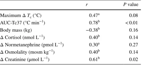
An association between copeptin and osmolality PRE (r = 0.46, P = 0.08) was strengthened POST (r = 0.70,
P < 0.01; Fig. 2). The change in copeptin from PRE to POST (Δ copeptin) correlated positively with Δ creatinine,
whereas its showed no significant association with Δ cor-tisol, Δ normetanephrine or Δ osmolality. There was also a strong correlation between Δ copeptin and AUC-Tc37 (r = 0.78, P < 0.01; Fig. 2) but no significant association between Δ copeptin and Δ body mass (Table 1).
From two-way ANOVA for repeated measures (Time by
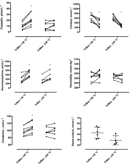
Tc), significant interactions existed for changes in copeptin (interaction F = 13.09, P < 0.05) and sCr (F = 9.7, P < 0.01), but not for cortisol, normetanephrine or osmolality (Fig. 3). For GT38 only, significant changes were observed for Δ copeptin (GT38 vs. LT38: 10.4 vs. 2.4 pmol L−1, 95% CI of
difference 6.6 to 14.2 vs. −1.7 to 6.5 pmol L−1) and Δ sCr
(GT38 vs. LT38: 10 vs. 2 μmol L−1, 95% CI of difference 6
to 15 vs. −3 to 6 μmol L−1). This represented effect sizes of
1.9 and 1.6 for higher Tc group (GT38 vs. LT38) on copeptin and sCr, respectively. In contrast, normetanephrine increased in both groups (GT38 vs. LT38: 275 vs. 169 pmol L−1, 95%
CI of difference 167–383 vs. 54–285 pmol L−1, effect size
0.9), cortisol decreased in both groups (GT38 vs. LT38: −247 vs. −354 nmol L−1, 95% CI of difference −357 to
−136 vs. −472 to −236 nmol L−1, effect size 0.9) and
osmolality did not change in either group (GT38 vs. LT38: 2 vs. −1 mosm kg−1, 95% CI of difference −2 to 6 vs. −5 to
3 mosm kg−1, effect size 0.7). In those in whom sweat
sam-ples were available, GT38 (n = 6) vs. LT38 (n = 4) tended to have a higher sweat sodium but this did not reach statistical significance (42.5 vs. 28.8 mmol L−1, 95% CI of difference
28.1 to −0.6 mmol L−1, effect size 1.5).
Discussion
This is the first study to investigate relationships between copeptin and Tc responses. The principal finding of the study was that both copeptin and sCr discriminated groups by 0 30 60 90 120 150 180 210
36.0 36.5 37.0 37.5 38.0 38.5 39.0 39.5
Time, min
Tc
,
°
C
0 30 60 90 120 150 180 210
36.0 36.5 37.0 37.5 38.0 38.5 39.0 39.5
Time, min
Tc
,
°
C
Minimum/Maximum 25% & 75% Percentile Median
Fig. 1 Core temperature (Tc) response recorded during simulated
field assault, 8.00 a.m.–11.30 a.m. Top: 8 volunteers with maxi-mum core temperature (TcMax) greater than 38 °C during the assault (GT38). Bottom: 7 volunteers with TcMax no greater than 38 °C (LT38)
0 50 100 150 200
-10 0 10 20
AUC-Tc37, °C.min
Copeptin, pmol.
L
-1 r=0.78, P=0.0007
280 285 290 295 300
0 10 20 30 40
Osmolality, mosm.kg-1
Copeptin, pmol.L
-1
POST, r =0.70, P=0.004 PRE, r=0.46, P=0.08
Fig. 2 Relationships of copeptin to osmolality (left: measured before, PRE, and after, POST, exposure) and area under the core body
[image:6.595.51.290.51.368.2] [image:6.595.85.509.518.682.2]TcMax, showing significant changes for GT38 but not LT38. Despite a rise in normetanephrine across the assault expo-sure and evidence of a strong relationship between copeptin and osmolality, changes in cortisol, normetanephrine and osmolality did not reflect TcMax as well as copeptin and sCr. As described in the introduction, copeptin has been found to be of prognostic value in various conditions such as sepsis (Seligman et al. 2008), septic and haemorrhagic shock (Mor-genthaler et al. 2007), in lower respiratory tract infections (Müller et al. 2007), heart failure (Stoiser et al. 2006) and myocardial infarction (Marston et al. 2016). We report for the first time an initial association between increased copep-tin and higher physiological strain under thermal stress.
During submaximal exercise in the heat, osmolality is known to increase in proportion to the reduction in total body water from evaporative sweating. This has been shown to impair mechanisms involved in the dissipation of body heat, even in the presence of high aerobic fitness or advanced acclimatisation status (Cadarette et al. 1984; Fortney et al. 1984; Sawka et al. 1983). Osmolality and AVP couple closely following submaximal exercise in a hot laboratory environment—despite varying hydration, exercise intensity and Tc response (Montain et al. 1997)—and before and after prolonged submaximal running in a hot field environment (Mudambo et al. 1997). Significant relationships have also been reported between copeptin and osmolality, both at rest (Szinnai et al. 2007) and after prolonged endurance running (Bracher et al. 2012). In the present study, the strong asso-ciation between copeptin and osmolality POST (r = 0.70,
P = 0.004) indicated a persistent relationship between AVP and osmolality in the warm field environment.
Although threshold osmolality for AVP release shows marked inter-individual variation and may rarely exceed 290 mosm kg−1 H
2O, it is expected that osmotic stimulus
to copeptin release existed in the majority of volunteers
during the assault. Comparison of the regression lines for copeptin and osmolality (PRE vs. POST) however sug-gested a change in the nature of the relationship between copeptin and osmolality over the course of the expo-sure. The increase in copeptin was 0.83 pmol L−1 per
unit increase in osmolality PRE, but almost doubled to 1.64 pmol L−1 per mosm kg−1 H
2O POST. The PRE
rela-tionship was not significant but did represent an important tendency (r = 0.46, P = 0.08, n = 15), where it is specu-lated that rise in Tc increased the sensitivity for osmotic release of AVP/copeptin from PRE to POST. This would be consistent with laboratory observations of the effect of passive heating upon the relationship between AVP and osmolality (Takamata et al. 1995), whereby Tc rise of up to 1.0 °C progressively augmented AVP secretion above a threshold osmolality of 290 mosm kg−1. This
mecha-nism could also account for increases in copeptin without significant elevation in osmolality observed for the GT38 group. The relationship of copeptin response to Tc excur-sion is further supported by the strong correlation shown between Δ copeptin and AUC-Tc37 (r = 0.78, P = 0.0007).
The finding that sweat sodium tended to be higher in GT38 argues against osmotic stimulation being the only significant driver of copeptin response in this study, as rela-tively higher sweat tonicity would diminish the rise in serum osmolality from hypotonic sweat losses. Rather, our results confirm the advancing acclimatisation status of the volun-teers—with group sweat sodium measures being in the lower part of the expected range—and raise the possibility that copeptin responses may have reflected differences in the rate of heat adaptation between GT38 and LT38, with LT38 hav-ing lesser copeptin response while appearhav-ing better adapted both by Tc and sweat sodium concentration.
An alternate or complementary explanation for the reported findings is that true ‘non-osmotic’ secretion of AVP and copeptin occurred. When Maresh et al. (2004) assessed the effect of drinking ad libitum upon osmolality and AVP responses during low-intensity exercise in a warm laboratory environment, significant AVP release was evi-dent despite unchanged osmolality (~ 287 mosm kg−1 H
2O)
and only 1% loss of body mass across a 90 min exposure. It was suggested that non-osmotic peripheral secretion of AVP occurred secondary to activation of the sympathetic nervous system. Mellor et al. (2015) showed a rise in copep-tin with exercise under progressively more stressful condi-tions during a high altitude trek. As for AVP this occurred independently of osmolality, but whereas increasing altitude (and physiological stress) was shown to have an effect on copeptin response, the corollary was not significant for AVP. This disparity may have reflected known difficulties in the assay of AVP; it is also possible that copeptin better reflected non-osmotic stimuli, including headache, nausea, anxiety and stress from exertion in conditions of hypobaric hypoxia.
Table 1 Association of Δ copeptin (PRE to POST) with
correspond-ing changes in other parameters among 15 volunteers undergocorrespond-ing simulated field assault
Tc core temperature, AUC-Tc37 area under the curve (temperature– time), baseline Tc = 37 °C
a Spearman r b Pearson r
r P value
Maximum Δ Tc (°C) 0.47a 0.08 AUC-Tc37 (°C min−1) 0.78b < 0.01
Body mass (kg) −0.38b 0.16
[image:7.595.51.291.91.205.2]1 3
In the present study, assay of plasma free norme-tanephrine was undertaken to provide information about changes in sympathetic outflow and norepinephrine metab-olism (Deutschbein et al. 2010; Woods et al. 2017). As a means of assessing sympathoadrenal activation during
exercise in field conditions, this approach may have avoided certain pitfalls associated with norepinephrine measurement, relating to changes in posture or stress stimuli proximal to collection. Whereas an interaction between time and Tc
group existed for copeptin responses, no such relationship
TcMax >38 °C TcMax
<38 °C 280
285 290 295 300 305 310
O
smolalit
y,
mosm.k
g
-1
TcMax >38 °C TcMax <38 °C
0 200 400 600 800 1000 1200
Normetanephrine, pmol.L
-1
* *
TcMax >38 °C TcMax
<38 °C 60
80 100 120 140
C
reatinine,
mol.L
-1 *
TcMax >38 °C TcMax
<38 °C
0 10 20 30 40 50
C
opeptin, pmol.L
-1 *
TcMax >38 °C TcMax <38 °C
0 200 400 600 800 1000 1200
Cortisol, nmol.L
-1
* *
TcMax >38 °C TcMax
<38 °C
20 30 40 50 60 70
Sweat sodium, mmol.
L
-1
Fig. 3 Responses in GT38 and LT38 groups, for plasma
copep-tin, serum cortisol, plasma normetanephrine, serum osmolality and serum creatinine (before, PRE, and after, POST, field assault) and
[image:8.595.86.513.53.597.2]was demonstrated for the normetanephrine or cortisol during the exposure. This finding would favour a genuine enhanced secretion of AVP/copeptin in consequence of greater relative thermal strain, rather than from more general stress asso-ciated with sympathoadrenal stimulation. Indeed, while copeptin has been found to rise after prolonged endurance exercise (over 160 km endurance races) (Hew-Butler et al. 2011) the GPS and cortisol data show that the exercise undertaken in our study was far from intense, and unlikely to explain the changes seen. And despite various other physi-cal discomforts and psychologiphysi-cal stressors that might be expected to serve as non-osmotic stimuli during simulated field combat, a clear interaction between training and Tc
group was evident. These observations strengthen the sug-gestion that copeptin may be of use in characterising physi-ological strain under heat stress in particular and that the relationship warrants further investigation.
In addition to Tc rise, thermal strain encompasses the cardiovascular thermo-effector responses to exertion in a warm environment, which must meet the competing needs of increased blood supply to exercising muscle (metabolic demand) and skin (thermoregulation). These changes occur at the cost of blood flow to the kidneys and acute kidney injury is a well-recognised complication of severe heat ill-ness (Leon and Bouchama 2015). The rise in sCr demon-strated by GT38 may reflect greater thermoregulatory strain relative to LT38, and it is possible that reduced renal blood flow and glomerular filtration rate (GFR) contributed to the differential copeptin response between the two groups. The robust correlation between copeptin and sCr supports this alternate hypothesis. Interestingly, recent work using the synthetic analogue of AVP, desmopressin, has also sug-gested a potential role for AVP in inducing chronic kidney disease following recurrent heat stress in mice (Roncal-Jimenez et al. 2016). It is also therefore possible that what we have seen in association with GT38 could reflect a patho-physiological mechanism to cause impaired kidney function in humans with thermal stress. Chronic kidney disease is epidemic among workers subject to dehydration in hot agri-cultural environments (Bodin et al. 2016), with one recent investigation showing an increased risk of acute kidney injury from higher thermal strain during shifts in the field (Moyce et al. 2017). The mean maximum Tc in that larger sample was 38 °C.
While copeptin measurement presently requires the use of laboratory-based assay platforms, no more than 50 μL of sample is required, no extraction step is needed and results can be ready in as little as 30 min (Morgenthaler 2012). This suggests that point-of-care assay will be feasible in the near future. In the meantime, the stability of the molecule (stable for ≥ 7 days at room temperature) reduces the logis-tics required for sample transfer from field/working environ-ments to the laboratory, such that copeptin assay could be
used to further investigate cumulative physiological strain in relation to next-day working practices and prevention of heat-related illnesses.
We acknowledge several limitations to our study. In the austere environment in which it was conducted we were unable to accurately measure changes in plasma volume. In addition to increased production and impaired clearance, a reduction in plasma volume from pre to post could further account for elevated plasma copeptin and sCr. However, it would seem unlikely to explain the 400–500% differences in copeptin and sCr responses between GT38 and LT38. In addition, the temporal relationships of the reported biochem-ical parameters to other aspects of the field assault should be addressed. In order to undertake study measures in a field setting with the necessary standardisation and control, the investigators co-located with volunteers during the ‘live-fir-ing’ phase of a busy infantry training exercise. Geographi-cal and time constraints necessitated pre sampling being completed 5 h before the start of the field assault, though volunteers did not engage in strenuous physical exertion and were exposed to much cooler environmental temperatures in the intervening period (3.00 a.m.–8.00 a.m.), such that peak Tc remained below the level observed during the field assault. The advantage of sampling volunteers shortly after rising was that pre results could be regarded as true ‘steady state’ values, which would otherwise be impossible to obtain during a military deployment of this kind. The subsequent change in copeptin reflected the stressors of the intervening period rather than circadian rhythmicity, as copeptin does not synchronise with the light–dark cycle in healthy humans (Darzy et al. 2010). While we report greater rises in copeptin in GT38 vs. LT38, suggesting that copeptin reflects thermal strain, there were no cases of heat illness in our cohort. As the participants followed an appropriate period of acclima-tisation and exertion levels were modest this is as expected. Further, it would have been unethical to specifically aim to induce heat casualties. Cardiovascular strain—which may also contribute to collapse during thermal stress—was not measured directly and further work is required to determine how this and other relevant factors, such as inflammatory responses, relate to copeptin and whether a single measure-ment of copeptin might have diagnostic/prognostic value at higher levels of heat strain and in heat illness cases.
In conclusion, the change in copeptin reflected Tc
1 3
global physiological strain and possibly also pathophysio-logical mechanisms contributing to heat-related morbidity. A point-of-care assay for copeptin would afford opportunity for in-field evaluation of a potential marker of integrated physiological strain alongside more conventional thermal stress monitoring. Further work is required to delineate the copeptin response to greater thermal stress both in health and ultimately in heat-related illnesses, as well as the effects of hydration and acclimatisation status.
Acknowledgements The following organisations and individuals
are acknowledged for their support: the study volunteers and other enabling elements of 3rd Battalion The Parachute Regiment; Surgeon General’s Research Steering Committee and Joint Medical Command; the Army Recruiting and Training Division and Collective Training Group, including enabling elements of the British Army Training Unit Kenya; the Army Directorate of Operations and Commitments; the staff of the Biochemistry Department at the Royal Victoria Infirmary, Newcastle-upon-Tyne.
This work was supported by the National Institute for Health Research (NIHR) Comprehensive Biomedical Research Centre at Imperial College Healthcare NHS Trust and Imperial College Lon-don. The views expressed are those of the authors and not necessarily those of the NIHR, the National Health Service or the UK Department of Health.
Author contributions MS, SD and SB collected the data; all authors
contributed to the conception of the study, analysis of the data and writing of the manuscript.
Compliance with ethical standards
Conflict of interest MS, SK, SB, AA, SB, JF and DW have nothing
to declare.
Ethical approval All procedures performed in studies involving
human participants were in accordance with the ethical standards of the institutional and/or national research committee and with the 1964 Helsinki declaration and its later amendments or comparable ethical standards.
Informed consent All volunteers gave written informed consent.
Open Access This article is distributed under the terms of the
Creative Commons Attribution 4.0 International License ( http://crea-tivecommons.org/licenses/by/4.0/), which permits unrestricted use, distribution, and reproduction in any medium, provided you give appro-priate credit to the original author(s) and the source, provide a link to the Creative Commons license, and indicate if changes were made.
References
Armstrong LE, Casa DJ, Millard-Stafford M, Moran DS, Pyne SW, Roberts WO (2007) American College of Sports Medicine posi-tion stand. Exerposi-tional heat illness during training and competiposi-tion. Med Sci Sports Exerc 39:556–572
Bodin T, Garcia-Trabanino R, Weiss I et al (2016) Intervention to reduce heat stress and improve efficiency among sugarcane work-ers in El Salvador: phase 1. Occup Environ Med 73:409–416 Bracher A, Knechtle B, Gnadinger M et al (2012) Fluid intake and
changes in limb volumes in male ultra-marathoners: does fluid overload lead to peripheral oedema? Eur J Appl Physiol 112:991–1003
Cadarette BS, Sawka MN, Toner MM et al (1984) Aerobic fitness and the hypohydration response to exercise-heat stress. Aviat Space Environ Med 55:507–512
Cheuvront SN, Kenefick RW (2014) Dehydration: physiology, assess-ment, and performance effects. Compr Physiol 4(1):257–285 Christ-Crain M, Fenske W (2016) Copeptin in the diagnosis of
vaso-pressin-dependent disorders of fluid homeostasis. Nat Rev Endo-crinol 12:168–176
Commission for Thermal Physiology of the International Union of Physiological Sciences (1987). Glossary of thermal physiology Pflügers. Arch 410:567–587
Darzy KH, Dixit KC, Shalet SM et al (2010) Circadian secretion pat-tern of copeptin, the C-terminal vasopressin precursor fragment. Clin Chem 56:1190–1191
Deutschbein T, Unger N, Jaeger A et al (2010) Influence of various confounding variables and storage conditions on metanephrine and normetanephrine levels in plasma. Clin Endocrinol 73:153–160
Fortney SM, Wenger CB, Bove JR et al (1984) Effect of hyperos-molality on control of blood flow and sweating. J Appl Physiol 57:1688–16895
Garzon-Villalba XP, Mbah A, Wu Y, Hiles M, Moore H, Schwartz SW, Bernard TE (2016) Exertional heat illness and acute injury related to ambient wet bulb globe temperature. Am J Ind Med 59:1169–1176
Gonzalez-Alonso J, Teller C, Andersen SL et al (1999) Influence of body temperature on the development of fatigue during pro-longed exercise in the heat. J Appl Physiol 86:1032–1039 Greisenegger S, Segal HC, Burgess AI et al (2015) Copeptin and
long-term risk of recurrent vascular events after transient ischemic attack and ischemic stroke: population-based study. Stroke 46:3117–3123
Hew-Butler T, Hoffman MD, Stuemple KJ et al (2011) Changes in copeptin and bioactive vasopressin in runners with and without hyponatraemia. Clin J Sport Med 21:211–217
Katan M, Morgenthaler N, Widmer I et al (2008a) Copeptin, a stable peptide derived from the vasopressin precursor, correlates with the individual stress level. Neuroendocrino Lett 29:341–346 Katan M, Müller B, Christ-Crain M (2008b) Copeptin: a new and
promising diagnostic and prognostic marker. Crit Care 12:117 Leon LR, Bouchama A (2015) Heat stroke. Compr Physiol
5:611–647
Maresh CM, Gabaree-Boulant CL, Armstrong LE et al (2004) Effect of hydration status on thirst, drinking and related hormonal responses during low-intensity exercise in the heat. J Appl Physiol 97:39–44 Marston NA, Shah KS, Mueller C et al (2016) Serial sampling of
copeptin levels improves diagnosis and risk stratification in patients presenting with chest pain: results from the CHOPIN trial. Emerg Med J 33:23–29
Meade RD, Poirier MP, Flouris AD, Hardcastle SG, Kenny GP (2016) Do the threshold limit values for work in hot conditions ade-quately protect workers? Med Sci Sports Exerc 48:1187–1196 Mellor AJ, Boos CJ, Ball S, Burnett A, Pattman S, Redpath M, Woods
DR (2015) Copeptin and arginine vasopressin at high altitude: relationship to plasma osmolality and perceived exertion. Eur J Appl Physiol 115:91–98
Morgenthaler N (2012) Copeptin—the steady and reliable copilot of vasopressin. Regul Pept 177:S11–S12
Morgenthaler NG, Struck J, Alonso C, Bergmann A (2006) Assay for the measurement of copeptin, a stable peptide derived from the precursor of vasopressin. Clin Chem 52:112–119
Morgenthaler NG et al (2007) Copeptin, a stable peptide of the argi-nine vasopressin precursor, is elevated in hemorrhagic and septic shock. Shock 28:219–226
Moyce S, Mitchell D, Armitage T et al (2017) Heat strain, volume depletion and kidney function in California agricultural workers. Occup Environ Med. doi:10.1136/oemed-2016-103848
Mudambo KSMT, Coutie W, Rennie MJ (1997) Plasma arginine vas-opressin, atrial natriuretic peptide and brain natriuretic peptide responses to long-term field training in the heat: effects of fluid ingestion. Eur J Appl Physiol 75:219–225
Müller B, Morgenthaler N, Stolz D et al (2007) Circulating levels of copeptin, a novel biomarker, in lower respiratory tract infections. Eur J Clin Investig 37:145–152
Noakes TD (2008) A modern classification of the exercise-related heat illnesses. J Sci Med Sport 11:33–39
Rhind SG, Gannon GA, Shek PN et al (1999) Contribution of exer-tional hyperthermia to sympathoadrenal-mediated lymphocyte subset redistribution. J Appl Physiol 87:1178–1185
Roncal-Jimenez CA, Milagres T, Andres-Hernando A et al (2016) Effects of exogenous desmopressin on a model of heat stress nephropathy in mice. Am J Physiol Renal Physiol 312:F418–F426 Sawka MN, Toner MM, Francesconi RP et al (1983) Hypohydration
and exercise: effects of heat acclimation, gender, and environment. J Appl Physiol 55:1147–1153
Seligman R, Papassotiriou J, Morgenthaler NG et al (2008) Copeptin, a novel prognostic biomarker in ventilator-associated pneumonia. Crit Care 12:R11. doi:10.1186/cc6780
Sladek CD, Johnson AK (2013) Integration of thermal and osmotic regulation of water homeostasis: the role of TRPV channels. Am J Physiol Regul Integr Comp Physiol 305:R669-78
Stoiser B, Mortl D, Hulsmann M et al (2006) Copeptin, a fragment of the vasopressin precursor, as a novel predictor of outcome in heart failure. Eur J Clin Investig 36:771–778
Sun H, Sun T, Ma B et al (2015) Prediction of all-cause mortality with copeptin in cardio-cerebrovascular patients: a meta-analysis of prospective studies. Peptides 69:9–18
Szinnai G, Morgenthaler NG, Berneis K et al (2007) Changes in plasma copeptin, the C-terminal portion of arginine vasopressin during water deprivation and excess in healthy subjects. J Clin Endo-crinol Metab 92:3973–3978
Takamata A, Mack GW, Stachenfeld NS et al (1995) Body temperature modification of osmotically induced vasopressin secretion and thirst in humans. Am J Physiol 269:R874–R880
Velho G, El Boustany R, Lefèvre G et al (2016) Plasma copeptin, kidney outcomes, ischemic heart disease, and all-cause mortal-ity in people with long-standing type 1 diabetes. Diabetes Care 39:2288–2295
Wallace RF, Kriebel D, Punnett L, Wegman DH, Wenger CB, Gardner JW, Gonzalez RR (2005) The effects of continuous hot weather training on risk of exertional heat illness. Med Sci Sports Exerc 37:84–90
Woods DR, O’Hara JP, Boos CJ, Hodkinson PD, Tsakirides C, Hill NE, Jose D, Hawkins A, Phillipson K, Hazlerigg A, Arjomand-khah N, Gallagher L, Holdsworth D, Cooke M, Green ND, Mellor A (2017) Markers of physiological stress during exercise under conditions of normoxia, normobaric hypoxia, hypobaric hypoxia, and genuine high altitude. Eur J Appl Physiol Mar. doi:10.1007/ s00421-017-3573-5(Epub ahead of print)
Wyndham CH, Strydom NB, Morrison et al (1965) Criteria for physi-ological limits for work in heat. J Appl Physiol 20:37–45 Yeoh WK, Lee JKW, Lim HY, Gan CW, Liang W, Tan KK (2017)