Copyright © 2003, American Society for Microbiology. All Rights Reserved.
Quantitative Microbiologic Models for Preterm Delivery
Andrew B. Onderdonk,* Mei-Ling Lee, Ellice Lieberman,
Mary L. Delaney, and Ruth E. Tuomala
Channing Laboratory, Departments of Pathology and Medicine, Brigham and Women’s Hospital, Harvard Medical School, Boston, Massachusetts
Received 20 August 2002/Returned for modification 8 October 2002/Accepted 26 November 2002
Preterm delivery (PTD) is the leading cause of infant morbidity and mortality in the United States. An epidemiological association between PTD and various bacteria that are part of the vaginal microflora has been reported. No single bacterial species has been identified as being causally associated with PTD, suggesting a multifactorial etiology. Quantitative microbiologic cultures have been used previously to define normal vaginal microflora in a predictive model. These techniques have been applied to vaginal swab cultures from pregnant women in an effort to develop predictive microbiologic models for PTD. Logistic regression analysis with microbiologic information was performed for various risk groups, and the probability of a PTD was calculated for each subject. Four predictive models were generated by using the quantitative microbiologic data. The area under the curve (AUC) for the receiver operating curves ranged from 0.74 to 0.94, with confidence intervals (CI) ranging from 0.62 to 1. The model for the previous PTD risk group with the highest percentage of PTDs had an AUC of 0.91 (CI, 0.79 to 1). It may be possible to predict PTD by using microbiologic risk factors measured once the gestation period has reached the 20-week time point.
The complex microflora of the human vagina is thought to play an important role in both health and disease. Although a great deal of quantitative and qualitative data describing the vaginal microflora are available, it is only recently that statis-tical models defining normal vaginal microflora have been de-veloped (33). It has also been possible to predict, on the basis of these quantitative microbiologic models, when the vaginal microflora is abnormal (50, 51). Part of the modeling process is to identify microbial risk factors, such as an abnormal vaginal microflora (i.e., vaginal candidiasis or bacterial vaginosis [BV]), that are associated with a specific outcome. The appli-cation of these predictive statistical modeling techniques, using microbiologic data, to other outcomes such as preterm births is appealing because women at risk for adverse pregnancy out-comes might then be identified early during their pregnancies. Early identification of women at high risk for an adverse out-come would allow for interventional therapy to be directed only at such women.
Preterm delivery (PTD) is the leading cause of infant mor-bidity and mortality in the United States (2, 12, 19). Preterm labor, premature rupture of membranes, and low birth weight have been associated with either specific maternal genital tract infections or an altered vaginal microflora during pregnancy (1, 8, 35, 37, 47, 48). Because bacteria are thought to contribute to PTD, substances of bacterial origin thought to act as mediators of PTD have been sought. Organisms implicated in PTD
in-clude specific agents, such as Chlamydia trachomatis,
Urea-plasma urealyticum, andBacteroidesand yeast species (13, 30, 32, 33, 54, 55). A constellation of organisms representative of those associated with BV have also been cited as having the potential to produce inflammatory mediators, including phos-pholipases, leading to increased production of prostaglandins
(9, 11, 16, 18, 22, 28, 56). Despite the epidemiological associ-ation between PTD and an altered vaginal microflora during pregnancy, the actual mechanism(s) by which preterm labor and delivery occur remains to be identified.
Based on these previous observations, interventional ther-apy has met with variable success (4, 10, 15, 20, 23, 31, 36). Antimicrobial agents directed at specific organisms, such as
Ureaplasma, Chlamydia, Gardnerella vaginalis, and obligately anaerobic microorganisms are generally able to eradicate the organism(s) in question but have little effect on pregnancy outcome. Although certain antibiotic regimens have been shown in some studies to decrease the rate of preterm births in specific groups, such as women with BV, the effects were lim-ited to only a subset of women delivering preterm and were not consistently observed. These observations indicate that, while a variety of abnormal microbiologic findings have been noted in women giving birth after a gestation period of less than 37 weeks, there may also be other risk factors or combinations of risk factors not accounted for by these previous microbiologic and clinical studies.
As the first phase of an ongoing clinical trial designed to determine microbiologic risk factors associated with preterm births, we performed quantitative and qualitative analysis of vaginal microbiologic cultures obtained from 229 pregnant subjects (gestation time point, 20 weeks) that were stratified by risk group. Analysis of the data identified several microbiologic risk factors that were used to construct multivariate predictive microbiologic models for PTD. The second phase of this on-going trial will be to determine how well such predictive mod-els work in identifying women at risk for preterm births.
MATERIALS AND METHODS
Subjects.Women presenting for their first prenatal care examination at Fac-ulty and Resident Practices, Brigham and Women’s Hospital, over a 28-month period were given an enrollment package containing a description of the study. The study nurse identified eligible subjects and approached patients for
enroll-* Corresponding author. Mailing address: Channing Laboratory, 181 Longwood Ave., Boston, MA 02115. Phone: (617) 525-2506. Fax: (617) 731-1541. E-mail: [email protected].
1073
on May 15, 2020 by guest
http://jcm.asm.org/
ment during their routine prenatal visits (each visit occurred when the gestation time had reached between 18 and 22 weeks). The study nurse asked each woman if she was interested in participating in the study and answered any questions regarding the clinical study and sampling procedures. Informed consent was obtained using an institutional review board-approved consent form. Any of the following criteria were considered grounds for exclusion at enrollment: young age (i.e., age of less than 15 years); receipt of antimicrobial therapy within 4 weeks of initial sampling; multiple gestation; previous pregnancy loss (after a
gestation period of less than 24 weeks) due to sepsis,cervical cerclage, placenta
previa, or Rh isoimmunization;or any other obstetric problem or chronic
med-ical condition predisposing to PTD, including chronic hypertension on medica-tion, pregnancy-induced hypertension, insulin-requiring diabetes, renal disease
with a baseline creatinine level of⬎2.0, or autoimmune disease requiring
ste-roids. These criteria were designed to exclude women likely to have medically indicated preterm births. Subjects were placed into one of four risk groups: (i) women with no apparent risks, (ii) women with a prior preterm birth, (iii) women with vaginal bleeding during the current pregnancy, and (iv) women with a prior PTD and vaginal bleeding during the current pregnancy. Group sizes for this phase of the study were based on historic data for preterm births of patients at the Faculty and Resident Practice and prior quantitative microbiologic studies
designed to detect quantitative differences (i.e., 0.5 log10CFU/g of sample) in
bacterial populations of the different groups (44, 45), as gauged by the vaginal swab sampling technique.
Determination of gestational age.Gestational age was determined by using the last menstrual period (LMP) and earliest ultrasound. If the ages determined by using the LMP and the earliest ultrasound were concordant within the defined limits of ultrasound (within 7 days of each other for a gestation period ranging from 0 to 12 weeks, within 10 days of each other for a gestation period ranging from 12 to 20 weeks, within 2 weeks of each other for a gestation period ranging from 20 to 28 weeks, and within 3 weeks of each other for a gestation period of longer than 28 weeks), then the age determined by using the LMP was taken to be correct. If the results were discordant, then the age determined by using the earliest ultrasound was taken to be correct.
Demographic data.Demographic, behavioral, and obstetric data were col-lected through the use of a questionnaire administered to each subject by the study nurse. Additional demographic and obstetric information was abstracted from each subject’s medical record.
Collection of samples for microbiologic analysis.Vaginal swab samples were obtained for laboratory-based microbiologic analysis from enrolled subjects in the antenatal clinic at the Center for Women and Newborn, Brigham and Women’s Hospital. The study nurse collected the first vaginal swab samples when the gestational age was between 18 and 22 weeks. To ensure patient confiden-tiality and to minimize possible bias in the interpretation of laboratory-based tests, all samples were labeled with a numeric code before being sent to the laboratory. The key to the code was kept in a locked file, housed at the Center for Women and Newborn. The code was not linked to the microbiologic data until final outcome data and medical record data were abstracted.
Vaginal swab samples were collected by using the duplicate swab sampling technique for quantitative and qualitative microbiology (7). Briefly, the study nurse inserted two sterile cotton swabs simultaneously into the vaginal vault. The swabs were rotated against the upper vaginal walls to achieve saturation and carefully removed. One swab (the preweighed swab) was returned to a sterile preweighed tube, and the other swab was placed into 10.5 ml of prereduced Amies transport medium without charcoal (PML Microbiologicals, Tualatin, Oreg.). Both swabs were transported to the microbiology laboratory for process-ing within 2 h. The preweighed swab and tube were reweighed, and the difference between the final weight (of swab and tube together) and the preswab weight (of swab and tube together) was recorded as the sample weight. This method has been validated for obtaining sample weight in previous studies (6, 7, 43). Two additional samples were obtained with sterile cotton swabs: one was used for measuring pH, making a smear for Nugent scoring, and detection of volatile
amines, and the other swab was used forTrichomonasculture.
Isolation of microorganisms and determination of microbiologic counts.
Upon arrival at the microbiology laboratory (within 2 h of sample collection), the swab for microbiologic analysis was placed into an anaerobic chamber for pro-cessing. The swab in transport medium was vigorously vortexed to disperse the
sample, and serial decimal dilutions (to 10⫺5) were made in phosphate buffered
saline, pH 7.2. A 0.1-ml aliquot of each dilution was plated onto enrichment or selective agar media, and the aliquot was spread over the surface of the medium with a sterile plastic tube. The media used for recovery of obligate anaerobes included BMB (prereduced brucella base blood agar containing 5% sheep blood,
hemin, and menadione) and BMB containing 5% laked sheep blood, 100g of
kanamycin per ml, and 7.5g of vancomycin per ml (PML Microbiologicals).
Facultative organisms were isolated with tryptic soy base–5% sheep blood agar, MacConkey’s agar, and mannitol-salt agar (all from PML Microbiologicals). Fastidious organisms were isolated on chocolate agar (PML Microbiologicals)
(42, 44–46). A7 medium was used for the isolation ofMycoplasmaand
Urea-plasmaspecies (Northeast Laboratory, Waterville, Maine).Trichomonas organ-isms were detected with Diamond’s medium (PML Microbiologicals). Testing for
C. trachomatisis routinely performed for all antenatal subjects in the Clinical Microbiology Laboratory at the Brigham and Women’s Hospital with the use of a nucleic acid probe system (GenProbe, San Diego, Calif.).
Identification of microorganisms. Following incubation under appropriate atmospheric conditions, colonies were enumerated on the various agar media and individual colony types were selected for identification based on colony morphology and Gram stain results. A variety of phenotypic methods, including long-chain fatty acid analysis, short-chain fatty acid analysis, and traditional biochemical testing, were used for identification purposes as described previously
(14, 43–46). All counts were recorded as the numbers of log10CFU per gram of
sample.
Determination of H2O2production.Following identification, the numerically
predominantLactobacillusisolate for each sample was tested for production of
hydrogen peroxide (H2O2) by a modified method described previously (17). The
medium included DeMan-Rogosa-Sharpe agar (for detection of lactobacilli; Difco Laboratories, Detroit, Mich.) as the base, and tetra methyl benzidine hydrochloride hydrate (0.25 mg/ml; Sigma, St. Louis, Mo.) and horseradish peroxidase (0.02 mg/liter; Sigma) were added. Each isolate was inoculated onto the prepared plates and incubated in an anaerobic chamber for 2 to 3 days. The
plates were removed from the chamber and exposed to ambient air. H2O2was
determined to be present if blue pigment was visible in and around the H2O2
-producing colonies.
Diagnosis of BV.The results of testing by the clinical criteria of Amsel (pH
⬎4.5, presence of an amine odor upon addition of 10% KOH to the swab sample,
presence of clue cells detected microscopically in wet mounts, and presence of a thin, homogenous vaginal discharge) were recorded for all subjects (2). In addi-tion, the Nugent Gram stain scoring system was used (this sytem scores the
numbers of lactobacilli, gram-negative and gram-variable rods [i.e.,Prevotella
andG. vaginalis], and curved gram-variable rods [i.e.,Mobiluncus]) (26, 40, 41, 53). Unstained smears were obtained at each visit and sent to the microbiology laboratory for Gram staining and scored by two independent evaluators (A.B.O. and M.L.D.).
Statistical methods.All data were maintained in relational databases created with commercial software (Access 97; Microsoft), with a common sample num-ber linking demographic and outcome data to microbiologic data. Possible mi-crobiologic risk factors were identified by using Fisher’s exact test for
observa-tional data and thet test for independent means as well as for correlation
coefficients for numeric data (Statistica; Softstat, Tulsa, Okla.). Predictive mod-els were generated by stepwise logistic regression analysis. Cross-validations were performed by the leave-one-out procedure.
RESULTS
Demographic information.A total of 231 women were en-rolled in the study; however, 2 subjects were not included in the analysis (1 subject was excluded following an in utero death due to fetal cytomegalovirus infection before the gestation period reached 24 weeks, and the sample from 1 subject was lost in transit to the laboratory). Of the 229 remaining subjects, 48 (21%) were in the vaginal-bleeding-only risk group, 37 (16%) were in the previous-PTD risk group, 19 (8%) were in the previous-PTD-and-vaginal-bleeding risk group, and 125 (55%) were in the no-risk group. A total of 36 (15.7%) spon-taneous preterm births occurred during this study. Women in the risk group with a prior preterm birth history (with or without vaginal bleeding) had the highest rate (33.9%) of PTD during this study period (for women with no obvious risk
fac-tors, the PTD rate was 8.9% [P⬍0.001], and for women with
only vaginal bleeding during this pregnancy, the PTD rate was
14.5% [P⬍0.03]). The distribution of births by gestational age
is shown in Table 1. Subjects ranged in age from less than 20 years old (0.5% of subjects) to more than 40 years old (4.5% of
on May 15, 2020 by guest
http://jcm.asm.org/
subjects), with 95% of subjects being between the ages of 20 and 39. The mean age for women delivering at less than 37
weeks was 29.8 years (standard deviation,⫾5.7 years), and for
women delivering at term, the mean age was 29.9 years
(stan-dard deviation, ⫾5.6 years). Approximately 55% of subjects
had private medical insurance, with the rest being either self-insured or unself-insured. A comparison of pregnancy outcomes by insurance status indicated that women who are uninsured or self-insured have a higher rate of preterm births than do
women with insurance (22.2 versus 10.7%; P ⬍ 0.03). No
significant differences in outcome based on ethnic group, Nu-gent score, parity (nulliparous, multiparous, and prima gravi-da), smoking history, or alcohol history were noted.
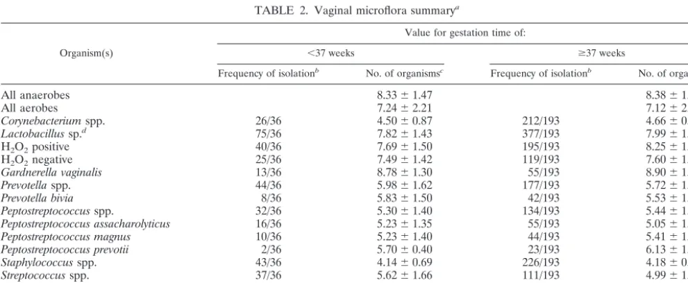
Descriptive microbiologic information.A comparison of fre-quencies of isolation and mean counts for the major genera
comprising the vaginal microflora is shown in Table 2. For the 229 subjects included in this study, 2,059 isolates representing 113 phenotypes were characterized. The mean values for total anaerobic bacterium counts, total aerobic bacterium counts, vaginal pH, and Nugent scores were similar for women deliv-ering at less than 37 weeks and for those delivdeliv-ering at term. No statistically significant differences between the PTD group and the term delivery group were noted in frequencies of isolation and mean counts for the major microbiologic categories. The
frequencies of isolation and mean total counts forG. vaginalis,
Prevotella spp., Peptostreptococcus spp.,Mycoplasma hominis, andUreaplama urealyticumwere similar for women delivering
at less than 37 weeks and those delivering at term.
Trichomo-nassp. strains were isolated from two subjects (0.1%). Neither
subject delivered preterm.
Evaluation of the multiple isolates of lactobacilli encoun-tered during this study revealed that women who were
simul-taneously harboring strains that produced H2O2 and strains
that did not produce H2O2 were much more likely to have
PTDs. While no statistical difference was noted for women
harboring either strains that produced H2O2or strains that did
not produce H2O2(10 of 33 women who had PTDs and 59 of
154 women who had at-term deliveries [P⬎0.40] for strains
producing H2O2; 5 of 33 women who had PTDs and 42 of 154
women who had at-term deliveries [P⬎ 0.18] for strains not
producing H2O2), it was noted that a significantly higher
per-centage of women harboring both strain types had PTDs (15 of
33 women versus 39 of 154 women [P ⬍ 0.03]). (Results of
testing for H2O2production were not available for 3 women
who had PTDs or for 39 women who had at-term deliveries.) In
addition, the total number of H2O2-producing lactobacilli was
significantly lower in women who had PTDs than in those who
[image:3.603.44.281.73.248.2]had at-term deliveries (mean ⫾standard error of the mean,
TABLE 1. Gestational age at birth
Week No. of birthsobserved
21... 1
24... 1
28... 1
31... 1
33... 4
34... 9
35... 6
36... 13
37... 21
38... 43
39... 56
40... 50
41... 20
42... 2
43... 1
Total ...229
TABLE 2. Vaginal microflora summarya
Organism(s)
Value for gestation time of:
⬍37 weeks ⱖ37 weeks
Frequency of isolationb No. of organismsc Frequency of isolationb No. of organisms
All anaerobes 8.33⫾1.47 8.38⫾1.57
All aerobes 7.24⫾2.21 7.12⫾2.29
Corynebacteriumspp. 26/36 4.50⫾0.87 212/193 4.66⫾0.88
Lactobacillussp.d 75/36 7.82⫾1.43 377/193 7.99⫾1.30
H2O2positive 40/36 7.69⫾1.50 195/193 8.25⫾1.21e
H2O2negative 25/36 7.49⫾1.42 119/193 7.60⫾1.36
Gardnerella vaginalis 13/36 8.78⫾1.30 55/193 8.90⫾1.28
Prevotellaspp. 44/36 5.98⫾1.62 177/193 5.72⫾1.80
Prevotella bivia 8/36 5.83⫾1.50 42/193 5.53⫾1.50
Peptostreptococcusspp. 32/36 5.30⫾1.40 134/193 5.44⫾1.43
Peptostreptococcus assacharolyticus 16/36 5.23⫾1.35 55/193 5.05⫾1.26
Peptostreptococcus magnus 10/36 5.23⫾1.40 44/193 5.41⫾1.55
Peptostreptococcus prevotii 2/36 5.70⫾0.40 23/193 6.13⫾1.08
Staphylococcusspp. 43/36 4.14⫾0.69 226/193 4.18⫾0.70
Streptococcusspp. 37/36 5.62⫾1.66 111/193 4.99⫾1.32
Mycoplasma hominis 7/36 6.15⫾0.81 36/193 6.03⫾1.02
Ureaplasma urealyticum 5/36 6.26⫾0.88 45/193 5.86⫾0.73
aVaginal pH and Nugent scores were as follows (values are means⫾standard deviations): for a gestation period of⬍37 weeks, 4.76⫾0.78 and 2.8⫾2.75,
respectively, and for a gestation period of⬎37 weeks, 4.74⫾0.79 and 2.45⫾2.60, respectively.
bValues are numbers of observations/numbers of subjects.
cValues are log
10CFU/gram of sample⫾standard deviations.
dA total of 10 strains in the preterm group and 63 strains in the term group were not characterized for H
2O2production.
eP⬍0.01 when compared to value for gestation period of⬍37 weeks.
on May 15, 2020 by guest
http://jcm.asm.org/
[image:3.603.44.540.471.675.2]7.69⫾ 0.24 versus 8.25⫾ 0.09 log10CFU/g of sample; P⬍
0.01).
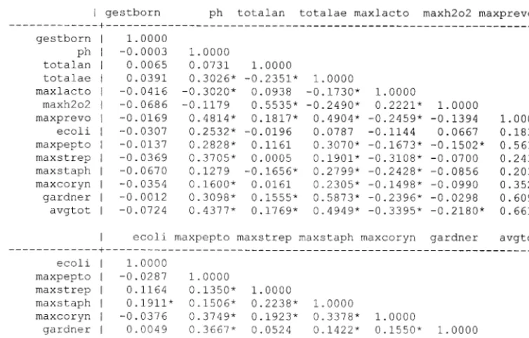
Predictive statistical model for PTD.A correlation matrix containing the major microbiologic members of the vaginal microflora was constructed as a preliminary step in developing statistical models for preterm birth (Fig. 1). There was a strong overall correlation between the total aerobic bacterium counts
and the average total bacterium count (avgtot) (r⫽0.49;P⬍
0.01). An even stronger correlation existed between these two variables for the subset of women delivering after a gestation
period of less than 37 weeks (r⫽0.62;P⬍0.01). The
maxi-mum counts of H2O2-producing lactobacilli and total
anaero-bic bacteria were noted to show a significant correlation (r⫽
0.55;P⬍0.01). An even stronger correlation existed between
these two variables for women delivering after a gestation
period of less than 37 weeks (r⫽0.61;P⬍0.01). Vaginal pH
levels had a positive correlation with the total aerobic counts (r
⫽ 0.30; P ⬍ 0.01) and a negative correlation with the total
counts forLactobacillusspp. (r⫽ ⫺0.30;P⬍0.01) (as the pH
decreased, the count of lactobacilli increased).
For women in different risk groups, we examined whether PTD might be related to somewhat different microbial profiles. Therefore, we considered separate models for women with and without prior PTDs.
For the women who had had a prior PTD, the rate of preterm birth was 37.8% for the period of this study. For these women, we considered two models: model 1 included women who had no vaginal bleeding during the current pregnancy, and model 2 included women with or without vaginal bleeding during the current pregnancy.
On the other hand, for women who had not previously had a PTD, the rate of preterm birth for women with vaginal
bleeding was 14.5% and for women with no obvious risk factor the rate of preterm birth was 8.9%. Hence, these two groups were combined and models 3 and 4 were instead constructed
according to the presence or absence of H2O2-positive
Lactoba-cillusspp., as follows: model 3 included women with H2O2
-posi-tiveLactobacilusspp. present, and model 4 included women from
whom H2O2-positiveLactobacilusspp. were absent.
Models 2, 3, and 4 are based on mutually exclusive groups of patients. Model 1 is a subset of Model 2 that focused on women without vaginal bleeding during the current pregnancy. A total of 206 (90%) of the 229 study subjects were evaluated by one of the models. Subjects with missing data were not evaluated.
For the previous-PTD risk group, the model was constructed as shown below, using numerous iterations to find the best fit.
Ifpis taken to be the probability of PTD, then logit (p) can be
calculated as follows:
log关p/共1⫺p兲兴⫽a0⫹a1⫻共risk factor 1兲⫹a2
⫻共risk factor 2兲. . .⫹an⫻共risk factorn兲 (1)
wherea1. . .anare estimated regression coefficients. If the
max-imum counts for H2O2-producing lactobacilli were not
avail-able, then the maximum counts for Lactobacillus spp. were
used in the regression.
[image:4.603.103.480.70.310.2]One measure for the overall performance of the predictive logistic regression model is the area under the receiver oper-ating curve (AUC). Subjects with predicted probability values above a specified threshold value were considered to be likely to have a PTD during the current pregnancy. The threshold probability was chosen so that the sensitivity and specificity values of the predictive model could be optimized.
FIG. 1. Correlation matrix for all subjects.ⴱ, significance atPvalue of⬍0.05. Abbreviations: gestborn, gestational age at birth; ph, pH; totalan, all anaerobes; totalae, all aerobes; maxlacto, maximum count ofLactobacillusspp.; maxh2o2, maximum count of H2O2-producingLactobacillus spp.; maxprevo, maximum count ofPrevotellaspp.; ecoli,Escherichia colicount; maxpepto, maximum count ofPeptostreptococcusspp.; maxstrep, maximum count ofStrepotococcusspp.; maxstaph, maximum count ofStaphylococcusspp.; maxcoryn, maximum count ofCorynebacteriumspp.; gardner,Gardnerellasp. count; avgtot, average total number of bacteria.
on May 15, 2020 by guest
http://jcm.asm.org/
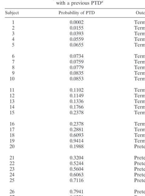
For women with a prior PTD but without vaginal bleeding during the current pregnancy, the logistic regression function
for model 1 is as follows: logit (p)⫽20.86⫺1.32⫻pH⫺2.61
⫻total anaerobic count ⫹0.58⫻maximum count of H2O2
-positive lactobacilli⫹0.70⫻maximum count of
Peptostrepto-coccusspp. Using this model, the predicted probability (p) is a number between 0 and 1. The area under the curve (AUC) has a value of 0.94, with a confidence interval (CI) of 0.79 to 1. Cross-validation by the leave-one-out procedure was per-formed, and the results were relatively stable. Comparison of the results predicted on the basis of the logistic regression model to the outcomes actually observed indicated that the predictive logistic model with a cutoff probability of 0.3 yielded a sensitivity of 88.9% and a specificity of 89.5% in predicting which subjects would have PTDs. The microbiologic risk fac-tors included in the regression equation and the AUC mea-sures for the four models are shown in Table 3. Predicted probabilities and actual outcomes for model 1 based on sub-jects with a prior PTD are shown in Table 4.
DISCUSSION
Previous studies conducted in our laboratory have exten-sively characterized the vaginal microflora of overtly healthy women (42, 44, 45). Based on the accumulated data from these earlier studies, a predictive model for normal vaginal micro-flora was constructed and tested (34). Prospective use of this model to predict an abnormal vaginal microflora has proven to be a practical application of these methods (49–52). The present study was an attempt to apply similar microbiologic and statistical methods to the prediction of preterm births, based on quantitative analysis of the vaginal microflora anal-ysis after 20 weeks of gestation.
Our previous studies have demonstrated that, for any given bacterial phenotype, the range of concentrations varies widely from subject to subject as well as for the same subject sampled longitudinally. This may account for the difficulty in correlating the presence or absence of any specific species or total bacte-rial counts with an outcome such as preterm birth. It was noted during the development of a statistical model for normal vag-inal microflora that the most predictive models included
mul-tiple bacterial risk factors. In the present study, we elected to use only microbiologic data or vaginal pH levels as risk factors for predicting PTD. Although a variety of demographic data for each subject were available, only the risk group was
con-TABLE 3. Regression models by risk groups
Risk group characteristic(s) m/na Regression model AUC CI for
AUC
Previous PTD but no
vaginal bleeding 9/28 logit (maximum Hp)⫽20.862O2⫺-positive1.33⫻LactobacilluspH⫺2.6⫻total anaerobic countsp. count⫹0.70⫻maximum⫹0.58⫻
Peptostreptococcussp. count
0.91 0.79–1.00
Previous PTD with or without vaginal bleeding
11/32 logit (p)⫽27.35⫺3.27⫻pH⫹1.92⫻bothb⫺2.22⫻total anaerobic
bacterium count⫹0.64⫻maximum H2O2-positiveLactobacillussp. count⫹0.22⫻maximumPeptostreptococcussp. count⫹0.21⫻
Gardnerellasp. count⫺0.32⫻maximumStaphylococcussp. count
0.82 0.65–0.98
Bleeding or no obvious risks; H2O2-positive lactobacilli present
15/139 logit (p)⫽2.05⫺0.75⫻total anaerobic bacterium count⫺0.48⫻total aerobic bacterium count⫹0.29⫻maximum H2O2-positive
Lactobacillussp. count⫹0.21⫻maximumStaphylococcussp. count⫹ 0.27⫻maximumCorynebacteriumsp. count⫹0.34⫻maximum
Gardnerellasp. count
0.74 0.62–0.87
Bleeding or no obvious risks; H2O2-positive lactobacilli absent
2/35 logit (p)⫽ ⫺81.14⫺0.45⫻total aerobic bacterium count⫹8.72⫻ maximumLactobacillussp. count⫹0.74⫻maximumStreptococcussp. count
0.94 0.81–1.00
am, number of PTD cases;n, number of subjects with all covariates.
[image:5.603.35.543.81.246.2]bboth, indicator variable for having both vaginal bleeding and previous PTD.
TABLE 4. Probability calculations and actual outcomes for women with a previous PTDa
Subject Probability of PTD Outcome
1 0.0002 Term
2 0.0155 Term
3 0.0393 Term
4 0.0559 Term
5 0.0655 Term
6 0.0734 Term
7 0.0759 Term
8 0.0779 Term
9 0.0835 Term
10 0.0853 Term
11 0.1102 Term
12 0.1149 Term
13 0.1336 Term
14 0.1766 Term
15 0.2378 Term
16 0.2378 Term
17 0.2881 Term
18 0.6093 Termⴱb
19 0.9414 Termⴱb
20 0.1988 Pretermⴱb
21 0.3204 Preterm
22 0.5244 Preterm
23 0.5604 Preterm
24 0.6063 Preterm
25 0.7116 Preterm
26 0.7941 Preterm
27 0.9221 Preterm
28 0.9422 Preterm
aData shown are for 28 of 36 subjects without any missing data.
bⴱ, probability calculation with a cutoff of 0.3 varies from actual outcome. All
three subjects thus denoted had total anaerobic counts that were significantly
higher than those observed for the rest of the group (P⬍0.034).
on May 15, 2020 by guest
http://jcm.asm.org/
[image:5.603.303.542.371.693.2]sidered during construction of the predictive models. This ap-proach allows for the use of observed microbiologic data in the absence of other confounding variables, such as a history of smoking and alcohol use, that have been suggested to influence pregnancy outcome. Subsequent statistical modeling has in-cluded both demographic data (such as age, ethnic group, insurance status, history of smoking and/or alcohol use), and parity. While these models may be more complete in that they use all of the available data, the AUC for these models is not significantly better than that obtained when only microbiologic data are used (unpublished data).
Among the organisms most commonly present as vaginal
microflora are members of the genus Lactobacillus.
Lactoba-cillus acidophilus is often reported to be the member of this genus most commonly isolated from vaginal secretions. Most of these previous reports have relied exclusively on conven-tional methods of phenotyping. However, recent studies using
16S rRNA-based ribotyping indicate thatLactobacillus
crispa-tus, Lactobacillus jensenii, and Lactobacillus gasseri are the species most commonly present as part of the vaginal micro-flora (3). The role of lactobacilli within the vaginal environ-ment is generally regarded as beneficial, with the production of lactic acid being responsible, in part, for the low pH of the vagina. This low pH is inhibitory for many potentially patho-genic organisms that also colonize the vaginal environment in low concentrations. More recently, it has been reported that
the production of H2O2 by some members of the genus
Lac-tobacillusis also associated with a healthy vaginal environment
(3, 17, 25, 27). The inhibitory effect in vitro of H2O2for
or-ganisms not producing catalase is well known. With regard to
the vaginal microflora, the inhibitory effect of H2O2 for G.
vaginalis has been documented, although the effect of H2O2
within the vaginal environment for other non-catalase-produc-ing species, includnon-catalase-produc-ing anaerobic streptococci and other obli-gate anaerobes, is not known. Women with BV have decreased
colonization rates for H2O2-producing strains of lactobacilli
and increased colonization rates for non-H2O2-producing
strains. It has also been reported that non-H2O2-producing
strains of lactobacilli are present as part of the normal vaginal
microflora (21, 24, 25, 29). In many women, both H2O2- and
non-H2O2-producing strains can be isolated. While the
pres-ence or abspres-ence of H2O2- and non-H2O2-producing strains has
been reported with respect to pregnancy outcome, this is the first attempt to use such information to develop a predictive model for PTD. Of particular note during the construction of the predictive models is the central role for both the type and total numbers of lactobacilli present as part of the vaginal
microflora. The observation that women harboring both H2O2
-producing and non-H2O2-producingLactobacillus strains are
at increased risk for PTD is a clear indication that this genus is an important microbiologic risk factor for predicting outcome. Although the characterization of lactobacilli in this initial
phase of the study included only genus designation and H2O2
production, our continuing study includes characterization of isolates by restriction fragment length polymorphism and pulsed-field gel electrophoresis analyses, species-level identifi-cation by 16S ribosomal DNA-based sequencing, and testing for the presence of lactocin genes by highly specific molecular probes. Further characterization of these strains by molecular techniques may provide additional useful information.
Many gram-positive organisms produce small peptides that are antibacterial in nature, variously called bacteriocins, lan-tibiotics, and lactocins. In the case of lactobacilli, such sub-stances are often directed at suppressing growth of closely related species. It has been reported, for example, that a
bac-teriocin produced byL. acidophilusis directed at inhibition of
Lactobacillus debrueckii, a closely related species (5). The char-acteristics of these small peptide inhibitors produced by lacto-bacilli are remarkably similar among gram-positive species and often contain posttranslational modifications of specific amino acids (38). Some of these compounds, such as lactocin S, can act as both bacteriocin and as cytolysin (38). While lactobacilli may be useful markers for a healthy vaginal environment, the possibility that specific strains or combinations of strains are harmful should not be ignored, particularly in light of their ability to produce substances capable of inhibiting other nor-mal vaginal microflora organisms. The elevated vaginal pH associated with pregnancy, coupled with the presence of mul-tiple strains of lactobacilli, may provide an ideal environment for production of bacteriocins. It has also been reported that a bacteriocin similar to those produced by lactobacilli is capable of inducing cellular injury (39) via the generation of superoxide anions. Among the genera present as part of the vaginal
mi-croflora, Lactobacillus is almost unique in that it has never
been explored as a possible mediator of tissue inflammation. The reliability of the models defined here to prospectively predict PTD is currently being evaluated in an ongoing clinical trial. If the models are able to prospectively predict preterm births reliably, the ability to use targeted interventional therapy only for those women at risk for preterm birth becomes a genuine possibility. The ability to construct these models using only microbiologic data suggests a primary role for bacteria in the etiology of preterm births. Understanding the mechanisms by which bacteria contribute to PTD is the basis for our con-tinued research efforts.
ACKNOWLEDGMENTS
This research was supported by grant RO1 HD35667 from the In-stitute of Child Health and Human Development, National InIn-stitutes of Health.
A.B.O., M.-L.L., E.L., M.L.D., and R.E.T. are members of the Microbiology and Prematurity Study Group. Other participants in-clude Robin Ross, Andrea DuBois, Linda Steele, Dorothy Pender, and Amy Cohen.
REFERENCES
1. Abele-Horn, M., J. Peters, O. Genzel-Boroviczeny, C. Wolff, A. Zimmer-mann, and W. Gottschling.1997. Vaginal Ureaplasma urealyticum coloni-zation: influence on pregnancy outcome and neonatal morbidity. Infection
25:286–291.
2. Amsel, R., P. A. Totten, C. A. Spiegel, K. C. Chen, D. Eschenbach, and K. K. Holmes.1983. Nonspecific vaginitis. Diagnostic criteria and microbial and
epidemiologic associations. Am. J. Med.74:14–22.
3. Antonio, M. A., S. E. Hawes, and S. L. Hillier.1999. The identification of vaginal Lactobacillus species and the demographic and microbiologic
char-acteristics of women colonized by these species. J. Infect. Dis.180:1950–
1956.
4. Antsaklis, A., G. Daskalakis, S. Michalas, and D. Aravantinos.1997. Eryth-romycin treatment for subclinical Ureaplasma urealyticum infection in
pre-term labor. Fetal Diagn. Ther.12:89–92.
5. Barefoot, S. F., and T. R. Klaenhammer.1983. Detection and activity of
lactacin B, a bacteriocin produced byLactobacillus acidophilus. Appl.
Envi-ron. Microbiol.45:1808–1815.
6. Bartlett, J. G., N. E. Moon, P. R. Goldstein, B. Goren, A. B. Onderdonk, and B. F. Polk.1978. Cervical and vaginal bacterial flora: ecologic niches in the
female lower genital tract. Am. J. Obstet. Gynecol.130:658–661.
on May 15, 2020 by guest
http://jcm.asm.org/
7. Bartlett, J. G., A. B. Onderdonk, E. Drude, C. Goldstein, M. Anderka, S. Alpert, and W. M. McCormack.1977. Quantitative bacteriology of the
vag-inal flora. J. Infect. Dis.136:271–277.
8. Bergstrom, S., and A. Libombo.1995. Low birthweight and post partum
endometritis-myometritis. Acta Obstet. Gynecol. Scand.74:611–613.
9. Briselden, A. M., B. J. Moncla, C. E. Stevens, and S. L. Hillier.1992. Sialidases (neuraminidases) in bacterial vaginosis and bacterial
vaginosis-associated microflora. J. Clin. Microbiol.30:663–666.
10. Carey, J. C., M. A. Klebanoff, J. C. Hauth, S. L. Hillier, E. A. Thom, J. M. Ernest, R. P. Heine, R. P. Nugent, M. L. Fischer, K. J. Leveno, R. Wapner, M. Varner, et al.2000. Metronidazole to prevent preterm delivery in
preg-nant women with asymptomatic bacterial vaginosis. N. Engl. J. Med.342:
534–540.
11. Chaim, W., M. Mazor, and J. R. Leiberman.1997. The relationship between bacterial vaginosis and preterm birth. A review. Arch. Gynecol. Obstet.
259:51–58.
12. Chambers, S., J. C. Pons, A. Richard, M. Chiesa, J. Bouyer, and E. Pa-piernik.1991. Vaginal infections, cervical ripening and preterm delivery.
Eur. J. Obstet. Gynecol. Reprod. Biol.38:103–108.
13. Cotch, M. F., S. L. Hillier, R. S. Gibbs, D. A. Eschenbach, et al.1998. Epidemiology and outcomes associated with moderate to heavy Candida
colonization during pregnancy. Am. J. Obstet. Gynecol.178:374–380.
14. Delaney, M. L., A. M. DuBois, and A. B. Onderdonk.1995. Qualitative and quantitative assessment of vaginal microflora following the use of cotton
tampons for two or twelve hours. Microecol. Ther.23:18–15.
15. Duff, P., M. L. Lee, S. L. Hillier, L. M. Herd, M. A. Krohn, and D. A. Eschenbach.1991. Amoxicillin treatment of bacterial vaginosis during
preg-nancy. Obstet. Gynecol.77:431–435.
16. Eschenbach, D. A.1994. Vaginitis including bacterial vaginosis. Curr. Opin.
Obstet. Gynecol.6:389–391.
17. Eschenbach, D. A., P. R. Davick, B. L. Williams, S. J. Klebanoff, K. Young-Smith, C. M. Critchlow, and K. K. Holmes.1989. Prevalence of hydrogen
peroxide-producingLactobacillusspecies in normal women and women with
bacterial vaginosis. J. Clin. Microbiol.27:251–256.
18. Flynn, C. A., A. L. Helwig, and L. N. Meurer.1999. Bacterial vaginosis in
pregnancy and the risk of prematurity: a meta-analysis. J. Fam. Pract.48:
885–892.
19. Garbaciak, J. A., Jr.1992. Prematurity prevention: who is at risk? Clin.
Perinatol.19:275–289.
20. Hauth, J. C., R. L. Goldenberg, W. W. Andrews, M. B. DuBard, and R. L. Copper.1995. Reduced incidence of preterm delivery with metronidazole
and erythromycin in women with bacterial vaginosis. N. Engl. J. Med.333:
1732–1736.
21. Hawes, S. E., S. L. Hillier, J. Benedetti, C. E. Stevens, L. A. Koutsky, P. Wolner-Hanssen, and K. K. Holmes.1996. Hydrogen peroxide-producing
lactobacilli and acquisition of vaginal infections. J. Infect. Dis.174:1058–
1063.
22. Hay, P. E., D. J. Morgan, C. A. Ison, S. A. Bhide, M. Romney, P. McKenzie, J. Pearson, R. F. Lamont, and D. Taylor-Robinson.1994. A longitudinal
study of bacterial vaginosis during pregnancy. Br. J. Obstet. Gynaecol.101:
1048–1053.
23. Hillier, S., M. A. Krohn, D. H. Watts, P. Wolner-Hanssen, and D. Eschen-bach.1990. Microbiologic efficacy of intravaginal clindamycin cream for the
treatment of bacterial vaginosis. Obstet. Gynecol.76:407–413.
24. Hillier, S. L., M. A. Krohn, E. Cassen, T. R. Easterling, L. K. Rabe, and D. A. Eschenbach.1995. The role of bacterial vaginosis and vaginal bacteria in amniotic fluid infection in women in preterm labor with intact fetal
mem-branes. Clin. Infect. Dis.20(Suppl. 2):S276–S278.
25. Hillier, S. L., M. A. Krohn, S. J. Klebanoff, and D. A. Eschenbach.1992. The relationship of hydrogen peroxide-producing lactobacilli to bacterial
vagino-sis and genital microflora in pregnant women. Obstet. Gynecol.79:369–373.
26. Hillier, S. L., M. A. Krohn, R. P. Nugent, R. S. Gibbs, et al.1992. Charac-teristics of three vaginal flora patterns assessed by gram stain among
preg-nant women. Am. J. Obstet. Gynecol.166:938–944.
27. Hillier, S. L., M. A. Krohn, L. K. Rabe, S. J. Klebanoff, and D. A. Eschen-bach.1993. The normal vaginal flora, H2O2-producing lactobacilli, and
bacterial vaginosis in pregnant women. Clin. Infect. Dis.16(Suppl. 4):S273–
S281.
28. Hillier, S. L., R. P. Nugent, D. A. Eschenbach, M. A. Krohn, R. S. Gibbs, D. H. Martin, M. F. Cotch, R. Edelman, J. G. Pastorek II, A. V. Rao, et al.
1995. Association between bacterial vaginosis and preterm delivery of a
low-birth-weight infant. N. Engl. J. Med.333:1737–1742.
29. Holst, E., A. R. Goffeng, and B. Andersch.1994. Bacterial vaginosis and vaginal microorganisms in idiopathic premature labor and association with
pregnancy outcome. J. Clin. Microbiol.32:176–186.
30. Ismail, M. A., G. Pridjian, J. U. Hibbard, C. Harth, and A. A. Moawad.1992. Significance of positive cervical cultures for Chlamydia trachomatis in pa-tients with preterm premature rupture of membranes. Am. J. Perinatol.
9:368–370.
31. Joesoef, M. R., S. L. Hillier, G. Wiknjosastro, H. Sumampouw, M. Linnan, W. Norojono, A. Idajadi, and B. Utomo.1995. Intravaginal clindamycin
treatment for bacterial vaginosis: effects on preterm delivery and low birth
weight. Am. J. Obstet. Gynecol.173:1527–1531.
32. Kimpen, J., E. Bosmans, J. Janssen, J. Lambrechts, A. Van Hoof, J. Gielen, H. Vandeput, and A. Van Waes.1986. Screening for Ureaplasma urealyticum infections in the neonate and the association with prematurity. Eur. J.
Ob-stet. Gynecol. Reprod. Biol.22:53–60.
33. Lamont, R. F., D. Taylor-Robinson, J. S. Wigglesworth, P. M. Furr, R. T. Evans, and M. G. Elder.1987. The role of mycoplasmas, ureaplasmas and chlamydiae in the genital tract of women presenting in spontaneous early
preterm labour. J. Med. Microbiol.24:253–257.
34. Lee, M.-L., R. A. Ross, M. L. Delaney, and A. B. Onderdonk.1995.
Mathe-matical modeling of the vaginal microflora. Microecol. Ther.23:18–21.
35. McGregor, J. A., J. I. French, D. Lawellin, and J. K. Todd.1988. Preterm birth and infection: pathogenic possibilities. Am. J. Reprod. Immunol.
Mi-crobiol.16:123–132.
36. McGregor, J. A., J. I. French, R. Richter, M. Vuchetich, V. Bachus, K. Seo, S. Hillier, F. N. Judson, J. McFee, J. Schoonmaker, et al.1990. Cervicovagi-nal microflora and pregnancy outcome: results of a double-blind,
placebo-controlled trial of erythromycin treatment. Am. J. Obstet. Gynecol.163:
1580–1591.
37. Minkoff, H., A. N. Grunebaum, R. H. Schwarz, J. Feldman, M. Cummings, W. Crombleholme, L. Clark, G. Pringle, and W. M. McCormack.1984. Risk factors for prematurity and premature rupture of membranes: a prospective
study of the vaginal flora in pregnancy. Am. J. Obstet. Gynecol.150:965–972.
38. Nes, I. F., and J. R. Tagg.1996. Novel lantibiotics and their pre-peptides.
Antonie Leeuwenhoek69:89–97.
39. Novak, R. M., T. J. Holzer, and C. R. Libertin.1993. Human neutrophil oxidative response and phagocytic killing of clinical and laboratory strains of
Enterococcus faecalis. Diagn. Microbiol. Infect. Dis.17:1–6.
40. Nugent, R. P., S. L. Hillier, et al.1992. Mucopurulent cervicitis as a predictor of chlamydial infection and adverse pregnancy outcome. Sex. Transm. Dis.
19:198–202.
41. Nugent, R. P., M. A. Krohn, and S. L. Hillier.1991. Reliability of diagnosing bacterial vaginosis is improved by a standardized method of Gram stain
interpretation. J. Clin. Microbiol.29:297–301.
42. Onderdonk, A. B., M. L. Delaney, G. R. Zamarchi, M. L. Hirsch, A. Munoz, and E. H. Kass.1989. Normal vaginal microflora during use of various forms
of catamenial protection. Rev. Infect. Dis.11(Suppl. 1):S61–S67.
43. Onderdonk, A. B., B. F. Polk, N. E. Moon, B. Goren, and J. G. Bartlett.1977. Methods for quantitative vaginal flora studies. Am. J. Obstet. Gynecol.
128:777–781.
44. Onderdonk, A. B., G. R. Zamarchi, M. L. Rodriguez, M. L. Hirsch, A. Munoz, and E. H. Kass.1987. Qualitative assessment of vaginal microflora during use of tampons of various compositions. Appl. Environ. Microbiol.
53:2779–2784.
45. Onderdonk, A. B., G. R. Zamarchi, M. L. Rodriguez, M. L. Hirsch, A. Munoz, and E. H. Kass.1987. Quantitative assessment of vaginal microflora during use of tampons of various compositions. Appl. Environ. Microbiol.
53:2774–2778.
46. Onderdonk, A. B., G. R. Zamarchi, J. A. Walsh, R. D. Mellor, A. Munoz, and E. H. Kass.1986. Methods for quantitative and qualitative evaluation of vaginal
microflora during menstruation. Appl. Environ. Microbiol.51:333–339.
47. Papiernik, E.1990. Preterm labor, preterm delivery, intrauterine infection,
and preterm rupture of membranes. Curr. Opin. Obstet. Gynecol.2:8–12.
48. Romero, R., F. Shamma, C. Avila, C. Jimenez, R. Callahan, J. Nores, M. Mazor, C. A. Brekus, and J. C. Hobbins.1990. Infection and labor. VI. Prevalence, microbiology, and clinical significance of intraamniotic infection
in twin gestations with preterm labor. Am. J. Obstet. Gynecol.163:757–761.
49. Ross, R. A., M. L. Delaney, and A. B. Onderdonk.1995. Candida albicans in
an in vitro model of the vaginal ecosystem. Microecol. Ther.25:320–323.
50. Ross, R. A., M.-L. Lee, M. Delaney, and A. Onderdonk.1995. Use of con-tinuous culture growth systems for modeling vaginal microflora behavior.
Microecol. Ther.23:16–17.
51. Ross, R. A., M. L. Lee, M. L. Delaney, and A. B. Onderdonk.1994. Mixed-effect models for predicting microbial interactions in the vaginal ecosystem.
J. Clin. Microbiol.32:871–875.
52. Ross, R. A., M. L. Lee, and A. B. Onderdonk.1995. Effect of Candida albicans infection and clotrimazole treatment on vaginal microflora in vitro.
Obstet. Gynecol.86:925–930.
53. Schwebke, J. R., S. L. Hillier, J. D. Sobel, J. A. McGregor, and R. L. Sweet.
1996. Validity of the vaginal gram stain for the diagnosis of bacterial
vagi-nosis. Obstet. Gynecol.88:573–576.
54. Sweet, R. L., D. V. Landers, C. Walker, and J. Schachter.1987. Chlamydia
trachomatis infection and pregnancy outcome. Am. J. Obstet. Gynecol.156:
824–833.
55. Van Winter, J. T., J. A. Ney, P. L. Ogburn, Jr., and R. V. Johnson.1994. Preterm labor and congenital candidiasis. A case report. J. Reprod. Med.
39:987–990.
56. Woodrow, N., and R. F. Lamont.1998. Bacterial vaginosis: its importance in
obstetrics. Hosp. Med.59:447–450.