Short Communication
Corresponding author: Dr. Shatrah Othman. e-mail: [email protected]
Received 24 May 2016 Accepted 8 September 2016
Subversion of immunoproteasome subunit expression
in dengue virus serotype 2-infected HepG2 cells
Chye Sheng Gan
[1],[2], Pei Jean Lim
[3], Muhammad Fazril Mohamad Razif
[1],
Rohana Yusof
[1],[2]and Shatrah Othman
[1],[2][1].Department of Molecular Medicine, Faculty of Medicine, University of Malaya, Kuala Lumpur, Malaysia. [2]. Drug Design and Development Research Group, University of Malaya, Kuala Lumpur, Malaysia.
[3]. Institute of Graduate Studies, University of Malaya, Kuala Lumpur, Malaysia.
Abstract:
Introduction: Infection with all serotypes of dengue virus (DV) results in augmented antigen presentation by MHC class I molecules. However, the upregulation of immunoproteasome subunits only results from infection with two serotypes. This study aims to elucidate changes in the expression of immunoproteasome subunits resulting from infection with DV, particularly DV serotype 2 (DV2). Methods: HepG2 cells were grown in various culture milieu. Total cellular RNA and proteins were extracted
and quantified. Results: Results demonstrated sequestration of immunoproteasome subunits LMP2 and LMP7 in DV2-infected cells. Conclusions: This study provides insights into the mechanisms underlying immune evasion by DV.
Keywords: Dengue virus. LMP2. LMP7.
Dengue is considered the most important arboviral disease globally. Hundreds of millions of infections occur annually worldwide, with approximately 75% of cases being endemic
to Asia-Pacific1. The dengue virus (DV) belongs to the genus
Flavivirus. Infection with all four DV serotypes elicits an increase in cell surface expression of major histocompatibility complex (MHC) class I molecules, similar to that observed in cells infected with the Japanese Encephalitis virus and West Nile virus2-3. In addition, the antigen presentation machinery
(APM) has also been shown to be affected by DV infection,
specifically at the proteasomal level3.
In order to escape the host’s immune response, viruses develop unique mechanisms to hijack the APM. The upregulation of MHC molecules in DV-infected cells does not appear to stimulate cytolysis by patrolling immune cells4.
Expression of immunoproteasome subunits low molecular weight polypeptide-2 (LMP2) and low molecular weight polypeptide-7 (LMP7) is pivotal for eliciting an immune response. The expression of these two proteins in DV-infected cells, as well as in those expressing DV non-structural proteins, has been found to be dysregulated3-4. The present study aimed
to investigate the effects of DV2 infection on LMP2 and LMP7 expression in human HepG2 cells using YK73, which has been shown to act as an antiviral agent against DV5.
HepG2 and Vero cell lines were obtained from the American Type Culture Collection (ATCC, USA). Dengue virus was isolated from a patient with dengue, and the presence of serotype
2 was confirmed by reverse transcription polymerase chain
reaction (RT-PCR)3 and sequencing (ABI, USA). Virus stock
titer was determined via plaque assay, as previously described3.
A preformed monolayer of HepG2 cells was infected with DV2 at a multiplicity of infection (m.o.i) of one. Media containing DV2 was aspirated after two hours of incubation at
37°C with humidified 5% CO2 and cells were rinsed with saline buffer. Culture medium, with or without 5µg/ml of antivirus YK735, was added to the infected cells and further incubated
for an additional 48 hours. The antiviral compound YK73 has a maximum non-toxic dose (MNTD) of 25µg/ml and a 90%
efficiency in inhibiting viral replication at a concentration of
5µg/ml5.
Cells were harvested 48 hours after post-infection using trypsin-ethylenediaminetetraacetic acid (EDTA), and ribonucleic acid (RNA) was isolated using the RNeasy mini kit (Qiagen, Germany) according to the manufacturer’s instructions. Then, 600ng of purified RNA was reverse-transcribed into double stranded complimentary DNA (cDNA) using high-capacity RNA-to-cDNA kit (ABI, USA). The
genes were quantified by real time quantitative PCR
(RT-qPCR) using the following TaqMan assays (ABI, USA): LMP2 (HS00160610_m1), LMP7 (HS00544760_g1), human leukocyte antigen type-A (HLA-A) (HS01058806_g1), and human leukocyte antigen type-B (HLA-B) (forward:
/03 /03 +/$$ +/$%
PRFN '9 '9<.
¨
[image:2.595.29.540.423.676.2]
5HODWLYHIROGFKDQJH
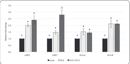
FIGURE 1. Gene expression profiles of LMP2, LMP7, HLA-A, and HLA-B. HepG2 cells were cultured under three different conditions: I) infected with
mock infection media (negative control), II) infected with DV2, and III) infected with DV2 and treated with antiviral agent YK73. Cells were incubated for 48 hours; then, total RNA was extracted and RT-qPCR was performed. Average expression of three independent experiments (n = 3) are presented. Error bars are
expressed as standard deviation. Values with different symbols are significantly different (at p < 0.05) between groups. DV2: dengue virus type 2; HLA-A: human leukocyte antigen type A; HLA-B: human leukocyte antigen type B; LMP2: low molecular weight polypeptide-2; LMP7: low molecular weight polypeptide-7;
RT-qPCR: real time quantitative polymerase chain reaction; RNA: ribonucleic acid.
and probe NEDtacgtggacgacaccaMGBNFQ-3′). The expression
levels of all four genes were normalized against that of the housekeeping gene GAPDH (4326317E).
LMP2 and LMP7 proteins were visualized via western blot using the following antibodies: anti-proteasome 20S LMP2 antibody ab3328 (Abcam, USA), anti-proteasome 20S LMP7 antibody (Abcam, USA), anti-beta actin antibody (Santa Cruz Biotechnology Inc. USA), goat anti-mouse immunoglobulin G type 1 conjugated with horseradish peroxidase (IgG1-HRP) antibody (Santa Cruz Biotechnology Inc. USA), and Anti-rabbit immunoglobulin G conjugated with horseradish peroxidase
(IgG-HRP) antibody (CST, Germany). Briefly, intracellular proteins
were extracted using cell lysis buffer and quantitated with Bradford reagent. Next, 80µg of protein was separated on a 12% acrylamide gel and transferred onto a nitrocellulose blotting membrane (PALL, USA). The membranes were blocked with 5% skimmed milk (Bio Rad, USA) in tris-buffered saline with Tween 20 (TBST) solution prior to incubation with primary antibodies. Subsequently, membranes were washed with TBST and further incubated with a secondary antibody. Bands on the developed membranes were analyzed using Image-J software (http://rsbweb.nih.gov/ij/).
Results are expressed as means ± standard deviation. Comparisons between groups were made using the Mann-Whitney U-test with the statistical software package Statistical Package for the Social Sciences (SPSS) version 17.0 software for Windows (IBM, USA).
The expression of immunoproteasome subunits LMP2 and
LMP7 has been reported to exhibit contrasting profiles when
cells are infected by different DV serotypes3: cells infected by
either DV2 or DV4 demonstrated increased expression of these genes compared with uninfected cells, while cells infected by
DV1 and DV3 exhibited no significant difference in LMP2
expression and reduced expression of LMP7 compared with uninfected cells, respectively.
In this study, LMP2 and LMP7 expression in cells following treatment with antiviral agent YK73 was investigated5. The
expression of LMP2 following antiviral treatment was found to be comparable to that in untreated cells; YK73-treated DV2-infected cells demonstrated a 1.2 fold difference (p = 0.487) compared with untreated DV2-infected cells, while YK73-treated DV2-infected cells demonstrated a 2.4 fold difference (p = 0.037) compared with uninfected cells. DV2-infected cells demonstrated only a two-fold increase (p = 0.037) relative to uninfected cells (Figure 1).
Interestingly, a 2.8-fold (p = 0.037) and a 1.5-fold (p = 0.037) increase in LMP7 expression was observed in YK73-treated DV2-infected cells and untreated DV2-infected cells, respectively, in comparison with uninfected cells. Additionally, an approximately 2-fold increase (p = 0.037) in LMP7 expression was observed between the YK73-treated and untreated DV2-infected cells.
Notably, the expression of MHC class I molecules was
/03 /03
'9 '9<.
'9
/03
/03
ȕDFWLQ
'9<.
5HODWLYHIROG
[image:3.595.58.567.249.686.2]
FIGURE 2. Protein expression of LMP2 and LMP7 following antiviral treatment. HepG2 cells were infected with DV2, supplemented with or without the
antiviral compound, and incubated for 48 hours. Then, cells were harvested and protein lysate was subjected to western blot. A. Western blot of LMP2, LMP7,
and β actin. B. The densitometry-quantified intensity of LMP2 and LMP7 bands normalized against that of β actin. Average expression determined by three independent experiments (n = 3) is presented. Error bars are expressed as standard deviation. Values with different symbols are significantly different at p < 0.05
between the groups. DV2: dengue virus type 2; LMP2: low molecular weight polypeptide-2; LMP7: low molecular weight polypeptide-7. A
B
infected cells relative to uninfected cells. The HLA-A molecule presentation was approximately 1.5-fold higher (p = 0.037) in both YK73-treated and untreated DV2-infected cells compared with that in uninfected cells, independently. Similarly, HLA-B displayed an approximately two-fold increase (p=0.037) in expression in both YK73-treated and untreated DV2-infected
cells relative to uninfected cells, respectively. These findings
strongly suggest that MHC class I cell surface protein expression remains enhanced despite antiviral treatment.
In order to determine whether immunoproteasome subunit
LMP2 and LMP7 profiles are congruent with the enhanced
transcription levels observed upon antiviral treatment,
western blot was conducted to compare these protein levels in YK73-treated and untreated DV2-infected cells (Figure 2). The expression of LMP7 was markedly increased in YK73-treated DV2-infected cells (approximately 2.5-fold higher, p = 0.037). As anticipated, there was only a slight increase (~1.3-fold change, p = 0.034) in the expression of the LMP2
subunit in YK73-treated DV2-infected cells. These findings
clearly indicate that DV2 plays an active role in regulating the production of the immunoproteasome subunits.
proteins, these multi-subunit proteasome complexes degrade defective ribosomal translational products6. Antigenic
proteins are processed by the APM, almost immediately after synthesis, via the transformation of the proteasome into the immunoproteasome; this enables rapid generation of the host immune response. It is notable that modulation of LMP2 and
LMP7 expression was found to affect the efficiency of APM7.
Additionally, a study by Khu et. al. demonstrated the role of the NS3 protein in downregulating LMP7 in the hepatitis C virus (HCV), another member of the genus Flavivirus8.
Similar to the previous report3, this study also demonstrated
elevation of both LMP2 and LMP7 expression during DV2 infection. Moreover, the increase in gene expression was more pronounced upon administration of antiviral treatment
to DV2-infected cells. This finding clearly demonstrates the
subversion of immunoproteasome subunit expression, despite the augmentation of these subunits during DV2 infection relative to that in uninfected cells. It is plausible that the virulence of the present DV2 serotype led to higher expression of immunoproteasome subunits in comparison to that on infection with other DV serotypes. Nevertheless, the results strongly
YK73
DV2 replication
Proteasome
Immunoproteasome
Generation of
[image:4.595.30.541.343.676.2]‘self’ peptides
Generation of
antigenic peptides
FIGURE 3. Putative effect of antivirus YK73 on the transformation of the proteasome into the immunoproteasome in DV2-infected cells. Active
replication of DV2 prevents efficient transformation of the immunoproteasome by limiting the transcription of both LMP2 and LMP7 genes. The absence of the
immunoproteasome results in impaired generation of antigenic peptides, which leads to poor surface presentation of the antigen epitopes. Conversely, increased
expression of both LMP2 and LMP7 was observed when virus replication was impeded by the antiviral compound YK73. This finding may indicate the recovery
of the host response, which enables the transformation of the immunoproteasome. DV2: dengue virus type 2; LMP2: low molecular weight polypeptide-2;
LMP7: low molecular weight polypeptide-7.
suggest that DV2 NS2B-NS3 plays a pivotal role in suppressing the expression of immunoproteasome subunits during infection.
In conclusion, this study provides evidence for, and elucidates the mechanisms underlying, the suppression of immunoproteasome subunit expression by DV. This conclusion was particularly
evident when the expression of LMP7 was intensified subsequent
to antiviral treatment. Suppression of LMP2 and LMP7 expression may consequently prevent the transformation of the immunoproteasome9. As a result, the constitutive proteasomes available were unable to generate antigenic peptides efficiently.
DV-infected cells were considered to ultimately present ‘self’ peptides as a result of crippled antigen processing9, despite the observed
augmentation in MHC class I expression3 (Figure 3). The antiviral
treatment is shown to reduce viral replication with simultaneous
reduction in the efficiency of manipulation of the host immune
response by the virus. This phenomenon may indicate the recovery of appropriate host response, thus allowing the transformation of the immunoproteasome. Further in-depth study is warranted in order to elucidate the relationship between the antiviral compound,
dengue infection, and the expression profiles of immunoproteasome
Acknowledgements
We offer our deepest gratitude to the University of Malaya that provided technical support for the development and implementation of this study. We additionally thank Dr. Yean Kee Lee for providing the antiviral compounds.
Conflict of interests
The authors declare that there is no conflict of interest.
Financial support
This work was supported by grants from the University of Malaya Research Grant (UMRG), RP027D-15AFR, and University of Malaya Postgraduate Grant, PG034-2014B.
REFERENCES
1. Murray NEA, Quam MB, Wilder-Smith A. Epidemiology of dengue: past, present and future prospects. Clin Epidemiol. 2013;5(1):299-309.
2. Othman S, Rahman NA, Yusof R. All serotypes of dengue virus induce HLA-A2 major histocompatibility complex class I promoter activity in human liver cells. Trans R Soc Trop Med Hyg. 2010;104(12):806-8.
3. Gan CS, Yusof R, Othman S. Different serotypes of dengue viruses differently regulate the expression of the host cell antigen processing machinery. Acta Trop. 2015; 149:8-14.
4. Hershkovitz O, Zilka A, Bar-Ilan A, Abutbul S, Davidson A, Mazzon M, et al. Dengue virus replicon expressing the
nonstructural proteins suffices to enhance membrane expression
of HLA class I and inhibit lysis by human NK cells. J Virol. 2008;82(15):7666-76.
5. Muhamad M, Kee LY, Abd Rahman N, Yusof R. Antiviral actions
of flavanoid-derived compounds on dengue virus type-2. Int J Biol.
Sci 2010;6(3):294-302.
6. Hewitt EW. The MHC class I antigen presentation pathway: strategies for viral immune evasion. Immunology. 2003;110(2):163-9.
7. Khanna R, Busson P, Burrows SR, Raffoux C, Moss DJ, Nicholls JM, et al. Molecular characterization of antigen-processing function in nasopharyngeal carcinoma (NPC): evidence for
efficient presentation of Epstein-Barr virus cytotoxic T-cell
epitopes by NPC cells. Cancer Res. 1998;58(2):310-14.
8. Khu YL, Tan YJ, Lim SG, Hong W, Goh PY. Hepatitis C virus non-structural protein NS3 interacts with LMP7, a component of the immunoproteasome, and affects its proteasome activity. Biochem J. 2004;384(2):401-9.