http://dx.doi.org/10.4236/jbnb.2015.63020
Environmental Toxicity and Antimicrobial
Efficiency of Titanium Dioxide Nanoparticles
in Suspension
Muriel Bonnet1, Christophe Massard2, Philippe Veisseire1, Olivier Camares1,
Komla Oscar Awitor2
1Clermont Université, Université d’Auvergne, Laboratoire de Biologie, Aurillac Cedex, France 2Clermont Université, Université d’Auvergne, C-BIOSENSS, Clermont-Ferrand Cedex, France
Email: [email protected]
Received 4 May 2015; accepted 13 July 2015; published 17 July 2015
Copyright © 2015 by authors and Scientific Research Publishing Inc.
This work is licensed under the Creative Commons Attribution International License (CC BY).
http://creativecommons.org/licenses/by/4.0/
Abstract
The aim of this work was to evaluate the photokilling efficiency of synthesized titanium dioxide nanoparticles in suspension. Two strains of Escherichia coli, Lactobacillus casei rhamnosus and Staphylococcus aureus were used as probes to test the photokilling activities of the nanoparticles. The toxicity effects of TiO2 nanoparticles on the environment were determined by a standard test
using gram-negative bioluminescent bacteria Vibrio fischeri. The antimicrobial activity of these nanoparticles (NPs) was then investigated versus NPs concentration, UV irradiation time and mi-cro-organism strains. We evaluated the LC50 values of the nanoparticles suspension by counting the Colony-Forming Units. Results highlighted the differences in bacteria sensitivity facing photo-killing treatment induced by the irradiation of anatase TiO2 nanoparticles suspension. At the
con-centration of 1 g·L−1 TiO2, tested bacteria were killed after 30 minutes of photo-treatment. Using
different TiO2 concentrations, the Staphylococcus aureus gram-positive/catalase-positive bacteria
were more resistant than gram-negative/catalase-positive ones or gram-positive/catalase-nega- tive bacteria. An effect of UV irradiation was evaluated by the quantification of hydrogen peroxide generated by the photolysis of water molecules in presence of the nanoparticles with or without the most resistant bacterium (S. aureus). After 30 minutes with UV irradiation in these two condi-tions, the concentration of hydrogen peroxide was 35 µM in presence of 1.2 g·L−1 TiO2 suspension.
This result suggested that the resistance mechanism of S. aureus was not due to an extracelullar H2O2 enzymatic degradation.
Keywords
1. Introduction
Photokilling of pathogen species is a promising alternative compared to conventional disinfection process. In particular, when chemical cleaning products are not effective or dangerous, a disinfection protocol based on the irradiation of photoactive species can be interesting. Contrary to other cleaning treatments, such as chlorination [1] [2] and ozonation [3]-[5], less toxic by-products are generated and the process can remain effective for a long time. The photokilling disinfection is mainly based on photoinduced oxidative reactions. Among all the photoactives species, TiO2 anatase is widely studied [6] [7] under UV irradiation. The use of photoactive tita-nium opens the way to the development of self-cleaning materials [8]-[11]. Works have been done to improve the process, concerning the antimicrobial selectivity. Nanocomposite materials with magnetic nanoparticles [12] have been used to enhance the photoactivity of silver/titanium oxide [13] [14]. Within environmental toxicity assessments, the supervising of the effects of nanoparticles on micro-organisms is still very limited. The biolu-minescence test Microtox® is often chosen as the first test in a test battery based on speed and cost consideration [15]. It is a standardized toxicity test (AFNOR T90-320, EN ISO 11348-3) system which is also sensitive and reproducible. It is recognized and used throughout the world as a standard test for aquatic toxicity testing [16] to determine EC50 (half maximal Effective Concentration). The photoinactivation of bacteria is a complex and multifaceted phenomenon. Currently many factors are taken into consideration regarding TiO2 nanoparticles toxicity. According to Cai et al. [17], the bactericidal activity of TiO2 NPs, in the presence of UV light, was due to oxidative stress. Gogniat et al. [18] suggested a sequence of nanoparticle interactions with the cell membrane followed by cell membrane oxidation facilitated by Reactive Oxygen Species (ROS). Accordingly, many studies have attributed to ROS production, the nanoparticles bactericidal effect generated under UV light [19] [20]. Furthermore, recent reports have shown that TiO2 nanoparticles can induce the oxidative stress defense of the cell against endogenous ROS like H2O2, which can sequentially elicit lipids, proteins and DNA damage [21]- [23]. Many studies also investigated the possibility of nanoparticle penetration inside the bacterial cell mem-brane as a possible toxicity mechanism [24] [25]. The cell is surrounded by the plasma membrane, a lipid bilay-er which contains opposing monolaybilay-ers, or leaflets, of phospholipids with the hydrophilic head groups facing the extracellular and intracellular solutions, and the hydrophobic tails facing each other. Generally three routes for nanoparticle entry into cells exist: diffusion, endocytosis and channel implication [26]-[28]. When entering the cell, nanoparticles can probably produce intracellular H2O2. Cells naturally produce this metabolite. This is the reason why a specific mechanism exists to counteract the presence of hydrogen peroxide for detoxifying the cell. Catalase is a tetrameric heme-containing enzyme, and is one of the key antioxidant enzymes present in al-most every aerobic organisms, catalyzing the breakdown of hydrogen peroxide to water and molecular oxygen to protect cells against the toxic effects of hydrogen peroxide [29].
In this study, we synthesized an original and stable anatase-crystallized suspension of TiO2 nanoparticles. Esche-richia coli strains LE392 and ETEC H10407 (gram-negative/catalase-positive bacteria), Lactobacillus casei rham-nosus strain Lcr35® (gram-positive/catalase-negative bacteria) and Staphylococcus aureus (SA51, gram-posi- tive/catalase-positive bacteria) were used as probes to test the photokilling efficiency of the nanoparticles in suspension. In particular, the resistance behaviour of different bacteria strains was evaluated using LC50 tests, focusing on two different parameters: the bacteria wall thickness (gram+ or gram−) and the presence or absence of the catalase gene (catalase+ or catalase−). Bioluminescent tests were performed to investigate the environ-mental toxicity of TiO2 in suspension. The quantification of H2O2 allowed a better understanding of the inactiva-tion mechanism involved in the photokilling process.
2. Material and Methods
2.1. Synthesis of the Nanoparticles Suspension
2.2. Transmission Electron Microscopy
The morphology and the particle sizes were characterized using a Philips CM 20 transmission electron micro-scope (TEM). The accelerating voltage was 200 kV. The samples were dispersed in methanol by ultrasonication. A drop of the suspension was then laid on a carbon-coated grid and dried. Selected Area Electron Diffraction (SAED) was performed to determine the crystalinity of the structure. The interplanar spacings were evaluated from the SAED patterns using the following formula:
λL = Rd (1) where λL is the constant of the microscope, R is the ring radius, and d is the interplanar spacing. The constant of the microscope was calculated by measuring the radius of a gold standard pattern whose interplanar spacings were well documented in scientific publications [30].
2.3. Bacterial Culture
Four micro-organisms were used for photokilling experiments: Escherichia coli LE392, Enterotoxigenic Esche-richia coli H10407, Lactobacillus casei rhamnosus Lcr35® and Staphylococcus aureus (SA51). These bacteria have a size comprised between 0.5 and 5 µm. E. coli cells were cultured at 37˚C for 24 h in Nutrient Broth me-dium at pH 7.2 (Biokar diagnostics) containing Tryptone (10 g·L−1), Meat extract (5 g·L−1) and Sodium Chlo-ride (5 g·L−1) after 12 h of pre-culture in the same conditions. Lactobacillus casei rhamnosus Lcr35® was cul-tured in De Man, Rogosa, Sharpe (MRS) medium (Bio-Rad, Mitry Mory, France) and S. aureus in Brain Heart Broth (Brain Heart Infusion 17.5 g·L−1, Pancreatic digest of gelatin 10 g·L−1, Sodium Chloride 5 g·L−1, Dis-odium phosphate 2.5 g·L−1, Glucose 2 g·L−1, Biokar diagnostics) under the same conditions than E. coli strains. Cells were centrifuged at 2500 g for 15 min at 4˚C and the pellet was re-suspended in de-ionized water to pre-vent unintentional increase in cell numbers. The initial population of bacteria was determined by enumeration with a Petroff-Hausser Counting Chamber.
2.4. Bioluminescent Tests
The Microtox® Procedure employs the bioluminescent marine gram-negative bacterium Vibrio fischeri as test organism. The bacteria are exposed to a range of concentration of the TiO2 in suspension being tested. The re-duction in intensity of light emitted from the bacteria is measured along with standard solutions and control samples. Toxicity is, then, inversely proportional to the intensity of the light emitted after contact with the toxic substances. The change in light output and concentration of the toxicant produce a dose/response relationship. The results are normalized and the EC50 (concentration producing a 50% reduction in light) is calculated.
The basic test protocol (consisting of four test dilutions) was carried out to evaluate the ecotoxicity of the me-dium containing TiO2 nanoparticles. All tests were performed using the Microtox 500 Analyser, and biolumi-nescence measurements were monitored at 0, 5 and 15 min of exposure. The effective concentrations causing 50% of bioluminescence inhibition were computed using the software for Microtox Omni Azur (AZUR envi-ronmental, 1998). Toxicity tests were performed in triplicate each week during a two months period and the re-sults are expressed in mg·L−1.
2.5. Inactivation Kinetics Measurements and LC50 Tests
For inactivation kinetics measurements, an amount of 20 mL of de-ionized water was inoculated with Escheri-chia coli LE392 or Enterotoxigenic EscheriEscheri-chia coli H10407 suspension in order to achieve a concentration of 106 CFU·mL−1 (Colony-Forming Unit by mL). This suspension was placed in a Petri plate with TiO2 nanopar-ticles to achieve a final concentration in TiO2 of 1 g·L−1. The slurries were continuously mixed and irradiated with UV (polychromatic fluorescent UV lamps (©Philips TLD 8 W) providing a total power of 48 W, in a con-figuration delivering 1.5 mW·cm−2 at the liquid surface). A complete mixing was done with a sterilized Teflon magnetic stir bar placed in the Petri dish with a speed of 200 rpm.
pre-vent any contamination in the media. The counts from three independent experiments corresponding to a partic-ular sample were averaged. The method used for the LC50 tests was similar to that used for inactivation kinetics and was performed on all bacteria strains with nanoparticles concentrations from 50 to 1200 mg·L−1 TiO2 under 30 min UV irradiation at 1.5 mW·cm−2.
2.6. Hydrogen Peroxide Concentration Determination
Generation of hydrogen peroxide by TiO2 nanoparticules in an aqueous liquid suspension was determined as described by Batdorj et al. [31] with slight modifications. Aqueous solutions of TiO2 particles concentrations ranging from 0 to 1200 mg·L−1 were placed in Petri plates and continuously mixed in the dark or irradiated with UV for 30 minutes. H2O2 concentrations were measured after eliminating the nanoparticles by centrifugation for 15 min at 200000 g. One mL of supernatant was added to a solution containing 100 µl of 4-aminoantipyrine (4 mg·mL−1 solution of 4-amino-2, 3-dimethyl-1-phenyl-3-pyrazolin-5-one, Sigma), 40 µl of water-satured phenol, 60 µl of horseradish peroxidase type VI-A (Sigma, 500 U·mL−1 solution in sodium phosphate buffer pH 6) and 800 µl of phosphate buffer Na2HPO4/NaH2PO4 (0.1 M, pH 7). The reaction was allowed to proceed for 5 min and the absorbance was measured at 505 nm. The hydrogen peroxide was quantified using a standard curve per-formed with concentrations ranging from 10 to 200 µM.
3. Results and Discussion
3.1. Nanoparticles Suspension Synthesis
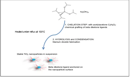
Stable titanium dioxide nanoparticles in suspension are fabricated using a derivate sol gel process. Figure 1 shows the main steps of the synthesis protocol. This soft chemistry process is a one-pot, low temperature and ef-ficient method to obtain highly dispersed colloids in a carrying liquid. The first step consists in the chelation of the titanium isopropoxide with an organic ligand, acetylacetone. This reaction is a substitution of alkoxy group of the titanium alcoxyd molecular species by beta diketone ligands. In consequence, the hydrolysis kinetic of the titanium precursor is lowered and undesirable precipitation avoided. Hydrolysis-condensation reactions were carried out by dropping acidified water in the homogeneous medium previously diluted in some isopropylic al-cohol. The reacting mixture was heated under reflux for almost 8 hours to obtain a stable dispersion of TiO2
Ti(OiPr)4
1. CHELATION STEP: with acetylacetone C5H8O2: chemical grafting of beta diketone ligands
2. HYDROLYSIS and CONDENSATION: titanium dioxide fabrication
Heated under reflux at 100˚C
Stable TiO2 nanoparticles in suspension
[image:4.595.87.538.439.706.2]Beta diketone ligand anchored on the nanoparticle surface
nanoparticles in an aqueous liquid. Taking into account that the pH of liquid carrier is low, the TiO2 mineral oxide nanoparticles have a positive surface charge.
3.2. Transmission Electron Microscopy
A TEM picture and the associated SAED pattern of our as-synthesized sample are presented inFigure 2. The TEM image (Figure 2(a)) shows that most of the particles are elongated, some of them are spherical. From these TEM pictures, the mean crystallite diameter is approximately 8 nanometers.
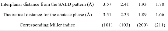
The SAED patterns of the most intense spots are shown in Figure 2(b). The comparison, inTable 1, between the interplanar distances calculated from the SAED patterns and the tabulated ones obtained for the anatase crystallographic structure exhibits a good agreement and confirms the anatase crystalline structure of our syn-thesized sample.
3.3. Bioluminescent Tests
The Microtox® test has been routinely applied to treated waste waters or single compounds and mixtures of in-organic and in-organic compounds [32]. Furthermore, bioluminescence test becomes a recognized tool to investi-gate ecotoxicity of nanoparticles [33]. No visible precipitate was observed during the test over the two months period, which confirmed nanoparticles suspension stability. Our results showed EC50 values of respectively 43.75 ± 23.38 mg·L−1 and 36.51 ± 20.55 mg·L−1 at 5 min and 15 min. The calculated EC50 after 5 and 15 mi-nutes exposure time are quite similar. The slight decrease could mean that the nanoparticles need a short time to diffuse into the cells and interact with lipids, carbohydrates, proteins and DNA [34]. Obtained EC50 values for TiO2 particles are much higher, relative to the literature [35] [36]. This may be due to our particular and original way of synthesis of nanoparticles with the use of acetylacetone which is known as a toxic molecule [37]. Our TiO2 nanoparticles with EC50 ranging from 36 to 44 mg·L−1, can be classified as harmful to aquatic micro-or- ganisms (EC50 in the range of 10 - 100 mg·L−1) according to the Commission Directive 93/67/EEC from the European Union for the assessment of risk to man and the environment of substances.
We have demonstrated the toxicity of our nanoparticle suspension in the dark on a very sensitive bacterium, Vibrio fischeri.
3.4. Inactivation Kinetics Measurements
[image:5.595.172.454.473.605.2]As in previous studies on Escherichia coli LE392 [38] where we clearly observed the total destruction of bacteria
Figure 2. TEM image of TiO2 nanoparticles (a) and SAED pattern of the par-ticles (b).
Table 1. Interplanar distances for the TiO2 nanoparticles deduced from the SAED patterns, compared to the expected ones for ideal anatase phase.
Interplanar distance from the SAED pattern (Å) 3.57 2.41 1.93 1.70
Theoretical distance for the anatase phase (Å) 3.51 2.33 1.89 1.66
[image:5.595.177.453.670.721.2]after only 1 hour of treatment with 1 g·L−1 TiO2 suspension under UV irradiation, we could wonder what hap-pens during this time duration. Figure 3 shows that after 10 min of treatment, approximately 40% of bacteria tested (E. coli LE392 and ETEC H10407) died. Ten minutes later, we can observe a drastic diminution of the population with around 80% of mortality. Finally, under these particular conditions, we clearly observed the to-tal destruction of both strains of bacteria after only 30 minutes.
Freshly grown bacterial cultures (106 CFU·mL−1) were treated with 1 g·L−1 of TiO2 and irradiated with UV (1.5 mW·cm−2). This experiment was carried out in triplicate. Wang et al. [39] found quite similar results show-ing that a lower TiO2 nanoparticles concentration (0.4 g·L−1) had a similar inactivation effect on E. coli but after 2 h UVA irradiation.
3.5. TiO2 Suspension Phototoxicity against Bacteria
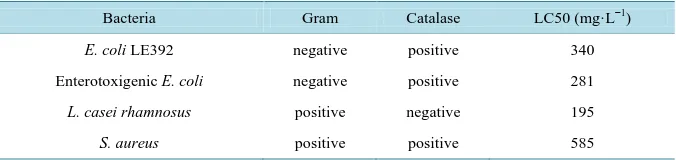
The 30 minutes-LC50 tests were then performed on all strains in order to make a comparison between bacteria differing in cell wall structure and detoxification system implicating the catalase enzyme (Table 2).
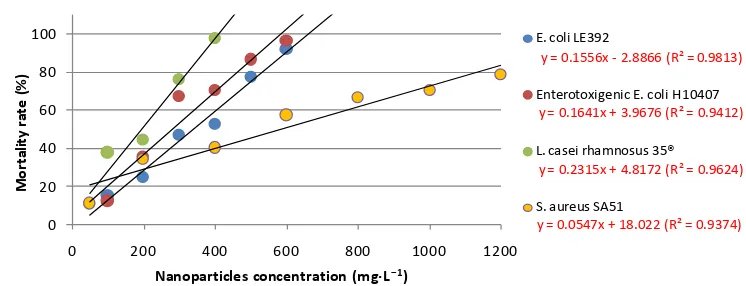
Concentration-dependent mortality in E. coli exposed to TiO2 suspension under 30 minutes UV irradiation (1.5 mW·cm−2) showed a linear profile for both strains at a concentration ranging from 100 to 600 mg·L−1 (Figure 4). The LC50 calculated by linear regression were 340 mg·L−1 for LE392 and 281 mg·L−1 for ETEC H10407 (Table 2). The TiO2 nanoparticles suspension had differing inactivation efficiency regarding Lactoba-cillus casei rhamnosus and Staphylococcus aureus. LC50 was calculated to be 195 mg·L−1 for L. casei rhamno-sus 35® whereas the value of 585 mg·L−1 was determinated for S. aureus. We can observe that concentration-de- pendent survival is higher for the S. aureus gram-positive catalase-positive bacteria compared to the other ones.
[image:6.595.144.488.409.587.2]The fact that TiO2 nanoparticles showed a lower effect on S. aureus than on the other ones, under the same conditions, indicates that the resistance of bacteria to TiO2 nanoparticles is species-dependent. These differences might be due to different structural properties of cell wall and/or a higher self-defense property [40] or self-repair ability of S. aureus than the other ones. Only focusing on the cell wall property of bacteria tested, we can see that the more resistant one is S. aureus which has a gram-positive cell wall. This is in accordance with previous
Figure 3.Influence of irradiation time on the mortality rate of Escherichia coli LE392 and
EnterotoxigenicEscherichia coli H10407.
Table 2. Wall type and catalase activity of different tested bacteria strains.
Bacteria Gram Catalase LC50 (mg·L−1)
E. coli LE392 negative positive 340
Enterotoxigenic E. coli negative positive 281
L. casei rhamnosus positive negative 195
S. aureus positive positive 585
0 20 40 60 80 100
0 10 20 30
M
o
rta
lity
r
a
te
(
%
)
Irradiation time (minutes)
[image:6.595.147.485.640.720.2]Figure 4. Determination of LC50 for Escherichia coli LE392, Enterotoxigenic Escherichia coli
H10407, Lactobacillus casei rhamnosus Lcr35® and Staphylococcus aureus SA51 when exposed to TiO2 nanoparticles with concentrations ranging from 50 to 1200 mg·L−1 under 30 min UV irradia-tion at 1.5 mW·cm−2. Experiments were carried out in triplicate. R2is a measure of goodness-of-fit of linear regression.
results [41] [42] respectively using ZnO and Ag nanoparticles, which exhibited a much stronger antibacterial effect on gram-negative bacteria. This difference in antimicrobial activity between gram-positive and gram- negative micro-organisms is often attributed to the structure of their perspective cell walls [43]. On the other hand, our results are not similar with another report using ZnO nanoparticles that showed a much stronger anti-bacterial effect on gram-positive bacteria than on gram-negative ones [44]-[46]. In addition, van Grieken et al. [47] observed no significant differences between the photocatalytic inactivation of gram-negative and gram- positive bacteria for all experiments and concluded that despite their differences in cell wall structure, both E. coli and E. faecalis showed similar reaction to the treatment. Moreover, in our study, the most sensitive bacte-rium is Lcr35® even if this micro-organism belongs to the gram-positive bacteria class. All these results confirm that the cell wall structure is not the primary factor involved in resistance to nanoparticles.
Major constituents of the cell wall are each specific strains and the surface charge of the bacteria is associated with the presence of the ionized groups of the macromolecules [48]. Generally, the cell wall of gram-positive bacteria has a stronger negative charge than gram-negative bacteria. This negative charge is due to the presence of teichoic acid in gram-positive bacteria and lipophosphate in gram-negative ones [49].
There are reports in the literature that show that electrostatic attraction between negatively charged bacterial cells and positively charged nanoparticles is crucial for the activity of nanoparticles as bactericidal materials. Nanoparticles are capable of penetrating bacterial cells and act as a catalyst, to inactivate enzymes that micro- organisms need for their metabolism by interacting with thiol groups of proteins, disrupt bacterial membranes and also affect DNA replication [50] [51]. In the case of nanoparticles of TiO2 illuminated with UV, the pro-duced hydrogen peroxide will contribute to this phenomenon [52]. Marugán et al. [40] found that bacteria by themselves had self-protection ability and could grow again after being injured. Therefore, the inactivation of bacteria requires a certain amount of cumulative damage.
In our study, we have to take into account the presence or absence in cells of an enzyme responsible for cata-lyzing the breakdown of hydrogen peroxide into water and molecular oxygen: catalase [53] [54]. The highest re-sistance of S. aureus encountered here could be explained by the combination of its cell wall gram+ and the di-minution of extracellular and/or intracellular H2O2 concentration by catalase.
3.6. H2O2 Measurements
TiO2 is a semiconductor [55] which can be excited by UV light. In these conditions, an electron of TiO2 receives photon energy and is excited [56]. It then reacts with H2O and/or O2 and produces hydroxyl radicals and/or ac-tive oxygen species [57]. The active species further react with bacteria and inactivate them. The damage cannot be completed in a short time, even though there are enough radicals produced by photocatalytic nano-TiO2 [39].
With the aim of evaluating the H2O2 production capacity by TiO2 nanoparticles in the dark or under UV ir-radiation after 30 minutes, we measured concentration of this molecule with regard with different nanoparticles concentrations (0 from 1200 mg·L−1, Figure 5). The influence of the presence of bacteria on this parameter was also evaluated.
y = 0.1556x - 2.8866 (R² = 0.9813)
y = 0.1641x + 3.9676 (R² = 0.9412)
y = 0.2315x + 4.8172 (R² = 0.9624)
y = 0.0547x + 18.022 (R² = 0.9374)
0 20 40 60 80 100
0 200 400 600 800 1000 1200
M
or
ta
lity
ra
te
(%
)
Nanoparticles concentration (mg·L−1)
E. coli LE392
Enterotoxigenic E. coli H10407
L. casei rhamnosus 35®
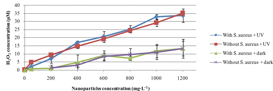
Figure 5. Influence of Staphylococcus aureus on the concentration of hydrogen peroxide (mg·L−1) after 30 minutes in the dark or under UV irradiation, for different nanoparticles concentrations. Experiment was carried out in triplicate.
For this experiment, we chose S. aureus because it was the most resistant bacterium among the four tested. Its resistance may be due to a detoxification capacity of the external environment by a catalase activity. Effectively, in order to counteract excess ROS, various antioxidant mechanisms are activated in the organisms. The initial mechanisms that act to adjust antioxidant levels to protect the cells include changes in antioxidant gene expres-sion [58].
We observed (Figure 5) that in the dark the presence of H2O2 was proportional to TiO2 concentration. The maximum value obtained was 13.23 ± 4.49 µmol·L−1 with 1200 mg·L−1 of nanoparticles in presence of S. au-reus. This result shows that even in the dark, the TiO2 nanoparticles cause the synthesis of hydrogen peroxide. Several studies indicate that certain nanomaterials, including metal oxide nanoparticles, have the potential to ex-hibit spontaneous ROS production based on material composition and surface characteristics [59]-[61]. The presence of S. aureus did not significantly affect this content. Under these conditions, it was not possible to show a detoxifying activity, by the micro-organism, in its environment.
Under UV irradiation, H2O2 concentration obtained was significantly greater than in dark condition. The maximum concentration (35 ± 1.66 µM hydrogen peroxide) was achieved with 1200 mg·L−1 of nanoparticles without S. aureus. As in the dark condition, the bacteria did not change the content of H2O2 in their extracellular environment.
The greatest resistance of S. aureus to TiO2 nanoparticles under UV irradiation is probably due to an intracel-lular detoxification process and wall thickness properties.
4. Conclusion
In this study, we synthesized stable anatase titanium dioxide nanoparticles in suspension. We evaluated the en-vironmental toxicity of suspension using Microtox® test. The Microtox® test using Vibrio fischeri has classified our nanoparticles as harmful to aquatic micro-organisms. The hydrogen peroxide quantification indicated that H2O2 was involved in the biological mechanism. The comparison between the bacteria strains showed a higher resistance with S. aureus than with E. coli and Lcr35®. This resistance may be due to the presence of the catalase gene in its genome and its thicker wall.
However, further studies are needed in order to elucidate mechanisms of toxicity induced by our TiO2 nano-particles, so it could be interesting to determine intracellular ROS concentration, lipid peroxidation level, mem-brane integrity and DNA damage. Gene expression analysis by RT-qPCR and/or RNA-Seq will also permit us to assess all the effects of our nanoparticles on the different metabolic pathways and especially on the oxidative pathway.
Acknowledgements
The authors acknowledge the University of Auvergne for its financial support and the company Probionov for the gift of Lactobacillus casei rhamnosus Lcr35® and Enterotoxigenic E. coli H10407. The authors acknowledge Yves Sibaud and Michelle Conry for their technical support.
0 5 10 15 20 25 30 35 40
0 200 400 600 800 1000 1200
H2 O2
c
o
n
c
e
n
tra
ti
o
n
(
µ
M
)
Nanoparticles concentration (mg·L−1)
With S. aureus + UV
Without S. aureus + UV
With S. aureus + dark
References
[1] Backer, L.C., Ashley, D.L., Bonin, M.A., Cardinali, F.L., Kieszak, S.M. and Wooten, J.V. (2000) Household Expo-sures to Drinking Water Disinfection By-Products: Whole Blood Trihalomethane Levels. Journal of Exposure Science and Environmental Epidemiology, 10, 321-326. http://dx.doi.org/10.1038/sj.jea.7500098
[2] Batterman, S., Zhang, L.Z. and Wang, S.Q. (2000) Quenching of Chlorination Disinfection By-Product Formation in Drinking Water by Hydrogen Peroxide. Water Research, 34, 1652-1658.
http://dx.doi.org/10.1016/S0043-1354(99)00294-8
[3] Miltner, R.J., Shukairy, H.M. and Summers, R.S. (1992) Disinfection By-Product Formation and Control by Ozonation and Biotreatment. Journal of the American Water Works Association, 84, 53-62.
[4] von Gunten, U. (2003) Ozonation of Drinking Water: Part II. Disinfection and By-Product Formation in Presence of Bromide, Iodide or Chlorine. Water Research, 37, 1469-1487. http://dx.doi.org/10.1016/S0043-1354(02)00458-X [5] Hammes, F., Salhi, E., Köster, O., Kaiser, H.P., Egli, T. and von Gunten, U. (2006) Mechanistic and Kinetic
Evalua-tion of Organic DisinfecEvalua-tion By-Product and Assimilable Organic Carbon (AOC) FormaEvalua-tion during the OzonaEvalua-tion of Drinking Water. Water Research, 40, 2275-2286. http://dx.doi.org/10.1016/j.watres.2006.04.029
[6] Sunada, K., Watanabe, T. and Hashimoto, K. (2003) Studies on Photokilling of Bacteria on TiO2 Thin Film. Journal of Photochemistry and Photobiology A: Chemistry, 156, 227-233. http://dx.doi.org/10.1016/S1010-6030(02)00434-3 [7] Tsuang, Y.H., Sun, J.S., Huang, Y.C., Lu, C.H., Chang, W.H.S. and Wang, C.C. (2008) Studies of Photokilling of
Bacteria Using Titanium Dioxide Nanoparticles. Artificial Organs, 32, 167-174. http://dx.doi.org/10.1111/j.1525-1594.2007.00530.x
[8] Guan, K.S. (2005) Relationship between Photocatalytic Activity, Hydrophilicity and Self-Cleaning Effect of TiO2/ SiO2 Films. Surface and Coatings Technology, 191, 155-160. http://dx.doi.org/10.1016/j.surfcoat.2004.02.022
[9] Mellott, N., Durucan, C., Pantano, C. and Guglielmi, M. (2006) Commercial and Laboratory Prepared Titanium Dio-xide Thin Films for Self-Cleaning Glasses: Photocatalytic Performance and Chemical Durability. Thin Solid Films, 502, 112-120. http://dx.doi.org/10.1016/j.tsf.2005.07.255
[10] Guan, H.M., Zhu, L.H., Zhou, H.H. and Tang, H.Q. (2008) Rapid Probing of Photocatalytic Activity on Titania-Based Self-Cleaning Materials Using 7-Hydroxycoumarin Fluorescent Probe. Analytica Chimica Acta, 608, 73-78.
http://dx.doi.org/10.1016/j.aca.2007.12.009
[11] Wu, D., Long, M., Zhou, J., Cai, W., Zhu, X., Chen, C. and Wu, Y. (2009) Synthesis and Characterization of Self- Cleaning Cotton Fabrics Modified by TiO2 through a Facile Approach. Surface and Coatings Technology, 203, 3728- 3733. http://dx.doi.org/10.1016/j.surfcoat.2009.06.008
[12] Chen, W.J., Tsai, P.J. and Chen, Y.C. (2008) Functional Fe3O4/TiO2 Core/Shell Magnetic Nanoparticles as Photo- Killing Agents for Pathogenic Bacteria. Small, 4, 485-491. http://dx.doi.org/10.1002/smll.200701164
[13] Choi, J.Y., Kim, K.H., Choy, K.C., Oh, K.T. and Kim, K.N. (2007) Photocatalytic Antibacterial Effect of TiO2 Film Formed on Ti and TiAg Exposed to Lactobacillus acidophilus. Journal of Biomedical Materials Research Part B: Ap-plied Biomaterials, 80, 353-359. http://dx.doi.org/10.1002/jbm.b.30604
[14] Kubacka, A., Ferrer, M., Martnez-Arias, A. and Fernández-Garca, M. (2008) Ag Promotion of TiO2-Anatase Disinfec-tion Capability: Study of Escherichia coli Inactivation. Applied Catalysis B: Environmental, 84, 87-93.
[15] Kahru, A., Tomson, K., Pall, T. and Külm, I. (1996) Study of Toxicity of Pesticides Using Luminescent Bacteria Pho-tobacterium phosphoreum. Water Science and Technology, 33, 147-154.
http://dx.doi.org/10.1016/0273-1223(96)00292-2
[16] Barrena, R., Casals, E., Colon, J., Font, X., Sanchez, A. and Puntes, V. (2009) Evaluation of the Ecotoxicity of Model Nanoparticles. Chemosphere, 75, 850-857. http://dx.doi.org/10.1016/j.chemosphere.2009.01.078
[17] Cai, R., Kubota, Y., Shuin, T., Sakai, H., Hashimoto, K. and Fujishima, A. (1992) Induction of Cytotoxicity by Pho-toexcited TiO2 Particles. Cancer Research, 52, 2346-2348.
[18] Gogniat, G., Thyssen, M., Denis, M., Pulgarin, C. and Dukan, S. (2006) The Bactericidal Effect of TiO2 Photocatalysis Involves Adsorption onto Catalyst and the Loss of Membrane Integrity. FEMS Microbiology Letters, 258, 18-24. http://dx.doi.org/10.1111/j.1574-6968.2006.00190.x
[19] Jang, H.D., Kim, S.K. and Kim, S.J. (2001) Effect of Particle Size and Phase Composition of Titanium Dioxide Nano-particles on the Photocatalytic Properties. Journal of Nanoparticle Research, 3, 141-147.
http://dx.doi.org/10.1023/A:1017948330363
[21] Xia, T., Kovochich, M., Brant, J., Hotze, M., Sempf, J. and Oberley, T. (2006) Comparison of the Abilities of Ambient and Manufactured Nanoparticles to Induce Cellular Toxicity According to an Oxidative Stress Paradigm. Nano Letters, 6, 1794-1807. http://dx.doi.org/10.1021/nl061025k
[22] Singh, N., Manshian, B., Jenkins, G.J.S., Griffiths, S.M., Williams, P.M., Maffeis, T.G.G., Wright, C.J. and Doak, S.H. (2009) NanoGenotoxicology: The DNA Damaging Potential of Engineered Nanomaterials. Biomaterials, 30, 3891- 3914. http://dx.doi.org/10.1016/j.biomaterials.2009.04.009
[23] Gou, N. and Gu, A.Z. (2011) A New Transcriptional Effect Level Index (TELI) for Toxicogenomics-Based Toxicity Assessment. Environmental Science and Technology, 45, 5410-5417. http://dx.doi.org/10.1021/es200455p
[24] Hu, C., Lan, Y.Q., Qu, J.H., Hu, X.X. and Wang, A.M. (2006) Ag/AgBr/TiO2 Visible Light Photocatalyst for Destruc-tion of Azodyes and Bacteria. Journal of Physical Chemistry B, 110, 4066-4072. http://dx.doi.org/10.1021/jp0564400 [25] Unfried, K., Albrecht, C., Klotz, L.O., Von Mikecz, A., Grether-Beck, S. and Schins, R.P.F. (2007) Cellular Responses
to Nanoparticles: Target Structures and Mechanisms. Nanotoxicology, 1, 52-71. http://dx.doi.org/10.1080/00222930701314932
[26] Verma, A., Uzun, O., Hu, Y., Hu, Y., Han, H.S., Watson, N., Chen, S., Irvine, D.J. and Stellacci, F. (2008) Surface- Structure-Regulated Cell-Membrane Penetration by Monolayer-Protected Nanoparticles. Nature Materials, 7, 588-595. http://dx.doi.org/10.1038/nmat2202
[27] Nel, A.E., Mädler, L., Velegol, D., Xia, T., Hoek, E.M.V., Somasundaran, P., Klaessig, F., Castranova, V. and Thompson, M. (2009) Understanding Biophysicochemical Interactions at the Nano-Bio Interface. Nature Materials, 8, 543-557. http://dx.doi.org/10.1038/nmat2442
[28] Verma, A. and Stellacci, F. (2010) Effect of Surface Properties on Nanoparticle Cell Interactions. Small, 6, 12-21. http://dx.doi.org/10.1002/smll.200901158
[29] Chance, B., Sies, H. and Boveris, A. (1979) Hydroperoxide Metabolism in Mammalian Organs. Physiological Reviews, 59, 527-605.
[30] Goodhew, P.J. and Humphrey, F.J. (1988) Electron Microscopy and Analysis. Taylor and Francis Inc., Philadelphia.
[31] Batdorj, B., Trinetta, V., Dalgalarrondo, M., Prevost, H., Dousset, X., Ivanova, I., Haertle, T. and Chobert, J.M. (2007) Isolation, Taxonomic Identification and Hydrogen Peroxide Production by Lactobacillus delbrueckii subsp. Lactis T31, Isolated from Mongolian Yoghurt: Inhibitory Activity on Food-Borne Pathogens. Journal of Applied Microbiology, 103, 584-593. http://dx.doi.org/10.1111/j.1365-2672.2007.03279.x
[32] Parvez, S., Venkataraman, C. and Mukherji, S. (2006) A Review on Advantages of Implementing Luminescence Inhi-bition Test (Vibrio fischeri) for Acute Toxicity Prediction of Chemicals. Environment International, 32, 265-268. http://dx.doi.org/10.1016/j.envint.2005.08.022
[33] Kahru, A., Dubourguier, H.C., Blinova, I., Ivask, A. and Kasemets, K. (2008) Biotests and Biosensors for Ecotoxicol-ogy of Metal Oxide Nanoparticles: A Minireview. Sensors, 8, 5153-5170. http://dx.doi.org/10.3390/s8085153
[34] Binaeian, E., Rashidi, A.M. and Attar, H. (2012) Toxicity Study of Two Different Synthesized Silver Nanoparticles on Bacteria Vibrio fischeri. World Academy of Science, Engineering and Technology, 67, 1219-1225.
[35] Garcia, A., Espinosa, R., Delgado, L., Casals, E., Gonzalez, E., Puntes, V., Barata, C., Font, X. and Sanchez, A. (2011) Acute Toxicity of Cerium Oxide and Iron Oxide Nanoparticles Using Standardized Tests. Desalination, 269, 136-141. http://dx.doi.org/10.1016/j.desal.2010.10.052
[36] Lopes, I., Ribeiro, R., Antunes, F.E., Rocha-Santos, T.A., Rasteiro, M.G., Soares, A.M., Gonçalves, F. and Pereira, R. (2012) Toxicity and Genotoxicity of Organic and Inorganic Nanoparticles to the Bacteria Vibrio fischeri and Salmo-nella typhimurium. Ecotoxicology, 21, 637-648. http://dx.doi.org/10.1007/s10646-011-0808-9
[37] Straganz, G.D., Glieder, A., Brecker, L., Ribbons, D.W. and Steiner, W. (2003) Acetylacetone-Cleaving Enzyme Dke1: A Novel C-C-Bond-Cleaving Enzyme from Acinetobacter johnsonii. Biochemistry Journal, 369, 573-581.
http://dx.doi.org/10.1042/BJ20021047
[38] Massard, C., Bonnet, M., Veisseire, P., Sibaud, Y., Caudron, E. and Awitor, K.O. (2013) Photokilling of Escherichia coli Using Hybrid Titania Nanoparticles Suspended in an Aqueous Liquid. Journal of Biomaterials and Nanobiotech-nology, 4,137-144. http://dx.doi.org/10.4236/jbnb.2013.42019
[39] Wang, J., Li, C., Zhuang, H. and Zhang, J. (2013) Photocatalytic Degradation of Methylene Blue and Inactivation of Gram-Negative Bacteria by TiO2 Nanoparticles in Aqueous Suspension. Food Control, 34, 372-377.
http://dx.doi.org/10.1016/j.foodcont.2013.04.046
[40] Marugán, J., van Grieken, R., Sordo, C. and Cruz, C. (2008) Kinetics of the Photocatalytic Disinfection of Escherichia coli Suspensions. Applied Catalysis B: Environmental, 82, 27-36. http://dx.doi.org/10.1016/j.apcatb.2008.01.002 [41] Nair, S., Sasidharan, A., Rani, V.V.D., Menon, D., Nair, S. and Manzoor, K. (2009) Role of Size Scale of ZnO
Materials in Medicine, 20, 235-241. http://dx.doi.org/10.1007/s10856-008-3548-5
[42] Tamboli, D.P. and Lee, D.S. (2013) Mechanistic Antimicrobial Approach of Extracellularly Synthesized Silver Nano-particles against Gram Positive and Gram Negative Bacteria. Journal of Hazardous Materials, 260, 878-884.
http://dx.doi.org/10.1016/j.jhazmat.2013.06.003
[43] Shrivastava, S., Bera, T., Roy, A., Singh, G., Ramachandrarao, P. and Dash, D. (2007) Characterization of Enhanced Antibacterial Effects of Novel Silver Nanoparticles. Nanotechnology, 18, 103-112.
[44] Premanathan, M., Karthikeyan, K., Jeyasubramanian, K. and Manivannan, G. (2011) Selective Toxicity of ZnO Nano-particles toward Gram-Positive Bacteria and Cancer Cells by Apoptosis through Lipid Peroxidation. Nanomedicine:
Nanotechnology, Biology and Medicine, 7, 184-192. http://dx.doi.org/10.1016/j.nano.2010.10.001
[45] Yadav, H.M., Otari, S.V., Koli, V.B., Mali, S.S., Hong, C.K., Pawar, S.H. and Delekar, S.D. (2014) Preparation and Characterization of Copper-Doped Anatase TiO2 Nanoparticles with Visible Light Photocatalytic Antibacterial Activi-ty. Journal of Photochemistry and Photobiology A: Chemistry, 280, 32-38.
http://dx.doi.org/10.1016/j.jphotochem.2014.02.006
[46] Yadav, H.M., Otari, S.V., Bohara, R.A., Mali, S.S., Pawar, S.H. and Delekar, S.D. (2014) Synthesis and Visible Light Photocatalytic Antibacterial Activity of Nickel-Doped Nanoparticles against Gram-Positive and Gram-Negative Bacte-ria. Journal of Photochemistry and Photobiology A: Chemistry, 294, 130-136.
http://dx.doi.org/10.1016/j.jphotochem.2014.07.024
[47] van Grieken, R., Marugán, J., Pablos, C., Furones, L. and López, A. (2010) Comparison between the Photocatalytic Inactivation of Gram-Positive E. faecalis and Gram Negative E. coli Faecal Contamination Indicator Microorganisms.
Applied Catalysis B: Environmental, 100, 212-220. http://dx.doi.org/10.1016/j.apcatb.2010.07.034
[48] Rijnaarts, H.H.M., Norde, W., Lyklema, J. and Zehnder, A. (1995) The Isoelectric Point of Bacteria as an Indicator for the Presence of Cell Surface Polymers That Inhibit Adhesion. Colloid Surface B, 4, 191-197.
http://dx.doi.org/10.1016/0927-7765(94)01164-Z
[49] Juang, D.F., Yang, P.C., Lee, C.H., Hsueh, S.C. and Kuo, T.H. (2011) Electrogenic Capabilities of Gram Negative and Gram Positive Bacteria in Microbial Fuel Cell Combined with Biological Wastewater Treatment. International Journal of Environmental Science and Technology, 8, 781-792. http://dx.doi.org/10.1007/BF03326261
[50] Klaine, S.J., Alvarez, P.J.J., Batley, G.E., Fernandes, T.F., Hande, R.D., Lyon, D.Y., Mahendra, S., McLaughlin, M.J. and Lead, J.R. (2008) Nanomaterials in the Environment: Behavior, Fate, Bioavailability, and Effects. Environmental Toxicology and Chemistry, 27, 1825-1851. http://dx.doi.org/10.1897/08-090.1
[51] Pelletier, D.A., Suresh, A.K., Holton, G.A., McKeown, C.K., Wang, W., Gu, B., Mortensen, N.P., Allison, D.P., Joy, D.C., Allison, M.R., Brown, S.D., Phelps, T.J. and Doktycz, M.J. (2010) Effects of Engineered Cerium Oxide Nano-particles on Bacterial Growth and Viability. Applied and Environmental Microbiology, 76, 7981-7989.
http://dx.doi.org/10.1128/AEM.00650-10
[52] Bartoli, M. and Dusseau, J.Y. (1995) Oxydants. In: Fleurette, J., Fresney, J. and Reverdy, M.E., Eds., Antiseptie et désinfection, Editions Eska, Paris, 305-314.
[53] Hamouda, T. and Baker Jr., J.R. (2000) Antimicrobial Mechanism of Action of Surfactant Lipid Preparations in Enteric Gram-Negative Bacilli. Journal of Applied Microbiology, 89, 397-403.
http://dx.doi.org/10.1046/j.1365-2672.2000.01127.x
[54] Sondi, I. and Salopek-Sondi, B. (2004) Silver Nanoparticles as Antimicrobial Agent: A Case Study on E. coli as a Model for Gram-Negative Bacteria. Journal of Colloid and Interface Science, 275, 177-182.
http://dx.doi.org/10.1016/j.jcis.2004.02.012
[55] Fujishima, A. and Honda, K. (1972) Electrochemical Photocatalysis of Water at a Semiconductor Electrode. Nature, 238, 37-38. http://dx.doi.org/10.1038/238037a0
[56] Carp, O., Huisman, C.L. and Reller, A. (2004) Photoinduced Reactivity of Titanium Dioxide. Progress in Solid State Chemistry, 32, 33-177. http://dx.doi.org/10.1016/j.progsolidstchem.2004.08.001
[57] Liu, H. and Yang, T.C. (2003) Photocatalytic Inactivation of Escherichia coli and Lactobacillus helveticus by ZnO and TiO2 Activated with Ultraviolet Light. Process Biochemistry, 39, 475-481.
http://dx.doi.org/10.1016/S0032-9592(03)00084-0
[58] Cushman, J.C. and Bohnert, H.J. (2000) Genomic Approaches to Plant Stress Tolerance. Current Opinion in Plant Bi-ology, 3, 117-124. http://dx.doi.org/10.1016/S1369-5266(99)00052-7
[59] Lovric, J., Cho, S.J., Winnik, F.M. and Maysinger, D. (2005) Unmodified Cadmium Telluride Quantum Dots Induce Reactive Oxygen Species Formation Leading to Multiple Organelle Damage and Cell Death. Chemistry and Biology, 12, 1227-1234. http://dx.doi.org/10.1016/j.chembiol.2005.09.008
Science and Technology, 40, 4346-4352. http://dx.doi.org/10.1021/es060589n