CASE REPORT
The Journal of Critical Care Medicine 2019;5(4):149-156
SIRS Triggered by Acute Right Ventricular
Function, Mimicked Septic Shock
Yingke He
1#, John Ong
2#*, Thuan Tong Tan
3, Brian K. P. Goh
4, Sharon G. K. Ong
5 1 Division of Anaesthesiology, Singapore General Hospital, Singapore2 Department of Engineering, Materials Engineering and Material-Tissue Interactions Group, University of Cambridge, United Kingdom
3 Department of Infectious Diseases, Singapore General Hospital, Singapore
4 Department of Hepato-pancreato-biliary and Transplant Surgery, Singapore General Hospital, Singapore 5 Department of Surgical Intensive Care, Singapore General Hospital, Singapore
Abstract
Background: The systemic inflammatory response syndrome (SIRS) is a complex immune response which can be precipitated by non-infectious aetiologies such as trauma, burns or pancreatitis. Addressing the underlying cause is crucial because it can be associated with increased mortality. Although the current literature associates chronic heart failure with SIRS, acute right ventricular dysfunction has not previously been reported to trigger SIRS. This case report describes the presentation of acute right ventricular dysfunction that triggered SIRS and mimicked septic shock.
Case presentation: A 70-year-old male presented to the Intensive Care Unit (ICU) with elevated inflammatory mark-ers and refractory hypotension after a robotic-assisted laparoscopic radical choledochectomy with pancreaticoduo-denectomy. Septic shock was misdiagnosed, and he was later found to have a pulmonary embolus. Thrombectomy and antimicrobials had no significant effect on lowering the elevated inflammatory markers or improving the per-sistent hypotension. Through Point of Care Ultrasound (POCUS), right ventricular dysfunction was diagnosed. Treat-ment with intravenous milrinone improved blood pressure, normalised inflammatory markers and led to a prompt discharge from the ICU.
Conclusion: Acute right ventricular dysfunction can trigger SIRS, which may mimic septic shock and delay appropriate treatment.
Keywords: Point of Care Ultrasound (POCUS), acute right ventricular failure, acute right ventricular dysfunction, sys-temic inflammatory response syndrome, septic shock, sepsis
Received: 31 May 2019 / Accepted: 23 September 2019
* Correspondence to: John Ong, Department of Engineering, Materials Engineering and Material-Tissue Interactions Group, University of Cambridge, Trumpington Street, CB2 1PZ, United Kingdom. E-mail: [email protected]
# Denotes joint first authorship.
Introduction
The Systemic Inflammatory Response Syndrome (SIRS) was first defined by Bone et al. (1992) [1] which describes widespread inflammation within the human body caused by a complex immune response to an in-sult [1,2]. It is characterized by the presence of two or more of the following features: (i) temperature >38°C or <36°C, (ii) heart rate >90 beats/minute, (iii) respira-tory rate >20/min or PaCO2 <32mmHg (4.3 kPa) (iv) white blood cell count >12 000/mm3 or <4000/mm3
or >10% immature bands. Although SIRS was initially thought to be within the spectrum of sepsis [3], other
precipitants of SIRS have since been acknowledged. Non-infectious causes of SIRS include, but are not limited to, pancreatitis, trauma, haemorrhage, burns, chronic heart failure, pulmonary embolism and ana-phylaxis [4-7]. Due to this non-specificity of SIRS in discriminating infection from other non-infectious causes, the definition of sepsis has since been revised to improve its management in clinical practice.
Recently, the 3rd International Consensus
Defini-tions for Sepsis and Septic Shock proposed new defini-tions for sepsis and septic shock to make it more distin-guishable from SIRS. Concerns with the sensitivity of
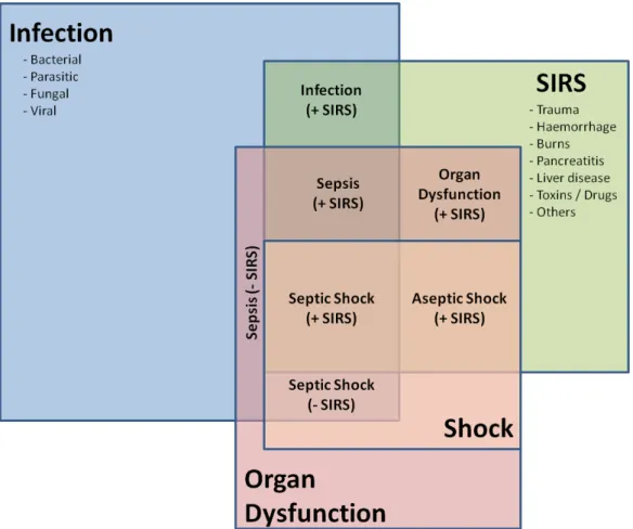
the previous definition of sepsis were raised since one in eight patients admitted to critical care units with in-fection, and recent organ failure failed to meet the cri-teria of SIRS to fulfil the definition of sepsis, but never the less experienced significant morbidity and mortal-ity [3,8]. As a result, the new definition for sepsis is now stated as “a life-threatening organ dysfunction caused by a dysregulated host response to infection”, character-ised by “an acute change of the sequential organ failure assessment (SOFA) score ≥ 2 due to infection” [3]. For septic shock, the proposed Sepsis-3 criteria are “sepsis with persisting hypotension requiring vasopressors to maintain mean arterial pressure (MAP) ≥ 65mmHg and having a serum lactate level ≥ 2 mmol/L (18mg/ dL) despite adequate fluid resuscitation [3].” However, even with these changes in definitions, clinicians must be aware that the SIRS response is still valid beyond sepsis and remain vigilant for the non-infectious caus-es that trigger it (Figure 1 summariscaus-es the relationship between infection, organ dysfunction, SIRS, sepsis and septic shock taking these revised definitions into ac-count).
The triggering of SIRS by acute isolated right ven-tricular (RV) dysfunction has not been previously re-ported. Here, we describe a case of acute RV dysfunc-tion, which through SIRS response and multi-organ failure, mimicked the presentation of septic shock. It was only through point of care ultrasound (POCUS) that the correct diagnosis was made and appropriate treatment was administered successfully.
Case Description
A 70-year-old male first presented at the accident and emergency department of a general district hospi-tal in Malacca, Malaysia, with a three-month history of painless jaundice. He also had a history of hyper-tension and morbid obesity with a body mass index (BMI) of 35kg/m2.
Magnetic resonance imaging (MRI) revealed a 1.6cm mass in the common bile duct suggestive of a cholangiocarcinoma. Endoscopic retrograde cholan-giopancreatography (ERCP) was performed, and a
bil-Fig. 1. Relationships between infection, organ dysfunction, sepsis, septic shock and SIRS taking into account revised definitions by Sepsis-3
iary stent was placed on the second day of admission to hospital as a temporary measure. The patient was discharged home the following day.
One month after his initial presentation, the patient sought further treatment in Singapore. He subsequent-ly underwent a robotic-assisted laparoscopic radical choledochectomy and pancreaticoduodenectomy at the Singapore General Hospital. A laparoscopic ap-proach was chosen to facilitate faster recovery and early mobilisation.
Due to the concern of bleeding following surgery, prophylactic anti-coagulation was omitted. Instead, the surgeon-in–charge opted for thrombo-embolic-deter-rent (TED) compression stockings and pneumatic calf compressors, which were utilised intra-operatively to minimise the risk of deep vein thrombosis. A two-gram single dose of ceftriaxone (PT Indofarma, Palembang, Indonesia) and metronidazole 500mg (Baxter, Deer-field, Illinois, USA) was administered intravenously as surgical antibiotic prophylaxis. The patient’s obese body habitus and difficult surgical access prolonged the duration of the operation, which lasted thirteen hours. Postoperatively, the patient was extubated and trans-ferred to the surgical high dependency unit (SHDU) for monitoring since he was haemodynamically stable.
On the first postoperative day, the patient developed acute kidney injury as indicated by a “kidney disease improving global outcome” (KDIGO) score of 2, and by oliguria, the urine output being <0.5ml/kg/hour over six hours. The serum creatinine levels rose from 97 to 235 µmmol/L. He was also mildly febrile with a temperature of 37.5 oC and tachycardic with a heart rate
of 110 beats/min. His blood pressure ranged between 91/59 mmHg to 116/72 mmHg. There was no overt blood loss or bilious fluid from the surgical drains.
Based on the clinical parameters of oliguria, tachy-cardia and borderline blood pressure suggesting a hypovolemic state, the on call surgeons in the SHDU administered fluid boluses totalling two litres. This did not result in any clinical improvement. Subsequently, the patient had an episode of desaturation to 91% as measured on pulse oximetry. He was mildly tachyp-noeic with a respiratory rate of 18 to 20 breaths per minute. The patient’s obese body habitus limited physi-cal examination, and it was cliniphysi-cally challenging to discern if the patient was euvolemic or hypervolemic. Chest auscultation revealed soft bilateral crepitations in the lung bases, but a chest X-Ray (CXR) demonstrated
only basal atelectasis. Given the positive fluid balance, bibasal crepitations, oliguria and rising creatinine lev-els, a provisional diagnosis of fluid overload and acute kidney injury was made. 80mg of intravenous furo-semide (Pfizer Ltd, Lake Forest, Illinois, USA) was given to facilitate diuresis, but no significant effect was observed.
On the second postoperative day, while awaiting re-nal replacement therapy, he developed transient supra-ventricular tachycardia (SVT). His heart rate was 180 beats/min and BP was 80/40 mmHg. The SVT reverted spontaneously to sinus tachycardia with a heart rate of 100-110 beats per minute and his blood pressure sub-sequently normalised. Oral bisoprolol (Sandoz, Cam-berley, UK) 1.25mg was started. However, a few hours later, the SVT recurred to 160-170 beats/min, and the BP decreased to 70/40 mmHg.
A single synchronised direct current shock of 50 Joules was administered which successfully converted the SVT to sinus rhythm and improved systolic blood pressure to 90-100mmHg.
At this point, he was then admitted to the ICU for closer monitoring. Laboratory tests showed raised in-flammatory markers; procalcitonin (PCT) 48µg/L, C-Reactive protein (CRP) 278mg/L and white cell count 3.96 x 10⁹/L. Arterial blood gas analysis
demon-strated a high anion gap metabolic acidosis (pH 7.21, BE-11, HCO3 15mmol/L and lactate 4.3mmol/L). The
laboratory blood chemistry results were as follows: urea 11.1mmol/L, creatinine 235 µmmol/L, sodium 139mmol/L, potassium 4.2mmol/L and magnesium 0.99mmol/L.
A 12-lead Electrocardiogram (ECG), apart from si-nus tachycardia post-cardioversion, did not reveal any ST changes and serial Troponin T levels were normal.
In the ICU, he had increasing oxygen requirements and was intubated by the ICU registrar on call, one hour after ICU admission. On controlled mechanical ventilation with a fractional inspired oxygen concen-tration (FiO2) of 0.8, the initial PaO2 was 69.9mmHg
and PaCO2 38 mmHg.
Given a positive culture of Enterococcus faecalis from his biliary stent taken intra-operatively, together with the clinical presentation and investigations, a presump-tive diagnosis of septic shock from intra-abdominal sepsis was made. Obstructive and cardiogenic shock were less likely to occur, given that there was no
pneu-mothorax detected on CXR, and serial ECGs together with troponin T levels were within the normal range.
Blood cultures were performed within an hour after ICU admission. Empirical broad-spectrum, intrave-nous antibiotics comprising tigecycline 50mg (Pfizer Ltd, Lake Forest, Illinois, USA) 12 hourly, meropenem 2g (Pfizer Ltd, Lake Forest, Illinois, USA) 8 hourly and anidulafungin 100mg (Merck Sharp & Dohme Ltd, Madrid, Spain) daily were started immediately after-wards.
Intravenous noradrenaline (Pfizer Ltd Lake Forest, Illinois, USA) 0.2µg/kg/min was required to maintain a mean arterial pressure above 65mmHg. However, his noradrenaline requirements began to rise soon after, and a repeat arterial blood gas analysis showed that the serum lactate had increased to 8.0 mmol/L. PCT re-mained elevated at 35 µg/L, and CRP rose to 334mg/L. Continuous renal replacement therapy was also initi-ated, given the acute kidney injury and haemodynamic instability.
Due to persistent hypotension together with esca-lating inotrope and vasopressor requirements of no-radrenaline (Pfizer Ltd, Lake Forest, Illinois, USA) 0.3µg/kg/min and vasopressin (Pfizer Ltd, Lake Forest, Illinois, USA) 0.03U/min, a point of care ultrasound (POCUS) and bedside echocardiography was per-formed two hours after ICU admission.
Ultrasonography of the chest and abdomen ruled out free fluid within the lungs and abdomen. Due to the patient’s obese habitus and overlying bowel gas, the aorta could not be visualised. Bedside echocardiog-raphy showed preserved left ventricular performance but exhibited severe right ventricular enlargement and systolic dysfunction with akinesia of the mid-ventric-ular segments but preserved contractility of the apex (McConnell sign). The inferior vena cava was partially visualized and noted to be plethoric on inspiration.
The RV was also dilated with a flattened septum, suggesting a “pressure overload” state (Figure 2). The left ventricular appeared normal. A CT pulmonary angiogram was performed within a few hours of the initial echocardiography assessment, which confirmed the diagnosis of a pulmonary embolism (PE) involving the right pulmonary artery (Figure 3). A CT scan of the abdomen and pelvis was done concurrently because of the limited views obtained by POCUS. This did not re-veal any significant intra-abdominal free fluid or source of infection, and the initial diagnosis was then revised
to “an acute RV dysfunction secondary to pulmonary embolism.”
Heparin (Baxter, Deerfield, Illinois, USA) infusion was started and titrated to maintain the partial throm-boplastin time (PTT) of about 60 seconds. An
inter-Fig. 2. Bedside echocardiography demonstrated dilated RV with flattened interventricular septum (white arrow) due to pulmonary embolism. Quality of the image was limited by patient's body habitus
Fig. 3. CT Pulmonary Angiography demonstrated a filling defect in the right pulmonary artery (white arrow) indicat-ing the presence of a pulmonary embolism
ventional radiologist performed a percutaneous pul-monary thrombectomy. Complete embolus removal was achieved, resulting in the restoration of normal blood flow in the right pulmonary artery.
However, the patient’s haemodynamic parameters and inflammatory markers continued to worsen. On the third postoperative day (ICU Day 2), bedside echo-cardiography demonstrated no improvement of the acute RV dysfunction despite complete clearance of the thrombus in the right pulmonary artery. This elucidat-ed severe RV dysfunction as the cause of persistent hy-potension. Left ventricular function remained normal. There were no significant valvular abnormalities. The diagnosis of SIRS secondary to RV dysfunction was suspected, and an intravenous milrinone (Pfizer Ltd, Lake Forest, Illinois, USA) infusion was started and titrated to a maximum dose of 0.5mcg/kg/min. Clini-cal recovery was noted a few hours later as the blood pressure was maintained above systolic 100mHg with reducing doses of vasoactive agents. The patient also started to diurese spontaneously from being anuric upon ICU admission.
On the fourth postoperative day (ICU Day 3), the noradrenaline and vasopressin infusions were ceased seventeen hours after milrinone (Pfizer Ltd, Lake For-est, Illinois, USA) was started.
Laboratory tests performed twelve hours later also showed that the inflammatory markers were normalis-ing, the base deficit had improved from -11.4 to -2.4 and lactate levels fell from 8.0 to 2.1mmol/L.
Another repeat bedside echocardiography, per-formed on ICU day three, demonstrated a significant improvement in right ventricular contractility. Oxygen requirements were also reduced with Fi02 from 0.8 to
0.4. There was improved oxygenation with the arte-rial blood gas PaO2 returning from 69mmHg to 131
mmHg. These responses to milrinone did not support the hypothesis that surgical trauma or infection was the cause of SIRS. Extubation and complete normalisation of PCT and CRP levels were achieved in the next for-ty-eight hours. The anti-microbials were stopped one week laterwhen blood cultures, urine cultures and re-peat CXR yielded no indication of infection.
The patient continued to make progress and was soon transferred to a step-down ward.
Discussion
This patient had multi-organ dysfunction with an acute rise in SOFA score from 0 to 7. His raised inflammatory markers and positive biliary stent culture, together with hemodynamic instability requiring inotropic support, fulfilled both definitions of sepsis and septic shock as defined in Sepsis-3. The combination of these factors led to his initial misdiagnosis of septic shock.
Acute right ventricular dysfunction was not sus-pected as a cause of his pro-inflammatory state because an association with SIRS had not been previously re-ported. However, given that no source of infection was found after repeated fluid cultures and imaging, it was concluded that the grossly elevated inflammatory markers were due to a SIRS response.
It was then hypothesised that the trigger of SIRS, in this patient, was RV dysfunction and not PE because no improvement was observed after treatment with heparin and the restoration of normal flow within the pulmonary artery after thrombectomy. On the con-trary, the patient continued to deteriorate despite the treatment for PE. A dramatic clinical recovery and rap-id improvement of inflammatory markers were only noted after the treatment of acute RV dysfunction with milrinone.
Hasper et al. (1998) [5] had earlier reported that the signs of systemic inflammation and SIRS-associated cytokines were elevated in patients with chronic heart failure, but interestingly both disappeared after patients were provided mechanical circulatory support. Our patient also had normal serial troponins and ECGs, which does not support myocardial infarction-induced cardiogenic shock and SIRS, and sepsis-related myo-cardial dysfunction [9-10]. Cardiogenic shock mainly arises from left ventricular dysfunction and rarely from right ventricular function [11]. Milrinone, as a bipy-ridine phosphodiesterase (PDE)-3 inhibitor, improves RV contractility and reduces pulmonary vascular re-sistance (PVR) by vasodilation. This reversed the acute RV dysfunction, which led to the resolution of SIRS and recovery of the patient.
This important observation of SIRS caused by acute RV dysfunction suggests clinicians must be vigilant for RV dysfunction mimicking or masked by septic shock because the treatment of both conditions can be
antag-onistic, e.g. intravenous fluid given for sepsis can exac-erbate right ventricular dysfunction. RV failure is clini-cally recognised as an acute RV dysfunction resulting in a low cardiac output state with hypoperfusion of major organs [12]. The classical physical findings, including ECG changes, may not always be present [13]. In acute RV pressure overload state, end-systolic flattening of the interventricular septum due to interventricular in-terdependence is usually an early sign. Sustained high RV afterload may lead to acute RV stunning, resulting in reduced contractility. This may progress to an RV volume overload state with tricuspid regurgitation and global RV enlargement [14].
Progressive venous congestion from RV dysfunction leads to impairment of renal, hepatic and intestinal function, resulting in increased morbidity and mortal-ity [15]. Therefore, the elevated inflammatory markers in our patient could also be due to the presence of he-patic and splenic congestion in right ventricular (RV) failure. Polsinelli et al (2017) and Sandek et al (2008) in reviewing RV-gut interactions in patients with heart failure, proposed that visceral venous congestion could result in hypoxia and acidosis in enterocytes, altera-tion of the gut microbiome leading to increased gut permeability and inflammation and alteration of renal haemodynamics with triggering of the cardiorenal syn-drome [15,16]. Hence, effective management of acute RV dysfunction requires accurate assessment of RV preload, contractility and afterload.
Classically, patients with RV failure are preload-de-pendent [17].However, over aggressive volume loading has the potential to over distend the RV and worsen wall tension and contractility [15]. Volume status must, therefore, be carefully evaluated, as both hypovolemia and hypervolemia can lead to a further decrease in cardiac output. In right heart dysfunction, the ideal vasoactive agent of choice is one that can reduce RV af-terload, increase RV contractility while preserving RV perfusion pressure [13]. Inodilators like milrinone and levosimendan have been used widely for the treatment of RV failure [13],[17]. Due to its vasodilatory proper-ties, milrinone is often used in conjunction with a va-sopressor.
It is clinically challenging to differentiate between sepsis and its mimickers. Studies on proteins and cy-tokines related to the host response to infection have no definitive conclusion on how to distinguish
inflam-mation from sepsis using biomarkers such as CRP, In-terleukin 6 and 8, soluble TREM1 and PLAUR[18-20].
A recent systematic review and meta-analysis by Tan et al. (2019) looking at the diagnostic accuracy of both PCT and CRP concluded that PCT was a better diagnostic tool for sepsis [21]. Their data gave an AU-ROC score CRP: 0.73 (95% CI 0.69 - 0.77) vs AUAU-ROC score PCT: 0.85 (95% CI 0.82 - 0.88). However, both biomarkers were found to have moderate specificity with wide confidence intervals. Interestingly, recent advancements in biotechnology have facilitated DNA-based molecular techniques which produce rapid mi-crobial identification and antimimi-crobial susceptibility with high sensitivity and specificity. This may prove to be another tool in the armamentarium for differentiat-ing between sepsis and sepsis mimickers [22].
In our experience, POCUS utilising the Rapid Ultra-sound in Shock (RUSH) protocol is another useful tool in differentiating the different types of shock in both emergency departments and critical care units [23-25]. Table 1 summarises the salient ultrasonographic fea-tures of the different types of shock. A meta-analysis of the diagnostic accuracy of using the RUSH protocol in the emergency department by Keikha et al. (2018) re-ported a higher sensitivity and specificity in diagnosing hypovolemic, cardiogenic and obstructive shock but lower sensitivity for shock with distributive or mixed aetiology [26]. Despite its limitations, there is growing popularity for the use of POCUS in the intensive care setting. Being portable and non-invasive, POCUS pro-vides physicians with useful information for the ma-jority of critically ill patients, especially in those whom heart failure, valvular heart disease, hypovolaemia and hypervolemia are suspected [27]. In our opinion, PO-CUS is an extremely useful and consideration should be made to incorporate it into intensive care training.
Conclusion
Despite recent revisions to the definition of sepsis and septic shock, clinicians should be aware that SIRS re-mains a clinical problem that can be precipitated by non-infectious aetiologies. Acute RV dysfunction can trigger a SIRS response that mimics sepsis, and septic shock if the RV dysfunction is severe.
Conflict of Interest
Table 1. Bedside Point of Care Ultrasound (POCUS) examination findings by systems to evaluate the aetiology of shock
SYSTEMS Hypovolemic Shock Cardiogenic Shock Distributive ShockSHOCK Obstructive Shock
Cardiovascular
Findings Hypercontractile heart with small Left Ventricle chamber size
Hypocontractile heart with dilated Left Ventricle
Hypercontractile heart in early sepsis
Hypocontractile heart in late sepsis
Cardiac Tamponade - Presence of pericardial fluid
- Right ventricle collapse during diastole
Presence of Cardiac Thrombus
Right Ventricle Dysfunc-tion (Pulmonary Embo -lism)
- Hypocontractile RV with dilatation - Moderate to severe tricuspid regurgitation - McConnell’s sign (re -duced right ventricular free wall motion but reserved apical motion) - D shaped ventricle with abnormal motion in interventricular septum Respiratory
Findings Hemothorax- Presence of ho-mogeneous echoic effusion/Hematocrit Sign
Pulmonary edema - Lung rockets with multiple diffuse B lines Pleural Effusion - Presence of free pleural fluid Pneumonia
- Presence of Air broncho-gram
- Loss of A lines,
- Presence of patchy B lines - Pleural effusion with/with -out multiple septations
Tension Pneumothorax - Absence of normal lung sliding - Absence of seashore sign on M mode - Presence of barcode sign on M mode Abdominal and Other Findings Collapsed Inferior Vena Cava (< l2mm) Ruptured/Leaking Abdominal Aortic Aneursym
- Focal aortic dilata -tion - Peri-aortic fluid, free intraperitoneal fluid, retroperitoneal fluid Aortic dissection - Presence of aortic root dilatation and intimal flap on the Parasternal Long Axis or longitudinal views (transthoracic or transabdominal) Distended Inferior Vena Cava (> 20mm)
Collapsed Inferior Vena Cava (<12mm)
Peritonitis
- Presence of peritoneal fluid with/without septation Pyelonephritis
- Swollen kidney with in-creased anechoic corticome -dullary area
Cholecystitis
- Presence of cholelithiasis - Sonographic Murphy sign - Gallbladder wall thicken-ing (>3mm) and presence of pericholecystic fluid
Cholangitis
- Common bile duct dilata-tion
- Thickening of wall of bile ducts
- Presence of debris in the common bile ducts (pus or sludge)
Distended Inferior Vena Cava (> 20mm)
Presence of Lower Limb Deep Vein Thrombosis
REFERENCES
1. Bone RC, Balk RA, Cerra FB, et al. Definitions for sepsis and organ failure and guidelines for the use of innovative therapies in sepsis. The ACCP/SCCM Consensus Conference Committee. American College of Chest Physicians/Society of Critical Care Medicine. Chest. 1992;101:1644-55.
2. Levy MM, Fink MP, Marshall JC, et al. SCCM/ESICM/ACCP/ATS/ SIS International Sepsis Definitions Conference. Intensive Care Med. 2003;29:530-8.
3. Singer M, Deutschman CS, Seymour CW, et al. The Third
International Consensus Definitions for Sepsis and Septic Shock (Sepsis-3). JAMA. 2016;315:801-10.
4. Bone RC. Toward a theory regarding the pathogenesis of the systemic inflammatory response syndrome: what we do and do not know about cytokine regulation. Crit Care Med. 1996;24:163-72.
5. Hasper D, Hummel M, Kleber FX, Reindl I, Volk HD. Systemic inflammation in patients with heart failure. Eur Heart J. 1998;19:761-5.
6. Kapoor T and Gutierrez G. Air embolism as a cause of the systemic inflammatory response syndrome: a case report. Crit Care. 2003;7:98-100.
7. Horeczko T, Green JP, Panacek EA. Epidemiology of the Systemic Inflammatory Response Syndrome (SIRS) in the Emergency Department. West J Emerg Med. 2014;15: 329-36.
8. Kaukonen KM, Bailey M, Pilcher D, Cooper DJ, Bellomo R. Systemic inflammatory response syndrome criteria in defining severe sepsis. N Engl J Med. 2015;372:1629-38.
9. Kohsaka S, Menon V, Lowe AM, et al. Systemic inflammatory response syndrome after acute myocardial infarction
complicated by cardiogenic shock. Arch Intern Med.
2005;165:1643-50.
10. Hussain N. Elevated cardiac troponins in the setting of systemic inflammatory response syndrome, sepsis, and septic shock. ISRN Cardiol. 2013;723435:1-7
11. Benedek T, Dobreanu D. Current Concepts and New Trends in the Treatment of Cardiogenic Shock Complicating Acute Myocardial Infarction. J Crit Care Med (Targu Mures). 2015;1:5-10.
12. Grignola JC, Domingo E. Acute Right Ventricular Dysfunction in Intensive Care Unit. BioMed Res Int. 2017;8217105:1-15. 13. Kholdani CA, Fares WH. Management of Right Heart Failure in
the Intensive Care Unit. Clin Chest Med. 2015;36:511-20. 14. Markley RR, Ali A, Potfay J, Paulsen W, Jovin IS. Echocardiographic
Evaluation of the Right Heart. J Cardiovasc Ultrasound. 2016;24:183-90.
15. Polsinelli VB, Sinha A, Shah SJ. Visceral Congestion in
Heart Failure: Right Ventricular Dysfunction, Splanchnic
Hemodynamics, and the Intestinal Microenvironment. Curr
Heart Fail Rep. 2017;14:519-28.
16. Sandek A, Rauchhaus M, Anker SD, von Haehling S. The
emerging role of the gut in chronic heart failure. Curr Opin Clin
Nutr Metab Care. 2008;11:632-9.
17. Harjola V-P, Mebazaa A, Čelutkienė J, et al. Contemporary management of acute right ventricular failure: a statement from the Heart Failure Association and the Working Group on Pulmonary Circulation and Right Ventricular Function of the European Society of Cardiology: Contemporary management of acute RV failure. Eur J Heart Fail. 2016;18:226-41.
18. Harbarth S, Holeckova K, Froidevaux C, et al. Diagnostic value of procalcitonin, interleukin-6, and interleukin-8 in critically ill patients admitted with suspected sepsis. Am J Respir Crit Care Med. 2001;164:396-402.
19. Latour-Perez J, Alcala-Lopez A, Garcia-Garcia MA, et al.
Diagnostic accuracy of sTREM-1 to identify infection in critically ill patients with systemic inflammatory response syndrome. Clin Biochem. 2010;43:720-4
20. Backes Y, van der Sluijs KF, Mackie DP, et al. Usefulness of suPAR as a biological marker in patients with systemic inflammation or infection: a systematic review. Intensive Care Med. 2012;38:1418-28.
21. Tan M, Lu Y, Jiang H, Zhang L. The diagnostic accuracy of procalcitonin and C-reactive protein for sepsis: A systematic review and meta-analysis. J. Cell. Biochem. 2019;120:5852–9.
22. Burrack-Lange SC, Personne Y, Huber M, et al. Multicenter
assessment of the rapid Unyvero Blood Culture molecular
assay. J Med Microbiol. 2018;67:1294-301.
23. Perera P, Mailhot T, Riley D, Mandavia D. The RUSH exam: Rapid ultrasound in shock in the evaluation of the critically ill. Emerg Med Clin North Am. 2010;28:29–56.
24. Jain SS, Toraskar KK, Khan AH, Loya YS. Application of rapid ultrasound in shock protocol in the ICU for management of
shock. Indian J Crit Care Med. 2014;18: 550–1.
25. Ghane MR, Gharib M, Ebrahimi A, et al. Accuracy of early
rapid ultrasound in shock (RUSH) examination performed by emergency physician for diagnosis of shock etiology in critically ill patients. J Emerg Trauma Shock. 2015;8:5–10.
26. Keikha M, Salehi-Marzijarani M, Soldoozi Nejat R, Sheikh Motahar Vahedi H, Mirrezaie SM. Diagnostic Accuracy of Rapid Ultrasound in Shock (RUSH) Exam; A Systematic Review and Meta-analysis. Bull Emerg Trauma. 2018;6:271-8.
27. Bernier-Jean A, Albert M, Shiloh AL, Eisen LA, Williamson D, Beaulieu Y. The Diagnostic and Therapeutic Impact of Point-of-Care Ultrasonography in the Intensive Care Unit. J Intensive Care Med. 2017;32:197-203