Adhesive capsulitis:
a case report
Mohsen Kazemi,
RN, DC, FCCSS(C)*
Adhesive capsulitis or frozen shoulder is an uncommon entity in athletes. However, it is a common cause of shoulder pain and disability in the general population. Although it is a self limiting ailment, its rather long, restrictive and painful course forces the affected person to seek treatment. Conservative management remains the mainstay treatment of adhesive capsulitis. This includes chiropractic manipulation of the shoulder, therapeutic modalities, mobilization, exercise, soft tissue therapy, nonsteroidal anti-inflammatory drugs, and steroid injections. Manipulation under anesthesia is advocated when the conservative treatment fails. A case of secondary adhesive capsulitis in a forty-seven-year-old female recreational squash player is presented to illustrate clinical presentation, diagnosis, radiographic assessment and conservative chiropractic management. The patient’s shoulder range of motion was full and pain free with four months of conservative chiropractic care.
(JCCA 2000; 44(3):169–176)
K E YW O R D S: active release techniques, adhesive capsulitis, adult, chiropractic, frozen shoulder, manipulation, rehabilitation, racquet sports injury.
La capsulite rétractile ou épaule bloquée se rencontre rarement chez les athlètes, mais elle est une cause fréquente de douleur à l’épaule et d’incapacité dans la population en général. Même s’il s’agit d’un trouble spontanément résolutif, son évolution plutôt longue, gênante et douloureuse incite les personnes atteintes à consulter un professionnel de la santé. Le traitement conservateur reste le pilier de la thérapeutique de la capsulite rétractile. Il comprend des manipulations chiropratiques de l’épaule, l’application de différentes formes de thérapie, la mobilisation, des exercices, le traitement des tissus mous, l’administration d’anti-inflammatoires non stéroïdiens et des injections de stéroïdes. Les manipulations sous anesthésie sont préconisées dans les cas d’échec du traitement conservateur. Voici le cas d’une femme de 47 ans souffrant de capsulite rétractile secondaire à la pratique de la balle au mur avec raquette durant ses heures de loisir; on y présente le tableau clinique, le diagnostic, l’évaluation radiographique et le traitement
chiropratique conservateur. L’amplitude de mouvement de l’épaule est redevenue normale et indolore au bout de quatre mois de traitement chiropratique conservateur.
(JACC 2000; 44(3):169–176)
M O T S C L É S : techniques de libération active, capsulite rétractile, adulte, chiropratique, épaule bloquée,
manipulations, réadaptation, blessures dues aux sports de raquette.
Introduction
Squash is a moderate to high intensity sport.1,2 Players are active 50 to 70% of the playing time. Eighty percent of the time, the ball is in play ten seconds or less and there are eight second rest intervals.2 The most common injuries in indoor racquet sports include contusions, sprains, strains, lacerations, eye injuries, bursitis and tendinitis.3,4 Squash players over the age of 40 as well as the beginners are at increased risk for injury.4 Increased frequency of play among females was also associated with elevated injury rates.4 An injured squash player rarely becomes perma-nently impaired.4 Chard and Lachmann5 in their 8-year retrospective study of the injuries in the racquet sports found that the proportion of squash injuries was higher than expected and properly related to higher physical stress and risk of contact in this sport. They also reported that the squash injuries occurred mainly in persons over 25 years of age. Adhesive capsulitis is one of the most com-mon causes of shoulder pain and disability in the general population.6 However, it is an uncommon entity in ath-letes.7 There is no report of frozen shoulder syndrome as a result of squash injury in the literature. The incidence of adhesive capsulitis in the general population is 2–5% and 10–20% in diabetics.8 It affects females slightly more than males and is usually seen in ages 40–70.8,9 The nondominant arm is more likely to be affected.8,9 About 12% of individuals affected develop the condition bilater-ally. Recurrence is rare in the same shoulder.10 Adhesive capsulitis is also called, frozen shoulder syndrome, peri-articular adhesions, pericapsulitis, irritative capsulitis, periarthritis of the shoulder, scapulohumerale periarthritis, humeroscapular fibrositis, bursitis calcerea, Duplay’s syn-drome, shoulder portion of shoulder-hand synsyn-drome, and stiff and painful shoulder.8
The frozen shoulder was first described as periarthritis involving the periarticular soft tissues of the shoulder by Duplay in 1872.11 Codman12 coined the term “frozen
arthroscopically as pre-adhesive, synovitis, maturation and the chronic stage. The etiology of adhesive capsulitis remains unknown. However, rotator cuff disease, im-pingement syndrome, chest or breast surgery, diabetes, prolonged immobilization, age, thyroid disease, various medical problems (pulmonary disease, myocardial infarc-tion, cerebrovascular accident), and autoimmune disease are associated with the development of adhesive cap-sulitis.15 The idiopathic adhesive capsulitis symptoms have been divided into three phases clinically: (1) the pain-ful phase, (2) the stiffening phase, and (3) the thawing phase.16 Adhesive capsulitis may complete its course in 12–42 months, but it may be as early as 6 months or as late as 10 years.8
Conservative management remains the mainstay treat-ment of the frozen shoulder.
A case report is presented to illustrate clinical presenta-tion, etiology, diagnosis, radiological assessment and conservative chiropractic management of a secondary ad-hesive capsulitis in a middle age female squash player.
Case report
ment of the left arm, lying on the left arm and she was awakened at night when she rolled onto the affected arm. The pain was slightly relieved by taking hot showers.
On examination, upper extremity deep tendon reflexes were 2+ bilaterally and light touch sensation examination was unremarkable. Cervical spine right lateral flexion, right rotation and flexion induced pulling sensation in the antagonist muscle groups (trapezius, levator scapulae and scaleni muscles). Aberrant motions with tenderness were detected in the left C2–3, C7–T1 and T3–4 facet joints upon static and motion palpation . However, the left shoul-der pain could not be reproduced by the neck examinations (i.e. range of motion, soft tissue palpation, Spurling’s, Jackson’s and maximal compression tests). Her left gleno-humeral joint active ranges of motion (ROM) were: inter-nal rotation 15 degrees, exterinter-nal rotation 10 degrees, forward flexion 20 degrees, extension 30 degrees and ab-duction 10 degrees. The resisted left glenohumeral joint flexion, abduction, internal and external rotations were graded 3/5. The left glenohumeral joint passive ROM was 5 degrees more in each direction. Posterior and poster-oinferior joint play of the left glenohumeral joint were restricted and painful. Her left scaleni, upper trapezius, infraspinatus, supraspinatus and teres muscles were hyper-tonic and tender upon palpation. There was a severe point tenderness over the left deltoid tubercle. The cervical spine
and the left shoulder radiographs were unremarkable. A clinical diagnosis of the left adhesive capsulitis was made with associated cervical and upper thoracic facet joint dysfunctions.
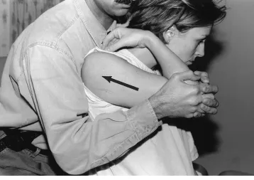
Initial treatment consisted of interferential current therapy, anteroposterior and long axis distraction mobili-zation, pendular home exercises for the left shoulder, soft tissue therapy (STT) and the spinal manipulation therapy (SMT) of the cervical and the thoracic spine three times per week for two weeks. At the sixth visit, the active left glenohumeral (initial range in brackets) abduction, flexion and external rotation were 35 (10), 60 (20) and 15 (10) degrees respectively. However, the passive ROM showed even more improvements where the passive abduction, flexion and external rotation were 70, 80 and 20 degrees respectively. At this point, strengthening exercises using rubber tubing including external rotation, forward flexion and abduction were introduced. In addition, the left gleno-humeral joint long axis distraction, anteroposterior and posteroinferior manual high speed low amplitude manipu-lation (Figures 1, 2 and 3),17 tender point therapy and ac-tive release techniques (ART)18 over the hypertonic and tender muscles ( trapezius, levator scapulae, lateral del-toid, supraspinatus and infraspinatus muscles) were started. Tender point therapy was also utilized by exerting digital compressive pressure over the tender points
pal-Figure 1 Glenohumeral long axis distraction adjustment.
Figure 3 Glenohumeral posteroinferior adjustment.
Patient is in supine position with the arm in forward flexion and the elbow bent. The practitioner grasps the arm with both hands, removes the joint slack and delivers a quick and shallow thrust inferiorly and posteriorly (arrowhead).
Figure 2 Glenohumeral anteroposterior adjustment.
Patient is seated with the arm in 900
forward flexion and the elbow fully flexed. Doctor stands behind the patient to stabilize the scapula, cups the olecranon with both hands, removing the joint slack and
delivering a quick and shallow thrust along the axis of humerus.
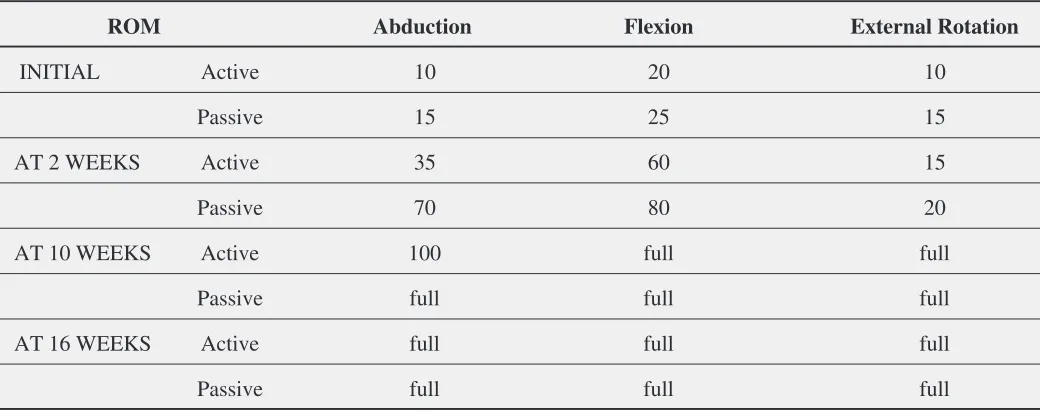
pated in the involved muscles. This pressure was gauged to the patient’s tolerance and was sustained until the patient reported the dissipation of the pain. The ART was done utilizing digital longitudinal tension along the involved muscle fibers. Starting at the shortest position of the mus-cle, the patient actively moves toward the longest position of the muscle while the tension is sustained by the treating clinician. This course of treatment was administered two times a week for eight weeks. At the end of this course of treatment, the patient’s left glenohumeral active abduction was 100 degrees and her flexion and external rotation were full. The left glenohumeral passive abduction was full but the resisted remained 4/5. The patient was assessed for isotonic strengthening exercises using free weights. A regimen of weight training exercises (isotonic) for shoul-der abduction, forward flexion, extension, internal and ex-ternal rotations three times per week were prescribed. In addition, the interferential current therapy was changed to microcurrent therapy to promote healing. The patient was seen once a week for six weeks there after. At the end of the sixteen weeks treatment, the patient had pain free full range of motion. (Table 1.)
Discussion
Lundberg19 classified patients suffering from frozen shoulder syndrome into “primary” and “secondary”. Pri-mary adhesive capsulitis pertains to those patients who present with no significant findings in the history, clinical examination, or radiographic evaluation to explain their motion loss and pain. However, patients with secondary adhesive capsulitis disclose a trauma or surgery to the af-fected upper extremity prior to their shoulder symptoma-tology.20 The patient in this case report, is classified as secondary adhesive capsulitis because of her neck in-volvement initially and squash trauma later. Reeves16 identified three phases in the natural history of the frozen shoulder syndrome: (1) an early painful phase lasting 10– 36 weeks; (2) an intermediate, stiff or frozen phase charac-terized mainly by limited range of motion lasting 4–12 months; (3) a recovery or thawing phase lasting 5–24 months or more. Our patient was in phase one at her initial visit to our office. However, she might had been in the recovery phase prior to her squash injury.
The historyusually indicates a gradual onset of stiffness and pain. The pain is quite intense and is often referred to
Table 1
Improvement of the patient’s left shoulder active and passive range of motion (ROM, in degrees) with treatment
ROM Abduction Flexion External Rotation
INITIAL Active 10 20 10
Passive 15 25 15
AT 2 WEEKS Active 35 60 15
Passive 70 80 20
AT 10 WEEKS Active 100 full full
Passive full full full
AT 16 WEEKS Active full full full
the insertion of the deltoid, the deltoid muscle region and the bicipital tendon.8,9,10 The pain is aggravated by the shoulder movements, especially external rotation, and sleeping on the involved side, and is relieved by limiting the use of the extremity.8,9,10 There is often soreness in the proximal upper back and neck which may be as a result of compensatory overuse of the accessory musculature (tra-pezius, scalene, levator scapulae, rhomboid muscles). The patient may complain of difficulty putting on a coat, reach-ing into the hip pocket for a wallet, combreach-ing his or her hair, and inability to fasten garments behind the back.8These symptoms closely resemble the patient in this case.
Some authors state that observation reveals guarded shoulder movements.8,9 At rest, the patient holds the in-volved arm in adduction and internal rotation.8 The arm swing in gait is usually limited or absent.8 Rounded shoul-ders, stooped posture with the involved shoulder elevated in a protective manner are commonly seen in these pa-tients8. Because of this altered posture, pain and trigger points often develop over the posterior aspect of the shoul-der, along the upper trapezius, and in the posterior cervical region.8
Disuse atrophy may be seen in the rotator cuff, deltoid,
biceps and triceps brachii muscles.10The active and pas-sive glenohumeral ROM are restricted.6,8–18 This limita-tion of ROM is characteristic of a capsular pattern; that is, external rotation is limited more than abduction, which is restricted more than internal rotation.10 In addition, for-ward flexion is also limited in patients with adhesive capsulitis. Naviaser and Naviaser9 stated: “At the limits of motion there is a sense of mechanical blockage or tether-ing of the joint rather than resistance because of pain.” Hence, it is not the patient resisting the motion because of the pain but rather there is a mechanical blockage (espe-cially in passive ROM) that limits the motion. Anterior, inferior glides and lateral distraction of the humerus on the glenoid are also restricted.8,10 These joint play findings
joints at the limit of the passive ROM is the most signifi-cant physical finding.
Radiographic examination of the shoulder is required in the patients with adhesive capsulitis to exclude other con-ditions.6,8–10,15,20 Plain x-rays of these shoulders ranges from normal to osteopenic with degenerative changes, cal-cium deposits, or cystic changes in long-standing cases.8 The shoulder radiographs in our patient were normal. Pearsall and Speer20recommend anteroposterior (AP), ax-illary and supraspinatus outlet views of the affected shoul-der. The AP film is assessed for osteopenia, bony abnormalities and superior migration of the head of the humerus. The axillary view is obtained to assess gleno-humeral subluxation or glenoid or gleno-humeral head articular damage. Finally, the supraspinatus outlet film is scruti-nized for supraspinatus outlet narrowing characterizing acromial impingement.14 This view was not taken in our patient.
Bone scans may show increased uptake in the affected shoulder; often a nonspecific finding.6,8,15 Shoulder ar-thrography shows loss of the normal axillary recess and a significantly decreased capsular volume.15 Although ar-thrography might be helpful, it is an invasive procedure that is painful and costly.8 MRI can be a better choice since it is not invasive and the MRI examination of the frozen shoulder in comparison to the normal shoulder shows thickened joint capsule and synovium.21
Many therapeutic regimens have been advocated for adhesive capsulitis. These include: TENS,22 interferential current therapy,23 therapeutic ultrasound,23 utilization of heat and ice,23 shoulder mobilization,8,10,24 manipula-tion of the neck and shoulder using activator,25,26 trigger point therapy,27,28 exercise therapy,6,8–10,15,22,25,26 anti-inflammatory medications,6,8,10,15,22,25 corticosteroid in-jections,6,8,29 arthrographic infiltration,1 and manipulation under anesthesia.6,8–10,30
ment period. Our patient was initially instructed to do pendular exercises (patient leans on the wall or on a table/ counter with the good arm and lets the affected arm hang, then moves the affected arm in flexion/extension, abduc-tion/adduction and circumduction clock-wise and counter clock-wise as a pendulum) wall walking with hands, utili-zation of pulleys and broom stick for stretching the tight muscles and structures. Emphasis was placed on the exter-nal rotation. Later on, as the pain decreased, strengthening exercises were added.
Polkinghorn25,26 reported successful treatment of two cases of adhesive capsulitis utilizing a mechanical force, manually assisted short lever adjusting device (activator) to manipulate the affected shoulder, cervical and thoracic spine. However, in the case presented, the author used high velocity low amplitude manual adjustments17 which were well tolerated by the patient. It is the author’s experience and opinion that a trained manual adjustor is skilled enough to modulate the force and the speed of the manipu-lation to achieve the desired outcome in the patient’s com-fort zone. Ferguson28 reported three cases of frozen shoulder which he successfully treated using trigger point therapy techniques. Although no trigger point was found in our patient, tender points were treated. Active Release Technique was utilized on the rotator cuff and the scapular stabilizing muscles to release the adhesions in the muscles caused by the restricted ROM and disuse associated with adhesive capsulitis.
Conclusions
Many authors suggest that adhesive capsulitis is a clinical diagnosis of exclusion. However, it can be identified by the restricted capsular pattern and forward flexion, pain, and severely decreased passive ROM with feeling of a mechanical block at the end range. A case report is presented to illustrate the potentials of conservative chiro-practic management and manual chirochiro-practic manipula-tion of the shoulder in a patient with adhesive capsulitis. However, further research is required to identify the effec-tiveness of this treatment.
References
1 Lock S, Colquhoun D, Briner M, Ellis L, O’Brien M, Wollstein J, Allen G. Squash racquets. A review of physiology and medicine. Sports Med 1997 Feb; 23(2):130–138.
2 Montpetit RR. Applied physiology of squash. Sports Med 1990 Jul; 10(1):31–41.
3 Silko GJ, Cullen PT. Indoor racquet sports injuries. Am Fam Physician 1994 Aug 50(2):374–378, 383–384. 4 Berson BL, Rolnick AM, Ramos CG, Thornton J. An
epidemiologic study of squash injuries. Am J Sports Med 1981 Mar–Apr; 9(2):103–136.
5 Chard MD, Lachmann SM. Racquet sports – patterns of injury presenting to a sports injury clinic. Br J Sports Med 1987 Dec; 21(4):150–153.
6 Anton H.A. Frozen shoulder. Canadian Fam Phys 1993; 39:1773–1778.
7 Reid DC. Sports injury assessment and rehabilitation. New York: Churchill Livingstone Inc. 1992; 942.
8 Grubbs N. Frozen shoulder: A review of literature. JOSPT 1993; 18(3):479–487.
9 Naviaser RJ Naviaser TJ. The frozen shoulder diagnosis and management. Clin Orthop and Related Research 1987; 223:59–64.
10 Wadsworth CT. Frozen shoulder. Phys Therapy 1986; 66(12):1878–1883.
11 Duplay ES. De la periarthrite scapulohumerale et des raideurs de l’epaule qui en son la consequence. Arch Gen Med 1872; 20:513–542.
12 Codman EA. The shoulder, Boston: Thomas & Todd Co., 1934.
13 Naviaser JS. Adhesive capsulitis of the shoulder: A study of the pathological findings in periarthritis of the shoulder. J Bone Joint Surg 1945; 27:211–222.
14 Naviaser TJ. Adhesive capsulitis. Orthop Clin of North Am 1987; 18(3):439–443.
15 Miller MD, Wirth MA, Rockwood CA. Thawing the frozen shoulder: The “Patient” patient. Orthopedics 1996; 19(10):849–853.
16 Reeves B. The natural history of the frozen shoulder syndrome. Scand J Rheumatol 1975; 4:193–196. 17 Bergman TF, Peterson DH, Lawrence DJ. Chiropractic
Technique. New York: Churchill Livingstone Inc. 1993; 555,564,565,568.
18 Leahy PM. Active release techniques, soft tissue management system for the upper extremity. Colorado Springs: Active Release Techniques, 1996; pages: 40,52,54,58,68.
19 Lundberg BJ. The frozen shoulder. Acta Orthop. Scand.(suppl.) 1969; 119:1–59.
21 Emig EW, Schweitzer ME, Karasick D, Lubowitz J. Adhesive capsulitis of the shoulder: MR diagnosis. AJR 1995; 164:1457–1459.
22 Rizk TE, Christopher RP, Pinals RS, Higgins AC, Frix R. Adhesive capsulitis (frozen shoulder): A new approach to its management. Arch Phys Med Rehabil 1983; 64:29–33. 23 Mao CY, Jaw WC, Cheng HC. Frozen shoulder:
Correlation between the response to physical therapy and follow-up shoulder arthrography. Arch Phys Med Rehabil 1997; 78:857–859.
24 Lawrence DJ. The frozen shoulder: conservative management. JACA 1981; 18:S49–S51.
25 Polkinghorn BS. Chiropractic treatment of frozen shoulder syndrome (adhesive capsulitis) utilizing mechanical force, manually assisted short lever adjusting procedures. JMPT 1995; 18(2):105–115.
26 Polkinghorn BS. Instrumental chiropractic treatment of frozen shoulder associated with mixed metastatic carcinoma. JMPT 1995; 7(3):98–102.
27 Goodheart GJ. The Afrozen shoulder” syndrome. The Dig of Chirop Econom 1969; 12:36–38.
28 Ferguson LW. Treating shoulder dysfunction and Afrozen shoulder”. Chirop Tech 1995; 7(3):73–81.
29 Baslund B, Thomsen BS, Jensen EM. Frozen shoulder: Current concepts. Scad J Rheumatology 1990;
19:321–325.
30 Hill JJ, Bogumill H. Manipulation in the treatment of frozen shoulder. Orthopedics 1988; 11(9):1255–1260.
CHIROPRACTIC FOUNDATION FOR SPINAL RESEARCH
The C.F.S.R. is a registered Charitable foundation dedicated to quality Chiropractic research. We appreciate your continued support.
Please send your
Tax Deductible Donation
TODAY:Donations
and/or