http://www.sciencepublishinggroup.com/j/ass doi: 10.11648/j.ass.20180602.14
ISSN: 2376-6174 (Print); ISSN: 2376-6182 (Online)
Full or Half-Thickness Continuous Suture for Right Atrium
Incision in Adult Patients Undergoing Open Cardiac
Surgery: the Practical Strategy to Reduce Incision Bleeding
Qing-Chun Zhang, Hai-Hui Yin, Jian-An Li, Hong Lei, Jian-Jun Ge
Department of Cardiac Surgery, the First Affiliated Hospital to University of Science and Technology of China, Hefei, China
Email address:
To cite this article:
Qing-Chun Zhang, Hai-Hui Yin, Jian-An Li, Hong Lei, Jian-Jun Ge. Full or Half-Thickness Continuous Suture for Right Atrium Incision in Adult Patients Undergoing Open Cardiac Surgery: the Practical Strategy to Reduce Incision Bleeding. Advances in Surgical Sciences. Vol. 6, No. 2, 2018, pp. 62-66. doi: 10.11648/j.ass.20180602.14
Received: June 4, 2018; Accepted: August 27, 2018; Published: October 10, 2018
Abstract:
Objective The aim of this study was to evaluate the beneficial effects and applicability of a half-thickness continuous suture outside the pectinate muscles used in the right atrial incision for adult patients undergoing cardiac surgery by cardiopulmonary bypass. Methods A total of 1040 consecutive adult patients undergoing cardiac surgery through a right atrium incision during the period of January 2010 to June 2014 were randomly allocated to the experimental group (n=522) and the control group (n=518). In the experimental group, a half-thickness continuous suture outside the pectinate muscles was used to close the right atrium incision, while the traditional method of a continuous full-thickness everting suture was used in the control group. The occurrences of bleeding spots in the right atrium sutures were recorded prior to the pericardial closure procedure. Results The occurrence rates of intra-operative bleeding from the atrial incision site for the experimental group were significantly lower than that in the control group (46 vs. 253 patients needed re-stitch procedures for the right atrial incision respectively, p=0.001). In the control group, two cases of re-exploration due to post-operative bleeding in the right atrial incision occurred, while none occurred in the experimental group. Conclusions The half-thickness continuous suture outside the pectinate muscles for closing right atrium incisions could effectively reduce the need for re-stitch procedures, as well as minimize the risks of bleedings for patients undergoing cardiac surgeries.Keywords:
Right Atrium Closure, Pectinate Muscles, Suturing, Bleeding, Cardiac Surgery1. Introduction
The most common factors attributed to post-operative bleeding events involve possible injuries to arteries, the cardiac incision and the chest wall [1-3]. Bleeding episodes from cardiac incision frequently occur in the right atrial sutures in patients who have undergone open-heart surgery through the right atrium path, and a higher incidence of local bleeding in the right atrial sutures has been found during the intra-operative examination before pericardiorrhaphy. Significant amount of patients had pulmonary hypertension and atrial fibrillation with heart remodelling and thin right atrial wall, the usual cause of anastomotic bleeding when suturing the right atrium incision site is the roughness and varied depths of the inner side of the atrial wall. The conventional method is to close the right atrium using the
2. Methods
2.1. Study Design
During the period of January 2010 and July 2014, adult patients undergoing open-heart surgeries via right atrial incision under cardiopulmonary bypass were recruited into this study. Using a set of random sequences, the consecutively admitted patients were assigned to either the experimental group or the control group. A computer-generated random sequence with the number “1” or “2” (1 = experimental group, 2 =control group) was applied to subject allocation. The inclusion criteria were: aged or above 18 years old, and scheduled for open heart surgery via the right atrium path under cardiopulmonary bypass. Patients not meeting these criteria were excluded. All participants received the surgical procedure by the same medical, anaesthetic teams, and a single blind trail technique was adopted in this study.
Ethical approval was obtained from the Institution Ethics Committee of Anhui Provincial Hospital. The patients were informed of their right to voluntarily participate or withdraw from the study at any time for any reason. The written consent form was then signed by the participant. Confidentiality was strictly maintained for all patients.
2.2. Suturing Techniques Used in Right Atrium Incisions
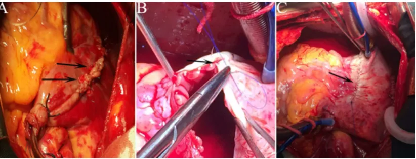
After standard general anaesthesia was achieved, a median sternotomy was performed, followed by routine aortic and right atrial cannulation. Cardiopulmonary bypass (CPB) was carried out using membrane oxygenators and moderate systemic hypothermia, with the aortic perfusion flow of 2.0~2.4 L·min-1·m-2. Meanwhile, myocardial protection was achieved by antegrade mild hypothermic (32°C) blood cardioplegia. When the open-heart surgery via the right atrium path was completed, the right atrial incision was closed with 4-0 prolene (Ethicon Inc). The control group, which was closed with the traditional continuous full-thickness everting suture, experienced frequent bleeding in the right atrial suture and needed stitching again in the bleeding sites (Figure 1A). In the experimental group, to prevent the pectinate muscles from interfering with the suturing-process, the right atrial incision was sewn using the continuous half-thickness suture outside the pectinate muscles (as illustrated in Figure 2). The stitching process was started at the inferior of incision(as illustrated in Figure 2A). Subsequently, the right atrial incision was closed using the continuous full-thickness suture, and the edge distance was 2-3 mm. When the suturing met the structures of the pectinate muscles, the site of the inserting needle and withdrawing needle was near the roots of the corresponding structures of the pectinate muscles which had been cut by the surgical incision (as indicated in Figure 2B and Figure1B). All these procedures attempted to keep the structures of the pectinate muscles away from the sew zone. Subsequently, the first suture was reinforced by a second continuous suture, and
the edge distance of the second continuous suture is longer 1-2mm than that of the first suture (as shown in Figure 2C and Figure 1C).
Heparin was neutralised by protamine in a ratio of 2 within 5 minutes after the completion of the CPB. All post-operative patients were transported to ICU for follow-up monitoring.
2.3. Outcome Measures
A self-designed questionnaire was used to collect the patients’ demographic data and disease characteristics, including body surface area, preoperative ejection fraction, preoperative hepatic and renal functions, and preoperative coagulation status. Another form was used to document post-operative data, such as open heart procedures, complications, cardiopulmonary bypass time, and aortic cross clamp time.
Before the pericardial closure procedure, the active bleeding spots in the right atrial sutures were counted, and the surgically controlled bleeding spots were defined as the applications of sutures and a fibrin sealant to the sites of sutures. The decision to perform re-exploration was made by the surgeon in accordance with current practice guidelines, such as the amount of blood loss as stipulated by Kirklin and Barratt-Boyes criteria [7], haemodynamic status, laboratory parameters and findings from echocardiographic examinations.
2.4. Statistical Analysis
Demographic and disease characteristics are presented using descriptive statistics, while comparisons between the experimental and the control groups were performed using the independent t-test and Chi-square test. In all cases, a P-value less than 0.05 was considered to be statistically significant.
3. Results
3.1. Demographic, Disease, and Pre-operative Data
Table 1. Demographic, disease and preoperative data for the participants.
Experimental Group (n=522) Control Group (n=518) P value
Male/Female 242/280 236/282 0.80
Age, year 45.75±15.45 44.55±16.80 0.17
BSA (m2) 1.69 ± 0.18 1.68±0.17 0.40
CHD,n(%) 56(10.7%) 62(12.0%) 0.55
VHD,n(%) 420(62.3%) 415(61.2%) 0.90
VHD+CAD,n(%) 41(26.1%) 35(25.7%) 0.51
LAM,n(%) 5(1.0%) 6(1.2%) 0.95
HT,n(%) 48(9.2%) 44(8.5%) 0.90
PH,n(%) 223(44.6%) 216(41.7%) 0.65
TR,n(%) 260(49.8%) 249(48.1%) 0.63
DM,n(%) 24(4.6%) 26(5.0%) 0.75
AF,n(%) 335(64.2%) 324(62.5%) 0.64
CAD,n(%) 22(4.2%) 19(3.7%) 0.70
Creatinine (mg/dL) 77.56±11.87 78.13±12.35 0.35
ALT (U/L) 38.04±19.22 36.57±18.51 0.22
AST (U/L) 44.45±19.60 42.23±16.34 0.09
LVEF(%) 50.50±7.25 48.25±8.50 0.21
PT-INR 1.10±0.11 1.08±0.13 0.41
ACC Time (min) 54.25±11.50 53.40±10.25 0.14
CPB Time (min) 80.75±23.25 82.25±18.50 0.20
CHD: congenital heart diseas; VHD:valvular heart diseas,; CAD: coronary artery disease;PH:pulmonary hypertension;DM: diabetes mellitus; HT:hypertension; AF: atrial fibrillation; LVEF: left ventricular ejection fraction; PT-INR; prothrombin time international ratio; ACC: aortic crossclamp; CPB: cardiopulmonary bypass.
3.2. Post-operative Data
All patients were to be operated under cardiopulmonary bypass. Five patients (0.5%) died of post-operative low cardiac syndrome: 2 cases in the control group, and 3 cases in the experimental group. This resulted in no significant difference in terms of the mortality rates between the two groups (p=0.9). Seventeen cases (1.6%) needed re-exploration due to bleeding or tamponade after cardiac operation, of which 10 (1.9%) occurred in the control group: 3 were diagnosed as massive bleeding, 2 were in right atrial incision, 2 were in bypass grafts or anastomosis, and the source of the remaining 3 were the chest wall, the thymus gland or the small artery of the heart surface, respectively. In the experimental group, 7 patients (1.3% vs.1.9% in the control group, p=0.62) needed re-exploration due to post-operative bleeding: 2 were diagnosed as massive bleeding, 2 were in bypass grafts or
anastomosis, and the source of the remaining 3 were the chest wall, pericardium or cannulation, respectively. Most significantly none was sited in the right atrium sutures in the experimental group (0/7 vs. 2/10 in the control group, p=0.33). In both groups, the right atrium sutures were examined carefully before the pericardial closure procedure. The total number of bleeding spots was significantly lower in the experimental group than in the control group (p=0.001). In the experimental group, only 46 cases (51 bleeding spots, mean 0.10±0.32) needed re-stitching by local continuous suture to stop the bleeding (as indicated in Table 2). While in the control group, a total of 253 cases were found with one or more active bleeding spots along the right atrium incisions, and all 492 bleeding spots (mean 0.95±1.04) were re-stitched with mat cotton-padded mattress sutures (Figure 1A ).
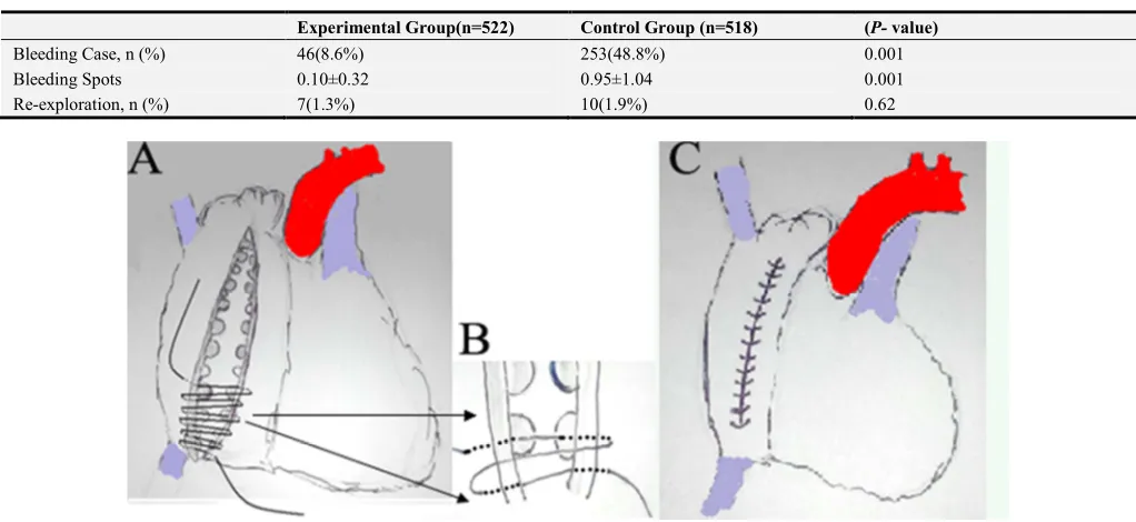
Table 2. Postoperative data about bleeding for the two groups.
Experimental Group(n=522) Control Group (n=518) (P- value)
Bleeding Case, n (%) 46(8.6%) 253(48.8%) 0.001
Bleeding Spots 0.10±0.32 0.95±1.04 0.001
Re-exploration, n (%) 7(1.3%) 10(1.9%) 0.62
Figure 2. The procedure of the half-thickness continuous suture outside of the structures of pectinate muscles in the atrial incision. The stitching process was started at the inferior of incision (A). Subsequently, the right atrial incision was closed using the continuous full-thickness suture, and the edge distance was 2-3 mm. When the suturing met the structures of the pectinate muscles, the site of the inserting needle and withdrawing needle was near the roots of the corresponding structures of the pectinate muscles which had been cut by the surgical incision (B). The first suture was reinforced by a second continuous suture, and the edge distance of the second continuous suture is longer 1-2mm than that of the first suture (C).
4. Discussion
Surgeons usually adopt different incision suture methods based on the characteristics of tissues and cut shape. Also, there are different methods in closing the right atrial incision. This study compared the full or half-thickness continuous suture outside the pectinate muscles for right atrium incision, and suggested that closing the right atrial incision using a continuous half-thickness suture outside the pectinate muscles was superior to the conventional continuous suturing method in preventing bleeding episodes in adult patients who undergone cardiac surgery by extracorporeal circulation. Bleeding episodes and risks of bleeding episodes in the right atrial incision decreased significantly during the intra-operative examination.
The depth of the atrial wall in the inherent right atrium is uneven at different places because of the structures of the pectinate muscles. Adult patients with valvular heart diseases or congenital heart diseases usually also suffer from pulmonary hypertension, tricuspid regurgitation and/or atrial fibrillation, which could cause the right atrial remodeling and/or right atrial enlargement [8-11] and make the right atrial wall even thinner in the parts that do not have the structures of the pectinate muscles. As a result, the right atrial wall’s tolerance of different tensions varies, and the same prolene tension could tear the right atrial wall in those parts that do not have the structure of the pectinate muscles. Upon completion of an open-heart surgery through the right atrium path, the conventional method of closing the right atrial incision was with a continuous full-thickness everting suture, but this resulted in frequent bleeding episodes in the right atrial
incision. The structure of the pectinate muscles was an important factor causing the bleeding episodes in the right atrial incision [12], which would increase the risk of bleeding at the needle hole in the thinner parts of the right atrium.
muscles from the suture zone and facilitated suturing of the cut edge in situ (Figure 1C). As a result, the occurrence rates of intra-operative bleeding sited in right atrial incision were reduced significantly (p=0.001, Table 2).
There were some limitations in our study, such as selection bias since only one hospital was involved and the technique was only performed on Chinese patients.
5. Conclusions
Bleeding episodes in the right atrial closure are still common in current clinical practice which uses the continuous everting suture for adult patients undergoing cardiac surgery by cardiopulmonary bypass. In this paper, we recommended to close the right atrium by a half-thickness continuous suture outside the structures of pectinate muscles in adult cardiac surgery patients with thin right atrial wall, which significantly reduced the occurrence rates of intra-operative bleeding in the right atrial incision.
Conflicts of Interest
All the authors do not have any possible conflicts of interest.
Author's Contribution
ZQC carried out the design of the study and performed the statistical analysis and drafted the manuscript. LJA, LH, LZ, ZL and YZY participated in the surgery operation including the experimental group and control group and recorded the primary data. YHH participated in ZQC conceived of the study, and participated in its design and coordination and helped to draft the manuscript. All authors read and approved the final manuscript.”
References
[1] Čanádyová J, Zmeko D, Mokráček A. Re-exploration for bleeding or tamponade. Interact Cardiovasc Thorac Surg, 2012; 14: 704-708.
[2] Hall TS, Brevetti GR, Skoultchi AJ, et al. Re-exploration for hemorrhage following open heart surgery differentiation on the causes of bleeding and the impact on patients outcomes. Ann Thorac Cardiovasc Surg, 2001; 7: 352-357.
[3] Kristensen KL, Rauer LJ, Mortensen PE, Kjeldsen BJ.
Reoperation for bleeding in cardiac surgery. Interact Cardiovasc Thorac Surg, 2012; 14: 709-713.
[4] Wu L, Sun ZQ, Jiang XG, Si JW, Xiao YC. Re-exploration for hemorrhage following open heart surgery: 94 cases report. Chin J Clin Thorac Cardiovasc Surg, 2005; 12: 372-373.
[5] Mataraci I, Polat A, Toker ME, Tezcan O, Erkin A, Kirali K. Postoperative revision surgery for bleeding in a tertiary heart center. Asian Cardiovasc Thorac Ann, 2010; 18: 266-271.
[6] Okonta K, Rajan S. Re-exploration after open heart surgery at the Madras medical mission, Chennai, India. J West Afr Coll Surg, 2011; 1: 1-17.
[7] Kirklin JW, Barratt-boys BG. Cardiac Surgery, 2nd edition. New York: Wiley, 1993; p224.
[8] Grapsa J, Gibbs JS, Cabrita IZ, Watson GF, Pavlopoulos H, Dawson D, et al. The association of clinical outcome with right atrial Eur Heart J Cardiovasc Imaging, 2012; 13:666-672.
[9] Fukuda Y, Tanaka H, Motoji Y, Ryo K, Sawa T, Imanishi J, et al. Utility of combining assessment of right ventricular function and right atrial remodeling as a prognostic factor for patients with pulmonary hypertension. Int J Cardiovasc Imaging, 2014; 30: 1269-1277.
[10] Szymański P, Klisiewicz A, Lubiszewska B, Lipczyńska M, Konka M, Kuśmierczyk M, et al. Functional anatomy of tricuspid regurgitation in patients with systemic right ventricles. J Am Soc Echocardiogr, 2010; 23: 504-510.
[11] Voigt N, Heijman J, Wang Q, Chiang DY, Li N, Karck M, et al. Cellular and molecular mechanisms of atrial arrhythmogenesis in patients with paroxysmal atrial fibrillation. Circulation, 2014; 129:145-156.
[12] Sergeant P, Kocharian R, Patel B, Pfefferkorn M, Matonick J. Needle-to-suture ratio, as well as suture material, impacts needle-hole bleeding in vascular anastomoses. Interact Cardiovasc Thorac Surg. 2016; 22:813-6.
[13] Haneya A, Diez C, Kolat P, Suesskind-Schwendi Mv, Ried M, Schmid C, et al. Re-exploration for bleeding or tamponade after cardiac surgery: impact of timing and indication on outcome. Thorac Cardiovasc Surg, 2015; 63:51-57.
[14] Moulton MJ, Creswell LL, Mackey ME, Cox JL, Rosenbloom M. Re-exploration for bleeding is a risk factor for adverse outcomes after cardiac operations. J Thorac Cardiovasc Surg, 1996; 111:1037-1046.