11 | P a g e
e- ISSN 0976-0342 Print ISSN 2229-7456
International Journal of Pharmacy & Therapeutics
Journal homepage: www.ijptjournal.com
IN VITRO
AND
IN VIVO
INVESTIGATION OF TOPICAL
FORMULATIONS OF ERYTHROMYCIN
Kripa Sagar Yadav*
Sun Pharmaceutical Industries Ltd, Western Express Highway, Goregaon (E), Mumbai 400063,Maharashtra, India.
ABSTRACT
The interest in in vivo/in vitro correlations in the dermal field of research has increased steadily. This study was designed to demonstrate the rate of release of Erythromycin from two different, marketed topical cream formulations; by performing In vitro and in vivo experiments. For In Vitro study the penetration of Erythromycin from two different formulations was evaluated using human cadaver skin in a Franz Diffusion cell setup. The release rate of Erythromycin from two different formulations in the receptor phase through human cadaver skin was monitored chromatographically. Whereas in In Vivo experiment the pharmacokinetic study of two marketed formulations of Erythromycin was performed by Dermatopharmacokinetic method using 12 healthy Indian male subjects. Pharmacokinetic parameters of Erythromycin were calculated as Cmax, Tmax, AUC (0-t) and AUC (0-∞). Erythromycin was estimated in Stratum Corneum using a validated chromatographic method. Different release profiles of Erythromycin were observed in both the formulations for both the studies. In conclusion, the linear In Vitro – In Vivo correlation for the release rate of Erythromycin was observed for both the two Formulations. But the In Vitro experiments using the cadaver skin is more time consuming than the In Vivo Dermatopharmacokinetic absorption experiments.
Key Words:- Erythromycin, Dermatopharmacokinetic, Franz diffusion cell, Human cadaver skin.
Access this article online
Home page:
http://ijptjournal.com/
DOI:
http://dx.doi.org/10.21276/ijpt.2017.8.1.2
Quick Response code
Received:25.10.16 Revised:12.11.16 Accepted:15.11.16
Corresponding Author
Kripa Sagar Yadav
Sun Pharmaceutical Industries Ltd, Western Express Highway, Goregaon (E), Mumbai 400063.
Email:[email protected]
INTRODUCTION
A continuous interest toward the dermal and transdermal products can be seen, offering several advantages (Foldvari, 2000; Asbill and Michniak, 2000; Barry, 2000a). Data from USA shows that out of 129 drug delivery candidates, 51 transdermal or dermal system products are listed; and 30% of 77
candidate products in preclinical development represent such drug delivery (Barry, 2002). Novel approaches, devices have been published in recent years (Barry, 200b); parallel with the development of in vitro and in vivo test methodologies (Neubert and Wohlrab, 1990; Segers and Zatz, 1998; Kierstan et al., 2000; Hanson, 1989; Wissing and Muller, 2002). Studying the multistep process of drug delivery through the skin involves many different methodologies both in vitro and in vivo (Leveque, 2004). Bioavailability study of a topical formulation begins with the in vitro investigation of the drug release from the compositions under evaluation (Abdou, 1989). In vitro release testing of the active ingredient has drawn much attention as a result of the edition of the SUPACSS (FDA, 1997), however as it was pointed out by FIP/AAPS (Siewert M and Dressman, 2003), there was no standard test protocol that can be applied to all formulations. Data resulting from these investigations can be used as quality indicators, and also for the screening of the compositions prior to in vivo animal testing. Delivery
12 | P a g e
of drugs to the skin is an effective and targeted therapy for local dermatological disorders. This route of drug delivery has gained popularity because it avoids first-pass effects, gastrointestinal irritation, and metabolic degradation associated with oral administration. Topical gel formulations provide a suitable delivery system for drugs because they are less greasy and can be easily removed from the skin. Percutaneous absorption of drugs from topical formulations involves the release of the drug from the formulation and permeation through skin to reach the target tissue. The penetration of a drug through the skin is a complex process typically rate-limited by the stratum corneum (SC). This external layer of the skin is composed of terminally differentiated corneocytes embedded in a complex lipid matrix comprising primarily ceramides, cholesterol, and free fatty acids. Delivery of drug by passive diffusion and the pharmacological effect elicited are dose-related: the better the drug permeates the skin, the greater the therapeutic effect. It follows, therefore, that formulation plays an important role in topical drug delivery. Erythromycin inhibits bacterial protein synthesis by binding to the 50S subunit of the ribosome. It has activity against Gram-positive aerobes and anaerobes as well as the Gram-negative anaerobes.
Erythromycin has been shown to be active against most of the isolates of the following microorganisms, both in vitro and in clinical infections (Herkenne et al., 2007). The objective of the present study was to evaluate rate of release of Erythromycin from the different formulations by In Vivo and In Vitro experiments.
MATERIAL AND METHOD Material:
Two marketed Erythromycin formulations were used for the study. The Franz diffusion cell was used to determine the amount of the drug diffused from different formulations.
Method
In vitro release testing methods:
Vertical Franz-type diffusion cells with a diffusional surface area of 1.76 cm2 were used to study the permeability of Erythromycin. The skin patches of 5 cm2 were collected from Government Medical College, Aurangabad. The skin was applied with the drug on its epidermal surface. 1 g of Acneclin and Erytop cream formulation of Erythromycin were packed into cell donor chamber, ensuring that there were no air bubbles between the formulation and donor surface of the human cadaver skin. The holders containing the skin and formulation were then placed on diffusion cells using a spring
clamp. The receptor phase was filled with phosphate buffer pH 6.8 and continuously stirred with a small magnetic bar at a speed of 100 rpm during the experiments to ensure homogeneity and maintained at 37±0.50C. The 2 ml samples were withdrawn at 0, 30, 60, 90 minutes respectively for each of the formulations of Erythromycin. Simultaneously equivalent amount of phosphate buffer pH 6.8 was added to maintain the volume of receptor Phase. The samples withdrawn from the port were taken in a 25 mL volumetric flask and acidified by adding 1 mL of 0.1 N HCl. To this solution, 10 ml of cyclohexane was added and the flask was stoppered and shaken vigorously for 10 minutes. The organic layer was separated and dried under vacuum. The dried residue was then dissolved in 5 mL of diluent, and the tubes were shaken vigorously for 15 minutes and sonicated. The solution was then filtered through 0.45 μ membrane filter and then analyzed. The concentration was measured by validated HPLC method. Treatment sample B was used as a reference sample (Sanna et al., 2009) in the analytical measurements.
In vivo percutaneous absorption studies:
13 | P a g e
not more than 20 days before dosing). Complete blood count. Urine – physical examinations, chemical examination, microscopic examination.
Subjects were admitted and housed in the clinical facility at least 2 hours before the application of the dose during the study. At the time of dosing, each of the marketed Erythromycin formulation was applied on the forearm of study subjects as per the dosing schedule. Treatment sample B was used as a reference sample. Subjects received a parallel treatment in the subsequent period of following dosing. The dosing procedure was as mentioned below:
1. Both the forearms were washed with mild soap and copious amount of water and dried in air.
2. Both the forearms were marked for total of 08 application sites of 1 sq. cm. area each.
3. 5 mm length product (semisolid dosage forms) or sufficient amount of drug sample was applied on all the sites so that the product completely and smoothly covered the site area.
4. Stratum corneum samples were collected from the sites on desired pre decided time.
5. SkinStratum Corneum samples were collected in sterile glass test tubes during each period. The samples were collected pre-dose and at 0.5, 1.0, 1.5, 2.0, 3.0, 4.0, and 6.0 hours post-dose application. The stratum corneum samples were analysed for Erythromycin concentrations only.
6. For each subject the total number of blood draws were 02 (01 for screening and another during post
study assessment); the total volume of blood withdrawn (10 ml for the prestudy evaluation and 10 ml for the post study) through the vein puncture did not exceed 20 ml.
7. The pre-dose Stratum corneum samples were collected within one hour prior to drug application. The post-dose samples were collected within 2 minutes of the scheduled time, where end time of collection to the nearest minute was recorded.
Individual Stratum corneum concentration VS time curves were constructed; Cmax and Tmax were directly obtained from these curves. AUC from time 0 (baseline) to 6 hours (AUC0–6) was calculated using the trapezoidal rule. Extrapolation of AUC from baseline to infinity (AUC0–∞) was calculated as follows: AUC0–∞= AUC0–6 + (C6/ke) where C6 was defined as concentration at 6 hours. The sample analysis was carried out using developed and validated HPLC method. The dermato pharma cokinetic samples collected in the test tubes were stored at -20ºC until analyzed. During analysis to each sample tube 5 ml of diluents were added and the tubes were shaken vigorously for 15 minutes and sonicated. The solution was then filtered through 0.45 μ membrane filter. To the filtrate 1 ml of 0.1 N HCl and 10 ml of Cyclohexane were added and the tubes were shaken vigorously. The organic layer was separated and dried under vacuum and the residue was then dissolved in 5ml of diluent solution and then analyzed by validated HPLC method.
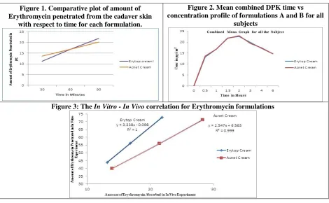
Figure 1. Comparative plot of amount of Erythromycin penetrated from the cadaver skin
with respect to time for each formulation.
Figure 2. Mean combined DPK time vs concentration profile of formulations A and B for all
subjects
14 | P a g e
Table 1. Amount of Erythromycin Penetrated through the In Vitro Skin Preparation
Time in Minutes Amount in µg of Erythromycin Penetrated through the In vitro Skin Preparation Formulations A Erytop Cream Formulations B Acnet Cream
30 11.184 13.66
60 16.822 16.77
90 21.853 20.12
Table 2. The In Vitro - In Vivo correlation for Erythromycin Topical formulations
The In vitro In vivo correlation for Erythromycin Topical formulations
Straight Line Characteristics Formulations A Erytop Cream Formulations B Acnet Cream
r2 1 0.999
Slope 3.338 2.547
Intercept -0.098 6.563
RESULTS AD DISCUSSION
In vitro penetration of Erythromycin formulation:
Two Erythromycin formulations were studied for their permeation through the human cadaver skin. The amount of drug permeated through the skin preparation was estimated by HPLC method and the results are represented in table 1 below.
In Vivo study of Erythromycin:
Twelve subjects were enrolled in the comparison between two formulations of Erythromycin (mean age, 25.16 years). The bioequivalence values of the test drug A were Cmax of 23.767±2.398 μg/ml, tmax of 1.75 ±0.261 h, AUC0-t of 100.507± 10.455 h. μg/ml, AUC0-∞ of 178.286± 22.859 h. μg/ml; of the test drug B Cmax of 24.084± 2.216 μg/ml, tmax of 1.75 ±0.261 h, AUC0-t of 100.586±11.15 h. μg/ml, AUC0-∞ of 179.887± 21.553 h. μg/ml.
In vitro – In vivo correlation:
The In vitro – In vivo correlation was established using the data obtained from the in vitro and in vivo experiments carried out. The results are represented by the following line chart in which the In Vivo results are plotted on X-axis and the In Vitro results are plotted on the Y-axis. The results of all the two formulations are plotted on the overlapping plot for the comparison. It was observed that, for each formulation the correlation could be represented as straight line equation. The characteristics of these straight lines representing the correlation are summarized in Table 2.
CONCLUSION
In invitro experiment for the penetration study of Erythromycin using the human cadaver skin,
the graph plotted against the amount of Erythromycin penetrated with respect to time for each formulation reveals that, the Erythromycin penetrates steadily by following the zero order kinetics. The results are in good agreement with the In Vivo experiments with little difference in the absorption characteristics of Acneclin and Erytop cream formulations.
The linear In Vitro - In Vivo correlation observed for all two Erythromycin Topical Formulations, reveals the fact that, the penetration and the dermal absorption of Erythromycin from these formulations are relative phenomenon and indicative of the results and predictions for each other. Even though, the experimental design. Time duration for the in-Vitro experiments using the cadaver skin is more time consuming than the In Vivo DPK absorption experiments. This is because in DPK experiments, multiple skins sampling for the same sampling time is possible and more importantly, the results are real time and not the predication from the results obtained using the dead tissue.
These results again strengthen the original idea that the DPK studies by skin stripping experiments are accurate, more reliable, easily reproducible, easy to set up, and are essentially the next needed incorporations in the regulatory guidelines for the Bioequivalence studies for the dermal products.
ACKNOWLEDGEMENT: None
CONFLICT OF INTEREST:
The authors declare that they have no conflict of interest.
REFERENCES
15 | P a g e
Asbill CS, Michniak BB. Percutaneous penetration enhancers, local versus transdermal activity. PSTT, 3, 2000, 36– 41.
Barry BW. Drug delivery routes in skin, a novel approach. Adv. Drug Del. Rev, 54, 2002, S31–S40.
Barry BW. Novel mechanisms and devices to enable successful transdermal drug delivery. Eur. J. Pharm. Sci, 14, 2001a, 101–114.
Barry W. Is transdermal drug delivery research still important today? DDT, 6, 2001b, 967.
FDA Guidance for Industry SUPAC-SS. Nonsterile Semisolid Dosage Forms. Scale-Up and Post approval Changes, Chemistry, Manufacturing, and Controls, In Vitro Release Testing and In Vitro Bioequivalence Documentation. May, 1997.
Foldvari M. Non-invasive administration of drugs through the skin, challenges in delivery system design. PSTT, 3, 2000, 417–425.
Hanson WA, State of the Art in Dissolution Testing of Transdermal Dosage Forms. Pham. Sci. Group Meeting, Scarborough, Ontario, December, 1989.
Herkenne C, Naik A, Kalia Y.N, Hadgraft J, Guy RH. Ibuprofen Transport into and through skin from topical formulations, In vitro- In vivo Comparison. Journal of Investigative Dermatology, 127, 2007, 135–142. Kierstan KTE, Beezer AE, Mitchell JC, Hadgraft J, Raghavan SL, Davis AF. Spectrophotometry study of membrane
transport processes with a novel diffusion cell. Int. J. Pharm, 229, 2001, 87–94.
Leveque N, Makki S, Hadgraft J, Humbert Ph. Comparison of Franz cells and microdialysis for assessing salicylic acid penetration through human skin. Int. J. Pharm, 269, 2004, 323–328.
Neubert R, Wohlrab W. In vitro methods for the biopharmaceutical evaluation of topical formulations. Acta Pharm. Technol, 36, 1990, 197–206.
Sanna V, Peana AT, Moretti MDL. Effect of Vehicle on Erythromycin Permeation from New Topical Formulations, In Vitro and In Vivo Studies. Current Drug Delivery, 6, 2009, 93-100.
Segers JD, Zatz JL. Techniques for Measuring In Vitro Release from Semisolids. Dissolution Technol, 5, 1998, 3–13. Siewert M, Dressman J. FIP/AASP Guidelines for Dissolution / In Vitro Release Testing of Novel/Special Dosage
Forms. Dissolution Technol, 10, 2003, 6–15.
Wissing SA, Muller RH. Solid lipid nanoparticles as carrier for sunscreens, in vitro release and in vivo skin penetration. J. Contr. Rel, 81, 2002, 225–235.
Cite this article:
Kripa Sagar Yadav. In Vitro and In vivo Investigation of Topical Formulations of Erythromycin. International Journal of Pharmacy & Therapeutics, 8(1), 2017,11-15.DOI:http://dx.doi.org/10.21276/ijpt.2017.8.1.2