University of Pennsylvania
ScholarlyCommons
Publicly Accessible Penn Dissertations
Fall 12-22-2010
A Study of Molecular Adsorption and Transport at
Cell Membrane and Biologically Relevant Surfaces
by Second Harmonic Generation
Jia Zeng
University of Pennsylvania, [email protected]
Follow this and additional works at:http://repository.upenn.edu/edissertations Part of theBiophysics Commons, and thePhysical Chemistry Commons
This paper is posted at ScholarlyCommons.http://repository.upenn.edu/edissertations/292
For more information, please [email protected].
Recommended Citation
Zeng, Jia, "A Study of Molecular Adsorption and Transport at Cell Membrane and Biologically Relevant Surfaces by Second Harmonic Generation" (2010).Publicly Accessible Penn Dissertations. 292.
A Study of Molecular Adsorption and Transport at Cell Membrane and
Biologically Relevant Surfaces by Second Harmonic Generation
Abstract
Most of the biological processes in living systems involve molecular adsorption and transport at biomembranes. It is highly desired to study the time-resolved transport kinetics through living cell membranes. In this thesis, an experimental means based on a nonlinear optical phenomenon, Second Harmonic Generation (SHG) has been demonstrated to detect the molecular adsorption and transport through living cell membranes in real time and to evaluate the salt ion effects on adsorption processes in biologically relevant colloidal systems. In the case of gram-negative bacteria, E.coli, a hydrophobic cation, Malachite Green (MG) has been observed to adsorb onto the cell surface and then sequentially transport across the double bilayer structures, the bacterial outer membrane and the cytoplasmic membrane. The adsorption characteristics as well as the transport rate constant at each of the membranes have been determined. In contrast to the prokaryotic E.coli cell, the molecular ion can adsorb onto the eukaryotic Murine Erythroleukemia (MEL) cell but cannot penetrate its membrane which has no hydrophobic ion permeable channels and is more tightly packed.
MG cation has been used as a SHG indicator to probe the effects of solvent ionic strength and ion specificity on molecular adsorption at model protein systems. Polystyrene sulfate (PSS) microspheres and polystyrene carboxyl (PSC) microspheres have been examined. The electrostatic force dominated molecule-surface interaction between MG cations and the sulfate terminations at PSS surface is largely affected by the ionic strength of the solution but is not sensitive to the ion identity. On the other hand, the hydrophobic force dominated molecule-surface interaction between hydrophobic regions of the MG dye and PSC microsphere shows pronounced specific ion effects but is less affected by ionic strength of the solution.
Degree Type
Dissertation
Degree Name
Doctor of Philosophy (PhD)
Graduate Group
Chemistry
First Advisor
Hai-Lung Dai
Keywords
cell membrane, molecular transport, ion channel, Hofmeister Series, hydrophobic interaction, Second Harmonic Generation
Subject Categories
Biophysics | Chemistry | Physical Chemistry
A STUDY OF MOLECULAR ADSORPTION AND TRANSPORT AT CELL MEMBRANE AND BIOLOGICALLY RELEVANT SURFACES
BY SECOND HARMONIC GENERATION
Jia Zeng
A DISSERTATION
in
Chemistry
Presented to the Faculties of the University of Pennsylvania in Partial Fulfillment of the Requirements for the Degree of Doctor of Philosophy
2010
__________________________________
Professor Hai-Lung Dai Supervisor of Dissertation
__________________________________
Professor Gary Molander Graduate Group Chair
Committee Members:
J. Kent Blasie, Walter H. & Leonore C. Annenberg Professor in the Natural Sciences Robin Hochstrasser, Donner Professor of Physical Sciences
A STUDY OF MOLECULAR ADSORPTION AND TRANSPORT AT CELL MEMBRANE AND BIOLOGICALLY RELEVANT SURFACES
BY SECOND HARMONIC GENERATION
COPYRIGHT
2010
iii
iv
ACKNOWLEDGEMENTS
There are many people without whom this thesis would not be possible. First, I
would express my sincere gratitude to Professor Hai-Lung Dai for his immeasurable
support and encouragement during my life at UPenn. His inspiring guidance and good
humor led me out the difficulty times in pursuing projects whose outcome was
unpredictable. There have been many expected and unexpected obstacles since the first
day we started to study the bio-related systems. His passion and creative thoughts on this
project has been the strongest backing of me. Under his supervision, I received rigorous
training required to become an independent researcher.
Second, I would like to thank the members of my committee: Professor J. Kent
Blasie, Professor Robin Hochstrasser, and Professor Michael L. Klein for their priceless
time, comments, and encouragements for my research. The questions they asked during
my committee meeting every year helped me think further in my research projects.
I must mention the collaborative laboratories that offered help to my research
projects. Without our previous neighbor Professor Mark Goulian’s group (especially Dr.
Tim Miyashiro), it wouldn’t be possible for me to initiate E.coli experiments. And later
Professor Gerd A. Blobel (especially Dr. Wulan Deng) was kind enough to provide us
MEL cell line. Dr. Luca D'Agostino of Temple University assisted me with culturing
techniques, for which I am very grateful.
I would also like to thank Professor Hongfei Wang for the useful discussion
during his visit to Dai Group. His knowledge and rich experience in this area were of
v
Special thanks goes out to Dr. Shih-Hui Jen for teaching me the precious
hands-on tricks of how to handle the lasers, which actually helped my first step out hands-on research.
I would also like to thank former and current Dai Group members, Heather
Eckenrode, Wei Gan, Grazia Gonella, Jun Han, Jianqiang Ma, Matt Nikow, Rosa
Qensinberry, Jonathan Smith, Michael Wilhelm, Bolei Xu, and Min Zhang for assistance
and discussion in my projects.
Finally, I would like to thank my family, especially, my mother and my 90 year
old grandmother for their continued and unselfish support all these years. I must also
vi
ABSTRACT
A STUDY OF MOLECULAR ADSORPTION AND TRANSPORT
AT CELL MEMBRANE AND BIOLOGICALLY RELEVANT SURFACES
BY SECOND HARMONIC GENERATION
Jia Zeng
Hai-Lung Dai
Most of the biological processes in living systems involve molecular adsorption
and transport at biomembranes. It is highly desired to study the time-resolved transport
kinetics through living cell membranes. In this thesis, an experimental means based on a
nonlinear optical phenomenon, Second Harmonic Generation (SHG) has been
demonstrated to detect the molecular adsorption and transport through living cell
membranes in real time and to evaluate the salt ion effects on adsorption processes in
biologically relevant colloidal systems. In the case of gram-negative bacteria, E.coli, a
hydrophobic cation, Malachite Green (MG) has been observed to adsorb onto the cell
surface and then sequentially transport across the double bilayer structures, the bacterial
outer membrane and the cytoplasmic membrane. The adsorption characteristics as well as
the transport rate constant at each of the membranes have been determined. In contrast to
the prokaryotic E.coli cell, the molecular ion can adsorb onto the eukaryotic Murine
Erythroleukemia (MEL) cell but cannot penetrate its membrane which has no
hydrophobic ion permeable channels and is more tightly packed.
MG cation has been used as a SHG indicator to probe the effects of solvent ionic
strength and ion specificity on molecular adsorption at model protein systems.
vii
have been examined. The electrostatic force dominated molecule-surface interaction
between MG cations and the sulfate terminations at PSS surface is largely affected by the
ionic strength of the solution but is not sensitive to the ion identity. On the other hand, the
hydrophobic force dominated molecule-surface interaction between hydrophobic regions
of the MG dye and PSC microsphere shows pronounced specific ion effects but is less
viii
TABLE OF CONTENTS
Chapter 1: Introduction 1
1.1 Colloidal Interfaces………1
1.2 Cell Membrane Interfaces………..3
1.3 Interface Studies by Second Harmonic Generation………...5
1.4 Aim and Scope of this Dissertation………...8
Chapter 2: Second Harmonic Generation at Interfaces 22
2.1 Introduction………..22
2.2 Theory of Second Harmonic Generation……….23
2.2.1 Phenomenological Mode and Maxwell’s Equation………25
2.2.2 Second Order Nonlinear Susceptibility………..28
2.2.2.1 Molecular Energy Level and Second Order Nonlinear Susceptibility…28 2.2.2.2 Second Order Nonlinear Susceptibility Tensor………..29
2.2.2.3 Electric Fields in the Interface Layer and Polarization Expressions of SHG………29
2.2.3 Transformation of Susceptibilities to Molecular Polarizabilities………...31
2.3 Second Harmonic Generation at Colloidal Interfaces……….35
Chapter 3: Molecular Adsorption and Time-Resolved Molecular Transport across E.coli Cell Membranes 48
3.1 Introduction………..48
3.2 Experiment………...51
3.2.1 The Laser System………51
ix
3.2.3 Malachite Green (MG) Dye ………...53
3.2.4 Cell Culture……….54
3.3 Modified Langmuir Adsorption Model………...54
3.3.1 Adsorption Isotherms……….54
3.3.2 Modified Langmuir Adsorption Model………..55
3.3.3 Application of the Modified Langmuir Model in Analyzing MG Adsorption under Physiological pH Value………56
3.3.4 Surface Coverage and SHG………58
3.4 Results and Discussion………59
3.4.1 Molecular Adsorption onto E.coli Cell Suface………..59
3.4.2 Time-Resolved Molecular Transport though E.coli Membranes………...62
3.4.3 Salt Effects on Molecular Adsorption at E.coli Cell Membrane………66
3.5 Summary………..67
Chapter 4: Molecular Adsorption on Murine Erythroleukemia (MEL) Cell Membrane 89
4.1 Introduction………..89
4.2 Experiment………...93
4.3 Results and Analysis………94
4.4 Discussion………97
4.4.1. Driving Force of Molecular Adsorption is Charge-Charge Interaction……….97
4.4.2. Adsorption Characteristics……….99
x
Chapter 5: Effects of Solvent Ionic Strength and Ion Specificity on
Molecule-Surface Interactions in Aqueous Colloids 119
5.1 Introduction………...119
5.2 Experiment……….124
5.3 Standard Langmuir Adsorption Model………..126
5.3.1 Standard Langmuir Adsorption Model……….126
5.3.2 General Application of the Modified Langmuir Model by Global Fitting……127
5.4 Results and Discussion………..129
5.4.1 Adsorption of MG on PSS/PSC Surfaces without Solvent Salts………129
5.4.2 Salt Ion Effects on MG Adsorption to Charged PSS Surface……….131
5.4.3 Salt Ion Effects on MG Adsorption to Neutral PSC Surface………..136
5.5 Summary………140
Chapter 6: Concluding Remarks 165
6.1 Perspective Applications………165
xi
LIST OF TABLES
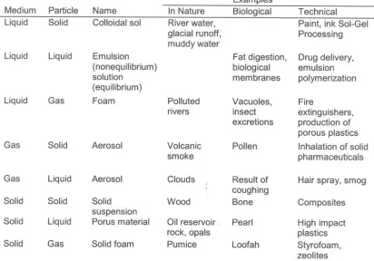
Table 1.1: Examples of different types of colloids based on the dispersed particles and
the continuous medium………..13
Table 3.1: Specifications of Malachite Green………...80
Table 3.2: Adsorption and Transport Characteristics (a) at E.coli Outer Membrane, (b) at
E.coli Cytoplasmic Membrane, and (c) Relative Results from Both Membranes in
Comparison with the Liposome Results………....81
Table 3.3: Adsorption isotherms of MG ions on the E.coli cell under different
salinities……….83
Table 4.1: Adsorption Characteristics of MG on MEL cell under Different
Conditions………111
Table 4.2: Adsorption Free Energy of MG on Polymeric Particles, Liposome and MEL
Cell………...112
Table 5.1: Global fitting results versus individual fitting results of MG adsorption at PSS
surface………..154
Table 5.2: Dependence of adsorption maximum density max
D
N and free energy ΔG on
NaCl concentration………..155
Table 5.3: Adsorption equilibrium constant and free energy of MG on PSC surface (1)
without NaCl (0mM); (2) at NaCl concentration (44mM) where SH intensity reaches
peak value; (3) at NaCl concentration (150mM) where SH intensity levels off……….156
Table 5.4: Adsorption equilibrium constant and free energy of MG on PSC surface at
featured ionic strengths: (a) 50mM and (b) 150mM, for the anion series:I, Cl,
2 4
xii
Table 5.5: Adsorption equilibrium constant and free energy of MG on PSC surface at
meatured ionic strengths: (a) 50mM and (b) 150mM, the cation series: Li, Na, K,
4
xiii
LIST OF ILLUSTRATIONS
Figure 1.1: A schematic representative of colloidal systems according to their typical
range of dimensions………...12
Figure 2.1: Schematic of the typical phenomenological model to treat the second
harmonic response from a planer interface between two isotropic bulk media………….39
Figure 2.2: The energy diagram of SHG in which the SH frequency 2ω is in resonant
enhancement with the electronic transition from the ground state g to the excited state
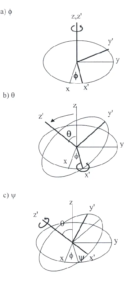
f . ………...40
Figure 2.3: Schematic of the rotations to transform from molecular coordinates to lab
coordinates using Euler angles………...41
Figure 2.4: Schematic of SHG from two SH producing molecules adsorbed at
centrosymmetric particles with opposite orientations..…...………...42
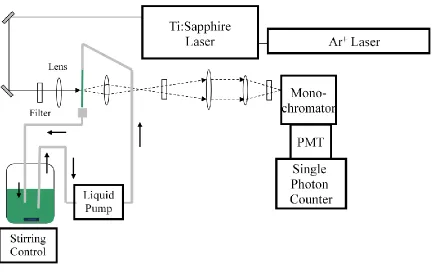
Figure 3.1: Schematic illustration of the experimental setup of the forward detection of
SHG signal……….69
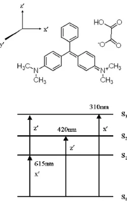
Figure 3.2: MG structure and its spectral features………70
Figure 3.3: Spectrum of the two-photon absorption and the two-photon excited
fluorescence for malachite green………...71
Figure 3.4: System of isotherm classification ………..72
Figure 3.5: Typical Langmuir adsorption isotherm curve expressed in SH intensity
versus molecular concentration………..73
Figure 3.6: Illustration of the two membranes of the E.coli bacteria………...74
Figure 3.7: Time profile of the SH intensity following addition of the MG ion (at
xiv
Figure 3.8: Nonlinear least-square fits (solid green lines) of the time profiles of the SH
intensity (normalized) following addition of MG ions at various concentrations into
solutions containing living E.coli cells………..76
Figure 3.9: Time profiles of membrane surface coverage simulated using the kinetic
model for MG=0.25μM……….77
Figure 3.10: Adsorption isotherm expressed in SH intensity for MG ion on the E.coli
cell……….78
Figure 3.11: Adsorption isotherms of MG ions on the E.coli cell under different salinities.
The solid lines are nonlinear least-square fits of a modified Langmuir model………….79
Figure 4.1: Molecular Structures of (a) Malachite Green (MG); (b) Bromocresol Purple
(BCP)………...104
Figure 4.2: SHG signal detected as a function of time following addition of MG ions into
the MEL cell solution at neutral pH……….105
Figure 4.3: Adsorption isotherms expressed in SH intensity for MG adsorbed on MEL
cell membrane surface at different conditions: (a) acidic pH value (pH=4.5); (b) neutral
pH value (pH=7.0); (c) MEL cell at a further differentiated stage………..107
Figure 4.4: SH signal detected with increasing BCP concentration added into a solution
containing MEL cells at acidic pH value (pH=4.5)……….108
Figure 4.5: Illustration of the MEL cell membrane structure………109
Figure 4.6: pH dependence of [COOH] and [COO -] of polystyrene carboxyl
xv
Figure 5.1: A theoretical simulation of the modified Langmuir model with the adsorption
equilibrium constant set to a small value (5×10⁷M-1) for different maximum surface
adsorption densities (1µM and 5µM)………..142
Figure 5.2: Theoretical simulations of the modified Langmuir isotherms expressed in SH
intensity as a function of MG concentration: (a) isotherms at a fixed value of adsorption
equilibrium constant (K1010) by varying maximum adsorption density; (b) isotherms at
a fixed value of maximum adsorption density (Nmax 0.2M) by varying adsorption
equilibrium constant……….143
Figure 5.3: An example of analyzing a set of adsorption isotherms of malachite green
(MG) molecules on polystyrene sulfate (PSS) surface by Global Fitting………...144
Figure 5.4: SH intensity detected as a function of MG concentration added in the
colloidal solution of (a) charged polystyrene sulfate microspheres (PSS); and (b) neutral
polystyrene carboxyl microspheres (PSC) particle surfaces, at pH= 3.5±0.2………….145
Figure 5.5: SH intensity recorded as a function of NaCl concentration added in the MG
fully covered PSS colloidal solutions………..146
Fi gure 5.6 : MG ads orption isot herms on PSS surfaces at different NaCl
concentrations………..147
Figure 5.7: Dependence of (a) adsorption maximum density NDmaxand (b) free energy ΔG
on NaCl concentration………..148
Figure 5.8: Titration profiles of a series of chloride salts: LiCl, NaCl, KCl, NH4Cl into
PSS colloids pre-covered with MG……….149
Figure 5.9: (a) SH intensity recorded as a function of NaCl concentration added in the
xvi
blank solution without NaCl. (b) MG adsorption isotherms of PSC (1) without NaCl
(0mM); (2) at NaCl concentration (44mM) where SH intensity reaches peak value; (3) at
NaCl concentration (150mM) where SH intensity levels off………..150
Figure 5.10: SH intensities as function of salt concentrations of (a) sodium salts of
different anions:I, Cl, 2 4
SO; and (b) chlorides with different cations: Li, Na, K,
4
NH; applied into PSC colloids pre-saturated with MG molecules……….151
Figure 5.11: MG adsorption isotherms on PSC surfaces at featured ionic strengths for the
anion series:I, Cl, 2 4
SO at (a) 50mM; (b) 150mM; and the cation series: Li, Na,
1
CHAPTER ONE
INTRODUCTION
1.1 Colloidal Interfaces
Stone Age paintings with stabilized colloidal pigments could be the earliest
evidence of man’s use of colloids [1]. In 1845, Francesco Selmi first defined common
properties of slightly turbid “pseudosolutions”, which was later named as “colloid” by
Thomas Graham in 1861 [1]. The low diffusion rates of colloidal particles implied the
low end of the particle size to be at least 1nm in diameter while the failure of the particles
to sediment under the influence of gravity implied the upper size limit to be
approximately 1µm. Most of the studies are focused on the monodispersed objects of
sizes ranging from 10nm to several miron meters [2-4]. Figure 1.1 is the schematic
reprehensive of various colloidal systems with their typical range and critical dimensions
[5]. According to the chart, the upper limit of the critical dimension for colloid definition
has been extended from 1µm to 100µm.
“Colloid” is no longer a modern term, however, colloidal science has become
ubiquitous, which encompasses many chemical and biological systems and influences
2
back to our earliest technological processes, such as paper making, pottery making, and
the fabrication of soaps and cosmetics. The extensive appearance of colloids in nature,
biology and technology clearly demonstrated its importance. A variety of colloidal
systems has long been fully developed industrial products, such as, papers, inks, paints,
coatings, and ceramics in chemical industry and clays, minerals, aerosols, semiconductors
and solar cells in material industry [8-20]. More recently, novel uses have been found for
newly developed biological systems [21-32]. Listed in Table 1.1 is the examples of how
colloids of all phased of matter are important in a highly industrialized society [1].
A typical colloidal system is often viewed as being comprised of two phases: the
dispersed phase (the colloidal particle) and the dispersing medium (the solvent) separated
by a well-defined interface. These interfaces are regions of high free energy, where many
surface phenomena, such as adsorption and the formation of electrical double layers, take
place; therefore, surface properties dominate the behavior of colloidal systems and play
an integral role in colloidal science. To understand the observation of interfacial
phenomenon is the key to solve practical issues and exploit the commercial use of
colloidal systems. A primary concern in the application of colloidal systems is the
dispersion stability, in which the inter-particle interactions and the particle-solvent
interactions play an integral role. Therefore, structures and kinetic/dynamic processes at
colloidal interfaces are of practical and fundamental interest.
Since the interfacial properties is deterministic for colloidal behavior, the most
common practice to probe and modify the surface of the colloids is to have molecule
species with specific properties (i.e., a surfactant, an emulsifier) adsorbed at the interface.
3
and predict colloidal behaviors. Many investigations have focused on characterizing
molecular adsorptions for the understanding of the effect of the molecular-interface
interactions on colloidal behaviors as well as the nature of those adsorption processes
[33-36]. One of the recent interests is to study the deterministic factors, such as the
solvent ionic strength and ion specificity, which could influence molecular adsorption at
colloidal surfaces. Both direct and indirect measurements of the characteristics of
molecular adsorption has been conducted the details of which will be described in section
1.3.
1.2 Cell Membrane Interfaces
The interests in colloidal systems not only have technological reason but also
have anthropological reason. Living systems are one of the most intriguing and
challenging scientific area in the nature. Significant advances have been made in
understanding and explaining various biological processes. It has long been recognized
that the physiological conditions, size and structure, and the intrinsic properties of living
systems makes it possible to be treated at the colloidal level from several perspectives:
Firstly, living systems are usually made up of proteins, polyelectrolytes, and
amphiphilic molecules contained in an aqueous medium. Such molecules, by their size
and structure, not necessary be classified as colloids, but do possess colloidal properties.
They may further self-assemble into a great variety of organized structures that perform
extremely sophisticated chemical and biological transformations. For example, many
4
colloidal assemblies [1]. To understand and control those life processes can be achieved
by monitoring the characteristics and properties at the colloidal level.
Secondly, it has long been the research focus to exploit appropriate colloidal
systems to achieve efficient drug delivery. More than one membrane must be permeated
before a drug molecule passes from the outside into a capillary. The quantitative study of
absorption, distribution, metabolism, and elimination of drugs is therefore closely
associated with colloidal science. Most drug candidates often suffer from the First Pass
Effect, that drugs may be metabolized (for example, by the liver) following absorption
but before reaching systemic circulation. Another major limitation is that drug molecules
as well as drug carriers usually get quickly removed from the blood, plasma or serum
while effective drug delivery systems require sustained drug concentration. Applications
of self-assembled vesicles as well as nanoparticles in controlled drug release have been
reported [37, 38]. The control of targeted drug delivery and medical diagnostic systems
are actually highly dependent on our ability to control fundamental colloidal interactions.
Last but not least, in this thesis, we consider cell suspensions as colloidal systems
as well and characterize the interfacial properties of the cell membrane interfaces under
different conditions. Almost all of the prokaryotic cells and a lot of the eukaryotic cells
are within the size range of colloidal definition. Except that cells intend to stick to glass
walls, they can be suspended in physiological buffers and stay as stable suspensions
without sediment for a relatively long period of time. This is due to the specific
composition and organization of the cell membrane structure, the lipid bilayer structures.
The outer surface of the cell membrane is hydrophilic phosphate groups that favor the
5
to be one of the best defined colloidal interfaces. Some of the cells would even have
special structures with the function to maintain certain cell shape. For example, the
murein layer in Gram-negative bacteria cells and the network of proteins that forms the
skeleton of the red blood cell (RBC) membrane. Uniformed cell shape makes cell
suspension a better model for colloidal characterizations. Almost all of the life processes
involve interfacial association with various molecules at the biological membranes as the
initial step. Further interactions occur at the cell membrane or transport through the cell
membranes can be all influenced by the very first interfacial adsorption processes. The
investigation of the cell membrane interface related chemical and biological processes
from a colloidal perspective will not only help to understand the intrinsic properties of
specific cell membrane interfaces and the associated interactions, but also provide a
model for predicting the general molecular adsorption and transport processes at
biological membranes. The interfacial studies require experimental capabilities for
characterizing the structure and kinetic/dynamic processes at the colloidal and biological
membrane interfaces.
1.3 Interface Studies by Second Harmonic Generation
Many powerful techniques, such as LEED, XPS, IR, AFM, etc. [39], have been
developed to study solid surfaces, providing information on adsorption geometry, surface
chemical composition, as well as vibrational structures. However, UHV conditions are a
requirement for these surface investigation techniques, which makes it a common
limitation in terms of their application to buried interfaces, often involving liquid phases.
6
interfaces are accessible to light. These optical techniques include absorption and
reflection spectroscopy, Raman and Infrared spectroscopy, fluorescence spectroscopy,
ellipsometry and light scattering [40].
However, for scientists immerged in colloidal systems, the conventional optical
techniques suffer the lack of surface sensitivity and selectivity. The barrier of probing
molecular interactions at colloidal surfaces is due to the fact that the spectroscopic signal
of a small surface population is often overwhelmed by a much larger contribution from
bulk species. The surface information is thus buried if the molecules of interest are
present both at the interface and in the bulk phase. However, the systems, including
vesicles, micelles, emulsions, droplets and cell suspensions have great importance in
nature and life sciences as discussed in the previous section. For these systems, the
adsorption of molecules at interfaces is described by the Gibbs adsorption equation and
the Langmuir adsorption isotherm. Only in very rare situations, the shifts in the spectra of
interfacial adsorbed molecules are sufficient to allow differentiation from the bulk
molecules [41-45], due to the inhomogeneous broadening in the condensed phase.
The arrival of interface specific non-linear optical techniques, i.e. Second
Harmonic Generation (SHG) and Sum Frequency Generation (SFG), overcome the main
obstacle of conventional linear optical techniques [46-50]. They are capable to achieve
selectivity between interfacial and bulk molecules and furthermore, the frequency of the
output optical signal lies in a different spectral region from that of the incident light,
which enables easy discrimination and detection of the desired signal. Therefore, SHG
and SFG have proven to be extremely useful in probing sub-monolayer of interfacial
7
molecular reaction and relaxation dynamic processes can be directly probed at interfaces
[48, 49, 51].
The nonlinear optical technique used in this thesis is Second Harmonic
Generation (SHG). SHG is the second order nonlinear optical process, in which two
photons of one frequency (ω) simultaneously interact with a non-linear medium and
generate radiation at the second harmonic frequency (2ω). Similar principle for the
technique of Sum Frequency Generation (SFG) is that two frequencies (ω1 and ω2) of
light interact with medium and the sum frequency (ω1+ω2) is generated. In this thesis,
only SHG technique is employed to probe colloidal and biological interfaces. The
production of SHG is sensitive to the symmetry of the medium. It is believed that SHG is
electric-dipole forbidden in centrosymmetric media but allowed at interfaces where the
inversion symmetry is broken down. SHG is generated only from the few layers which
constitute the interface; therefore, the sensitivity of SHG technique is not determined by
the optical penetration depth (optical wavelength) as conventional optical techniques. The
symmetry sensitive nature of SHG makes it surface specific technique for the study of all
kinds of interfaces.
The interface studies have been advancing rapidly with the interface specific
nonlinear optical techniques, i.e. SHG and SFG. However, the application of SHG and
SFG was restricted to planar surface for a long time. Given the fact that the SHG
efficiency is usually small, for macroscopically flat interfaces, almost all of the SH
photons generated from interfaces can be collected in order to achieve the sub-monolayer
sensitivity. It was believed that the application of SHG to curved interfaces was not
8
Eisenthal group first reported the observation of SHG signal originated from the interface
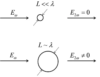
of spherical polystyrene particles with adsorbed dye molecules. Contrary to general
thinking, SHG turned out to be dipole allowed for centrosymmetric media as long as the
size scale is roughly within the range of tens of nanometers to microns. The light induced
second order polarizations from the “opposite” sides of the colloidal particle do not have
opposite phases and therefore do not cancel with each other. The investigation of buried
interfaces, such as vesicles, micelles, emulsions, droplets and cell suspensions, began to
grow since this major breakthrough. The effectiveness of SHG has been demonstrated on
a full scope of colloidal systems, including polymer particles [36, 52-54], nanoparticles
[55-58], carbon black particles [59], emulsions [53, 60], and living cells [61, 62].
1.4 Aim and Scope of this Dissertation
The non-destructive nature of SHG technique makes it ideal for the application to
systems of biological nature in an in-situ manner. In this thesis, we reported works on
investigating molecular adsorption at cell membrane interfaces as well as the subsequent
molecular transport kinetics through the cell membranes. We also present study on the
solvent salt effects on molecular adsorption at the colloidal interfaces. The study shed
light on the understanding of the interaction driving forces that control biological
processes and furthermore, the effects of local ionic conditions may have on interfacial
association of molecules.
Chapter 2 is the framework of the general theory of Second Harmonic Generation
(SHG) with the emphasis on interface studies. Both macroscopic and microscopic
9
In chapter 3, it is demonstrated for the first time that the nonlinear optical
phenomenon Second Harmonic Generation (SHG) can be used to observe in real time the
transport of molecules through living cell membranes. The adsorption and transport of a
molecular ion at the E.coli outer and cytoplasmic membranes were recorded in sequence.
Adsorption isotherms were obtained and analyzed to give molecular adsorption
characteristics of free energy and adsorption density values. A kinetic model has been
established to characterize the molecular transport processes through the double bilayer
structures of the E.coli membranes. Transport of this hydrophobic ion through the outer
membrane is much faster than the cytoplasmic membrane reflecting the effectiveness of
ion transport porins. These observations illustrate that SHG can serve as an experimental
means for studying interactions of small molecules with cell membranes, and the
importance of the ionic nature and the detailed membrane structure in regulating
molecular transport. The time resolved observations and characterizations have advanced
the application of SHG technique to study the real time biological processes rather than
limited to steady-state approximated equilibriums.
To highlight the possibility that different living cell membranes may have
different molecular adsorption and transport properties, we examine a eukaryotic cell, the
Murine Erythroleukemia (MEL) cell, in chapter 4. It is the first time to employ MEL cells
as a model system for studying molecular interaction with eukaryotic cells. The size and
shape of MEL cells favor SHG detection in the forward direction. Adsorption free energy
as well as the maximum number density of model probe molecules has been determined
under different conditions. Different cell membranes may have different constitutions and
10
The collective behavior observed for molecular interactions with MEL cell interfaces
together with those reported in chapter 3 for E.coli cell membrane interfaces, provides a
new level of knowledge for the general understanding of molecular interactions at
biological membrane interfaces.
In chapter 5, Second Harmonic Generation (SHG) – has been applied to
investigate the solvent salt ion effects on molecule-surface interactions in aqueous
colloids. Malachite Green (MG) cationic dye were used as a SHG indicator to probe ion
effects on two model colloidal systems: the negatively charged polystyrene sulfate (PSS)
microspheres and the neutral polystyrene carboxyl (PSC) microspheres in aqueous
solutions at pH<4.0. MG adsorption behavior under different ionic strength and ion
specificity were examined. Modified and standard Langmuir models were employed to
analyze the adsorption isotherms obtained for PSS and PSC, respectively. The
electrostatic force dominated molecule-surface interaction between MG cations and the
sulfate terminations at PSS surface is largely affected by the ionic strength of the solution
but is not sensitive to the ion identity. The electrostatic contribution of the adsorption free
energy depends on the interfacial electric potential, which can be related to ionic strength
of the solvent salts through Gouy-Chapman model. On the other hand, the hydrophobic
force dominated molecule-surface interaction between hydrophobic regions of the MG
dye and PSC microsphere shows pronounced specific ion effects but is less affected by
ionic strength of the solution. Surface acquired anions will provide extra adsorption sites
for the cationic dye and the change in adsorption free energy reflects the electrostatic
11
observations agree very well with both Hofmeister anion and cation series in the ability to
12
Figure 1.1: A schematic representative of colloidal systems according to their typical
13
Table 1.1: Examples of different types of colloids based on the dispersed particles and
14
Bibliography:
1. Evans, D.F. and H. Wennerström, The Colloidal Domain- where physics,
chemistry, biology, and technology meet. 2nd ed. 1999, New York: Wiley-VCH.
2. Matijevic, E., Preparation and properties of uniform size colloids. Chem. Mat.,
1993. 5(4): p. 412-426.
3. Matijevic, E., Uniform inorganic colloid dispersions - achievements and
challenges. Langmuir, 1994. 10(1): p. 8-16.
4. Ozin, G.A., Nanochemistry - synthesis in diminishing dimensions. Adv. Mater.,
1992. 4(10): p. 612-649.
5. Xia, Y.N., et al., Monodispersed colloidal spheres: old materials with new
applications. Adv. Mater., 2000. 12(10): p. 693-713.
6. Antonietti, M., Colloid Chemistry. 2003, Springer: New York.
7. Everett, D.H., Principles of Colloidal Science. 1988, London: Royal Society of
Chemistry.
8. Alivisatos, A.P., Semiconductor clusters, nanocrystals, and quantum dots.
Science, 1996. 271(5251): p. 933-937.
9. Bovey, F.A., Emulsion Ploymerization. 1955, New York: Interscience.
10. Henglein, A., Small-particle research -physicochemical properties of extremely
small colloidal metal and semiconductor particles. Chem. Rev., 1989. 89(8): p.
1861-1873.
11. Idler, R.K., The Chemistry of Silica. 1979, New York: Wiley.
15
13. Pieranski, P., L. Strzelecki, and B. Pansu, Thin colloidal crystals. Phys. Rev. Lett.,
1983. 50(12): p. 900-903.
14. Schick, M.J., Science and Technology of Polymer Colloids, G.W. Poehlein, R.H.
OTTEWILL, and J.W. Goodwin, Editors. 1983, Martinus Nijhoff: Amsterdam.
15. Schmid, G., Large clusters and colloids - metals in the embryonic state. Chem.
Rev., 1992. 92(8): p. 1709-1727.
16. Schmid, G. and L.F. Chi, Metal clusters and colloids. Adv. Mater., 1998. 10(7): p.
515-526.
17. Schon, G. and U. Simon, A fascinating new field in colloid science - small
ligand-stabilized metal-clusters and possible application in microelectronics. 1.
state-of-the-art. Colloid Polym. Sci., 1995. 273(2): p. 101-117.
18. Steigerwald, M.L. and L.E. Brus, Semiconductor crystallites - a class of large
molecules. Accounts Chem. Res., 1990. 23(6): p. 183-188.
19. Wang, Z.L., Structural analysis of self-assembling nanocrystal superlattices. Adv.
Mater., 1998. 10(1): p. 13-+.
20. Weller, H., Colloidal semiconductor Q-particles - chemistry in the transition
region between solid-state and molecules. Angew. Chem.-Int. Edit. Engl., 1993.
32(1): p. 41-53.
21. Kataoka, K., A. Harada, and Y. Nagasaki, Block copolymer micelles for drug
delivery: design, characterization and biological significance. Adv. Drug Deliv.
16
22. Moghimi, S.M., A.C. Hunter, and J.C. Murray, Long-circulating and
target-specific nanoparticles: theory to practice. Pharmacol. Rev., 2001. 53(2): p.
283-318.
23. Brigger, I., C. Dubernet, and P. Couvreur, Nanoparticles in cancer therapy and
diagnosis. Adv. Drug Deliv. Rev., 2002. 54(5): p. 631-651.
24. Martinek, K., et al., Micellar enzymology. Eur. J. Biochem., 1986. 155(3): p.
453-468.
25. Parak, W.J., et al., Biological applications of colloidal nanocrystals.
Nanotechnology, 2003. 14(7): p. R15-R27.
26. Yguerabide, J. and E.E. Yguerabide, Light-scattering submicroscopic particles as
highly fluorescent analogs and their use as tracer labels in clinical and biological
applications - I. Theory. Anal. Biochem., 1998. 262(2): p. 137-156.
27. Stolnik, S., L. Illum, and S.S. Davis, Long circulating microparticulate drug
carriers. Adv. Drug Deliv. Rev., 1995. 16(2-3): p. 195-214.
28. Kakizawa, Y. and K. Kataoka, Block copolymer micelles for delivery of gene and
related compounds. Adv. Drug Deliv. Rev., 2002. 54(2): p. 203-222.
29. Otsuka, H., Y. Nagasaki, and K. Kataoka, PEGylated nanoparticles for biological
and pharmaceutical applications. Adv. Drug Deliv. Rev., 2003. 55(3): p. 403-419.
30. Janes, K.A., P. Calvo, and M.J. Alonso, Polysaccharide colloidal particles as
delivery systems for macromolecules. Adv. Drug Deliv. Rev., 2001. 47(1): p.
17
31. Chiou, P.Y., A.T. Ohta, and M.C. Wu, Massively parallel manipulation of single
cells and microparticles using optical images. Nature, 2005. 436(7049): p.
370-372.
32. Penn, S.G., L. He, and M.J. Natan, Nanoparticles for bioanalysis. Curr. Opin.
Chem. Biol., 2003. 7(5): p. 609-615.
33. Radovic, L.R., et al., An experimental and theoretical study of the adsorption of
aromatics possessing electron-withdrawing and electron-donating functional
groups by chemically modified activated carbons. Carbon, 1997. 35(9): p.
1339-1348.
34. Matijevic, E., Interactions of solutes with monodispersed colloids - practical
aspects, in Adsorption and Its Applications in Industry and Environmental
Protection, Vol I: Applications in Industry. 1999, Elsevier Science Publ B V:
Amsterdam. p. 847-878.
35. Aray, Y., et al., Electrostatics for exploring the nature of water adsorption on the
laponite sheets' surface. J. Phys. Chem. B, 2003. 107(34): p. 8946-8952.
36. Eckenrode, H.M., et al., Adsorption of a cationic dye molecule on polystyrene
microspheres in colloids: Effect of surface charge and composition probed by
second harmonic generation. J. Phys. Chem. B., 2005. 109(10): p. 4646-4653.
37. Chambers, E. and S. Mitragotri, Prolonged circulation of large polymeric
nanoparticles by non-covalent adsorption on erythrocytes. J. Control. Release,
18
38. Chambers, E. and S. Mitragotri, Long circulating nanoparticles via adhesion on
red blood cells: mechanism and extended circulation. Exp. Biol. Med., 2007.
232(7): p. 958-966.
39. Somorjai, G.A., Chemistry in Two Dimensions: Surfaces. 1981, Ithaca: Cornell
Univeristy Press.
40. Light scattering by liquid surfaces and complementary techniques D. Langevin,
Editor. 1992, M. Dekker, Inc.: New York.
41. Zachariasse, K.A., N. Vanphuc, and B. Kozankiewicz, Investigation of micelles,
micro-emulsions, and phospholipid-bilayers with the pyridinium n-phenolbetaine
ET(30), a polarity probe for aqueous interfaces. J. Phys. Chem., 1981. 85(18): p.
2676-2683.
42. Warr, G.G. and D.F. Evans, Spectroscopic determination of the effective
dielectric-constant of micelle water interfaces between 15 and 85-degrees-C.
Langmuir, 1988. 4(1): p. 217-224.
43. Ueda, M. and Z.A. Schelly, Reverse micelles of aerosol-ot in benzene. 4.
investigation of the micropolarity using 1-methyl-8-oxyquinolinium betaine as a
probe. Langmuir, 1989. 5(4): p. 1005-1008.
44. Varadaraj, R., et al., Micropolarity and water penetration in micellar aggregates
of linear and branched hydrocarbon surfactants. Langmuir, 1990. 6(8): p.
1376-1378.
45. Zhu, D.M., X. Wu, and Z.A. Schelly, Reverse micelles and water in oil
19
dynamic light-scattering and turbidity studies. Langmuir, 1992. 8(6): p.
1538-1540.
46. Shen, Y.R., Optical 2nd harmonic-generation at interfaces. Annu. Rev. Phys.
Chem., 1989. 40: p. 327-350.
47. Richmond, G.L., J.M. Robinson, and V.L. Shannon, 2nd harmonic-generation
studies of interfacial structure and dynamics. Prog. Surf. Sci., 1988. 28(1): p.
1-70.
48. Eisenthal, K.B., Equilibrium and dynamic processes at interfaces by 2nd
harmonic and sum frequency generation. Annu. Rev. Phys. Chem., 1992. 43: p.
627-661.
49. Corn, R.M. and D.A. Higgins, Optical 2nd-harmonic generation as s probe of
surface-chemistry. Chem. Rev., 1994. 94(1): p. 107-125.
50. Shen, Y.R., Surface-properties probed by 2nd-harmonic and sum-frequency
generation. Nature, 1989. 337(6207): p. 519-525.
51. Dai, H.-L. and W. Ho, eds. Laser spectroscopy and photochemistry on metal
surfaces 1995, World Scientific: Singapore ; River Edge, N.J. .
52. Wang, H., et al., Second harmonic generation from the surface of
centrosymmetric particles in bulk solution. Chem. Phys. Lett., 1996. 259(1-2): p.
15-20.
53. Wang, H.F., et al., Energetics and population of molecules at microscopic liquid
20
54. Eckenrode, H.M. and H.L. Dai, Nonlinear optical probe of biopolymer adsorption
on colloidal particle surface: Poly-L-lysine on polystyrene sulfate microspheres.
Langmuir, 2004. 20(21): p. 9202-9209.
55. Malvezzi, A.M., et al., Melting-induced enhancement of the second-harmonic
generation from metal nanoparticles. Phys. Rev. Lett., 2002. 89(8).
56. Russier-Antoinc, I., et al., Multipolar contributions of the second harmonic
generation from silver and gold nanoparticles. J. Phys. Chem. C, 2007. 111(26):
p. 9044-9048.
57. Jen, S.H. and H.L. Dai, Probing molecules adsorbed at the surface of nanometer
colloidal particles by optical second-harmonic generation. J. Phys. Chem. B,
2006. 110(46): p. 23000-23003.
58. Jen, S.H., G. Gonella, and H.L. Dai, The effect of particle size in second harmonic
generation from the surface of spherical colloidal particles. I: experimental
observations. J. Phys. Chem. A, 2009. 113(16): p. 4758-4762.
59. Wang, H.F., et al., Adsorption at a carbon black microparticle surface in aqueous
colloids probed by optical second-harmonic generation. J. Phys. Chem. C, 2007.
111(25): p. 8708-8715.
60. Yan, E.C.Y., Y. Liu, and K.B. Eisenthal, New method for determination of
surface potential of microscopic particles by second harmonic generation. J. Phys.
Chem. B, 1998. 102(33): p. 6331-6336.
61. Zeng, J., H.M. Eckenrode, and H.-L. Dai, Time-Resolved Molecular Transport
21
62. Zeng, J., H.M. Eckenrode, and H.-L. Dai, Molecular interaction with murine
erythroleukemia (MEL) cell membrane probed by second harmonic generation. to
22
CHAPTER TWO
SECOND HARMONIC GENERATION AT INTERFACES
2.1 Introduction
Generally, polarization of the medium by an electromagnetic field can be
expressed as a power series [1]:
(1) (2) (3)
P E E E E E E (2.1)
where (1) represents the linear susceptibility, (2) the second order susceptibility, etc.
and Eω is the incoming electric field of frequency ω. The interaction of weak light field
with matter is dominated by linear process, but with intense field, higher order nonlinear
terms, second harmonic generation, third harmonic generation, etc. become observable.
Second harmonic generation (SHG) is a nonlinear process where a photon at the
frequency 2ω is generated from the interaction between intense light at frequency ω and a
nonlinear medium. In 1961, shortly after the invention of the optical laser as intense light
source, the first observation of optical second harmonic generation (SHG) was made
from the noncentrosymmetric quartz crystal [2]. SHG generated from the
centrosymmetric calcite crystal was also reported [3]. The field of nonlinear optics started
23
metal surface [4] and the liquid-air interface [5]. On the other hand, in order to interpret
the experimental observations, the theoretical treatments were also developed for
homogeneous nonlinear media [6] as well as the boundary of two centrosymmetric media
[7]. Initially, it was believed that the quadrupolar term in the nonlinear polarization that
acts as the source of SHG. However, observation of SH signal from the adsorbed layers
at silver surfaces showed that an additional surface dipole term must contribute. Since
then, theoretical description has started to attribute the origin of SHG to the
symmetry-breaking nature of the surface [8]. It was not until early 1980s that the basic SHG theory
was sophisticated enough to relate the experimental observables to the surface molecular
properties [9-12]. Exhibiting strong sensitivity and selectivity to the interfacial region,
SHG has been developed into a powerful technique for interface studies. SHG has been
successfully applied to all kinds of planar interfaces as well as buried colloidal surfaces.
Much attention has been given to the molecular adsorption at these interfaces. A variety
of dye molecules [13-15], surfactants [16-18], proteins [19], and other biomolecules [20,
21] have been investigated. Only recently has SHG been extended to biomembrane
surfaces. Conprehensive reviews have been written on the extensive applicability of this
technique [22-30].
2.2 Theory of Second Harmonic Generation
When intense light interacts with a nonlinear medium, higher order polarization
terms are induced in addition to the linear polarization. Second harmonic generation is
24
at the second harmonic frequency 2ω. As was described in the previous section, the
induced second order polarization P(2)can be expressed by itself as [9-12]:
(2) (2)
P E E (2.2)
Since the second order polarization (2)
P and the incoming electric field Eat
frequency ω are vectors, and (2)
is a second order 3×3×3 tensor, a more rigorous
expression for eq. 2.2 is:
(2) (2) ,
j k
i ijk
j k
P
E E (2.3)where i, j, k are the three normal coordinates of laboratory system, respectively.
As an even order term in the polarization expression, second harmonic generation,
is sensitive to the symmetry of the medium. An inversion symmetry operation will
change both polar vectors’ sign: the second order polarization P(2)and the electric field
E. However, for a centrosymmetric medium with inversion symmetry (2)does not
change sign upon inversion. Therefore, the expression for P(2)in a centrosymmetric
medium after inversion is:
(2) (2)
( )( )
P E E
(2.4)
Comparison of equation 2.2 and 2.4 implies (2) (2), which is impossible
unless (2)0 . Therefore, (2) vanishes in isotropic media and second harmonic
generation is forbidden. However, at interfaces where inversion symmetry is no longer
conserved, (2)is not necessarily equal to zero upon inversion operation, which makes
25
Complete treatment of SHG can be found in literature [6, 7, 10, 11, 25]. In this
chapter, we will focus on the theory of second harmonic generation at interfaces, from
both macroscopic and microscopic perspectives. The macroscopic second order nonlinear
response of the interface to the applied optical field is described by the macroscopic
second order nonlinear susceptibility,(2), which can be obtained from the experimental
observables. On the other hand, in order to obtain the microscopic properties of the
interface down to the molecular orientation level, we need to relate to the microscopic
hyperpolarizability (2)of the molecules at interface.
In the following sections, we first discuss the radiation from a sheet of
polarization governed by the Maxwell’s equations. Then a discussion on the second order
nonlinear susceptibility and how it relates to the molecular energy level structures.
Finally, the transformation of in the molecular coordinates at system into with the
experimental laboratory coordinates is presented.
2.2.1 Phenomenological Mode and Maxwell’s Equation
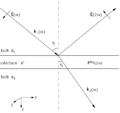
The typical phenomenological model that widely adopted to treat the second
harmonic response from a planer interface is shown in Fig. 2.1. The interfacial region is
in between two centrosymmetric bulk media. The incoming electric field Eat frequency
ω passes through medium 1 and gives rise to a nonlinear polarization (2) NL
P P at
frequency 2ω in the interfacial region. A small amount of fundamental is converted into
2ω in both the reflected direction in medium 1 and the transmitted direction in medium 2.
26
The interfacial region usually occurs over a distance of only a few Angstroms,
consisting of only one or several molecular layers, the thickness of which is much smaller
than the wavelength of the incoming light. We consider the nonlinearity of the interface
as a sheet of the nonlinear source polarization imbedded in a dielectric medium with a
dielectric constant as ε’ shown in Fig. 2.1. The induced nonlinear polarization of the sheet
is described by:
(2) (2)
(2 ) ( )
P z E E (2.5)
where ( )z is a delta function representing the thickness of the interfacial layer.
The nonlinear polarization P(2) PNLgenerated from the interface is governed by
the Maxwell’s equations, from which the wave equation can be derived as [1]:
2
2 2 2
2
2 2 2 2
4
( )
NL
E P
E
c t c t
(2.6)
and
4 NL
E P
(2.7)
Satisfying boundary conditions[10]:
2 4
x y
B i P
c
(2.8)
2 4
y x
B i P
c
(2.9)
0 z B
(2.10)
4 ' z x P E x
(2.11)
4 ' z y P E y
27 4 ( x y)
z P P D x y
(2.13)
Where Bxrepresents the difference of Bxacross the interface:
( 0 ) ( 0 )
x x x
B B z B z
(2.14)
And similar relations for all other boundary condition terms in eq. 2.9-2.13, respectively.
Solving the Maxwell’s equations (eq. 2.6 and 2.7) with boundary conditions (eq.
2.8 -2.13) gives the expressions for the p- (parallel to the table) and s- (perpendicular to
the laboratory table) polarized components of the reflected second harmonic electric field,
and, respectively [10]:
2 1 2
2 2 1 1 2
4
( )
'
NL NL
p z x x z
z z
i k
E k P k P
k k
(2.15)
2 1 1
1 2 1
4 NL
p y
z z
i k k
E P
k k
(2.16)
1z 1cos i
k k , k2z k2cosr, and k2x k2sinr, where k1 and k2are the wave vectors of
2
E in media 1 and 2; the subscripts x, y, and z denote x-, y-, and z- components in the
laboratory coordinate system; i is the incident angle and rthe refractive angle. NL x P ,
NL y
P and PzNL are the Cartesian components of the second order nonlinear polarization.
The nonlinear polarization, PNLat 2ω, should be expressed in terms of the local
electric field 'L
E in the layer of polarization sheet, which is different from the input
optical fields:
(2) ' '
:
NL L L
28 The components of PNL, i.e., NL
x
P PyNL and NL z
P , can be expressed with the
susceptibility tensor elements,ijk(2), as:
(2) ' ' ,
( ) ( )
NL L L
i ijk j k
j k
P
E E (2.18)
The tensor elements ijk(2)as well as the local field E'L need to be examined to give the
explicit expressions for the Cartesian components of the polarization NL
P , i.e., NL x P PyNL
andPzNL, which will be presented later in this chapter.
2.2.2 Second Order Nonlinear Susceptibility
2.2.2.1 Molecular Energy Level and Second Order Nonlinear Susceptibility
Second order nonlinear susceptibility (2)
describes the response of the medium
to the applied field. This macroscopic property of the interface can be related to the
molecular energy level structures [1]. The full expression for the nonlinear
susceptibilities under dipolar approximation can be expressed as [31]:
(2) ,
| | | | | | 2
i f gi ig gf fg
g i i f f g
i i
(2.19)Where g||i represents the transition dipole moment from g to i state, gi is the
corresponding optical frequency of the transition, igis the linewidth associated with the
transition, etc. Eq. 2.17 clearly demonstrated that the magnitude of the (2)will be greatly
enhanced when the fundamental or the second harmonic frequency is in resonance with
that of a molecular transition. This condition can be satisfied by selecting proper
29
an energy diagram in which the SH frequency 2ω is in resonant enhancement with the
electronic transition from the ground state to the excited state.
2.2.2.2 Second Order Nonlinear Susceptibility Tensor
The second order nonlinear susceptibility (2) is a third rank tensor with
3×3×3=27 elements. Symmetry operations can reduce the number of the elements [32,
33]. For an isotropic interface, though the inversion symmetry is broken along axis z,
other symmetries are still preserved. Due to the rotational symmetry around axis z, which
makes x and y indistinguishable, many components of disappear. Only 7 non-vanishing
tensor elements survive, with 4 independent values [32]:
(2)
zzz
(2.20a)
(2) (2)
zxx zyy
(2.20b)
(2) (2)
xzx yzy
(2.20c)
(2) (2)
xxz yyz
(2.20d)
For SHG, the number can be further reduced by recognizing the fact that the order of the
input electric fields does not matter since they are identical, ijk(2) ikj(2) . Plus the
equivalence of x and y at an isotropic surface, we have:
(2) (2) (2) (2)
xzx yzy xxz yyz
(2.20e)
Altogether 3 independent tensor elements remain: eq. 2.20a, 2.20b and 2.20e.
2.2.2.3 Electric Fields in the Interface Layer and Polarization Expressions of SHG
Given eq. 2.20, the expressions for the three Cartesian components of the
30
(2) ' '
2 ( ) ( )
NL L L
x xzx z x
P E E (2.21a)
(2) ' '
2 ( ) ( )
NL L L
y yzy z y
P E E (2.21b)
(2) ' 2 (2) ' 2 ' 2
( ) ( ( ) ( ) )
NL L L L
z zzz z zxx x y
P E E E (2.21c)
Here the electric fields Ex'L( ) ,Ey'L( ) , and Ez'L( ) in the interface layer are
different from the input optical fields Es and. Ep. The relation between the two are
given by the Fresnel transmission coefficients, and the appropriate continuity conditions
[32].
As was shown in Figure 2.1, the incoming electric field comes from medium 1 to
the interface at an anglei, and is then refracted into medium 2 at an angler. r can be
obtained by Snell’s Law:
sin sin
i i r r
n n (2.22)
The Fresnel transmission coefficients (which are the ratio of the transmitted to incident
fields) are given by [34]:
2 1
2 cos sin sin( ) s i r s s i r E T E (2.23a) 2 1
2 cos sin
sin( ) cos( )
p
i r
p p
i r i r
E T E
(2.23b)
Since E is continuous between the interface and medium 2, at the boundary the continuity
conditions yield:
2 '
( ) L( )
x x
E E (2.24a)
2 '
( ) L( )
y y
31
2 '
2
'
( ) L( )
E E
(2.24c)
Therefore, for expressions of the components ofE'L, we have:
'
( ) cos
L p
x p r
E T E (2.25a)
'
( )
L s
y s
E T E (2.25b)
' 2
( ) sin
'
L p
z p r
E T E
(2.25c)
The expressions in eq. 2.25 may be substituted into eq. 2.21, and then into eq. 2.15 and
2.16 to obtain the overall expressions for second harmonic field components Ep2 and
2
s
E as functions of input field components Es and Ep . The macroscopic nonlinear
susceptibility (2)can be extracted by selecting the incoming optical fields and measuring
the s- and p- polarized second harmonic generation components.
2.2.3 Transformation of Susceptibilities to Molecular Polarizabilities
The macroscopic susceptibility elements ijk(2)can be written as the sum of the
individual molecular polarizabilities i j k(2)' ' 'through a statistical average over molecular
orientations:
(2) (2)
s N
(2.26)
where is the number density of adsorbates at the interface and the brackets denote an
average over the orientation distribution of the adsorbates. Eq. 2.26 is valid for coherent
processes such as SHG from an interface, where the in phase individual oscillating


![Figure 3.4: System of isotherm classification [ref]. L2 represents the Langmuir isotherm](https://thumb-us.123doks.com/thumbv2/123dok_us/9261103.1464543/91.612.139.508.73.450/figure-isotherm-classification-ref-l-represents-langmuir-isotherm.webp)