R E V I E W
Open Access
AMD and the alternative complement
pathway: genetics and functional
implications
Perciliz L. Tan
1,2, Catherine Bowes Rickman
3,2and Nicholas Katsanis
1,2*Abstract
Age-related macular degeneration (AMD) is an ocular neurodegenerative disorder and is the leading cause of legal blindness in Western societies, with a prevalence of up to 8 % over the age of 60, which continues to increase with age. AMD is characterized by the progressive breakdown of the macula (the central region of the retina), resulting in the loss of central vision including visual acuity. While its molecular etiology remains unclear, advances in genetics and genomics have illuminated the genetic architecture of the disease and have generated attractive pathomechanistic hypotheses. Here, we review the genetic architecture of AMD, considering the contribution of both common and rare alleles to susceptibility, and we explore the possible mechanistic links between
photoreceptor degeneration and the alternative complement pathway, a cascade that has emerged as the most potent genetic driver of this disorder.
Background
Age-related macular degeneration is the third leading cause of vision loss worldwide. It is a late-onset disease with a complex etiology. Major risk factors contributing to susceptibility include age, family history, and smoking [1–3]. The earliest clinical manifestations of age-related macular degeneration (AMD) are focal deposits of debris, termed drusen, which are also considered to be a normal part of aging, present almost ubiquitously in the eyes of healthy individuals over the age of 50. Progression into the spectrum of pathological consequences begins with exces-sive accumulation of drusen in the central retina during the early/intermediate stages of AMD, followed by local-ized inflammation, and ultimately neurodegeneration in the macula that characterizes advanced stages of AMD.
Combining epidemiological and genetic approaches has enabled the identification of environmental and gen-etic contributors to AMD, both of which have tracked with technological advances in conceptual and practical statistical and genomic tools. Linkage of an AMD locus
to 1q32 [4, 5], and the genome-wide association at the complement factor H (CFH) locus [6] led to the identifi-cation of the first common genome-wide significant risk variant, Y402H, that has upon sequencing in ethnic stratified cohorts revealed differential frequencies ran-ging from 5 % in East Asian populations to 35 % in European populations [7, 8]. This discovery propagated numerous genetics and genomic studies that have contrib-uted to our understanding of the pathomechanisms con-tributing to AMD. Notably, the subsequent association of common and rare alleles at or near several additional complement genes (CFH, C2/CFB, C3,CFI, and C9) has had a significant impact of the formation of pathomecha-nistic hypotheses, with the cumulative evidence both from human genetics but also from histopathological studies highlighting a major role of the alternative complement pathway as a driver of AMD [9–21]. Here, we synthesize genetic evidence from rare- and common-allele studies in AMD, and we discuss the emergent picture of the genetic architecture of this atypical complex trait. Moreover, benefiting from the discovery of likely potent coding mu-tations in genes encoding complement components, we explore how these mutations might impede specific func-tions and discuss the potential contribution of aspects of this pathway to AMD pathogenesis.
* Correspondence:[email protected] 1
Center for Human Disease Modeling, Duke University Medical Center, Durham, NC 27710, USA
2Department of Cell Biology, Duke University Medical Center, Durham, NC
27710, USA
Full list of author information is available at the end of the article
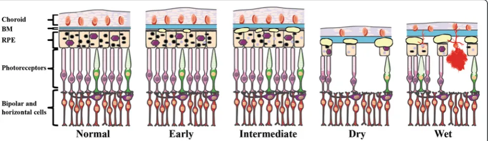
AMD histopathology
The retina is composed of five major layers: the neuro-sensory retina; the retinal pigment epithelium (RPE); Bruch’s membrane (BM); the choriocapillaris; and the choroid (Fig. 1). The sensory cells of the neural retina are the photoreceptors (rods and cones), which through phototransduction convert light into electrical signals. Adjacent to the photoreceptors is the RPE, a part of the blood-ocular barrier and has numerous functions, in-cluding photoreceptor outer segment phagocytosis; regulation of the transportation nutrients; and cytokine secretion [22]. Lining the basal side of the RPE is BM, which is a pentalaminar extracellular matrix (ECM) con-sisting of elastin semipermeable barrier between inner and outer collagenous zones that provides connective tissue-based structural support and transport of waste from photoreceptors and RPE to the choroid and nutri-ents from the choroid to the RPE. The outermost layer of BM is composed of the basement membrane of the choriocapillaris, a fenestrated capillary bed that together with the choroid, a highly vascularized, pigmented tissue, are responsible for supplying the high metabolic de-mands of oxygen and nutrients to the outer retina [23].
During the natural process of aging, the human eye undergoes physiological changes that include changes in the distribution of photoreceptors (30 % loss of rod pho-toreceptors [24]); thickening of BM [25]; and accumula-tion of sub-RPE debris, including drusen [22, 26–29]. Drusen are composed of esterified cholesterol, phospho-lipids, lipofuscin, inflammatory components (e.g., com-plement), and other intra- and extraocular degenerative materials [30, 31]. They are classified according to their appearance, with inert, “hard” drusen being small with well-demarcated borders in contrast to the generally pathogenic “soft” drusen that lack distinct borders and
can range in size from <63 to >124 μm in diameter, the larger diameters coinciding with disease progression [32].
The clinical pathology of AMD has been described ex-tensively [3, 33]. It involves the progressive degeneration of the macula, a small, pigmented area at the center of the retina that has the densest concentration of photore-ceptors and is responsible for visual acuity. Damage to the macula results in loss of central vision. Initial indica-tions of AMD are seen as focal hyperpigmentation of the RPE and accumulation of sub-RPE deposits, includ-ing drusen, both between BM and the RPE and within the RPE itself [34, 35].
Even though the degeneration of rods and cones does define the end-stage of the disorder, AMD is not a pri-mary disease of the photoreceptor; most of the candidate processes, biochemical pathways, and molecules exert their effects primarily on the RPE-choroid complex [36].
AMD subtypes
The anatomical histopathology and clinical progres-sion contribute to the clinical definition of four major AMD subtypes as categorized by the Age-Related Eye Disease Study (AREDS) severity scale grading system [37]: 1. Early AMD; 2. Intermediate AMD; 3. Ad-vanced non-neovascular (“Dry” or geographic atrophy) AMD; and 4. Advanced neovascular (“Wet” or exuda-tive) AMD [32, 38, 39]. Early AMD features few small (<63μm) or medium-sized drusen (63–124μm) and pig-mentary abnormalities in the RPE, resulting in either mild visual impairment (blurred vision or decreased contrast sensitivity) or can be asymptomatic. The progression from early to intermediate AMD is hallmarked by the appear-ance of at least one large druse (>125μm), along with nu-merous medium-size drusen. This pathology can progress to one of the two advanced forms of AMD: Dry AMD
Fig. 1Illustration of the anatomical retinal pathology associated with the various AMD subtypes. Diagram of the outer layers of the human central retina in normal and in AMD. As the disease progresses, Bruch’s membrane (BM) increases in thickness. Early AMD is associated with small drusen and retinal pigmented epithelium (RPE) pigment abnormalities. As the disease progresses to the intermediate form, additional drusen are observed. In the two late forms of AMD (Dry and Wet), there is extensive drusen and photoreceptor cell death, with atrophy of the RPE and choroid in the Dry form and choroidal neovascularization (CNV), hemorrhaging, and RPE detachment in the Wet form. In all forms, the underlying circuitry including the horizontal and bipolar cells remains intact initially. This figure was prepared using Servier Medical
(non-neovascular), characterized by presence of drusen and atrophy of the RPE and choroid; or Wet AMD (neovascular), defined by newly formed vessels (chor-oidal neovascular membranes (CNV)) and RPE de-tachment. While occurring gradually in the Dry form versus suddenly/profoundly in the Wet form, the end result of both of these processes leads to photorecep-tor cell death and vision loss (Fig. 1, Table 1).
Genetics of AMD: common variants versus rare variants
AMD exemplifies complex disorders: natural history and epidemiological studies have highlighted a prominent environmental role in disease risk with factors such as smoking resulting in a relative risk (RR) of >2, diet and obesity (RR > 2), nutritional supplements (odds ratio (OR) ~0.6) all contributing to pathogenicity [40]. In addition, family- and population-based studies have also highlighted that AMD carries a significant genetic bur-den, demonstrating >45 % concordance found in mono-zygotic and dimono-zygotic twins as well as a recurrence-risk ratio between three to six times greater in siblings than in the general population [41–46].
In many ways, the dissection of the genetic basis of AMD has been an early beneficiary of advances in gen-etic and genomic technologies, wherein the development of both analytic platforms (genotyping, sequencing) as well as statistical methods, have found early application in this field.
Before our ability to query the genome in toto, complex trait studies focused on more traditional tools such as linkage and candidate gene association studies. Utilizing
the latter, AMD was shown to be associated with two genes, ABCA4 (ATP-binding cassette subfamily A mem-ber 4) and APOE(apolipoprotein E), both of which only confer a minor risk contribution to AMD. ABCA4 is a causal gene for Stargardt disease, a retinopathy whose clinical symptoms overlap with AMD [47, 48];APOEis a gene associated with Alzheimer’s disease (AD), a neuro-logical degenerative disorder, whose pathology overlaps with AMD [49]. Nonetheless, and despite this initial discovery, the candidate gene approach was largely unsuc-cessful in identifying genetic contributors to AMD. This was due in part to limited cohort sizes, imperfect statis-tical methodologies, and unclear pathomechanisms hin-dering the ability to define candidate genes and in part due to our lack of appreciation of the extensive rare and common variation carried in human genomes [50–52].
In the era following the completion of the human gen-ome project and the International HapMap Project, traditional family-based studies retained utility, as exem-plified by the family-based linkage mapping of an AMD locus on 1q25-q31 and on 10q26 [4, 53]. However, trans-formative genetic progress in AMD was driven by the common-allele hypothesis explored through genome-wide association studies (GWAS) [6, 54]. Remarkably, modest GWAS analysis on 96 cases and 50 controls yielded a genome-wide significant signal (P< 10−7) on 1q32 of a common single nucleotide polymorphism (SNP) on a gene encoding CFH that revealed a poly-morphism in linkage disequilibrium, Y402H, that has proven to be a common, predisposing allele that confers a two- to sevenfold increased risk for developing AMD [6, 9, 55, 56].
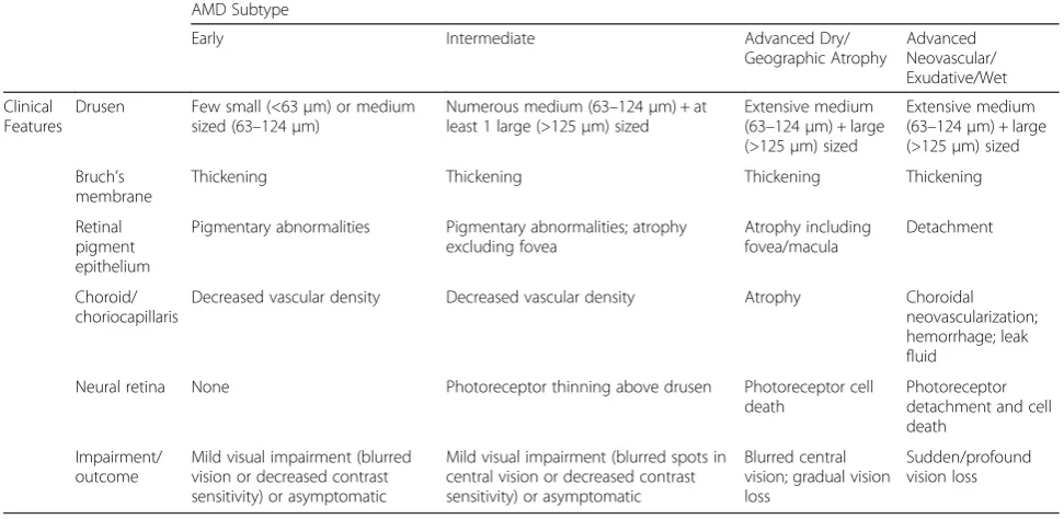
Table 1Characteristics of AMD Clinical Subtypes (based on AREDS)
AMD Subtype
Early Intermediate Advanced Dry/
Geographic Atrophy
Advanced Neovascular/ Exudative/Wet
Clinical Features
Drusen Few small (<63μm) or medium sized (63–124μm)
Numerous medium (63–124μm) + at least 1 large (>125μm) sized
Extensive medium (63–124μm) + large (>125μm) sized
Extensive medium (63–124μm) + large (>125μm) sized
Bruch’s membrane
Thickening Thickening Thickening Thickening
Retinal pigment epithelium
Pigmentary abnormalities Pigmentary abnormalities; atrophy excluding fovea
Atrophy including fovea/macula
Detachment
Choroid/ choriocapillaris
Decreased vascular density Decreased vascular density Atrophy Choroidal neovascularization; hemorrhage; leak fluid
Neural retina None Photoreceptor thinning above drusen Photoreceptor cell death
Photoreceptor detachment and cell death
Impairment/ outcome
Mild visual impairment (blurred vision or decreased contrast sensitivity) or asymptomatic
Mild visual impairment (blurred spots in central vision or decreased contrast sensitivity) or asymptomatic
Blurred central vision; gradual vision loss
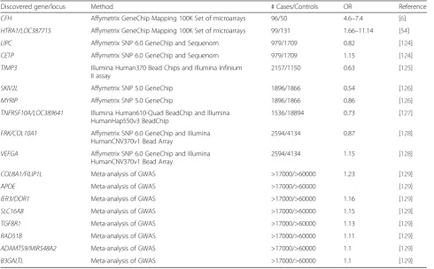
Soon after the identification of theCFH locus, another modestly sized GWAS performed on 96 cases and 130 controls yielded a genome-wide significant signal on 10q26, another primary locus for AMD that contains the candidate genes ARMS2/HTRA1(age-related maculopa-thy susceptibility 2/HtrA serine peptidase 1) coding for a protein of unknown function and a serine protease, re-spectively [54, 57]. Thereafter, additional GWAS studies with progressively increasing cohort size led to the iden-tification of more associated loci but generally resulted in diminishing returns with many of the loci having a minor contribution to AMD (Table 2).
The associated loci discovered also germinated tar-geted sequencing strategies with a goal of identifying rare variants that might (a) provide direct causal evi-dence for the gene(s) in associated regions; (b) improve our measure of the overall risk of such genes to AMD pathogenesis; and (c) inform the direction of effect. Tar-geted high-throughput sequencing in combination with genotyping of theCFH locus led to the identification of a highly penetrant, rare variant R1210C which was shown to be associated with advanced AMD and to po-tentially result in disease onset 6 years earlier [58]. Simi-larly, in a smaller discovery cohort of 84 unrelated AMD
patients, another penetrant, rare missense mutation in complement factor I (CFI), was unveiled, encoding G119R. This allele confers high risk to AMD (OR = 22.2), with patients with the heterozygous mutation having lower FI serum concentration [19].
With a goal of identifying elusive rare variants, a larger targeted exon sequencing study was undertaken in 2013, in which all exons of the 681 genes mapping at or close to all the AMD reported loci were sequenced in 1676 cases, 745 controls, and 36 siblings with discordant dis-ease status [59]. This study combined sequencing-based genotypes, with the exome chip genome-wide genotyp-ing data, with SNP genotypgenotyp-ing to uncover K155Q (OR = 2.8) in the complement component C3 gene (C3) and P167S (OR = 2.2) inC9 as contributors to AMD disease pathophysiology. Importantly, the study also showed an overall increase in the burden of rare variants in cases compared to controls. Much of that signal originated from specific genes, such as CFI (7.8 % in cases com-pared to 2.3 % in controls). However, the data suggested that there were variants in additional genes that were likely involved but could not be powered sufficiently to show association for specific alleles [59]. More recent studies have involved a targeted capture enriched for
Table 2Past genome-wide association studies of AMD
Discovered gene/locus Method # Cases/Controls OR Reference
CFH Affymetrix GeneChip Mapping 100K Set of microarrays 96/50 4.6–7.4 [6]
HTRA1/LOC387715 Affymetrix GeneChip Mapping 100K Set of microarrays 99/131 1.66–11.14 [54]
LIPC Affymetrix SNP 6.0 GeneChip and Sequenom 979/1709 0.82 [124]
CETP Affymetrix SNP 6.0 GeneChip and Sequenom 979/1709 1.15 [124]
TIMP3 Illumina Human370 Bead Chips and Illumina Infinium II assay
2157/1150 0.63 [125]
SKIV2L Affymetrix SNP 5.0 GeneChip 1896/1866 0.54 [126]
MYRIP Affymetrix SNP 5.0 GeneChip 1896/1866 0.86 [126]
TNFRSF10A/LOC389641 Illumina Human610-Quad BeadChip and Illumina HumanHap550v3 BeadChip
1536/18894 0.73 [127]
FRK/COL10A1 Affymetrix SNP 6.0 GeneChip and Illumina HumanCNV370v1 Bead Array
2594/4134 0.87 [128]
VEFGA Affymetrix SNP 6.0 GeneChip and Illumina HumanCNV370v1 Bead Array
2594/4134 1.15 [128]
COL8A1/FILIP1L Meta-analysis of GWAS >17000/>60000 1.23 [129]
APOE Meta-analysis of GWAS >17000/>60000 [129]
IER3/DDR1 Meta-analysis of GWAS >17000/>60000 1.16 [129]
SLC16A8 Meta-analysis of GWAS >17000/>60000 1.15 [129]
TGFBR1 Meta-analysis of GWAS >17000/>60000 1.13 [129]
RAD51B Meta-analysis of GWAS >17000/>60000 1.11 [129]
ADAMTS9/MIR548A2 Meta-analysis of GWAS >17000/>60000 1.1 [129]
B3GALTL Meta-analysis of GWAS >17000/>60000 1.1 [129]
complement components that led to discovery of enrich-ment of rare variants in CFH in AMD patients [60] as well as the largest AMD GWAS study to date that ex-amined >12 million variants in 16,144 patients and 17,832 controls [61]. This study leads to the identifica-tion of 52 common and rare variants across 34 loci, 16 of which reached genome-wide significance for the first time [61]. Taken together, these studies highlight two key points. First, they showed that, for AMD, a blend of rare and common alleles can contribute substantially to the disease burden and, as accepted by evolutionary the-ory, coding alleles of major functional effect can have a much larger contribution to susceptibility. Second, how-ever, was the more sobering observation that AMD is quite different from most other common traits studied to date. A few exceptions notwithstanding, the genetic architecture of AMD has intimated the presence of a few genes in which common, modestly penetrant alleles account for a significant fraction of the genetic burden, with rare coding variants in the same genes providing both causal evidence and adding further to the popula-tion burden [6, 58, 62, 63]. When juxtaposed with the >1000 GWAS executed for a variety of traits, this landscape has, for the most part, been confined to this disorder. The reason for this is unclear. These observations might underscore a “winner’s curse” of the first major complex trait’s GWAS successes. More importantly, they raise the possibility that the bio-chemical underpinnings of AMD might be fundamen-tally different from other complex traits and that understanding the reasons for such differences might inform our approach to both genetic discovery and therapeutic design.
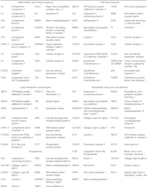
Also contrasting other GWAS approaches in other diseases, a significant fraction of the susceptibility sig-nal in AMD has mapped to, or near, genes encoding components of the complement cascade, although this is not the sole pathway implicated. Overall, the genes and alleles thought to confer significant susceptibility to AMD pathology can be clustered broadly into five major pathways (Table 3): (a) the inflammation and immune response, (b) lipid metabolism and transport, (c) extracellular matrix and cell adhesion, (d) angio-genesis, and (e) cellular stress responses. Among these, risk assessment analysis for either common or rare alleles has highlighted in genes encoding comple-ment pathway components. The common variants near six complement genes, CFH, C2/CFB, C3, CFI, and C9 together, account for almost 60 % of the AMD genetic risk [36]. Notably, for the rare alleles found in sufficient recurrent rates to empower mean-ingful studies, the individual risk to AMD is sharply higher and appears as if they are almost Mendelian, as observed in the case of CFI [19].
A pathogenic route to AMD: alternative complement pathway
One of the leading candidates for predisposition to AMD is the inflammatory pathogenesis theory, which hypothesizes dysregulation of the immune response, spe-cifically complement system [6, 9, 12, 14, 15, 20, 55, 56, 64–68]. Reports dating back to 1875 have hypothesized that macular lesions were due to inflammation and that disciform degeneration were associated with choroidal inflammation [69]. Since then multiple components of the complement pathway have been linked to AMD and its pathological consequences [15, 16, 30, 64, 70, 71]. The complement system is a specialized part of innate immunity that can respond to antigen-antibody com-plexes (classical pathway) or bacterial mannose groups (lectin pathway) and can also be active in a low-level continuous state (alternative pathway (AP)), to allow for an immediate amplified response [72]. All complement pathways culminate in the creation of the membrane at-tack complex (MAC) for cell lysis and organismal defense.
AMD is not the first human genetic disorder associ-ated with AP complement dysfunction. A number of dis-orders are thought to be the result of excessive AP activation, including membranoproliferative glomerulo-nephritis type II (MPGN type II); atypical hemolytic uremic syndrome (aHUS); and paroxysmal nocturnal hemoglobinuria (PNH, a rare form of hemolytic anemia). Of note, MPGN type II, which is characterized by renal disease and low serum C3 levels, is also associated with complete FH deficiency, arguing for a mechanistic simi-larity between MPGN and AMD [55, 73]. Similarly, aHUS has been associated with low FH levels in addition to low C3 levels and has mutations in several AP com-ponents, however, at a unique haplotype compared to AMD and MPGN [73]. PNH is caused by mutations in phosphatidylinositol glycan-complementation class A (PIGA) that is essential in the establishment of glyco-phosphatidylinositol anchors. Two known inhibitors of AP activation are glycolipid-anchored proteins that are required for the regulation at the C3 convertase step and MAC assembly [74].
Table 3Genes associated with AMD that cluster into five major pathways
Inflammation and immune response Cell stress response
C2 Complement
component 2
HLA-C Major histocompatibility complex, class I, C
ABCA4 ATP-binding cassette subfamily A member 4
HTRA1 HtrA serine peptidase 1
C3 Complement
component 3
IL8 Interleukin B ACE Angiotensin I
converting enzyme 1
RORA RAR-related orphan receptor alpha
CFB Complement
factor B
MMP9 Matrix metallopeptidase 9 APOE Apoliporotein E SOD2 Superoxide dismutase 2, mitochondrial
CFH Complement
factor H
PLEKHA1 Pleckstrin homology domain containing, family A member 1
ARMS2 Age-related maculopathy susceptibility 2
TF Transferrin
CFD Complement
factor D
RORA RAR-related orphan receptor alpha
CST3 Cystatin C TLR3 Toll-like receptor 3
CFHR1-5 Complement factor H-related 1-5
SERPING1 Serpin peptidase inhibitor, clade G, member 1
CX3CR1 Chemokine receptor 1 TLR4 Toll-like receptor 4
CFI Complement
factor I
TLR3 Toll-like receptor 3 CYP24A1 Cytochrome P450, family 24, subfamily A peptide 1
VLDLR Very low-density lipoprotein receptor
C9 Complement
component 9
TLR4 Toll-like receptor 4 GSTM1 Glutathione S-transferase mu 1
TNFRSF10A/ LOC389641
Tumor necrosis factor receptor superfamily, member 10a
CX3CR1 Chemokine receptor 1
VLDLR Very low-density lipoprotein receptor
GSTP1 Glutathione S-transferase pi 1
IER3 Immediate early response 3
F13B Coagulation factor XIII, B polypeptide
VTN Vitronectin GSTT1 Glutathione
S-transferase tau 1
TGFBR1 Transforming growth factor, beta receptor 1
Lipid metabolism and transport Extracellular matrix and cell adhesion
ABCA1 ATP-binding cassette, subfamily A, member 1
FADS1-3 Fatty acid desaturases 1-3
ACE Angiotensin 1 converting enzyme 1
ROBO1 Roundabout, axon guidance receptor, homolog 1
ABCA4 ATP-binding cassette, subfamily A, member 4
LIPC Hepatic lipase ARMS2 Age-related maculopathy susceptibility 2
TIMP3 Tissue inhibitor of metalloproteinase 3
APOE Apolipoprotein E LPL Lipoprotein lipases ADAMTS9 ADAM metallopeptidase with trhombospondin type 1 motif, 9
MMP19 Matrix
metallopeptidase 19
CETP Cholesteryl ester transfer protein, plasma
LRP6 Low-density lipoprotein receptor-related protein 6
COL8A1 Collagen, type VIII, alpha 1 PCOLCE Procollagen c-endopeptidase enhancer
CFHR1-5 Complement factor H-related 1-5
RORA RAR-related orphan receptor alpha
COL10A1 Collagen, type X, alpha 1 VTN Vitronectin
CYP24A1 Cytochrome P450, family 24, subfamily A peptide 1
VLDLR Very low-density lipoprotein receptor
CST3 Cystatin C ABCA7 ATP-binding cassette,
cubfamily A, member 7
ELOVL4 ELVL fatty acid elongase 4
PLTP Phospholipid transfer protein
CX3CR1 Chemokine receptor 1 ACTG1 Actin gamma 1
Angiogenesis F13B Coagulation factor XIII,
B polypeptide
BCAR1 Breast cancer anti-estrogen resistance 1
ACE Angiotensin I converting enzyme 1
LRP6 Low-density lipoprotein receptor-related protein 6
FBLN5 Fibulin 5 COL4A4 Collagen, type IV, alpha 4
COL10A1 Collagen, type X, alpha 1
MMP9 Matrix metallopeptidase 9 HMCN1 Hemicentin ITGA7 Integrin, alpha 7
COL8A1 Collagen, type VIII, alpha 1
RORA RAR-related orphan receptor alpha
HTRA1 HtrA serine peptidase 1 MYL2 Myosin, light chain 2, regulatory, cardiac, slow
CST3 Cystatin C SERPINF1 Serpin peptidase inhibitor, clade F
MMP9 Matrix metallopeptidase 1
AMD is associated with activators of alternative pathway components
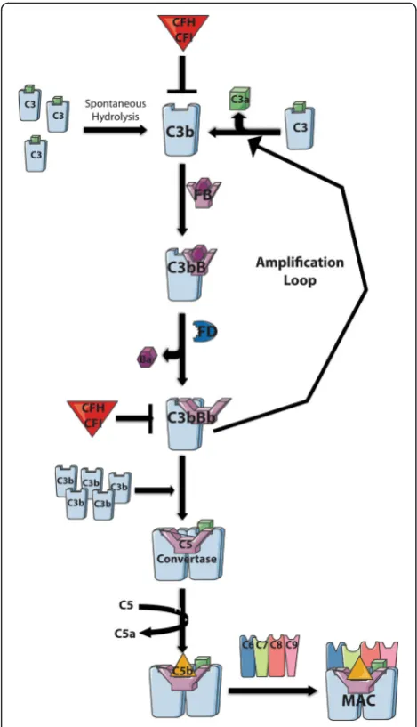
Low levels of constitutive complement activation via the AP allows for immediate immune response. It involves the central molecule of all three arms of the comple-ment pathway, C3. In the AP, C3, is activated in two ways (Fig. 2): (1) it is either cleaved by convertases/ plasma proteases to generate C3b or (2) a small portion is hydrolyzed spontaneously to C3H2O establishing a continuous “tick-over” ready for immediate C3b depos-ition on pathogens for target opsonization [76–79]. Fac-tor B (FB) binds to C3H2O or C3b and the complex is cleaved by the plasma protease factor D (FD) forming the essential C3 convertase (C3bBb or C3H2OBb), lead-ing to an amplification loop that cleaves and assembles C3 to C3b to C3bBb continuously. The accumulation of C3b leads to C3b binding to C3bBb, thereby creating a new enzyme, the C5 convertase (C3bBbC3b) that cleaves C5 to C5a (an anaphylatoxin and chemoattractant) and C5b, a component of the lytic pore which combines with C6–C9 to form the membrane attack complex (MAC) resulting in cell lysis [72].
The AP has the unique characteristic of being able to be activated spontaneously, allowing for an immediate immune response, which must be tightly regulated to prevent excessive activation. Intrinsically, the AP has two major negative regulators encoded byCFHandCFI. CFH is the soluble inhibitor of the complement cascade and encodes a secreted glycoprotein, FH that acts as co-factor for FI mediated cleavage of C3b as well as acceler-ating decay of the C3bBb convertase.
Many cells also express membrane-associated AP reg-ulators that hinder the C3 amplification loop and/or pre-vent the deposition/accumulation of C3b on self-tissue. These membrane-associated proteins include membrane cofactor protein (MCP), decay-accelerating factor (DAF), and complement receptor 1 (CR1) all of which are encoded by genes located within the RCA gene cluster on 1q32 [80]. MCP and CR1 have cofactor activity for CFI,while CR1 along with DAF have decay acceleration activity. Differential expression ofMCP has been linked to AMD and upon the addition of an environmental stimulus such as smoking, a risk factor of AMD, both MCP and DAF are down regulated [81, 82]. Both activat-ing and inhibitactivat-ing components have been reported to harbor variants that potentially have functional impacts
on the AP (Table 4), with an increase in overall muta-tional burden being reported in CFH and CFI in AMD patients [60, 83]. These perturbations of the components themselves or of their ability to interact with other AP components typically result in uncontrolled AP activation.
Pathogenic outcomes of the AP
Advanced age is the only risk factor common to all AMD patients. During the aging process, the physio-logical changes include redistribution of the photorecep-tors, thickening of BM, and accumulation of debris in the eye in conjunction with environmental factors and genetic variation contribute to cascading dysfunction of physiological pathways such as lipid transport, angiogen-esis, stress response, and ECM remodeling [24–26]. In turn, disruption of each of these pathways can lead to an inflammatory and an AP immune response ultimately culminating in cell death [72]. Each of these pathways has been hypothesized to alter components of the retina [36, 84] affecting the interdependence of the photore-ceptors, RPE, BM, and choriocapillaris making it chal-lenging to understand the model for AMD pathogenesis. During the early stages of AMD, sub-RPE deposit for-mation (drusen and soft basal linear deposits) between the RPE and BM occurs and is also thought to be the main site of immune complex formation in AMD. While containing over 40 % lipids, other components of sub-RPE deposits include TIMP3 (TIMP metallopepti-dase inhibitor 3), which plays a role in ECM mainten-ance and remodeling [85, 86]; amyloid beta (Aβ) that is produced either systemically or from the RPE and is proangiogenic and a known activator of comple-ment [87]; apolipoproteins, which are also generated systemically and by the RPE [88]); and CFH and other complement components [30].
Lipid accumulation, similar to that seen in atheroscler-osis, can increase choroidal vessel resistance preventing the choriocapillaris from properly clearing additional li-poproteins from the RPE and BM [89]. The accumula-tion of lipoproteins along with Aβlead to the formation of a lipid “wall” external to the RPE [90, 91] that is the precursor to basal linear deposits that forms between the RPE basement membrane and the inner collagenous zone of BM [92]. The accumulated, peroxidizable lipo-proteins are oxidized contributing to RPE damage [93]. Table 3Genes associated with AMD that cluster into five major pathways(Continued)
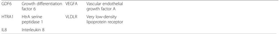
GDF6 Growth differentiation factor 6
VEGFA Vascular endothelial growth factor A
HTRA1 HtrA serine peptidase 1
VLDLR Very low-density lipoprotein receptor
Additionally, one byproduct of lipid peroxidation prod-uct is malondialdehyde (MDA), a marker for oxidative stress that contributes to RPE dysfunction [94] and a binding partner to CFH, allowing for an endogenous anti-inflammatory mediated response to this pro-anti-inflammatory byproduct [95]. Similar to MDA, another cholesterol ox-idation product having pro-inflammatory effects is 7-ketocholesterol (7KCh), which has been associated with cytotoxicity and has been shown to interact with retinal
microglia, possibly promoting choroidal neovascularization [96] [97]. Further, these oxidized low-density lipoproteins can bind C-reactive protein initiating an inflammatory re-sponse leading to complement activation [98, 99]. Supple-menting evidence has also been reported that complement activation leads to the recruitment of mononuclear phago-cytes that contribute to RPE pathology in AMD [100]. All points that support immune system activation as a conse-quence of pathologic lipid accumulation.
The majority of complement is synthesized primarily by the liver and is delivered through circulation; how-ever, some local tissues are also able to synthesize com-plement components, specifically the RPE and choroid [55]. Upon factors such as aging, oxidative stress (including cigarette smoke which that has been reported in vitro to activate C3 [101]), and lipid accumulation, increased AP activation in the RPE, which is thought to be the site of primary dysfunction in AMD, is primed for genetic predispositions to onset disease progression [14, 75, 101]. Upon AP activation, initiation of the terminal pathway ensues, forming MAC in BM and the choriocapillaris contributing to compromised function of the tight interaction of the RPE-BM-choroid com-plex [18].
Additional clues and emerging thoughts on pathogenic contributions to complex disease
There have been multiple insights implicating comple-ment components in the pathobiology of AMD including genetic associations and accumulation of complement components in sub-RPE deposits of patient retinas [30, 102]. With common variants accounting for 65 % of the heritability of AMD, the search for rare variant contribu-tors has only recently been undertaken through the advances of next generation sequencing [40]. Rare variants of C3, CFH, CFI, and C9 that have thus far been associ-ated with AMD have been shown to play a role in either the complement pathway, to have an impact on the muta-tional load, or to hold the promise of putative future therapeutic targets [19, 58, 59, 63, 65]. However, a leading limitation in understanding disease outcome has been the lack of comprehension of the functional impact of the genetic contributors themselves. By combining the com-mon variant comcom-mon disease and rare variant comcom-mon disease hypotheses [103–105], the idea of mutational bur-den emerges as a prognostic/diagnostic alternative for AMD, as exemplified by both common and rare variants in AP inhibitors leading to pathogenic dysregulated AP ac-tivity (Table 4). The latter highlights the necessity to func-tionally assess the role of a variant(s) in a gene(s) in order to more explicitly understand AMD pathogenesis. This highlights the need for development of animal models which are currently in development, for example, a trans-genic mouse model expressing the human normal (Y402)
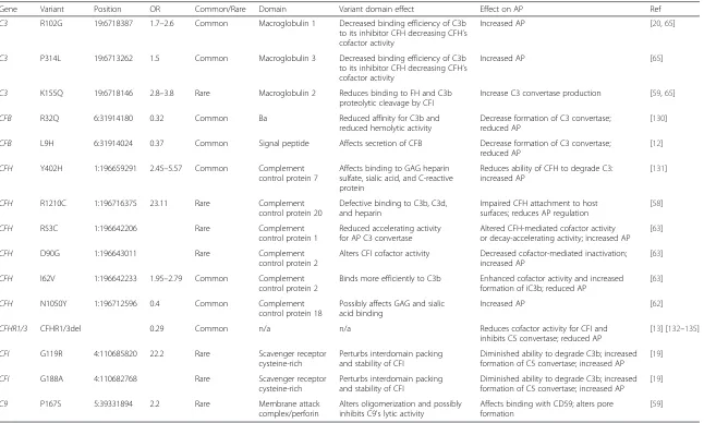
Table 4AP complement variants
Gene Variant Position OR Common/Rare Domain Variant domain effect Effect on AP Ref
C3 R102G 19:6718387 1.7–2.6 Common Macroglobulin 1 Decreased binding efficiency of C3b
to its inhibitor CFH decreasing CFH’s cofactor activity
Increased AP [20,65]
C3 P314L 19:6713262 1.5 Common Macroglobulin 3 Decreased binding efficiency of C3b
to its inhibitor CFH decreasing CFH’s cofactor activity
Increased AP [65]
C3 K155Q 19:6718146 2.8–3.8 Rare Macroglobulin 2 Reduces binding to FH and C3b
proteolytic cleavage by CFI
Increase C3 convertase production [59,65]
CFB R32Q 6:31914180 0.32 Common Ba Reduced affinity for C3b and
reduced hemolytic activity
Decrease formation of C3 convertase; reduced AP
[130]
CFB L9H 6:31914024 0.37 Common Signal peptide Affects secretion of CFB Decrease formation of C3 convertase;
reduced AP
[12]
CFH Y402H 1:196659291 2.45–5.57 Common Complement
control protein 7
Affects binding to GAG heparin sulfate, sialic acid, and C-reactive protein
Reduces ability of CFH to degrade C3: increased AP
[131]
CFH R1210C 1:196716375 23.11 Rare Complement
control protein 20
Defective binding to C3b, C3d, and heparin
Impaired CFH attachment to host surfaces; reduces AP regulation
[58]
CFH R53C 1:196642206 Rare Complement
control protein 1
Reduced accelerating activity for AP C3 convertase
Altered CFH-mediated cofactor activity or decay-accelerating activity; increased AP
[63]
CFH D90G 1:196643011 Rare Complement
control protein 2
Alters CFI cofactor activity Decreased cofactor-mediated inactivation; increased AP
[63]
CFH I62V 1:196642233 1.95–2.79 Common Complement
control protein 2
Binds more efficiently to C3b Enhanced cofactor activity and increased formation of iC3b; reduced AP
[63]
CFH N1050Y 1:196712596 0.4 Common Complement
control protein 18
Possibly affects GAG and sialic acid binding
Increased AP [62]
CFHR1/3 CFHR1/3del 0.29 Common n/a n/a Reduces cofactor activity for CFI and
inhibits C5 convertase; reduced AP
[13] [132–135]
CFI G119R 4:110685820 22.2 Rare Scavenger receptor
cysteine-rich
Perturbs interdomain packing and stability of CFI
Diminished ability to degrade C3b; increased formation of C5 convertase; increased AP
[19]
CFI G188A 4:110682768 Rare Scavenger receptor
cysteine-rich
Perturbs interdomain packing and stability of CFI
Diminished ability to degrade C3b; increased formation of C5 convertase; increased AP
[19]
C9 P167S 5:39331894 2.2 Rare Membrane attack
complex/perforin
Alters oligomerization and possibly inhibits C9's lytic activity
Affects binding with CD59; alters pore formation
[59]
Genomic
s
(2016) 10:23
Page
9
of
and AMD-risk associated (Y402H) variants of FH has re-cently been described [106].
Familial aggregation is observed in most complex dis-ease since there is greater likelihood of sharing disdis-ease- disease-predisposing genotypes; however, non-genetic factors can contribute to discordant phenotypes [40]. In an ef-fort to understand disease phenotypic outcomes, most studies have started by trying to identify causal variants. The identification of susceptibility variants is hindered by numerous confounding factors that have to be ac-knowledged to gain insight into disease mechanism. (1) Excluding a few cases such as AD and AMD, the vast majority of common variants exhibit a modest effect therefore large cohorts are required to power the find-ings [107, 108]. (2) Rare, disease-causing mutations, while numerous, are rarely observed in the general population [60, 83, 109, 110]. (3) SNP association of a locus does not imply that the causal variant itself is a SNP, as observed in Crohn’s disease where the causal mutations is a deletion upstream of the promoter [111]. (4) Multiple distinct alleles either common, rare, or both, can be present at a single locus [58, 112]. 5) Allele fre-quencies vary among ethnic groups, as not only ob-served in AMD but also described in Hirschsprung’s disease in which the causal variant has a minor allele fre-quency (MAF) ranging from 0.01 to 0.45 depending on the population [113]. (6) Causality due to non-coding variants is difficult to establish, similar to what was ob-served in coronary artery disease [114–116]. (7) Similar to pathways functioning in multiple processes, variants can also be pleiotropic, with their effect being dependent on the genetic content in which they are identified [117]. 8) Genes with causal variants identified need to be fur-ther studied for additional variation that could contrib-ute to disease, as was seen in the case when delving deeper into CFH and CFI in AMD [19, 58]. (9) Struc-tural variation cannot be ignored, as was observed in the case of Bardet-Biedl Syndrome in which copy number variations lead to disease progression (unpublished data). (10) Environmental factors can modify genetic effects and phenotypic outcomes [2, 101, 118].
Conclusions
Genetics has aided in the understanding of the complex, multifactorial nature of AMD. However, with modest signals differentiating the Dry versus Wet advanced forms of AMD, genetics has not revealed a strong pre-dictive value on phenotypic severity or progression. Des-pite the long road of discovery that lies ahead, various lines of evidence linking the AP to AMD disease patho-genesis including (a) clinical phenotypes that associate with deficiencies of AP regulators [119]; (b) linkage of SNPs in AP components to disease risk [11, 120]; and (c) the functional analysis of individual AP components
in the establishment of clinical phenotypes observed in both mouse and zebrafish models [74]. With numerous mouse models recapitulating at least 10 distinct human dis-orders, such as rheumatoid arthritis, traumatic brain injury, and AMD, the effects of AP on development and homeo-stasis have been established, making it clear that dysregula-tion/dysfunction of the process of innate immunity plays a contributory role in disease outcome [74]. Predominant functional assays for measuring the impact of variation on the complement pathway have been hemolytic and enzyme immunoassays [59, 121–123]; focus should and has begun to be placed on additional in vivo model systems to further understand the roles of AP disease contributors in a more systemic context [100, 106].
Abbreviations
7KCh:7-ketocholesterol; ABCA4: ATP-binding cassette subfamily A member 4; AD: Alzheimer’s disease; aHUS: atypical hemolytic uremic syndrome; AMD: age-related macular degeneration; AP: alternative pathway; APOE: apolipoprotein E; AREDS: Age-Related Eye Disease Study; ARMS2: age-related maculopathy susceptibility 2; Aβ: amyloid beta; BM: Bruch’s membrane; C2: complement component 2; C3: complement component 3; C9: complement component 9; CFB or FB: complement factor B; CFH or FH: complement factor H; CFI or FI: complement factor I; CNV: choroidal neovascular membranes;
CR1: complement receptor 1; DAF: decay-accelerating factor; ECM: extracellular matrix; FD: complement factor D; GWAS: genome-wide association studies; HTRA1: HtrA serine peptidase 1; MAC: membrane attack complex; MAF: minor allele frequency; MCP: membrane cofactor protein; MDA: malondialdehyde; MPGN: membranoproliferative glomerulonephritis; OR: odds ratio; PIGA: phosphatidylinositol glycan-complement class A; PNH: paroxysmal nocturnal Hemoglobinuria; RCA: regulator of complement activation; RPE: retinal pigment epithelium; RR: relative risk; SNP: single nucleotide polymorphism; TIMP3: TIMP metallopeptidase inhibitor 3.
Acknowledgements
We thank Allison Ashley-Koch, Maria Kousi, and Erik Madsen for their thoughtful comments on the manuscript.
Authors’contributions
The manuscript was written by PLT. All authors critically revised the manuscript. All authors read and approved the final manuscript.
Competing interests
The authors declare that they have no competing interests. Author details
1
Center for Human Disease Modeling, Duke University Medical Center, Durham, NC 27710, USA.2Department of Cell Biology, Duke University Medical Center, Durham, NC 27710, USA.3Department of Ophthalmology, Duke Eye Center, Duke University, Durham, NC 27710, USA.
Received: 25 May 2016 Accepted: 8 June 2016
References
1. Klein R, Peto T, Bird A, Vannewkirk MR. The epidemiology of age-related macular degeneration. Am J Ophthalmol. 2004;137:486–95. doi: 10.1016/j.ajo.2003.11.069.
2. Thornton J et al. Smoking and age-related macular degeneration: a review of association. Eye (Lond). 2005;19:935–44. doi:10.1038/sj.eye.6701978. 3. Young RW. Pathophysiology of age-related macular degeneration. Surv
Ophthalmol. 1987;31:291–306.
5. Weeks DE et al. Age-related maculopathy: an expanded genome-wide scan with evidence of susceptibility loci within the 1q31 and 17q25 regions. Am J Ophthalmol. 2001;132:682–92.
6. Klein RJ et al. Complement factor H polymorphism in age-related macular degeneration. Science (New York, NY). 2005;308:385–9. doi:10. 1126/science.1109557.
7. Kondo N, Bessho H, Honda S, Negi A. Complement factor H Y402H variant and risk of age-related macular degeneration in Asians: a systematic review and meta-analysis. Ophthalmology. 2011;118:339–44. doi:10.1016/j.ophtha. 2010.06.040.
8. Thakkinstian A et al. Systematic review and meta-analysis of the association between complement factor H Y402H polymorphisms and age-related macular degeneration. Hum Mol Genet. 2006;15:2784–90. doi:10.1093/hmg/ddl220.
9. Edwards AO et al. Complement factor H polymorphism and age-related macular degeneration. Science (New York, NY). 2005;308:421–4. doi:10.1126/ science.1110189.
10. Ennis S, Gibson J, Cree AJ, Collins A, Lotery AJ. Support for the involvement of complement factor I in age-related macular degeneration. Eur J Hum Genet. 2010;18:15–6. doi:10.1038/ejhg.2009.113.
11. Fagerness JA et al. Variation near complement factor I is associated with risk of advanced AMD. Eur J Hum Genet. 2009;17:100–4. doi:10. 1038/ejhg.2008.140.
12. Gold B et al. Variation in factor B (BF) and complement component 2 (C2) genes is associated with age-related macular degeneration. Nat Genet. 2006;38:458–62. doi:10.1038/ng1750.
13. Hageman GS et al. Extended haplotypes in the complement factor H (CFH) and CFH-related (CFHR) family of genes protect against age-related macular degeneration: characterization, ethnic distribution and evolutionary implications. Ann Med. 2006;38:592–604.
14. Hageman GS et al. An integrated hypothesis that considers drusen as biomarkers of immune-mediated processes at the RPE-Bruch’s membrane interface in aging and age-related macular degeneration. Prog Retin Eye Res. 2001;20:705–32.
15. Johnson LV, Leitner WP, Staples MK, Anderson DH. Complement activation and inflammatory processes in Drusen formation and age related macular degeneration. Exp Eye Res. 2001;73:887–96. doi:10.1006/exer.2001.1094. 16. Johnson LV, Ozaki S, Staples MK, Erickson PA, Anderson DH. A potential role
for immune complex pathogenesis in drusen formation. Exp Eye Res. 2000; 70:441–9. doi:10.1006/exer.1999.0798.
17. Li M et al. CFH haplotypes without the Y402H coding variant show strong association with susceptibility to age-related macular degeneration. Nat Genet. 2006;38:1049–54. doi:10.1038/ng1871.
18. Mullins RF et al. The membrane attack complex in aging human choriocapillaris: relationship to macular degeneration and choroidal thinning. Am J Pathol. 2014;184:3142–53. doi:10.1016/j.ajpath.2014.07.017. 19. van de Ven JP et al. A functional variant in the CFI gene confers a high risk
of age-related macular degeneration. Nat Genet. 2013;45:813–7. doi:10.1038/ ng.2640.
20. Yates JR et al. Complement C3 variant and the risk of age-related macular degeneration. N Engl J Med. 2007;357:553–61. doi:10.1056/ NEJMoa072618.
21. Zhan X et al. Identification of a rare coding variant in complement 3 associated with age-related macular degeneration. Nat Genet. 2013;45: 1375–9. doi:10.1038/ng.2758.
22. Strauss O. The retinal pigment epithelium in visual function. Physiol Rev. 2005;85:845–81. doi:10.1152/physrev.00021.2004.
23. Lutty GA et al. Development of the human choriocapillaris. Eye (Lond). 2010;24:408–15. doi:10.1038/eye.2009.318.
24. Curcio CA, Owsley C, Jackson GR. Spare the rods, save the cones in aging and age-related maculopathy. Invest Ophthalmol Vis Sci. 2000;41:2015–8. 25. Feeney-Burns L, Ellersieck MR. Age-related changes in the ultrastructure of
Bruch’s membrane. Am J Ophthalmol. 1985;100:686–97.
26. Curcio CA, Millican CL, Bailey T, Kruth HS. Accumulation of cholesterol with age in human Bruch’s membrane. Invest Ophthalmol Vis Sci. 2001;42:265–74.
27. Delori FC, Goger DG, Dorey CK. Age-related accumulation and spatial distribution of lipofuscin in RPE of normal subjects. Invest Ophthalmol Vis Sci. 2001;42:1855–66.
28. Eldred GE, Miller GV, Stark WS, Feeney-Burns L. Lipofuscin: resolution of discrepant fluorescence data. Science (New York, NY). 1982;216:757–9.
29. Moreira EF, Larrayoz IM, Lee JW, Rodriguez IR. 7-Ketocholesterol is present in lipid deposits in the primate retina: potential implication in the induction of VEGF and CNV formation. Invest Ophthalmol Vis Sci. 2009;50:523–32. doi:10. 1167/iovs.08-2373.
30. Mullins RF, Russell SR, Anderson DH, Hageman GS. Drusen associated with aging and age-related macular degeneration contain proteins common to extracellular deposits associated with atherosclerosis, elastosis, amyloidosis, and dense deposit disease. Faseb J. 2000;14:835–46.
31. Russell SR, Mullins RF, Schneider BL, Hageman GS. Location, substructure, and composition of basal laminar drusen compared with drusen associated with aging and age-related macular degeneration. Am J Ophthalmol. 2000; 129:205–14.
32. Jager RD, Mieler WF, Miller JW. Age-related macular degeneration. N Engl J Med. 2008;358:2606–17. doi:10.1056/NEJMra0801537.
33. Sarks SH. Ageing and degeneration in the macular region: a clinico-pathological study. Br J Ophthalmol. 1976;60:324–41.
34. Sarks SH, Arnold JJ, Killingsworth MC, Sarks JP. Early drusen formation in the normal and aging eye and their relation to age related maculopathy: a clinicopathological study. Br J Ophthalmol. 1999;83:358–68.
35. Spraul CW, Grossniklaus HE. Characteristics of Drusen and Bruch’s membrane in postmortem eyes with age-related macular degeneration. Arch Ophthal. 1997;115:267–73.
36. Fritsche LG et al. Age-related macular degeneration: genetics and biology coming together. Annu Rev Genomics Hum Genet. 2014;15:151–71. doi:10. 1146/annurev-genom-090413-025610.
37. Group, A.-R. E. D. S. R. Risk factors associated with age-related macular degeneration. A case-control study in the Age-Related Eye Disease Study: Age-Related Eye Disease Study report number 3. Ophthalmology. 2000;107:2224–32.
38. Davis MD et al. The Age-Related Eye Disease Study severity scale for age-related macular degeneration: AREDS report no. 17. Arch Ophthal. 2005;123: 1484–98. doi:10.1001/archopht.123.11.1484.
39. Ferris FL et al. A simplified severity scale for age-related macular degeneration: AREDS report no. 18. Arch Ophthal. 2005;123:1570–4. doi:10. 1001/archopht.123.11.1570.
40. Sobrin L, Seddon JM. Nature and nurture- genes and environment- predict onset and progression of macular degeneration. Prog Retin Eye Res. 2014; 40:1–15. doi:10.1016/j.preteyeres.2013.12.004.
41. Hammond CJ et al. Genetic influence on early age-related maculopathy: a twin study. Ophthalmology. 2002;109:730–6.
42. Klaver CC et al. Genetic risk of age-related maculopathy. Population-based familial aggregation study. Arch Ophthal. 1998;116:1646–51.
43. Klein ML, Mauldin WM, Stoumbos VD. Heredity and age-related macular degeneration. Observations in monozygotic twins. Arch Ophthal. 1994; 112:932–7.
44. Meyers SM, Greene T, Gutman FA. A twin study of age-related macular degeneration. Am J Ophthalmol. 1995;120:757–66.
45. Seddon JM, Ajani UA, Mitchell BD. Familial aggregation of age-related maculopathy. Am J Ophthalmol. 1997;123:199–206.
46. Seddon JM, Cote J, Page WF, Aggen SH, Neale MC. The US twin study of age-related macular degeneration: relative roles of genetic and environmental influences. Arch Ophthal. 2005;123:321–7. doi:10.1001/ archopht.123.3.321.
47. Allikmets R et al. Mutation of the Stargardt disease gene (ABCR) in age-related macular degeneration. Science (New York, NY). 1997;277:1805–7. 48. Zhang K et al. The ABCR gene in recessive and dominant Stargardt diseases:
a genetic pathway in macular degeneration. Genomics. 1999;60:234–7. doi: 10.1006/geno.1999.5896.
49. Klaver CC et al. Genetic association of apolipoprotein E with age-related macular degeneration. Am J Hum Genet. 1998;63:200–6. doi: 10.1086/301901.
50. Flint J, Mackay TF. Genetic architecture of quantitative traits in mice, flies, and humans. Genome Res. 2009;19:723–33. doi:10.1101/gr.086660.108. 51. Risch N, Merikangas K. The future of genetic studies of complex human
diseases. Science (New York, NY). 1996;273:1516–7.
52. Peter I, Seddon JM. Genetic epidemiology: successes and challenges of genome-wide association studies using the example of age-related macular degeneration. Am J Ophthalmol. 2010;150:450–452.e452. doi:10. 1016/j.ajo.2010.06.012.
54. Dewan A et al. HTRA1 promoter polymorphism in wet age-related macular degeneration. Science (New York, NY). 2006;314:989–92. doi:10.1126/science. 1133807.
55. Hageman GS et al. A common haplotype in the complement regulatory gene factor H (HF1/CFH) predisposes individuals to age-related macular degeneration. Proc Natl Acad Sci U S A. 2005;102:7227–32. doi:10.1073/pnas. 0501536102.
56. Haines JL et al. Complement factor H variant increases the risk of age-related macular degeneration. Science (New York, NY). 2005;308:419–21. doi: 10.1126/science.1110359.
57. Rivera A et al. Hypothetical LOC387715 is a second major susceptibility gene for age-related macular degeneration, contributing independently of complement factor H to disease risk. Hum Mol Genet. 2005;14:3227–36. doi: 10.1093/hmg/ddi353.
58. Raychaudhuri S et al. A rare penetrant mutation in CFH confers high risk of age-related macular degeneration. Nat Genet. 2011;43:1232–6. doi:10. 1038/ng.976.
59. Seddon JM et al. Rare variants in CFI, C3 and C9 are associated with high risk of advanced age-related macular degeneration. Nat Genet. 2013;45: 1366–70. doi:10.1038/ng.2741.
60. Triebwasser MP et al. Rare variants in the functional domains of complement factor H are associated with age-related macular degeneration. Invest Ophthalmol Vis Sci. 2015;56:6873–8. doi:10.1167/iovs. 15-17432.
61. Fritsche LG et al. A large genome-wide association study of age-related macular degeneration highlights contributions of rare and common variants. Nat Genet. 2016;48:134–43. doi:10.1038/ng.3448.
62. Boon CJ et al. Basal laminar drusen caused by compound heterozygous variants in the CFH gene. Am J Hum Genet. 2008;82:516–23. doi:10.1016/j. ajhg.2007.11.007.
63. Yu Y et al. Whole-exome sequencing identifies rare, functional CFH variants in families with macular degeneration. Hum Mol Genet. 2014;23:5283–93. doi:10.1093/hmg/ddu226.
64. Anderson DH, Mullins RF, Hageman GS, Johnson LV. A role for local inflammation in the formation of drusen in the aging eye. Am J Ophthalmol. 2002;134:411–31.
65. Despriet DD et al. Complement component C3 and risk of age-related macular degeneration. Ophthalmology. 2009;116:474–480.e472. doi:10.1016/ j.ophtha.2008.09.055.
66. Maller JB et al. Variation in complement factor 3 is associated with risk of age-related macular degeneration. Nat Genet. 2007;39:1200–1. doi:10. 1038/ng2131.
67. Jakobsdottir J, Conley YP, Weeks DE, Ferrell RE, Gorin MB. C2 and CFB genes in age-related maculopathy and joint action with CFH and LOC387715 genes. PLoS One. 2008;3:e2199. doi:10.1371/journal.pone.0002199. 68. Park KH, Fridley BL, Ryu E, Tosakulwong N, Edwards AO. Complement
component 3 (C3) haplotypes and risk of advanced age-related macular degeneration. Invest Ophthalmol Vis Sci. 2009;50:3386–93. doi:10.1167/ iovs.08-3231.
69. Pagenstecher, H. a. G., C. Atlas der Pathologischen Anatomie des Augapfels. CW Kreidel, Wiesbaden (1875).
70. Hanssen R. Zur anatomie der scheibenformigen degeneration der netzhaumitte.Z. Augenh.1930. 360-368.
71. Penfold PL, Killingsworth MC, Sarks SH. Senile macular degeneration: the involvement of immunocompetent cells. Graefes Arch Clin Exp Ophthalmol. 1985;223:69–76.
72. Ricklin D, Hajishengallis G, Yang K, Lambris JD. Complement: a key system for immune surveillance and homeostasis. Nat Immunol. 2010;11:785–97. doi:10.1038/ni.1923.
73. Pickering MC et al. Spontaneous hemolytic uremic syndrome triggered by complement factor H lacking surface recognition domains. J Exp Med. 2007; 204:1249–56. doi:10.1084/jem.20070301.
74. Holers VM. The spectrum of complement alternative pathway-mediated diseases. Immunol Rev. 2008;223:300–16. doi:10.1111/j.1600-065X.2008.00641.x. 75. Anderson DH et al. The pivotal role of the complement system in aging
and age-related macular degeneration: hypothesis re-visited. Prog Retin Eye Res. 2010;29:95–112. doi:10.1016/j.preteyeres.2009.11.003.
76. Pangburn MK, Schreiber RD, Muller-Eberhard HJ. Formation of the initial C3 convertase of the alternative complement pathway. Acquisition of C3b-like activities by spontaneous hydrolysis of the putative thioester in native C3. J Exp Med. 1981;154:856–67.
77. Pangburn MK, Muller-Eberhard HJ. Initiation of the alternative complement pathway due to spontaneous hydrolysis of the thioester of C3. Ann N Y Acad Sci. 1983;421:291–8.
78. Bexborn F, Andersson PO, Chen H, Nilsson B, Ekdahl KN. The tick-over theory revisited: formation and regulation of the soluble alternative complement C3 convertase (C3(H2O)Bb). Mol Immunol. 2008;45:2370–9. doi: 10.1016/j.molimm.2007.11.003.
79. Lachmann PJ, Halbwachs L. The influence of C3b inactivator (KAF) concentration on the ability of serum to support complement activation. Clin Exp Immunol. 1975;21:109–14.
80. Pardo-Manuel de Villena F, Heine-Suner D, Rodriguez de Cordoba S. Ordering of the human regulator of complement activation gene cluster on 1q32 by two-colour FISH. Cytogenet Cell Genet. 1996;72:339–41.
81. Fett AL, Hermann MM, Muether PS, Kirchhof B, Fauser S.
Immunohistochemical localization of complement regulatory proteins in the human retina. Histol Histopathol. 2012;27:357–64.
82. Wang L et al. Nrf2 signaling modulates cigarette smoke-induced complement activation in retinal pigmented epithelial cells. Free Radic Biol Med. 2014;70:155–66. doi:10.1016/j.freeradbiomed.2014.01.015.
83. Kavanagh, D. et al. Rare genetic variants in the CFI gene are associated with advanced age-related macular degeneration and commonly result in reduced serum factor I levels. Hum Mol Genet. 2015. doi:10.1093/ hmg/ddv091.
84. Miller JW. Age-related macular degeneration revisited—piecing the puzzle: the LXIX Edward Jackson memorial lecture. Am J Ophthalmol. 2013;155:1– 35 e13. doi:10.1016/j.ajo.2012.10.018.
85. Fariss RN, Apte SS, Olsen BR, Iwata K, Milam AH. Tissue inhibitor of metalloproteinases-3 is a component of Bruch’s membrane of the eye. Am J Pathol. 1997;150:323–8.
86. Kamei M, Hollyfield JG. TIMP-3 in Bruch’s membrane: changes during aging and in age-related macular degeneration. Invest Ophthalmol Vis Sci. 1999;40:2367–75.
87. Yoshida T et al. The potential role of amyloid beta in the pathogenesis of age-related macular degeneration. J Clin Invest. 2005;115:2793–800. doi:10. 1172/jci24635.
88. Anderson DH et al. Local cellular sources of apolipoprotein E in the human retina and retinal pigmented epithelium: implications for the process of drusen formation. Am J Ophthalmol. 2001;131:767–81.
89. Friedman E. A hemodynamic model of the pathogenesis of age-related macular degeneration. Am J Ophthalmol. 1997;124:677–82.
90. Ruberti JW et al. Quick-freeze/deep-etch visualization of age-related lipid accumulation in Bruch’s membrane. Invest Ophthalmol Vis Sci. 2003;44:1753–9.
91. Curcio CA, Johnson M, Rudolf M, Huang JD. The oil spill in ageing Bruch membrane. Br J Ophthalmol. 2011;95:1638–45. doi:10.1136/bjophthalmol-2011-300344.
92. Curcio CA, Johnson M, Huang JD, Rudolf M. Aging, age-related macular degeneration, and the response-to-retention of apolipoprotein B-containing lipoproteins. Prog Retin Eye Res. 2009;28:393–422. doi:10.1016/j.preteyeres. 2009.08.001.
93. Gordiyenko N et al. RPE cells internalize low-density lipoprotein (LDL) and oxidized LDL (oxLDL) in large quantities in vitro and in vivo. Invest Ophthalmol Vis Sci. 2004;45:2822–9. doi:10.1167/iovs.04-0074. 94. Kaemmerer E, Schutt F, Krohne TU, Holz FG, Kopitz J. Effects of lipid
peroxidation-related protein modifications on RPE lysosomal functions and POS phagocytosis. Invest Ophthalmol Vis Sci. 2007;48:1342–7. doi: 10.1167/iovs.06-0549.
95. Weismann D et al. Complement factor H binds malondialdehyde epitopes and protects from oxidative stress. Nature. 2011;478:76–81. doi: 10.1038/nature10449.
96. Indaram M et al. 7-Ketocholesterol increases retinal microglial migration, activation, and angiogenicity: a potential pathogenic mechanism underlying age-related macular degeneration. Sci Rep. 2015;5:9144. doi: 10.1038/srep09144.
97. Rodriguez IR, Alam S, Lee JW. Cytotoxicity of oxidized low-density lipoprotein in cultured RPE cells is dependent on the formation of 7-ketocholesterol. Invest Ophthalmol Vis Sci. 2004;45:2830–7. doi:10.1167/iovs.04-0075. 98. Chang MK, Binder CJ, Torzewski M, Witztum JL. C-reactive protein binds to
99. Rodriguez IR, Larrayoz IM. Cholesterol oxidation in the retina: implications of 7KCh formation in chronic inflammation and age-related macular degeneration. J Lipid Res. 2010;51:2847–62. doi:10.1194/jlr.R004820. 100. Toomey CB, Kelly U, Saban DR, Bowes Rickman C. Regulation of age-related
macular degeneration-like pathology by complement factor H. Proc Natl Acad Sci U S A. 2015;112:E3040–9. doi:10.1073/pnas.1424391112. 101. Kew RR, Ghebrehiwet B, Janoff A. Cigarette smoke can activate the
alternative pathway of complement in vitro by modifying the third component of complement. J Clin Invest. 1985;75:1000–7. doi:10.1172/ jci111760.
102. Gass JD. Drusen and disciform macular detachment and degeneration. Arch Ophthal. 1973;90:206–17.
103. Cirulli ET, Goldstein DB. Uncovering the roles of rare variants in common disease through whole-genome sequencing. Nat Rev Genet. 2010;11:415– 25. doi:10.1038/nrg2779.
104. Reich DE, Lander ES. On the allelic spectrum of human disease. Trends Genet. 2001;17:502–10.
105. Schork NJ, Murray SS, Frazer KA, Topol EJ. Common vs. rare allele hypotheses for complex diseases. Curr Opin Genet Dev. 2009;19:212–9. doi: 10.1016/j.gde.2009.04.010.
106. Ding JD et al. Expression of human complement factor H prevents age-related macular degeneration-like retina damage and kidney abnormalities in aged Cfh knockout mice. Am J Pathol. 2015;185:29–42. doi:10.1016/j. ajpath.2014.08.026.
107. Genome-wide association study of 14,000 cases of seven common diseases and 3,000 shared controls. Nature. 2007. 447, 661-678. doi:10.1038/ nature05911.
108. Zeggini E et al. Meta-analysis of genome-wide association data and large-scale replication identifies additional susceptibility loci for type 2 diabetes. Nat Genet. 2008;40:638–45. doi:10.1038/ng.120.
109. Cohen JC et al. Multiple rare alleles contribute to low plasma levels of HDL cholesterol. Science (New York, NY). 2004;305:869–72. doi:10.1126/science. 1099870.
110. Ji W et al. Rare independent mutations in renal salt handling genes contribute to blood pressure variation. Nat Genet. 2008;40:592–9. doi:10. 1038/ng.118.
111. McCarroll SA et al. Deletion polymorphism upstream of IRGM associated with altered IRGM expression and Crohn’s disease. Nat Genet. 2008;40:1107– 12. doi:10.1038/ng.215.
112. Schulte EC et al. Targeted resequencing and systematic in vivo functional testing identifies rare variants in MEIS1 as significant contributors to restless legs syndrome. Am J Hum Genet. 2014;95:85–95. doi:10.1016/j.ajhg.2014.06.005. 113. Emison ES et al. A common sex-dependent mutation in a RET enhancer
underlies Hirschsprung disease risk. Nature. 2005;434:857–63. doi:10.1038/ nature03467.
114. Helgadottir A et al. A common variant on chromosome 9p21 affects the risk of myocardial infarction. Science (New York, NY). 2007;316:1491–3. doi:10. 1126/science.1142842.
115. McPherson R et al. A common allele on chromosome 9 associated with coronary heart disease. Science (New York, NY). 2007;316:1488–91. doi:10. 1126/science.1142447.
116. Samani NJ et al. Genomewide association analysis of coronary artery disease. N Engl J Med. 2007;357:443–53. doi:10.1056/NEJMoa072366. 117. Jordan DM et al. Identification of cis-suppression of human disease
mutations by comparative genomics. Nature. 2015;524:225–9. doi:10.1038/ nature14497.
118. Hobbs RP, Bernstein PS. Nutrient supplementation for age-related macular degeneration, cataract, and dry eye. J Ophthalmic Vis Res. 2014;9:487–93. doi:10.4103/2008-322X.150829.
119. Botto M et al. Complement in human diseases: lessons from complement deficiencies. Mol Immunol. 2009;46:2774–83. doi:10.1016/j.molimm.2009.04.029. 120. Fisher SA et al. Meta-analysis of genome scans of age-related macular
degeneration. Hum Mol Genet. 2005;14:2257–64. doi:10.1093/hmg/ddi230. 121. Nilsson UR, Nilsson B. Simplified assays of hemolytic activity of the classical
and alternative complement pathways. J Immunol Methods. 1984;72:49–59. 122. Tortajada A et al. C3 glomerulopathy-associated CFHR1 mutation alters FHR oligomerization and complement regulation. J Clin Invest. 2013;123:2434– 46. doi:10.1172/jci68280.
123. Yoshida Y et al. A novel quantitative hemolytic assay coupled with restriction fragment length polymorphisms analysis enabled early diagnosis of atypical hemolytic uremic syndrome and identified unique
predisposing mutations in Japan. PLoS One. 2015;10:e0124655. doi:10. 1371/journal.pone.0124655.
124. Neale BM, et al. Genome-wide association study of advanced age-related macular degeneration identifies a role of the hepatic lipase gene (LIPC). Proc Natl Acad Sci USA. 2010;107:7395-7400. doi:10.1073/pnas.0912019107. 125. Chen W, et al. Genetic variants near TIMP3 and high-density
lipoprotein-associated loci influence susceptibility to age-related macular degeneration. Proc Natl Acad Sci USA. 2010;107:7401-6. doi:10.1073/pnas.0912702107. 126. Kopplin LJ, et al. Genome-wide association identifies SKIV2L and MYRIP as
protective factors for age-related macular degeneration. Genes and immunity. 2010;11:609–21. doi:10.1038/gene.2010.39.
127. Arakawa S, et al. Genome-wide association study identifies two susceptibility loci for exudative agerelated macular degeneration in the Japanese population. Nat Genet. 2011;43:1001–4. doi:10.1038/ng.938. 128. Yu Y, et al. Common variants near FRK/COL10A1 and VEGFA are associated
with advanced agerelated macular degeneration. Hum Mol Genet. 2011;20: 3699-709. doi:10.1093/hmg/ddr270.
129. Fritsche LG, et al. Seven new loci associated with age-related macular degeneration. Nat Genet. 2013;45:433-439. 439e431–432, doi:10.1038/ng. 2578.
130. Montes T, Tortajada A, Morgan BP, Rodriguez de Cordoba S, Harris, CL. Functional basis of protection against age-related macular degeneration conferred by a common polymorphism in complement factor B. Proc Natl Acad Sci USA. 2009;106:4366–71. doi:10.1073/pnas.0812584106. 131. Herbert AP, et al. Structure shows that a glycosaminoglycan and protein
recognition site in factor H is perturbed by age-related macular degeneration-linked single nucleotide polymorphism. The Journal of biological chemistry. 2007;282:18960–8. doi:10.1074/jbc.M609636200. 132. Eberhardt HU, et al. Human factor H-related protein 2 (CFHR2) regulates
complement activation. PLoS One. 2013;8:e78617. doi:10.1371/journal.pone. 0078617.
133. Fritsche LG, et al. An imbalance of human complement regulatory proteins CFHR1, CFHR3 and factor H influences risk for age-related macular degeneration (AMD). Hum Mol Genet. 2010;19:4694–704. doi:10.1093/hmg/ ddq399.
134. Hughes AE, et al. A common CFH haplotype, with deletion of CFHR1 and CFHR3, is associated with lower risk of age-related macular degeneration. Nat Genet. 2006;38:1173–7. doi:10.1038/ng1890.
135. Spencer KL, et al. Deletion of CFHR3 and CFHR1 genes in age-related macular degeneration. Hum Mol Genet. 2008;17:971–7. doi:10.1093/hmg/ ddm369.
• We accept pre-submission inquiries
• Our selector tool helps you to find the most relevant journal
• We provide round the clock customer support
• Convenient online submission
• Thorough peer review
• Inclusion in PubMed and all major indexing services
• Maximum visibility for your research
Submit your manuscript at www.biomedcentral.com/submit