Dendrites differ from axons in
patterns of microtubule stability and
polymerization during development
www.neuraldevelopment.com
Katherine M Kollins
et al
.
Neural
Open Access
Research article
Dendrites differ from axons in patterns of microtubule stability and
polymerization during development
Katherine M Kollins
†3, Robert L Bell
†1, Matthew Butts
1and
Ginger S Withers*
1,2Address: 1Department of Biology, Boyer Ave, Whitman College, Walla Walla, WA 99362, USA, 2Center for Research on Occupational and
Environmental Toxicology, Oregon Health and Science University, SW Sam Jackson Park Road, Portland, OR 97239, USA and 3Department of
Neurobiology and Anatomy, Drexel University College of Medicine, Queen Lane, Philadelphia, PA 19129, USA
Email: Katherine M Kollins - [email protected]; Robert L Bell - [email protected]; Matthew Butts - [email protected]; Ginger S Withers* - [email protected]
* Corresponding author †Equal contributors
Abstract
Background: Dendrites differ from axons in patterns of growth and development, as well as in morphology. Given that microtubules are key structural elements in cells, we assessed patterns of microtubule stability and polymerization during hippocampal neuron development in vitro to determine if these aspects of microtubule organization could distinguish axons from dendrites.
Results: Quantitative ratiometric immunocytochemistry identified significant differences in microtubule stability between axons and dendrites. Most notably, regardless of developmental stage, there were high levels of dynamic microtubules throughout the dendritic arbor, whereas dynamic microtubules were predominantly concentrated in the distal end of axons. Analysis of microtubule polymerization using green fluorescent protein-tagged EB1 showed both developmental and regional differences in microtubule polymerization between axons and dendrites. Early in development (for example, 1 to 2 days in vitro), polymerization events were distributed equally in both the anterograde and retrograde directions throughout the length of both axons and dendrites. As development progressed, however, polymerization became biased, with a greater number of polymerization events in distal than in proximal and middle regions. While polymerization occurred almost exclusively in the anterograde direction for axons, both anterograde and retrograde polymerization was observed in dendrites. This is in agreement with predicted differences in microtubule polarity within these compartments, although fewer retrograde events were observed in dendrites than expected.
Conclusion: Both immunocytochemical and live imaging analyses showed that newly formed microtubules predominated at the distal end of axons and dendrites, suggesting a common mechanism that incorporates increased microtubule polymerization at growing process tips. Dendrites had more immature, dynamic microtubules throughout the entire arbor than did axons, however. Identifying these differences in microtubule stability and polymerization is a necessary first step toward understanding how they are developmentally regulated, and may reveal novel mechanisms underlying neuron maturation and dendritic plasticity that extend beyond the initial specification of polarity.
Published: 14 July 2009
Neural Development 2009, 4:26 doi:10.1186/1749-8104-4-26
Received: 3 February 2009 Accepted: 14 July 2009
This article is available from: http://www.neuraldevelopment.com/content/4/1/26 © 2009 Kollins et al; licensee BioMed Central Ltd.
Background
A fundamental problem in cell biology is how morpho-logically and functionally distinct compartments are assembled and maintained in polarized cells. For hippoc-ampal neurons developing in vitro, polarization occurs as structurally equivalent immature minor processes differ-entiate into mature axonal and dendritic arbors [1,2]. During this developmental progression, dendrites diverge from axons through a stereotypic sequence of morpho-genesis, suggesting that at some level the cytoskeleton is being organized differently [3]. In fact, evidence suggests that local control of microtubule polymerization [4,5] and stability [6] both play a role in the initial specification of the axon and in minor process outgrowth [7,8]. Thus, it may be that differences in local regulation of microtubule polymerization or stability also underlie the faster rate of growth observed in axons [2,9,10], as well as contribute to the delayed formation and maturation of the dendritic arbor [3,11,12].
Previous work has shown that net axon growth involves both dynein motor-driven transport of short microtu-bules [13,14] and the formation of microtubule polymers within the axonal growth cone [5,15-17]. Correspond-ingly, molecular markers for newly assembled microtu-bules are concentrated at growing ends, while markers for more mature microtubules are found in the stable proxi-mal and middle regions of axons [18-21]. Few studies have tested whether dendritic growth depends on tubulin subunit addition to the same extent, but overexpression of cypin, a protein that promotes microtubule polymeriza-tion, has been shown to increase the size of the dendritic arbor [22,23].
One characteristic difference that arises during maturation of the axon and dendrites is their intrinsic microtubule polarity. Previous electron microscopic analysis showed that axons of mature vertebrate neurons contained micro-tubules that were oriented exclusively with plus-ends pro-jecting away from the soma, whereas the dendrites contained a combination of microtubules with mixed polarity [24]. Current imaging techniques allow microtu-bule polymerization to be examined directly in living cells, and can be used to determine how well this static view represents the dynamic nature of the microtubule network. Microtubule binding proteins that associate spe-cifically with the distal plus-ends of actively polymerizing microtubules (called +TIPS), when tagged with green flu-orescent protein (GFP), produce fluflu-orescent 'comets' that track in the direction of polymerization [25,26]. Several in vitro studies have characterized GFP-+TIP dynamics, con-firming that real-time localization of these proteins directly reflects the polarity of microtubule assembly in a variety of neuronal types [13,27-31]. One such protein, EB1, binds microtubules with high affinity and has been
used in live imaging studies to study patterns of polymer-ization [32-35]. Thus, these fluorescent tools have the potential to reveal 'hot regions' of microtubule polymeri-zation associated with active growth, as well as the orien-tation of microtubule polymerization, with the caveat that expression of a fusion protein can interfere with the func-tion of the native protein.
The current study takes advantage of fluorescent imaging approaches to determine whether the spatial and tempo-ral patterns of microtubule stability and polymerization differ between developing axons and dendrites of cultured hippocampal neurons. In this well-characterized model system, morphological stages of development can be observed readily [3]. In these cells, axons form first and grow rapidly. After the initial specification of the axon, development proceeds with an extended period of den-dritic growth and maturation, during which dendrites become tapered, the arbor becomes highly branched, and postsynaptic specializations (for example, dendritic spines), appear. Quantitative immunofluorescence was used to compare the distribution of tyrosinated (Tyr) and acetylated (Ac) tubulin, post-translational modifications that reflect newly assembled (that is, dynamic) and mature stable microtubules, respectively [36,37]. Local patterns of microtubule polymerization were determined by measuring the distribution and directionality of fluo-rescent comets in living neurons expressing GFP-tagged EB1. Our analyses showed that axons and dendrites develop significant differences in both regional stability and ongoing polymerization of microtubules, each fol-lowing distinct time courses. Dendrites could be distin-guished from axons in patterns of microtubule stability within the first 2 days in vitro (DIV), whereas differences in patterns of polymerization arose later. Understanding how these differences are developmentally regulated may reveal novel mechanisms underlying neuron maturation and dendritic plasticity that extend beyond the initial specification of polarity.
Materials and methods
Hippocampal cultures
Primary neuron cultures were prepared from hippocampi of embryonic day 18 Sprague-Dawley rats (Taconic, Rens-selaer, NY, USA) as previously described [38] except where noted below. Dissociated neurons were plated onto poly-L-lysine coated glass coverslips at approximately 15,000 cells/cm2 for transfection experiments and at
lower densities of 2,000 to 4,000 cells/cm2 for
Louis, MO, USA) was added to the co-cultures to prevent further glial cell division.
PC12 cultures
PC12 rat pheochromocytoma cells (ATCC number: CRL-1721) were grown and treated with nerve growth factor (50 ng/ml; Sigma-Aldrich) to induce a neuron-like pheno-type, as described previously [40,41]. After 7 to 10 days, differentiated PC12 cells were maintained in Dulbecco's modified Eagle's medium (DMEM) supplemented with 1% horse serum (HS) and 50 ng/ml nerve growth factor, which was refreshed by exchanging one-third of the plat-ing volume every other day durplat-ing the culture period.
Antibodies and immunocytochemistry
To study the distribution of newly assembled and stable microtubules, monomeric tubulin was extracted by plac-ing neuronal cultures in microtubule stabilizplac-ing buffer (80 mM PIPES pH 6.8, 1 mM MgCl2, 5 mM EGTA with 0.5% Triton X-100) for 30 s followed immediately by fix-ation in -20°C methanol for 3 to 5 minutes, and rehydra-tion in 0.15 M Tris HCl, 0.15 M NaCl, and 0.1% Triton X-100. To ensure that there were no artifacts associated with methanol fixation, additional neurons were extracted by briefly rinsing in the microtubule stabilizing buffer PHEM (60 mM PIPES, 25 mM HEPES, 10 mM EGTA, 2 mM MgCl2, pH 6.9), followed by 0.5% Triton X-100 and 10
μm taxol in PHEM followed by fixation in 4% formalde-hyde, 4% sucrose in phosphate-buffered saline (pH 7.4) [42]. Similar results were observed between these two extraction methods. Neurons were double-immunos-tained as described previously [43] using anti-acetylated tubulin (Sigma-Aldrich, clone 6–11B-1 mouse purified immunoglobulin, 1:1,000) and anti-tyrosinated tubulin (Chemicon, Temecula, CA, USA, clone YL1/2 rat mono-clonal antibodies, 1:500). Total tubulin was assessed using anti-mouse alpha tubulin (DM1A, Sigma-Aldrich, 1:1,000), or anti-rabbit neuron-specific beta III tubulin (Covance Research Products, Princeton, NJ, USA). Sec-ondary antibodies used were: Alexafluor goat anti-rabbit 546 (Molecular Probes, Invitrogen, Carlsbad, CA, USA; 1:500); FITC goat anti-rat (Jackson Immunoresearch Lab-oratories, West Grove, PA, USA; 1:500); and Alexafluor goat anti-mouse 488 (Molecular Probes; 1:400). All fixed and immunostained coverslips were rinsed, and mounted with Elvanol anti-fade medium (133.5 mM Tris HCl, 9.5 mM poly-vinyl alcohol, 45 mM Dabco, 3.6 mM glycerol).
GFP-fusion constructs and transient transfection
The GFP-tagged EB1 plasmid (pEGFP-N1 vector, Clon-tech, Palo Alto, CA, USA) was obtained from Lynn Cas-simeris, Lehigh University, Bethlehem, PA, USA [34]. Cells were transfected with 1.6 to 2 μg/ml purified DNA per 6-cm dish, in glial-conditioned medium. DOTAP (Roche Applied Science, Indianapolis, IN, USA) was used
to transfect EB1-GFP into hippocampal neurons immedi-ately after their isolation, and expression was examined between 1 and 17 DIV. In addition, Effectene (Qiagen, Valencia, CA, USA) and Lipofectamine 2000 (Gibco, Inv-itrogen) were used for transfection of hippocampal neu-rons performed between 3 and 10 DIV, and for PC12 cells, with EB1-GFP expression examined 24 to 48 h thereafter. For all lipid-mediated transfections, after a 4-h incubation in the transfection mixture at 37°C, neuronal coverslips were transferred to glial-feeder co-culture dishes and maintained in Neurobasal medium supplemented with 1 mM kynurenic acid (Sigma-Aldrich) to reduce excitotoxic-ity, and 2 mM sodium butyrate (Sigma-Aldrich) to enhance expression of cytomegalovirus (CMV) promoter-driven genes.
Fluorescence microscopy and live cell imaging
Images of immunostained neurons were acquired using a Leica DM-RA2 epifluorescence microscope (Leica Micro-systems, Bannockburn, IL, USA) with a high resolution motorized stage (ProScan, Prior Scientific, Rockland, MA, USA) coupled to a cooled CCD camera (CoolSnap fx 20 MHz Camera, Photometrics, Tucson, AZ, USA), using identical camera settings for fluorescent images (500 ms exposure). Cells were selected for acquisition using a forced sampling scheme. Briefly, the stage was moved at fixed intervals and digital images of the cells nearest the center of the field were acquired with Fluotar 20× and 40× objectives using MetaMorph software (MDS Analytical Technologies, Downingtown, PA, USA).
Image processing and data analysis
Ratio imaging of immunostained neurons was performed using MetaMorph software. First, to control for nonuni-formity in the image field, shading corrections and back-ground subtraction were applied, using an image from a blank slide acquired under the same acquisition parame-ters. Next, to compare fluorescent intensity of the mid-shaft with the distal tip of dendrites, each dendrite was traced from cell body to tip to determine its length. A
5-μm segment from the midpoint, and a 5-μm segment from within the last 10 microns of the tip were selected (purposefully excluding the growth cone), and the 'line scan' function was used to obtain the mean pixel intensity for each of these regions for each antibody. The ratio of mid-shaft to distal region for both acetylated and tyrosi-nated forms of tubulin was calculated by dividing the value obtained at the mid-shaft region by the value obtained at the distal region. The same method was used to quantify immunofluorescence in axons.
EB1-GFP comets in axons and dendrites were analyzed from digital image stacks using MetaMorph software. In 1-day-old cultures, only neurons that had developed a sin-gle axon that was at least 10 μm longer than the other minor processes of the cell were analyzed (in accordance with previous work that shows this length difference is predictive of the axon) [3]. In older cultures, the identity of axons and dendrites was based on standard morpho-logical criteria and was unambiguous. Regions along axons and dendrites were categorized and grouped for analysis based on location with respect to the cell body or growth cone. For cells where the entire length of a process could be contained within the field of view, the process was divided into three equal regions: proximal, middle and distal. This was the case for cells at 1 to 2 DIV and for the dendrites at all ages. In older cells where the axon length was beyond the field of view, proximal was defined as within 50 μm of the soma, distal was within 50 μm of the growth cone, and the remainder of the axon was defined as the middle region. Only comets that could be detected in multiple, consecutive frames were selected for analysis. Individual EB1-GFP comet trajectories were determined using the 'kymograph' function of Meta-Morph imaging software, which generates a cross-sec-tional view of pixel-intensity values throughout the stack of planes captured over time (Figure 1; Additional file 1). Briefly, lines were drawn along the entire length of the neuronal process, or the entire segment within the field of view. The line was then expanded to bracket the width of the process. Next, the kymograph function was used to find maximal pixel intensities at each point along the expanded cross-sectional transept for each plane in the image stack. These values were then plotted for each plane, with the time indicated on the x-axis and position along the neurite on the y-axis. Thus, moving comets,
indicative of tubulin polymerization, appeared as diago-nal lines whose slopes were a measure of the rate of polymerization and the direction of polymerization (anterograde, or toward the growth cone, versus retro-grade, or toward the soma). Region statistics were deter-mined for neuronal populations at each time point, including the number of polymerization events per unit length for both axons and dendrites, and the proportion of events that occurred in the anterograde versus retro-grade direction. Individual events identified with the kymographs were corroborated manually by tracking movements frame by frame.
Results
Tyrosinated microtubules are abundant in distal ends of axons and tips of developing dendrites
To assess spatial and temporal patterns of microtubule stability in axons and dendrites of developing hippocam-pal neurons, the distribution of dynamic and stable microtubules was determined using antibodies to tyrosi-nated (Tyr) and acetylated (Ac) tubulin, respectively
(Fig-Quantitative analysis of microtubule polymerization in axons and dendrites was performed using kymographs
Figure 1
ure 2). Key stages of dendritic development chosen for quantitative analysis were, first, minor process (that is, immature dendrite) growth following axon specification (2 DIV; Figure 2A–C), and second, differentiation of the dendritic arbor characterized by the development of taper and formation of branches (4 and 7 DIV; Figure 2D–F and 2G–I, respectively). A third late stage of dendrite matura-tion (19 DIV; Figure 2J–L) was examined qualitatively, but not quantitatively, because axon fasiculation along dendrites at this time-point prevented individual proc-esses from being measured.
Within axons, Tyr-tubulin was enriched in growth cones (for example, note strong staining in axonal growth cones in Figure 2A; arrow in corresponding merged image, Fig-ure 2C) as well as in the distal region of the axon (FigFig-ure 2D, F). Ac-tubulin was proportionally higher within the proximal and middle segments of the axon (Figure 2E, H; red in the corresponding merged images, Figure 2F, I). Similar patterns have been reported for axons of sympa-thetic neurons, suggesting common spatial regulation of microtubule stability among developing neuronal cell types [18-21].
During the first week in culture, dendritic growth cones stained intensely for Tyr-tubulin (Figure 2, arrowheads), similar to the distal Tyr-tubulin enrichment observed in axons. By 19 DIV, growth cones on dendrites could no longer be detected. At this late stage of development, growth cones could be present, though not apparent with immunostaining of the crowded neuronal field; alterna-tively, a loss of dendritic growth cones could reflect a change in the maturational state of the arbor.
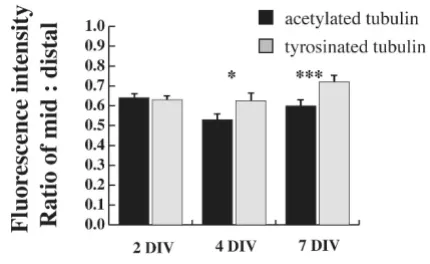
Not every immature dendrite had a well-defined growth cone, but even those without showed Tyr-tubulin immu-noreactivity. To determine whether the proportion of Tyr-tubulin was significantly increased over Ac-Tyr-tubulin in dis-tal dendrites, we determined the ratio of the fluorescence intensity for each antibody between two defined regions of the dendrite: the middle segment of the dendrite and 10 μm from the visible end of the tip, purposefully exclud-ing the growth cone, which is typically motile and was clearly enriched in Tyr-tubulin compared to Ac-tubulin. In fact, the relative amount of Tyr-tubulin at the distal den-drite was significantly greater than that for Ac-tubulin at both 4 and 7 DIV (Figure 3). This time period coincides with a period of robust outgrowth of the dendritic arbor in cultured hippocampal neurons [3], and so increased levels of dynamic microtubules could contribute to that growth. No significant changes were detected at 2 DIV, but this is a time when there is little net extension of dendrites and so this finding also fits well with the prediction that dynamic microtubules are associated with outgrowth.
Differential distribution of dynamic microtubules in shafts of dendrites and axons
Regardless of developmental time in culture, the entire dendritic arbor exhibited pronounced immunofluores-cence for Tyr-tubulin (Figure 2A, D, G, J) as well as Ac-tubulin (Figure 2B, E, H, K), whereas Tyr-Ac-tubulin was clearly less abundant in the proximal and middle regions of axons. This suggested that the distribution of acetylated and tyrosinated microtubules along the dendrite shaft was more closely parallel than reciprocally proportional. To test this, we determined the ratio of immunofluorescence intensity of dynamic and stable microtubules at the mid-point and distal ends of the axon and dendrites of individ-ual cells, and then compared the magnitude of difference (midshaft-to-distal). To control for potential variability in staining intensity between cells, this analysis was limited to cells at 2 DIV, where both the entire axon and dendritic field were contained within a single digital micrograph. With this comparison, the mean ratio of Ac-tubulin to Tyr-tubulin was 40% lower in dendrites than in axons (mean value of ratio Ac:Tyr ± standard error: dendrites, 4.68 ± 0.3; axons, 7.23 ± 1.79). Thus, there was proportionally more dynamic tubulin within dendrite shafts than within axons at this developmental time-point.
Previous analyses of microtubule stability in growing axons has led to the hypothesis that there is a predictable relationship between increased length and increased sta-bility along the existing shaft [18,44]. If this is the case, then one might expect that with increased length, the intensity of staining for dynamic microtubules should decrease within the shaft. To test this prediction, the fluo-rescence intensity of Tyr-tubulin was measured at mid-shaft and plotted against process length for both axons and dendrites of neurons at 2 DIV (Figure 4). This predic-tion was supported by measures of dynamic microtubules in axons, but not in dendrites. The longer the axon, the weaker the intensity of dynamic Tyr-tubulin staining in the middle segment of the axon (Figure 4A). There was no evidence for a similar inverse relationship between length and dynamic Tyr-tubulin fluorescence in dendrites (Fig-ure 4B).
Localization of dynamic and stable microtubules
Figure 2
Figure 2. Within the middle segment of dendrites, Tyr-tubulin intensity did not decrease as dendrites matured and became longer (correlation coefficient between length and fluorescence intensity at midshaft = 0.21, data not shown). In addition, quantitative analysis of fluores-cence intensity at the midshaft and distal dendrites of a subset of dendrites that were clearly isolated at 7 DIV showed that, while Tyr-tubulin is slightly decreased com-pared to Ac-tubulin, immunofluorescence for Tyr-tubulin is still relatively high when compared as a percentage of initial fluorescence in the proximal-most region (Figure 5). Tyr-tubulin did not appear to decrease substantially within the dendritic shaft of mature neurons either (Fig-ure 2J–L), but as axons became intertwined with den-drites, it was not possible to identify dendrites and axons in isolation in order to make the same quantitative meas-urements. Combined, all these measures suggest that den-drites maintain a greater pool of dynamic microtubules in proportion to stable ones throughout development than do axons, which may have implications for ongoing plas-ticity.
Microtubule polymerization is not restricted to growing ends of axons and dendrites
Imaging of living cells transfected with EB1-GFP enables comparison of the spatial distribution of microtubule
polymerization within the axonal and dendritic compart-ments of a single cell. To determine if polymerization occurs preferentially in regions of active growth, or differs between axons and dendrites during development, we recorded and analyzed EB1-GFP comets in polarized neu-rons at different stages of dendritic maturity. Live cell imaging of neurons expressing EB1-GFP at 1 to 17 DIV revealed bright comets along neurites (Figure 6). Regard-less of age, polymerization was evident not only at the dis-tal growing ends of axons and dendrites, but throughout the arbors of both immature (Figure 6A; Additional files 2 and 3) and mature (Figure 6B; Additional files 4, 5 and 6) neurons. The majority of comets traveled unidirectionally for short distances (up to 3 μm) before disappearing. Treatment with nocodozole (1 μM) to interfere with microtubule polymerization eliminated the appearance of EB1-GFP comets, leaving only diffuse fluorescence throughout the soma and neuronal processes (data not shown), similar to previous observations [30]. These find-ings support previous reports that EB1-GFP binds selec-tively to acselec-tively polymerizing microtubule plus-ends within both developing axons and dendrites, similar to endogenous EB1, thus providing a reliable marker for tracking physiological changes in microtubule dynamics. Moreover, these data also confirm that active growth of microtubules is not restricted to the growing tips of neu-ronal processes, but rather, occurs throughout the length of both axons and dendrites.
Microtubule polymerization becomes biased toward distal regions with neuronal development
A longstanding hypothesis is that microtubule polymeri-zation supports process outgrowth, and, thus, regions of active growth should show preferential polymerization even though lower level 'maintenance' polymerization might also persist throughout a process. If this is the case, then once an arbor is built, the number of polymerization events should decrease in stable regions, but might remain high (or increase) in distal regions that are still actively growing. To determine whether there was any regional bias in hippocampal neurons to changes in polymerization associated with development, we com-pared the spatial distribution of comets within the first 2 days in culture (1 to 2 DIV) with that of more mature neu-rons (7 to 17 DIV). Proximal-middle and distal (non-growth cone) regions of axons and dendrites were sam-pled randomly, and the number of fluorescent comets per unit length was determined (Figure 7).
At 1 to 2 DIV, comets were distributed equally across the proximal-middle and distal regions of both axons and dendrites. In more mature neurons (7 to 17 DIV), how-ever, more polymerization events per unit length were observed in the distal regions of both axons and dendrites. These data suggest that with maturation, overall
microtu-Tyr-tubulin is increased in distal dendrites at stages of devel-opment when dendrite outgrowth is robust
Figure 3
bule polymerization declines within the established por-tion of both the axonal and dendritic shafts, whereas distal regions of active growth maintain higher levels of polymerization, as predicted.
The orientation of polymerizing microtubules changes with development
At the electron microscopic level following conditions allowing free-tubulin decoration, microtubules appear uniformly oriented with plus-ends distal within axons, but are oriented with both plus- and minus-ends distal in dendrites [24]. In the mature dendritic arbor, the propor-tion of microtubules with each polarity orientapropor-tion is about equal. Such an analysis cannot reveal whether any of these microtubules are in a dynamic or stable state, and so leaves open the question of whether the directionality of active polymerization is proportional to the static
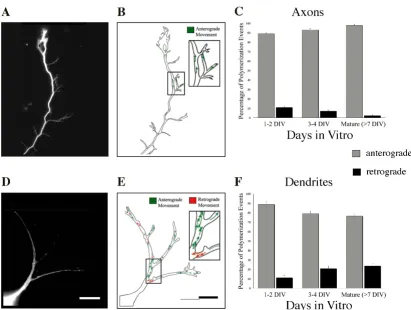
dis-tribution of microtubule polarity observed with the elec-tron microscope. We tested the hypothesis that the overall directional pattern of polymerization would reflect the predicted distribution of microtubule polarity by quanti-fying the proportion of polymerization events that were anterograde (reflecting plus-end distal microtubules) and retrograde (reflecting minus-end distal microtubules) within developing axons and dendrites (Figure 8). In these analyses, we found that the relative proportion of anterograde and retrograde directed comets differed as a function of the ongoing development of axons and den-drites (Figure 8C, F; Additional files 7 and 8).
During the first 2 days in culture there was no difference in the overall directionality of microtubule polymeriza-tion between immature axons and dendrites of EB1-expressing polarized neurons, with most polymerization
Relationship between fluorescence intensity (in arbitrary units) of Tyr-tubulin and Ac-tubulin at the mid-shaft and process length in 2-DIV neurons
Figure 4
events occurring anterogradely. Approximately 11% of all comets traveled in the retrograde direction, a somewhat larger percentage than would have been predicted based on electron microscopy analyses of microtubule orienta-tion (3 to 6%) [45]. As neurons matured, axons exhibited polymerization events that became nearly uniformly ori-ented in the anterograde direction, with fewer than 2% of the comets traveling in the retrograde direction (Figure 8C). Within dendrites, the proportion of retrograde events increased with time in culture (Figure 8F) as expected, but only reached 24% at the most advanced time-points measured (7 to 17 DIV) rather than the 50% expected. Taken together, these findings indicate that the direction-ality of microtubule polymerization mirrors the predicted distribution of anterogradely oriented microtubules in mature axons closely. Even with an increasing proportion of retrograde polymerization events, however, dendrites may have separate mechanisms controlling the gradual attainment of the characteristic mixed microtubule polar-ity and the distribution of microtubule polymerization.
Analysis of rate and comparison with a neuron-like cell line, PC12 cells
Despite changes in the spatial distribution of EB1-GFP comets throughout development, the rates of polymeriza-tion did not differ between anterograde and retrograde events, nor did they differ between axons and dendrites (Table 1). Within axons, the rates of anterograde-directed microtubule polymerization ranged from 0.23 ± 0.02 μm/ s at 1 to 2 DIV to 0.33 ± 0.02 μm/s at maturity. For den-drites, anterograde polymerization ranged from 0.25 ± 0.02 μm/s at 1 to 2 DIV to 0.22 ± 0.03 μm/s at maturity. The small number of retrograde events noted in axons after 3 DIV precluded statistical analysis of rates at matu-rity. In contrast, dendrites displayed increasing numbers of retrograde-directed EB1-GFP comets over the course of development, and the corresponding retrograde polymer-ization rates were similar with maturation. There is clearly variability in the observed rates. When considered together, however, similar anterograde and retrograde microtubule polymerization rates observed for axons and dendrites over development suggest that the kinetics of tubulin assembly are not affected by maturational
Dynamic microtubules remain high in dendrites at 7 DIV
Figure 5
EB1-GFP dynamics
Figure 6
changes in hippocampal neurons; moreover, these rates are in agreement with previously published rates in neu-rons [27,28,30].
For comparison with another cell type that extends proc-esses, we measured the rate and orientation of polymeri-zation events in PC12 cells after they had been induced to take on a neuron-like phenotype. Although these cells extend processes, they do not differentiate into axons or dendrites, but instead have features characteristic of each.
Similar to neurons, microtubule polymerization in PC12 cells was evident throughout the length of the neurites (Additional file 9). Quantification of the number of EB1-GFP comets showed that while 82% of the polymeriza-tion events occurred in the anterograde direcpolymeriza-tion, 18% were oriented in the retrograde direction (Table 1), sug-gesting similarity to mature dendrites rather than to axons in this measure.
Discussion
Our findings suggest that even before the dendrites of cul-tured hippocampal neurons began to show signs of matu-ration (2 DIV), they contained a higher proportion of dynamic Tyr-tubulin throughout their arbor than did axons, and these high levels of Tyr-tubulin were main-tained for several weeks. From these data, it appears that differential control over stability not only contributes to axon formation during the initiation of neuron polarity [6], but it also continues to distinguish dendrites from axons into maturity as well. The directionality of microtu-bule polymerization within axons and dendrites diverged later in development (3 to 4 DIV), with an increased inci-dence of retrogradely oriented polymerization that is closely aligned with the time-course of dendrite morpho-genesis. This lag supports the hypothesis that changes in the orientation of microtubule polarity do not contribute to the initial specification of polarity, but may be impor-tant for later stages of dendritic maturation. Further strengthening this argument, interactions between retro-gradely oriented microtubules and motor proteins, partic-ularly dynein, have recently been shown to play a significant role in the transport and localization of den-dritic proteins and organelles in mammalian models [35], as well as dendritic growth and branching in Drosophila
neurons [46].
Post-translational modifications of microtubules and the development of neuronal polarity
High levels of dynamic microtubules were present throughout the shaft of dendrites during development, as well as in mature neurons. This clearly contrasted with the distribution of microtubules in axons, where microtu-bules in the shafts were predominantly stable, whereas the microtubules in the distal ends were more dynamic, sim-ilar to axons of peripheral neurons [18-21]. These results fit well with previous reports of differential vulnerability to microtubule depolymerizing drugs between axons and dendrites of sympathetic neurons [20,47]; microtubules in the proximal and middle shaft of axons showed resist-ance to nocodozole treatment, whereas microtubules in dendrites were more vulnerable. More recent evidence suggests that interfering with post-translational modifica-tions (for example, acetylation and detyrosination [36]) that are associated with microtubule stability can have sig-nificant effects on branching in axons [48] and cause
aber-The spatial distribution of microtubule polymerization becomes biased towards distal regions as axons and den-drites develop
Figure 7
The spatial distribution of microtubule polymeriza-tion becomes biased towards distal regions as axons and dendrites develop. (A, B) The mean number of com-ets per micron was determined within segments representing the proximal-middle versus distal region (excluding the growth cone) of axons (A) and dendrites (B). For both axons and dendrites, microtubule polymerization was distributed equally within all regions at the earliest stages of develop-ment, but predominated within distal segment as neurons matured. Error bars represent standard error of the mean; P
As neurons mature, the proportion of microtubules polymerizing in the anterograde direction increases to nearly 100% in axons, whereas the number of retrogradely oriented events observed in dendrites increases
Figure 8
As neurons mature, the proportion of microtubules polymerizing in the anterograde direction increases to nearly 100% in axons, whereas the number of retrogradely oriented events observed in dendrites increases. (A, D) Single frames from the distal region of an axon (A) and a dendritic arbor (D) at 3 DIV, and (B, E) composite tracings of observed polymerization events, with individual EB1-GFP comet trajectories identified in the anterograde (green arrows) or retrograde (red arrows) directions. Insets depict enlarged views of the boxed regions. (C, F) The percentage of microtubule polymerization events occurring in an anterograde or retrograde direction was determined for each population of axons (C) and dendrites (F) observed throughout the developmental time-course. In axons, the total numbers of events tracked were: 833, 1 to 2 DIV; 892, 3–4 DIV; 284, 7+ DIV. For dendrites, the total numbers of events tracked were: 748, 1 to 2 DIV; 955, 3 to 4 DIV; 232, 7+ DIV. Scale bar, 10 μm. Error bars represent standard error of the mean. See Additional files 7 (axon) and 8 (dendritic arbor) for accompanying movies.
Table 1: Microtubule polymerization rates in developing neurons and PC12 cells
Cell population ANTEROGRADE EVENTS Mean Velocity (μm/sec ± SE)
RETROGRADE EVENTS Mean Velocity (μm/sec ± SE)
Hippocampal neurons Axons Dendrites Axons Dendrites
1–2 DIV 0.23 ± 0.02 (142) 0.25 ± 0.02 (114) 0.29 ± 0.05 (22) 0.20 ± 0.05 (9) 3–4 DIV 0.26 ± 0.01 (214) 0.36 ± 0.02 (199) 0.43 ± 0.20 (5) 0.27 ± 0.03 (44) > 7 DIV 0.33 ± 0.02 (86) 0.22 ± 0.03 (68) 0.35 (1) 0.18 ± 0.03 (21)
rant outgrowth [49], potentially impacting the association of some +TIPs with microtubule ends [50]. Post-transla-tional modifications of tubulin also contribute to the reg-ulation of differential subcellular transport, in particular by altering the affinity of some transport vesicles and motors for particular microtubules [51,52]. Thus, local asymmetric increases in microtubule acetylation or dety-rosination could bias kinesin-mediated transport to par-ticular neurites, generating positive feedback signaling to allow for continued outgrowth and differentiation.
Subcellular changes in microtubule stability or transport could help to explain the different patterns of outgrowth that have been observed in different domains of cultured hippocampal neurons. While both axons and dendrites elongate and add collateral branches along their arbor, they can be distinguished by the rates of growth and pat-terns of motility once the axon becomes specified [9,10]. The axon extends rapidly, growing as much as several hundred microns per day, whereas the net growth rate of dendrites is very slow. Individual dendritic branches increase in length by about 10 μm/day and show frequent bouts of retraction as well as extension. A higher propor-tion of immature, and thus less stable, tubulin polymers within the shaft could contribute to the frequent retrac-tions, and slow net growth of dendrites. Likewise, biased delivery of kinesin-associated vesicles to the more acetylated microtubules of the axon could also facilitate its rapid growth.
Considered together, the present data and earlier findings demonstrate that from the earliest specification of polarity and continuing into maturity, the axonal shaft is com-posed of relatively stable microtubules, whereas the den-dritic arbor maintains a greater pool of dynamic microtubules. This is likely to reflect different require-ments for dynamic instability that arise between the axonal and dendritic compartments, presumably impor-tant for establishing their distinct physiologies as neurons mature.
Microtubule polymerization and neuron growth
Imaging of EB1-GFP comets revealed that microtubule polymerization occurs throughout the length of hippoc-ampal neuron axons and dendrites, rather than being restricted solely to regions of active growth, such as axonal growth cones and dendritic tips. As these neurons contin-ued to grow in vitro, however, the number of polymeriza-tion events declined within the older established porpolymeriza-tion of both axonal and dendritic shafts, whereas newer distal regions maintained higher levels of polymerization. Since distal ends constitute regions of active growth, such increased local polymerization suggests a shift toward ongoing assembly of the cytoskeleton on site, with lower level 'maintenance' polymerization along the shaft. Such
a mechanism for regulating outgrowth in axons is well-supported (see, for example, [53]). The findings reported here suggest a similar mechanism for dendrites. Analyses of immunofluorescence showed concentrations of Tyr-tubulin at the distal tips of dendrites, and others have shown that regulation of tubulin polymerization within dendrite tips promotes dendritic growth and arborization [22,54]. Collectively, these data suggest a critical role for ongoing microtubule polymerization in dendritic arbors as well as axons as neuronal circuits are developing.
Orientation of polymerization and differentiation of dendrites and axons
At the earliest time-points after hippocampal neurons became morphologically polarized, axons and dendrites could not be distinguished by directional patterns of microtubule polymerization. Rather, they began to diverge in the proportion of anterograde to retrograde polymerization events around 3 to 4 DIV. By 7 DIV, axons attained a nearly uniform distribution of anterograde polymerizing microtubules, while dendrites exhibited increased numbers of retrograde polymerization events. Retrograde polymerization had been detected in dendrites previously [30], a finding anticipated by previous electron microscopy studies [24,45]. What was unexpected, how-ever, was that the directionality of microtubule polymeri-zation in dendrites never reached roughly equal proportions of anterograde and retrograde events. Even neurons grown for more than 2 weeks in vitro, an age typ-ically deemed mature by morphological and molecular criteria [1], continued to show a bias toward polymeriza-tion in the anterograde direcpolymeriza-tion. One possible interpreta-tion of this finding is that microtubules oriented with minus ends distal (thus polymerizing in the retrograde direction) are more stable than those with plus-ends distal (anterograde polymerizing). Regardless of the explana-tion for why the proporexplana-tion of observed retrograde events is less than the expected total distribution of retrogradely oriented microtubules, these data suggest that the direc-tionality of microtubule assembly is not simply propor-tional to the number of plus-end or minus-end distal microtubules present.
Implications for regulation of growth and plasticity
the dendritic arbor, including transient forays into spines, could be important for spine formation, morphology, and activity-dependent changes in synapses [58-60]. Axons make collateral branches during pathfinding in develop-ment, but once the path is established, maintaining stabil-ity might be more important than retaining a capacstabil-ity sprouting all along the axon tract. In contrast, it appears that the entire dendritic arbor is capable of growth, branch addition, and synapse formation throughout life. Thus, the differences in microtubule stability and dynamic polymerization between axons and dendrites reported here suggest a structural substrate that can account not only for differences in patterns of outgrowth, but also the capacity for plasticity.
Conclusion
Significant differences in the organization of microtu-bules between dendrites and axons arise during develop-ment, but are maintained into maturation. Most notably, higher levels of dynamic tubulin, measured as Tyr-tubulin immunofluorescence, are retained throughout the den-dritic arbor, whereas Tyr-tubulin is concentrated at distal growing ends of axons. While differences in microtubule stability can be detected even as neurons are polarizing, the proportion of retrograde to anterograde oriented polymerization diverges 1 to 2 days later, suggesting this aspect of microtubule organization may be more impor-tant for later phases of growth and maturation rather than in the initial specification of axons and dendrites.
Abbreviations
Ac: acetylated; DIV: days in vitro; GFP: green fluorescent protein; Tyr: tyrosinated.
Competing interests
The authors declare that they have no competing interests.
Authors' contributions
Quantitative immunofluorescence experiments were done by RB and GW; imaging and analysis of EB1-trans-fected cells were conducted by KK, RB, MB and GW; KK, RB and GW contributed to the manuscript. All authors approved the final manuscript.
Additional material
Additional file 1
Movement of EB-1 comets in a dendrite at 2 DIV. Recording of a por-tion of a dendrite (at 2 DIV) from Figure 1 shows EB-1 comets traveling in both anterograde and retrograde directions (the accompanying kymo-graph is shown in Figure 1B, C). Movie duration, 10 s, with frames (500 ms exposure) acquired in a continuous stream.
Click here for file
[http://www.biomedcentral.com/content/supplementary/1749-8104-4-26-S1.mov]
Additional file 2
Movement of EB-1 comets in a dendrite at 1 DIV. Segment of a den-drite (shown in Figure 6a') at 1 DIV showing most EB-1 comets traveling in the anterograde direction (toward the right), although retrograde events (toward the left) are clearly apparent as well.
Click here for file
[http://www.biomedcentral.com/content/supplementary/1749-8104-4-26-S2.mov]
Additional file 3
Movement of EB-1 comets in an axonal shaft at 1 DIV. Segment of axonal shaft (shown in Figure 6a") at 1 DIV. Both anterograde (down-ward) and retrograde (up(down-ward) events are clearly evident in this segment, and could be detected throughout the entire axon length.
Click here for file
[http://www.biomedcentral.com/content/supplementary/1749-8104-4-26-S3.mov]
Additional file 4
Movement of EB-1 comets in a dendritic arbor at 15 DIV. Dendritic arbor from a cell at 15 DIV. EB-1 comets can be detected throughout the arbor, traveling in both retrograde (toward the left) and anterograde (toward the right) directions, as well as entering a portion of dendritic spines.
Click here for file
[http://www.biomedcentral.com/content/supplementary/1749-8104-4-26-S4.mov]
Additional file 5
Movement of EB-1 comets in an axonal arbor at 15 DIV. Anterogradely oriented EB-1 comets could also be observed throughout the axonal arbor at 15 DIV, although with less frequency than was evident in younger cells. This recording was taken from the middle region of an axon, with the cell body to the right, and growth cone to the left.
Click here for file
[http://www.biomedcentral.com/content/supplementary/1749-8104-4-26-S5.mov]
Additional file 6
Recording from the distal region and growth cone at 15 DIV. Record-ing from the distal region, and growth cone (from the same cell as in Addi-tional file 5) at 15 DIV.
Click here for file
[http://www.biomedcentral.com/content/supplementary/1749-8104-4-26-S6.mov]
Additional file 7
Movement of EB-1 comets in an axon at 3 DIV. Accompanying movie of the 3-DIV axon shown in Figure 8A, B. Anterograde events are evident throughout the shaft of the axon, as well as the branches and filopodia that arise from the shaft.
Click here for file
Acknowledgements
The EB1-GFP construct was generously provided by Lynn Cassimeris. The authors would like to thank members of the Withers lab and Christopher Wallace for helpful discussions and critical reading of the manuscript. This work was supported by a National Science Foundation CAREER award (IBN-0135985) to GW.
References
1. Craig AM, Banker G: Neuronal polarity. Ann Rev Neurosci 1994,
17:267-310.
2. Bradke F, Dotti CG: Establishment of neuronal polarity: les-sons from cultured hippocampal neurons. Curr Opin Neurobiol
2000, 10:574-581.
3. Dotti CG, Sullivan CA, Banker GA: The establishment of polarity by hippocampal neurons in culture. J Neurosci 1988,
8:1454-1458.
4. Inagaki N, Chihara K, Arimura N, Menager C, Kawano Y, Matusuo N, Nishimura T, Amano M, Kaibuchi K: CRMP-2 induces axons in cultured hippocampal neurons. Nat Neurosci 2001, 4:781-782. 5. Fukata Y, Itoh TJ, Kimura T, Ménager C, Nishimura T, Shiromizu T,
Watanabe H, Inagaki N, Iwamatsu A, Hotani H, Kaibuchi K: CRMP-2 binds to tubulin heterodimers to promote microtubule assembly. Nat Cell Biol 2002, 4:583-591.
6. Witte H, Neukirchen D, Bradke F: Microtubule stabilization specifieds initial neuronal polarization. J Cell Biol 2008,
180:619-632.
7. Dehmelt L, Nalbant P, Steffen W, Halpain S: A microtubule-based, dynein-dependent force induces local cell protrusions: Impli-cations for neurite initiation. Brain Cell Biol 2006, 35:39-56. 8. Dehmelt L, Halpain S: Actin and microtubules in neurite
initia-tion: are MAPs the missing link? J Neurobiol 2004, 58:18-33. 9. Esch T, Lemmon V, Banker G: Local presentation of substrate
molecules directs axon specification by cultured hippocam-pal neurons. J Neurosci 1999, 19:6417-6426.
10. Ruthel G, Banker G: Role of moving growth cone-like 'wave' structures in the otugrowth of cultured hippocampal axons and dendrites. J Neurobiol 1999, 39:97-106.
11. Fletcher T, DeCamilli P, Banker G: Synaptogenesis in hippocam-pal cultures: evidence indicating the axons and dendrites become competent to form synapses at different stages of neuronal development. J Neurosci 1994, 14:6695-6706. 12. Craig A, Graf E, Linhoff M: How to build a central synapse: clues
from cell culture. Trends Neurosci 2006, 29:8-20.
13. Ahmad FJ, He Y, Myers K, Hasaka T, Francis F, Black MM, Baas PW:
Effects of dynactin disruption and dynein depletion on axonal microtubules. Traffic 2006, 7:524-537.
14. Baas PW, Karabay A, Qiang L: Microtubules cut and run. Trends Cell Biol 2005, 15:518-524.
15. Bamburg JR, Bray D, Chapman K: Assembly of microtubules at the tip of growing axons. Nature 1986, 321:788-790.
16. Dent EW, Callaway JL, Szebenyi G, Baas PW, Kalil K: Reorganiza-tion and movement of microtubules in axonal growth cones and developing interstitial branches. J Neurosci 1999,
19:8894-8908.
17. Gallo G, Letourneau PC: Different contributions of microtubule dynamics and transport to the growth of axons and collat-eral sprouts. J Neurosci 1999, 19:3860-3873.
18. Brown A, Li Y, Slaughter T, Black MM: Composite microtubules of the axon: quantative analysis of tyrsoinated and acetylated alpha-tubulin alon axonal microtubules. J Cell Sci
1993, 104:339-352.
19. Brown A, Slaughter T, Black MM: Newly assembled microtubules are concentrated in the proximal and distal regions of grow-ing axons. J Cell Biol 1992, 119:867-882.
20. Baas PW, Ahmad FJ, Pienkowski TP, Brown A, Black MM: Sites of microtubule stabilization for the axon. J Neurosci 1993,
13:2177-2185.
21. Shea TB: Selective stabilization of microtubules within the proximal region of developing axonal neurites. Brain Res Bull
1999, 48:255-261.
22. Akum BF, Chen M, Gunderson SI, Riefler GM, Scerri-Hansen MM, Firestein BL: Cypin regulates dendrite patterning in hippoc-ampal neurons by promoting microtubule assembly. Nat Neurosci 2004, 7:145-152.
23. Chen H, Firestein BL: RhoA regulates dendrite branching in hippocampal neurons by decreasing cypin protein levels. J Neurosci 2007, 27:8378-8386.
24. Baas PW, Deitch JS, Black MM, Banker GA: Polarity orientation of microtubules in hippocampal neurons: uniformity in the axon and nonuniformity in the dendrite. Proc Natl Acad Sci USA
1988, 85:8335-8339.
25. Morrison EE: Action and interactions a microtubule ends. Cell Mol Life Sci 2007, 64:307-317.
26. Lansbergen G, Akhmanova A: Microtubule plus end: a hub of cel-lular activities. Traffic 2006, 7:499-507.
27. Ma Y, Shakiryanove D, Vardya I, Popov SV: Quantitative analysis of microtubule transport in growing nerve processes. Curr Biol 2004, 14:725-730.
28. Kim T, Chang S: Quantative evaluation of the mode of micro-tubule transport in Xenopus neurons. Mol Cells 2006, 21:76-81. 29. Nakata T, Hirokawa N: Microtubules provide directional cues for polarized axonal transport through interaction with kinesin motor head. J Cell Biol 2003, 162:1045-1055.
30. Stepanova T, Slemmer J, Hoogenraad CC, Lansbergen G, Dortland B, Zeeuw CID, Grosveld F, Cappellen Gv, Akhmanova A, Galjart N: Vis-ualization of microtubule growth in cultured neurons via the use of EB3-GFP (end-binding protein 3-green fluorescent protein). J Neurosci 2003, 23:2655-2664.
31. Rolls MM, Satoh D, Clyne PJ, Henner AL, Uemura T, Doe CQ: Polar-ity and intracellular compartmentalization of Drosophila
neurons. Neural Dev 2007, 2:7.
32. Morrison EE, Wardleworth BN, Askham JM, Markham AF, Meredith DM: EB1, a protein which interacts with the APC tumour suppressor, is associated with the microtubule cytoskeleton throughout the cell cycle. Oncogene 1998, 17:3471-3477. 33. Morrison EE, Moncur PM, Askham JM: EB1 identifies sites of
microtubule polymerisation during neurite development.
Brain Res Mol Brain Res 2002, 98:145-152.
34. Piehl M, Cassimeris L: Organization and dynamics of growing microtubule plus ends during early mitosis. Mol Biol Cell 2003,
14:916-925.
35. Zheng Y, Wildonger J, Ye B, Zhang Y, Kita A, Younger SH, Zimmer-man S, Jan LY, Jan YN: Dynein is required for polarized dendritic transport and uniform microtubule orientaation in axons.
Nat Cell Biol 2008, 10:1172-1180.
36. Bulinski JC, Gungersen GG: Stabilization and post-translational modification of microtubules during cellular morphogenesis.
Bioessays 1991, 13:285-293.
37. Piperno G, LeDizet M, Chang XJ: Microtubules containing acetylated alpha-tubulin in mammalian cells in culture. J Cell Biol 1987, 104:289-302.
38. Banker GA, Goslin K: Culturing Nerve Cells 2nd edition. Cambridge, MA: MIT Press; 1998.
Additional file 8
Movement of EB-1 comets in a dendritic arbor at 3 DIV. Accompany-ing movie of the 3-DIV dendritic arbor shown in Figure 8D, E. Both anterograde and retrograde events throughout the length of the arbor are shown.
Click here for file
[http://www.biomedcentral.com/content/supplementary/1749-8104-4-26-S8.mov]
Additional file 9
Recording from a PC12 cell differentiated to take on a neuron-like phenotype. Both anterograde and retrograde events were observed in these cells, in about the same proportions as were observed in the dendritic arbors of more mature neurons.
Click here for file
Publish with BioMed Central and every scientist can read your work free of charge "BioMed Central will be the most significant development for disseminating the results of biomedical researc h in our lifetime."
Sir Paul Nurse, Cancer Research UK
Your research papers will be:
available free of charge to the entire biomedical community
peer reviewed and published immediately upon acceptance
cited in PubMed and archived on PubMed Central
yours — you keep the copyright
Submit your manuscript here:
http://www.biomedcentral.com/info/publishing_adv.asp
BioMedcentral 39. Bottenstein JE, Sato GH: Growth of a rat neuroblastoma cell
line in serum-free supplemented medium. Proc Natl Acad Sci USA 1979, 76:514-517.
40. Greene LA, Tischler AS: Establishment of a noradrenergic clonal line of rat adrenal pheochromocytoma cells which respond to nerve growth factor. Proc Natl Acad Sci USA 1976,
73:647-648.
41. Lambeng N, Michel PP, Brugg B, Agid Y, Ruberg M: Mechanisms of apoptosis in PC12 cells irreversibly differentiated with nerve growth factor and cyclic AMP. Brain Res 1999, 821:60-68. 42. Ahmad FJ, Yu W, McNally FJ, Baas PW: An essential role for
katanin in severing microtubules in the neuron. J Cell Biol 1999,
145:305-315.
43. Withers GS, Banker G: Characterizing and studying neuronal cultures. In Culturing Nerve Cells 2nd edition. Edited by: Banker G, Goslin K. Cambridge: MIT Press; 1998:113-151.
44. Baas PW, Black MM: Individual microtubules in the axon con-sist of domains that differ in both composition and stability.
J Cell Biol 1990, 111:495-509.
45. Baas PW, Black MM, Banker GA: Changes in microtubule polar-ity orientation during the development of hippocampal neu-rons in culture. J Cell Biol 1989, 109:3085-3094.
46. Satoh D, Sato D, Tsuyama T, Saito M, Ohkura H, Rolls M, Ishikawa F, Uemura T: Spatial control of branching within dendritic arbors by dynein-dependent transport of Rab5-endosomes.
Nat Cell Biol 2008, 10:1164-1171.
47. Baas PW, Slaughter T, Brown A, Black MM: Microtubule dynamics in axons and dendrites. J Neurosci Res 1991, 30:134-153. 48. Creppe C, Malinouskaya L, Volvert ML, Gillard M, Close P, Malaise O,
Laguesse S, Cornez I, Rahmouni S, Ormenese S, Belachew S, Mal-grange B, Chapelle JP, Siebenlist U, Moonen G, Chariot A, Nguyen L:
Elongator controls the migration and diferentiation of corti-cal neurons through acetylation of alpha-tubulin. Cell 2009,
136:551-564.
49. Erck C, Peris L, Andrieux A, Meissirel C, Gruber AD, Vernet M, Sch-weitzer A, Saoudi Y, Pointu H, Bosc C, Salin PA, Job D, Wehland J: A vital role of tubulin-tyrosine-ligase for neuronal organiza-tion. Proc Natl Acad Sci USA 2005, 102:7853-7858.
50. Peris L, Thery M, Fauré J, Saoudi Y, Lafanechère L, Chilton JK, Gor-don-Weeks P, Galjart N, Bornens M, Wordeman L, Wehland J, Andrieux A, Job D: Tubulin tyrosination is a major factor affecting the recruitment of CAP-Gly proteins at microtu-bule ends. J Cell Biol 2006, 174:839-849.
51. Dunn S, Morrison EE, Liverpool TB, Molina-Paris C, Cross RA, Alonso MC, Peckham M: Differential trafficking of Kif5c ontyro-sinated and detyroontyro-sinated microtubules in live cells. J Cell Sci
2008, 121:1085-1095.
52. Reed N, Cai D, Blasius T, Jih G, Meyhofer E, Gaertig J, Verhey K:
Microtubule acetylation promotes kinesin-1 binding and transport. Curr Biol 2006, 16:2166-2172.
53. Kalil K, Dent EW: Hot +TIPS: guidance cues signal directly to microtubules. Neuron. 2004, 42(6):877-879.
54. Chen M, Lucas KG, Akum BF, Balasingam G, Stawicki TM, Provost JM, Riefler GM, Jornsten RJ, Firestein BL: A novel role for snapin in dendrite patterning: interaction with cypin. Mol Biol Cell 2005,
16:5103-5114.
55. Wong RO, Ghosh A: Activity-dependent regulation of den-dritic growth and patterning. Nat Rev Neurosci 2002, 3:803-812. 56. Klintsova AY, Greenough W: Synaptic plasticity in cortical
sys-tems. Curr Opin Neurobiol 1999, 9:203-208.
57. Cline H, Haas K: The regulation of dendritic arbor develop-ment and plasticity by glutamatergic synaptic input: a review of the synaptotrophic hypothesis. J Physiol 2008, 586:1509-1517. 58. Gu J, Firestein BL, Zheng JQ: Microtubules in dendritic spine
development. J Neurosci 2008, 28:12120-12124.
59. Hu X, Viesselmann C, Nam S, Merriam E, Dent EW: Activity-dependent dynamic microtubule invasion of dendritic spines. J Neurosci 2008, 28:13094-13105.