O R I G I N A L R E S E A R C H
Long Noncoding RNA NEAT1 Promotes Cell
Proliferation And Invasion And Suppresses
Apoptosis In Hepatocellular Carcinoma By
Regulating miRNA-22-3p/akt2 In Vitro And In Vivo
This article was published in the following Dove Press journal:
OncoTargets and Therapy
Xichang Zhou1,* Xiang Wang2,* Yizhou Zhou2 Long Cheng1 Youwei Zhang2 Yangmei Zhang2,3
1Department of Intervention, Xuzhou
Central Hospital, Xuzhou Medical University, XuZhou 221009, People’s Republic of China;2Department of
Medical Oncology, Xuzhou Central Hospital, Xuzhou Medical University, XuZhou 221009, People’s Republic of China;3Department of Medical
Oncology, The First Affiliated Hospital of Soochow University, Suzhou 215006, People’s Republic of China
*These authors contributed equally to this work
Background:Hepatocellular carcinoma (HCC) is one of the most aggressive cancers that is associated with cirrhosis and other chronic liver diseases. Although remarkable progress has been made in past decades, it is still necessary to continue exploring the pathology and development of HCC.
Objective:In this study, we elucidated the effect of long noncoding RNA (lncRNA) NEAT1 on HCC development and underlying mechanisms.
Methods:Clinicopathological features of HCC patients were collected and the correlations
with NEAT1 expression were assessed. To determine cell activities, CCK-8,flow cytometry,
invasion assays, and TUNEL assays were performed. Real-time PCR, Western blot, and luciferase reporter assays were performed to investigate the related mechanism of HCC.
Results:The results revealed that NEAT1 expression was associated with tumor size and differentiation where NEAT1 was upregulated in both HCC tissues and cell lines. Overexpression of NEAT1 promoted proliferation and invasion while inhibited apoptosis in HCC cells, which was opposite to the effect of NEAT1 knockdown. Also, AKT2 was increased in HCC tissues. Downregulation of AKT2 was associated with reduced cell proliferation and invasion while increased apoptosis, while overexpression of AKT2 exerted opposite roles. In addition, the expression of miRNA-22-3p displayed an inverse association with NEAT1. miRNA-22-3p mimic and inhibitor suppressed and promoted HCC develop-ment, respectively. The luciferase assay revealed that both NEAT1 and AKT2 were direct target genes of miRNA-22-3p. Furthermore, knockdown and overexpression of NEAT1 suppressed and promoted tumor growth in the HCC mouse model, which were abolished by the miRNA-22-3p inhibitor and mimic, respectively.
Conclusion:In conclusion, the results demonstrate that NEAT1 promotes the development of HCC, both in vitro and in vivo, through regulating miRNA-22-3p/AKT2, and provides insight into developing a new strategy for HCC treatment.
Keywords: hepatocellular carcinoma, NEAT1, miRNA-22-3p, AKT2, apoptosis, cell proliferation
Introduction
Hepatocellular carcinoma (HCC) is the most common and aggressive liver
malig-nancy in the world and is a leading cause of tumor-associated death,1accounting for
the top seven diagnosed cancers in men and women.2Despite remarkable progress
in prevention, diagnosis and treatment, the high incidence and mortality of HCC
Correspondence: Youwei Zhang Xuzhou Central Hospital, No. 199, Jiefang South Road, Quanshan district, XuZhou 221009, People’s Republic of China Email [email protected]
Yangmei Zhang
Department of Medical Oncology, Xuzhou Central Hospital, Xuzhou Medical University, XuZhou 221009, People’s Republic of China Email [email protected]
OncoTargets and Therapy
Dove
press
open access to scientific and medical research
Open Access Full Text Article
OncoTargets and Therapy downloaded from https://www.dovepress.com/ by 118.70.13.36 on 25-Aug-2020
continue to elevate.3 Thanks to high heterogeneity and barriers of symptoms, diagnosis and high frequency of metastasis or recurrence, only around 40% of patients
with HCC are suitable for curative resection.3,4 Thus, it
is essential to explore potential biomarkers for diagnosis and developing an HCC treatment.
Long non-coding RNAs (lncRNAs), over 200 nucleo-tides in length, are a class of transcripts without amino
acid-coding potential.5Moreover, lncRNAs impact
mole-cular and cellular processes, including gene regulation, ligand-receptor activity, and cellular signaling, thereby playing fundamental roles in various physiological and
pathological processes.6,7 As a multifunctional regulator,
lncRNA NEAT1 participates in the regulation of various
cancers, including breast cancer,8 colorectal cancer9 and
gastric cancer.10 Regarding HCC, previous studies have
reported that NEAT1 is associated with tumorigenesis and
metastasis11–13 and sorafenib sensitivity14 in HCC cells.
Therefore, further investigation is needed to better under-stand the role of NEAT1 as a promoting target molecule in HCC development.
In the protein kinase B family, AKT includes three
isoforms, AKT1, AKT2, and AKT3.15 Increasing
evi-dence shows that AKT signaling is important in cell
growth, cell cycle, and metabolism.15 In addition,
AKT2 is a target gene of miRNA-493-3p to promote
apoptosis in ovarian cancer cells.16 Cyclin-dependent
kinase 9 modulates cell proliferation and apoptosis via
the AKT2/p53 pathway in cervical cancer.17The
expres-sion of AKT2, not ATK1, is found to be associated with
the prognosis of HCC.18
Thus, in the present study, the objective was to further investigate the effects of NEAT1 on apoptosis, invasion and cell proliferation in HCC in vivo and in vitro and related mechanisms underlying these processes.
Materials And Methods
Ethics Statement
In the study, all subjects were informed before their inclu-sion and written consents were given. The study protocol was approved by the Ethics Committee of Xuzhou Medical University and the experiments were performed in accordance with the Declaration of Helsinki Principles. All animal experiments were approved by the Institutional Animal Care and Use Committee of Xuzhou Medical University and the experimental procedures were con-ducted according to the approved guidelines.
Patients And Samples
The clinicopathological features were assessed
accord-ing to earlier studies.19,20 Forty-seven primary patients
with HCC treated in Xuzhou Central Hospital between July 2016 and June 2017 were selected based on the
criteria19,20 and the patient information was summarized
in Table 1. All samples were evaluated by at least two
professional pathologists. HCC and adjacent tissues
were collected and stored at −80°C before processing.
Cell Culture
The human HCC cell lines (97H, Hep3B, HepG2, SMMC-7721, and SNU423) and the human normal liver cell line (L02) were obtained from the Liver Cancer
Institute, Fudan University, Shanghai. The identification for
cell lines was conducted by STR profiling. Cells were
cul-tured in DMEM medium (Thermo Scientific, Madison, CA,
USA) supplemented with 10% fetal bovine serum (100μg/
mL streptomycin and 100 μg/mL penicillin; Gibco, Grand
Island, USA) at 37 °C with 5% CO2.
Real-Time PCR
Total RNA was extracted from the liver tissues and cell lines using TRIzol reagent (Invitrogen, CA, USA)
accord-ing to the manufacturer’s instructions. cDNA was
synthe-sized using the M-MLV Reverse Transcriptase (RNase H) kit (GeneCopoeia, MD, USA). Real-time PCR was
per-formed using a 7500 real-time system (Applied
Biosystems, CA, USA) with the recommended conditions for each reaction. The primers used were previously
described and included: GAPDH,21 NEAT1,21
miRNA-22-3p,22 U6,23 and AKT2.24 Gene expression data were
analyzed using the 2−ΔΔCtmethod25 with GAPDH as the
reference gene and miRNA-154 expression was normal-ized to those of U6.
Cell Transfection
All siRNAs were synthesized by GenePharma Co., Ltd (Shanghai, China). The sequences of siRNA were
pre-viously described: nonsense control,19 NEAT1,26 and
AKT2.27 The pLV-CMV-Not/BamHI–GFP–puro-NEAT1
(pLV-CMV-NEAT1) and pLV-CMV-AKT2 was synthe-sized by GenePharma Co., Ltd (Shanghai, China). The miRNA-22-3p mimic, inhibitor and negative control were
purchased from Thermo Scientific Dharmacon (Lafayette,
USA). The transfection of HepG2 cells were performed
according to the manufacturer’s instructions of the
OncoTargets and Therapy downloaded from https://www.dovepress.com/ by 118.70.13.36 on 25-Aug-2020
Lipofectamine™ 3000 Transfection Reagent (Invitrogen, Waltham, USA). After 48 hrs, transfected cells were used in subsequent experiments.
Luciferase Reporter Assay
The sequences containing the predicted binding sites of
miRNA-22-3p were synthesized from the 3ʹUTR of
NEAT1 and AKT2, respectively, and then inserted into
the firefly luciferase reporter gene in pMIR (Ambion,
Austin, USA). The sequences containing mutated
miRNA-22-3p binding sites was inserted into the same
luciferase reporter to test binding specificity. The
engineered luciferase reporter plasmids were transfected with miRNA-22-3p mimic or miRNA-control into HepG2
by using the Lipofectamine™ 3000 kit (Invitrogen, CA,
USA) in accordance with the manufacturer’s instructions.
After 24 hrs, relative luciferase activity was analyzed using the luciferase assay kit (Promega, Madison, WI, USA).
Flow Cytometer
HepG2 cells (1 × 10⁵cells/well) were used for cell cycle
analysis. The detailed protocol was described in a previous
study.28 The cell cycle was evaluated from the flow
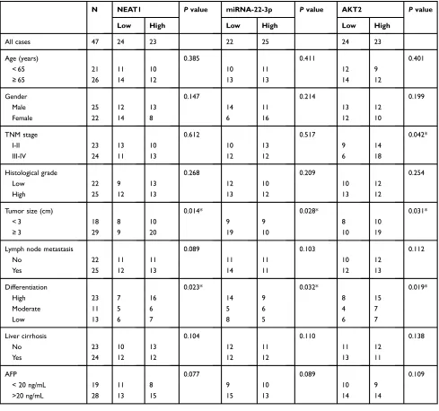
Table 1 Correlation Between Clinicopathological Features Of Patients With HCC And NEAT1, miRNA-22-3p, And AKT2 Expressions. *P<0.05
N NEAT1 Pvalue miRNA-22-3p Pvalue AKT2 Pvalue
Low High Low High Low High
All cases 47 24 23 22 25 24 23
Age (years) 0.385 0.411 0.401
< 65 21 11 10 10 11 12 9
≥65 26 14 12 13 13 14 12
Gender 0.147 0.214 0.199
Male 25 12 13 14 11 13 12
Female 22 14 8 6 16 12 10
TNM stage 0.612 0.517 0.042*
I-II 23 13 10 10 13 9 14
III-IV 24 11 13 12 12 6 18
Histological grade 0.268 0.209 0.254
Low 22 9 13 12 10 10 12
High 25 12 13 13 12 13 12
Tumor size (cm) 0.014* 0.028* 0.031*
< 3 18 8 10 9 9 8 10
≥3 29 9 20 19 10 10 19
Lymph node metastasis 0.089 0.103 0.112
No 22 11 11 11 11 10 12
Yes 25 12 13 14 11 12 13
Differentiation 0.023* 0.032* 0.019*
High 23 7 16 14 9 8 15
Moderate 11 5 6 5 6 4 7
Low 13 6 7 8 5 6 7
Liver cirrhosis 0.104 0.110 0.138
No 23 10 13 12 11 11 12
Yes 24 12 12 12 12 13 11
AFP 0.077 0.089 0.109
< 20 ng/mL 19 11 8 9 10 10 9
>20 ng/mL 28 13 15 15 13 14 14
OncoTargets and Therapy downloaded from https://www.dovepress.com/ by 118.70.13.36 on 25-Aug-2020
cytometer assay (FACSort; Becton Dickinson). The cell population in each stage was evaluated by ModFit soft-ware (Verity Softsoft-ware House,Top-sham, USA).
CCK-8 Assay
Cell proliferation was evaluated using CCK-8 (Dojin Laboratories, Kumamoto, Japan) according to the
manu-facturer’s instructions. The infected HepG2 cells were
seeded (1 × 10⁵cells/well) in a 96-well cell culture dish.
OD values were determined at 0, 24, 48, 72, and 96 h.
Transwell Invasion Assay
HepG2 cells were treated with a corresponding treatment and were subjected to the invasion assay (BD Biosciences,
NJ, USA) as previously described.28,29
Western Blot
HepG2 cell protein was isolated by using the cell lysis buffer (Beyotime Institute of Biotechnology, Shanghai, China). The detailed protocol was performed as previously
reported.29The primary antibodies against AKT2 (1:1000)
and GAPDH (1:1000) (Santa Cruz Biotechnology, Santa Cruz, CA) were incubated with protein samples at 4 °C
overnight. Quantification of optical density was evaluated
by the Uvitec Alliance software (Eppendorf, Germany).
TUNEL Assay
HepG2 cells were fixed in 4% paraformaldehyde in PBS
containing 0.12 mM sucrose for 15 mins. Apoptosis was determined by the terminal deoxynucleotidyl transferase dUTP nick end labeling (TUNEL) apoptosis detection kit (Beyotime Institute of Biotechnology, Nantong, China)
according to the manufacturer’s instructions. The
apopto-sis level was evaluated byfluorescence microscopy (Carl
Zeiss, Oberkochen, Germany).
HCC Mouse Model And In Vivo Study
Male BALB/c athymic nude mice (6 weeks old) were eachinoculated with HepG2 cells (1.0 × 10⁷) in 200 µl of PBS
by subcutaneous injection into theirflanks. Tumor growth
was monitored daily. After 8 days, the transplanted mice were randomly divided into six groups (n=5): si-NEAT1 control, si-NEAT1, si-NEAT1+miRNA-22-3p inhibitor, plV-CMV-control, plV-CMV-NEAT1, and miRNA-22-3p mimic+plV-CMV-NEAT1. Each treatment was directly injected into the tumor in 20 ul PBS (1 nmol). The injec-tion was performed every 4 days for a total of 7 times. Tumor volume (V) was evaluated by measuring the length
(L) and width (W) with caliper and calculated with the
formula V = (L × W2) × 0.5.
Statistical Analysis
Data were presented as means ± SD. Statistical analysis was performed using SPSS v.18.0 software (SPSS, IL, USA). There were at least 3 replicates in each treatment. The correlations between NEAT1 expression and
clinico-pathological features, NEAT1 and miRNA-22-3p,
miRNA-22-3p and AKT2, as well as NEAT1 and AKT2, were analyzed with the Spearman rank correlation.
OncoLnc online platform30 was used to predict overall
survival rate. Differences between groups were analyzed
with Student’s t-test and one-way analysis of variance.
Differences were considered to be significant atP<0.05.
Results
Upregulation Of NEAT1 Is Associated
With The Development Of HCC
NEAT1 expression was increased in HCC tissue
(Figure 1A) and several cell lines (Figure 1C), where the greatest expression was found in the HepG2 cell line. Therefore, the HepG2 was selected for subsequent experi-ments. Regarding the correlation between NEAT1 and clinicopathological features in HCC, the high expression of NEAT1 was associated with larger tumor size and greater levels of differentiation and NEAT1 expression
was significantly higher in HCC tissues compared with
healthy tissues (Table 1) (Figure 1B). To determine the
effect of upregulation of NEAT1 in HCC cells, si-NEAT1s and plV-CMV-NEAT1s were applied to knockdown and
overexpress NEAT1, respectively and the efficacy was
evaluated by real-time PCR (Figure 1DandE). Also, the
expression level of NEAT1 was shown to be associated
with the overall survival of patients with HCC (Figure 1F).
NEAT1 Is Essential For The Development
Of HCC
The inhibited NEAT1 expression suppressed cell
prolifera-tion (Figure 2A), invasion (Figure 2C) and increased the
number of cells in the S-phase (Figure 2B).
Downregulation of NEAT1 was also associated with
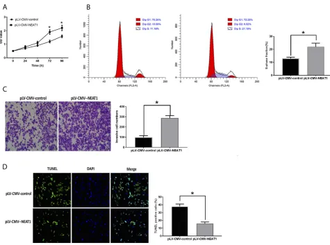
inhib-ited cell apoptosis (Figure 2D). Conversely, HepG2 cells
transfected with plV-CMV-NEAT1 displayed opposite trends compared with those with downregulation of
NEAT1 (Figure 3A–D).
OncoTargets and Therapy downloaded from https://www.dovepress.com/ by 118.70.13.36 on 25-Aug-2020
Increased AKT2 Is Involved In HCC
Development
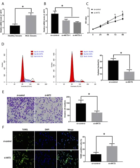
The mRNA expression of AKT2 was increased in HCC
tissues compared with those in healthy tissues (Figure 4A).
To investigate the function of AKT2 in HCC, si-AKT2s were applied to knockdown AKT2 and the knockdown
efficiencies were evaluated by RT-PCR (Figure 4B). The
downregulation of AKT2 led to the inhibitory effect on
HCC development. Specifically, HepG2 cells transfected
with si-AKT2 showed reduced cell proliferation
(Figure 4C), the number of cells in the S-phase
(Figure 4D), and invasion (Figure 4E) while elevated
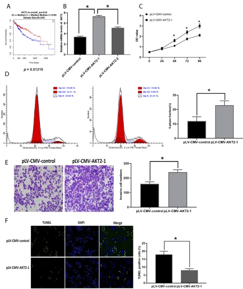
apoptosis levels (Figure 4F). Also, the correlation between
the expression of AKT2 and the overall survival rate was
demonstrated by online bioinformatics analysis
(Figure 5A). Furthermore, we tested the effect of
over-expression of AKT2 in the development of HCC
(Figure 5B) and we found that forced AKT2 expression
by plV-CMV-AKT2 further enhanced progression of HCC,
that is elevated cell proliferation (Figure 5C), number of
cells in the S-phase (Figure 5D), and invasion (Figure 5E)
whereas downregulated apoptosis levels (Figure 5F).
Moreover, the expression of AKT2 was correlated with tumor size, tumor differentiation grade, and TNM stage (Table 1). Collectively, these results demonstrated that the important effect of AKT2 in HCC.
NEAT1 Is Negatively Associated With
miRNA-22-3p In HCC
To further determine the mechanism associated with func-tions of NEAT1 and AKT2 in HCC, the potential target genes of NEAT1 and AKT2 were predicted by using online
databases: PicTar (http://pictar.mdc-berlin.de/), Miranda
(http://microRNA.org) and TargetScan (www.targetscan.
org/index.html). Both NEAT1 and AKT2 contained the
presumed binding site of miRNA-22-3pin 3ʹUTR and then
these predictions were confirmed by results from the
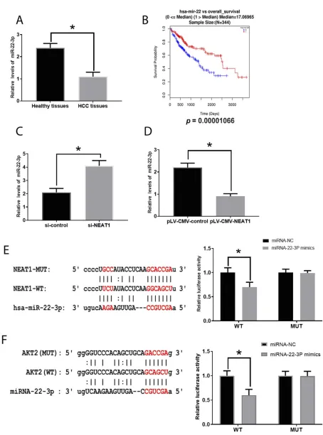
luci-ferase reporter assay (Figure 6EandF). Also, we found the
decreased miRNA-22-3pexpression in HCC tissues
com-pared with those in healthy tissues (Figure 6A) and the
Figure 1Involvement of NEAT1 in HCC. (A). NEAT1 expression in healthy and HCC tissues. (B). Association between NEAT1 and tumor size in HCC. (C). NEAT1 expression in several HCC cell lines. (D). Efficacy evaluation of si-NEAT1s. (E). Efficacy evaluation of pLV-CMV-NEAT1. (F). Correlation between NEAT1 expression and overall survival rate of patients with HCC (0=alive; 1=dead). Values are means ±SD. (*) denotes the difference between groups (P<0.05).
OncoTargets and Therapy downloaded from https://www.dovepress.com/ by 118.70.13.36 on 25-Aug-2020
expression of miRNA-22-3pdisplayed inverse patterns in those treated with siRNA- NEAT1 and plV-CMV-NEAT1,
respectively (Figure 6C and D), indicating NEAT1 can
directly target miRNA-22-3pin HCC. Moreover, through analysis in OncoLnc, the low level of miRNA-22-3pwas associated with a poor overall survival rate in patients with
HCC (Figure 6B)
MiRNA-22-3p Is Associated With HCC
Development
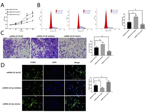
Then, we further investigated the roles of miRNA-22-3pby application of mimic and inhibitor. The results showed that the upregulation of miRNA-22-3pinhibited cell
prolifera-tion (Figure 7A), number of cells in the S-phase
(Figure 7B), and invasion (Figure 7C) and promoted cell
apoptosis (Figure 7D), which is opposite to the effects of
downregulation of miRNA-22-3pon HCC cells, indicating the essential roles of miRNA-22-3p in the development of HCC. Also, like NEAT1, the abundance of miRNA-22-3p
is associated with two clinicopathological features, tumor
size and differentiation grade (Table 1).
MiRNA-22-3p/akt2 Is Involved In The
Effect Of NEAT1 On Modulating HCC
Development
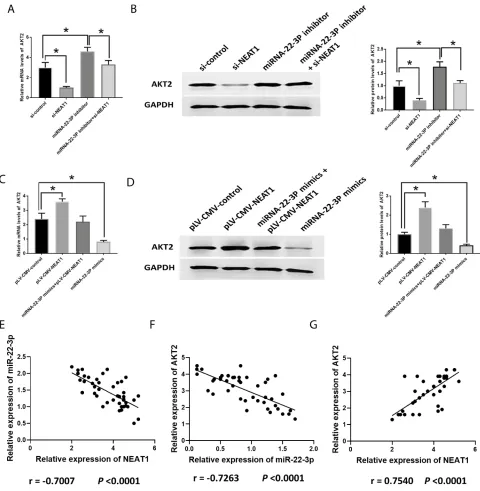
The knockdown of NEAT1 and the inhibition of
miRNA-22-3pdecreased and increased AKT2, respectively,
similarly in both mRNA and protein levels (Figure 8A
and B). The combination of si-NEAT1 and
miRNA-22-3pinhibitor abolished the effect of their respective roles on AKT2, showing no differences compared to the control
group (Figure 8A and B). The overexpression of NEAT1
and miRNA-22-3pmimic increased and decreased AKT2,
respectively, in both mRNA and protein levels (Figure 8C
and D). Similarly, the combination of plV-CMV-NEAT1
and miRNA-22-3pmimic reversed their respective effects on AKT2, displaying similar expression to those of control
group. Furthermore, Pearson’s correlation analysis showed
Figure 2Inhibition of NEAT1 suppresses HCC development. (A). OD values of HCC cells. (B). Cell cycle analysis in HCC cells. (C) Cell invasion in HCC cells. (D). Apoptosis in HCC cells. Values are means ±SD. (*) denotes the difference between groups (P<0.05).
OncoTargets and Therapy downloaded from https://www.dovepress.com/ by 118.70.13.36 on 25-Aug-2020
that the negative correlation existed between NEAT1 and
miRNA-22-3p (Figure 8E), and miRNA-22-3p and AKT2
(Figure 8F) while the positive correlation was found
between NEAT1 and AKT2 (Figure 8G).
Roles Of NEAT1in HCC In Vivo
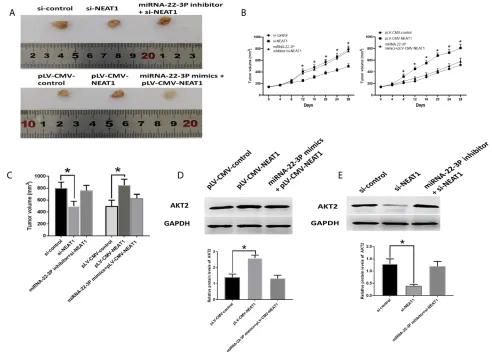
The downregulation of NEAT1 inhibited tumor growth, which was reversed by the combination of si-NEAT1 and
miRNA-22-3pinhibitor (Figure 9A–C). Meanwhile,
AKT2 protein expression was reduced by si-NEAT1 treatment but was not affected by the combination of
si-NEAT1 and miRNA-22-3P inhibitor (Figure 9A–C). On
the other hand, overexpression of NEAT2 promoted tumor growth, which was abolished by the combination
of plV-CMV-NEAT1 and miRNA-22-3P mimic.
Accordingly, overexpression of NEAT1 caused elevated
AKT2 protein expression (Figure 9D). while the
combi-nation of plV-CMV-NEAT1 and miRNA-22-3P mimic
did not impact AKT2 abundance (Figure 9E).
Discussion
In this study, we uncovered NEAT1 interacts with miRNA-22-3p regulate apoptosis and proliferation of HCC cells by targeting AKT2. The regulatory effect of NEAT1 and related mechanisms were demonstrated in vivo using an HCC mice model. Furthermore, NEAT1, miRNR-22-3p, and AKT2 correlated with clinicopatholo-gical features and overall survival in patients with HCC, respectively.
NEAT1 is tightly associated with HCC, such as the downregulation of NEAT1 promotes cytolysis activity to
suppress cell growth in HCC mice,31 the oncogenic roles
of NEAT1 in HCC are associated with its interaction with
miRNA-384,12 and NEAT1 decreases the sensitivity for
sorafenib, an effective drug for HCC, in HCC cells along
with downregulation of miRNA-335.14These observations
together imply the critical functions of NEAT1 in the development of HCC. Consistent with these previous reports, the present study showed an increased level of
Figure 3Upregulation of NEAT1 promotes HCC development. (A). OD values of HCC cells. (B). Cell cycle analysis in HCC cells. (C) Cell invasion in HCC cells. (D). Apoptosis in HCC cells. Values are means ±SD. (*) denotes the difference between groups (P<0.05).
OncoTargets and Therapy downloaded from https://www.dovepress.com/ by 118.70.13.36 on 25-Aug-2020
NEAT1 in both HCC tissues and cell lines. Moreover,
knockdown and overexpression of NEAT1 exerted
opposite roles in apoptosis, cell proliferation, and invasion in HCC cells. Accordingly, the hypothesis was supported
Figure 4Suppression of AKT2 inhibits in HCC development. (A). AKT2 expression in healthy and HCC tissues. (B). Efficacy evaluation of si-AKT2s. (C). OD values of HCC cells. (D). Cell cycle analysis in HCC cells. (E). Cell invasion in HCC cells. (F). Apoptosis in HCC cells. Values are means ±SD. (*) denotes the difference between groups (P<0.05).
OncoTargets and Therapy downloaded from https://www.dovepress.com/ by 118.70.13.36 on 25-Aug-2020
in vivo where the downregulation of NEAT1 could
sig-nificantly decrease tumor growth in HCC mice. Notably,
this is the first study reporting the involvement of NEAT1
in apoptosis in HCC cells, which suggests that NEAT1 may be a regulator hub mediating various cellular activ-ities in HCC cells.
Figure 5Enhanced expression of AKT2 promotes HCC development. (A). Correlation between AKT2 expression and overall survival rate of patients with HCC (0=alive; 1=dead). (B). Efficacy evaluation of pLV-CMV-AKT2s. (C). OD values of HCC cells. (D). Cell cycle analysis in HCC cells. (E). Cell invasion in HCC cells. (F). Apoptosis in HCC cells. Values are means ±SD. (*) denotes the difference between groups (P<0.05).
OncoTargets and Therapy downloaded from https://www.dovepress.com/ by 118.70.13.36 on 25-Aug-2020
Figure 6Involvement of miRNA-22-3p in HCC. (A). miRNA-22-3p in healthy and HCC tissues. (B) Correlation between miRNA-22-3p expression and overall survival rate of patients with HCC (0=alive; 1=dead). (C). MiRNA-22-3p expression in HCC cells treated with si-NEAT1. (D). MiRNA-22-3p expression in HCC cells treated with pLV-CMV-NEAT1. (E). The predictive sequence of miRNA-22-3p binding sites in the 3ʹUTR of NEAT1 and relative luciferase activity. (F). The predictive sequence of miRNA-22-3p binding sites in the 3ʹUTR of AKT2 and relative luciferase activity. Values are means ±SD. (*) denotes the difference between groups (P<0.05).
OncoTargets and Therapy downloaded from https://www.dovepress.com/ by 118.70.13.36 on 25-Aug-2020
To further investigate the mechanism underlining the regulatory effect of NEAT1 in HCC, the potential microRNA (miRNA) mediating the functions of NEAT1 in HCC was determined. In this study, the expression of miRNA-22-3p was decreased in HCC tissues and nega-tively correlated with NEAT1 expression. Based on the predictive evidence from online databases, miRNA-22-3p was found to be a presumed miRNA functioning down-stream factor of NEAT1, which was subsequently
con-firmed via a luciferase reporter assay. Moreover, the
functional evidence revealed that inhibition and enhance-ment of miRNA-22-3p played opposite functions in HCC cell activities. Furthermore, Chen et al reported that ber-berine-induced upregulation of miRNA-22-3p inhibits
HCC cell proliferation.22 Collectively, NEAT1 targets
miRNA-22-3p32 and miRNA-22-3p to modulate their
expressions and may function as an oncogenic regulator in HCC.
In the present study, the results also revealed that AKT2 was increased in HCC tissue and miRNA-22-3p
could bind to 3ʹUTR of AKT2. Also, functional studies
demonstrated that downregulation of AKT2 significantly
inhibited cell proliferation and invasion as well as pro-moted apoptosis in HCC cells. Furthermore, there was an inverse correlation on expressions between miRNA-22-3p and AKT2. Therefore, the results together indicated that AKT2 is a direct target gene of miRNA-22-3p and that AKT2 participates in NEAT1-associated cell activities in HCC. The serine/threonine kinase AKT functions as a
downstream mediator of the phosphatidylinositol 3′kinase
(PI3K) pathway and have been reported to be essential for
cell growth and survival.33,34 Overexpression of AKT2
Figure 7Roles of miRNA-22-3p in HCC development. (A). OD values of HCC cells. (B) Cell cycle analysis in HCC cells. (C). Cell invasion in HCC cells. (D). Apoptosis in HCC cells. Values are means ±SD. (*) denotes the difference between groups (P<0.05).
OncoTargets and Therapy downloaded from https://www.dovepress.com/ by 118.70.13.36 on 25-Aug-2020
promotes cell invasion and metastasis in ovarian and
breast cancer.35 Subsequently, the similar role of AKT2
is found in other cancers, such as colorectal cancer36and
lung cancer.37Taken together, the onco-effect of AKT2 is
modulated, or in part, by NEAT1/miRNA-22-3p axis in the development and progression of HCC.
In addition to evidence from molecular and cellular studies, the present study revealed that the expression of NEAT1 was positively correlated with clinicopathological
features in patients with HCC, such as tumor size and differentiation. Increased expression of NEAT1 is asso-ciated with enhanced clinical features of HCC, including
metastasis, vascular invasion, and the TNM stage.11 In
addition, the prognostic significance of NEAT1 in HCC is
observed where the upregulation of NEAT1 is tightly asso-ciated with liver cirrhosis, the TNM stage, and
microvas-cular invasion.38Therefore, NEAT1 may act as a favorable
prognostic marker for tumorigenesis and invasion of HCC.
Figure 8Involvement of AKT2 in HCC. (A, B). mRNA and protein expression of AKT2 in HCC cells transfected with si-NEAT1, miRNA-22-3p inhibitor, or both. (C, D). mRNA and protein expression of AKT2 in HCC cells transfected with pLV-CMV-NEAT1, miRNA-22-3p mimics or both. (E–G). Pearson’s correlation between NEAT1 and miRNA-22-3p, miRNA-22-3p and AKT2, and AKT2 and NEAT1. Values are means ±SD. (*) denotes the difference between groups (P<0.05).
OncoTargets and Therapy downloaded from https://www.dovepress.com/ by 118.70.13.36 on 25-Aug-2020
In conclusion, NEAT1 modulates apoptosis and cell viability in HCC cells in both in vitro and in vivo through interacting miRNA-22-3p and AKT2, suggest-ing the promissuggest-ing prognostic value of NEAT1 in patients
with HCC. Our finding may provide novel evidence to
support applying NEAT1 as a target for the treatment of HCC.
Acknowledgments
This study was funded by the Natural Science
Foundation of China (No. 81502435 and No. 81472615), the Xuzhou Science and Technology Project (KC18040),
andthe “The Six Top Talents” of Jiangsu Province
(YY-102).
Disclosure
The authors report no conflicts of interest in this work.
References
1. Adami H-O, Hunter DJ, Trichopoulos D. Textbook of Cancer Epidemiology. USA: Oxford University Press;2008.
2. Jemal A, Bray F, Center MM, Ferlay J, Ward E, Forman D. Global cancer statistics.CA Cancer J Clin.2011;61:69–90.
3. Balogh J, David Victor EHA III, Burroughs SG, et al. Hepatocellular carcinoma: a review.J Hepatocell Carcinoma.2016;3:41. doi:10.2147/ JHC.S61146
4. Han D, Li J, Wang H, et al. Circular RNA circMTO1 acts as the sponge of microRNA-9 to suppress hepatocellular carcinoma progres-sion.Hepatology.2017;66:1151–1164. doi:10.1002/hep.29270 5. Schmitt AM, Chang HY. Long noncoding RNAs in cancer pathways.
Cancer Cell.2016;29:452–463. doi:10.1016/j.ccell.2016.03.010 6. Wang KC, Chang HY. Molecular mechanisms of long noncoding
RNAs.Mol Cell.2011;43:904–914. doi:10.1016/j.molcel.2011.08.018 7. Yang G, Lu X, Yuan L. LncRNA: a link between RNA and cancer.
Biochim Biophys Acta Gene Regul Mech.2014;1839:1097–1109. 8. Müller V, Oliveira-Ferrer L, Steinbach B, Pantel K, Schwarzenbach H.
Interplay of lncRNA H19/miR-675 and lncRNA NEAT1/miR-204 in breast cancer.Mol Oncol.2019;13:1137–1149. doi:10.1002/1878-0261.12472 9. Yu HM, Wang C, Yuan Z, Chen GL, Ye T, Yang BW. LncRNA
NEAT1 promotes the tumorigenesis of colorectal cancer by sponging miR-193a-3p.Cell Prolif.2019;52:e12526. doi:10.1111/cpr.12526
Figure 9Roles of NEAT2/miRNA-22-3p/AKT2 axis in vivo study. (A) Representative images of isolated tumors. (B) Tumor growth curve. (C) Tumor volume. (D) Protein expression of AKT2 in HCC tissues of xenograft mice model treated with pLV-CMV-NEAT1, and combination of pLV-CMV-NEAT1 and miRNA-22-3p mimics. (E) Protein expression of AKT2 in HCC tissues of xenograft mice model treated with si-NEAT1, and combination of NEAT1 and miRNA-22-3p inhibitor. Values are means ±SD. (*) denotes the difference between groups (P<0.05).
OncoTargets and Therapy downloaded from https://www.dovepress.com/ by 118.70.13.36 on 25-Aug-2020
10. Tan HY, Wang C, Liu G, Zhou X. Long noncoding RNA NEAT1-modulated miR-506 regulates gastric cancer development through targeting STAT3. J Cell Biochem. 2019;120:4827–4836. doi:10. 1002/jcb.26691
11. Guo S, Chen W, Luo Y, et al. Clinical implication of long non-coding RNA NEAT1 expression in hepatocellular carcinoma patients.Int J Clin Exp Pathol.2015;8:5395.
12. Zhu L, Yang N, Li C, Liu G, Pan W, Li X. Long noncoding RNA NEAT1 promotes cell proliferation, migration, and invasion in hepa-tocellular carcinoma through interacting with miR-384. J Cell Biochem.2019;120:1997–2006.
13. Yang X, Qu S, Wang L, et al. PTBP3 splicing factor promotes hepatocellular carcinoma by destroying the splicing balance of NEAT1 and pre-miRNA-612.Oncogene.2018;37:6399–6413. doi:10. 1038/s41388-018-0416-8
14. Chen S, Xia X. Long noncoding RNA NEAT1 suppresses sorafenib sensitivity of hepatocellular carcinoma cells via regulating miR-335– c-Met.J Cell Physiol.2019.
15. Pascual J, Turner N. Targeting the PI3-kinase pathway in triple-negative breast cancer.Ann Oncol.2019.
16. Kleemann M, Schneider H, Unger K, et al. Induction of apoptosis in ovarian cancer cells by miRNA-493-3p directly targeting AKT2, STK38L, HMGA2, ETS1 and E2F5. Cell Mol Life Sci. 2019;76:539–559. doi:10.1007/s00018-018-2958-x
17. Li H, Lu S, Chen Y, et al. AKT2 phosphorylation of hexokinase 2 at T473 promotes tumorigenesis and metastasis in colon cancer cells via NF-κB, HIF1α, MMP2, and MMP9 upregulation. Cell Signal. 2019;58:99–110. doi:10.1016/j.cellsig.2019.03.011
18. Xu X, Sakon M, Nagano H, et al. Akt2 expression correlates with prognosis of human hepatocellular carcinoma. Oncol Rep. 2004;11:25–32.
19. Wang J, Li J, Shen J, Wang C, Yang L, Zhang X. MicroRNA-182 downregulates metastasis suppressor 1 and contributes to metastasis of hepatocellular carcinoma. BMC Cancer. 2012;12:227. doi:10. 1186/1471-2407-12-227
20. Li Q, Wang J, Juzi JT, et al. Clonality analysis for multicentric origin and intrahepatic metastasis in recurrent and primary hepatocellular carcinoma.J Gastrointest Surg.2008;12:1540. doi:10.1007/s11605-008-0591-y
21. Cooper D, Carter G, Li P, Patel R, Watson J, Patel N. Long non-coding RNA NEAT1 associates with SRp40 to temporally regulate PPARγ2 splicing during adipogenesis in 3T3-L1 cells. Genes. 2014;5:1050–1063. doi:10.3390/genes5041050
22. Chen J, Wu F-X, Luo H-L, et al. Berberine upregulates miRNA-22-3p to suppress hepatocellular carcinoma cell proliferation by target-ing Sp1.Am J Transl Res.2016;8:4932.
23. Xu Y, Li F, Zhang B, et al. MicroRNAs and target site screening reveals a pre-microRNA-30e variant associated with schizophrenia.Schizophr Res.2010;119:219–227. doi:10.1016/j.schres.2010.02.1070
24. Li J, Chen Y, Zhao J, Kong F, Zhang Y. miRNA-203 reverses chemoresistance in p53-mutated colon cancer cells through down-regulation of Akt2 expression. Cancer Lett. 2011;304:52–59. doi:10.1016/j.canlet.2011.02.003
25. Livak KJ, Schmittgen TD. Analysis of relative gene expression data using real-time quantitative PCR and the 2(-Delta Delta C(T)) method.Methods.2001;25:402–408. doi:10.1006/meth.2001.1262 26. Wang P, Wu T, Zhou H, et al. Long noncoding RNA NEAT1
pro-motes laryngeal squamous cell cancer through regulating miRNA-107/CDK6 pathway. J Exp Clin Cancer Res. 2016;35:22. doi:10.1186/s13046-016-0297-z
27. Liu -L-L, Lu S-X, Li M, et al. FoxD3-regulated microRNA-137 suppresses tumour growth and metastasis in human hepatocellular carcinoma by targeting AKT2.Oncotarget.2014;5:5113. doi:10.18 632/oncotarget.v5i13
28. Wang S-H, Wu X-C, Zhang M-D, Weng M-Z, Zhou D, Quan Z-W. Long noncoding RNA H19 contributes to gallbladder cancer cell proliferation by modulated miRNA-194-5p targeting AKT2.Tumor Biol.2016;37:9721–9730. doi:10.1007/s13277-016-4852-1 29. Pulito C, Mori F, Sacconi A, et al. Metformin-induced ablation of
microRNA 21-5p releases Sestrin-1 and CAB39L antitumoral activ-ities.Cell Discovery.2017;3:17022. doi:10.1038/celldisc.2017.22 30. Vasaikar SV, Straub P, Wang J, Zhang B. LinkedOmics: analyzing
multi-omics data within and across 32 cancer types.Nucleic Acids Res.2017;46:D956–D963. doi:10.1093/nar/gkx1090
31. Yan K, Fu Y, Zhu N, et al. Repression of lncRNA NEAT1 enhances the antitumor activity of CD8+ T cells against hepatocellular carci-noma via regulating miRNA-155/Tim-3. Int J Biochem Cell Biol. 2019;110:1–8. doi:10.1016/j.biocel.2019.01.019
32. Seitz H. Redefining microRNA targets.Current Biol.2009;19:870– 873. doi:10.1016/j.cub.2009.03.059
33. Yuan ZQ, Sun M, Feldman RI, et al. Frequent activation of AKT2 and induction of apoptosis by inhibition of phosphoinositide-3-OH kinase/Akt pathway in human ovarian cancer. Oncogene. 2000;19:2324. doi:10.1038/sj.onc.1203598
34. Graff JR, Konicek BW, McNulty AM, et al. Increased AKT activity contributes to prostate cancer progression by dramatically accelerat-ing prostate tumor growth and diminishaccelerat-ing p27Kip1 expression. J Biol Chem.2000;275:24500–24505. doi:10.1074/jbc.M003145200 35. Arboleda MJ, Lyons JF, Kabbinavar FF, et al. Overexpression of
AKT2/protein kinase Bβ leads to up-regulation of β1 integrins, increased invasion, and metastasis of human breast and ovarian cancer cells.Cancer Res.2003;63:196–206.
36. Rychahou PG, Kang J, Gulhati P, et al. Akt2 overexpression plays a critical role in the establishment of colorectal cancer metastasis.Proc National Acad Sci.2008;105:20315–20320. doi:10.1073/pnas.08107 15105
37. Lee MW, Kim DS, Lee JH, et al. Roles of AKT1 and AKT2 in non-small cell lung cancer cell survival, growth, and migration.Cancer Sci.2011;102:1822–1828. doi:10.1111/j.1349-7006.2011.02025.x 38. Liu Z, Chang Q, Yang F, et al. Long non-coding RNA NEAT1
overexpression is associated with unfavorable prognosis in patients with hepatocellular carcinoma after hepatectomy: a Chinese popula-tion-based study. Eur J Surg Oncol. 2017;43:1697–1703. doi:10.1016/j.ejso.2017.06.013
OncoTargets and Therapy
Dove
press
Publish your work in this journal
OncoTargets and Therapy is an international, peer-reviewed, open access journal focusing on the pathological basis of all cancers, potential targets for therapy and treatment protocols employed to improve the management of cancer patients. The journal also focuses on the impact of management programs and new therapeutic
agents and protocols on patient perspectives such as quality of life, adherence and satisfaction. The manuscript management system is completely online and includes a very quick and fair peer-review system, which is all easy to use. Visit http://www.dovepress.com/ testimonials.php to read real quotes from published authors.
Submit your manuscript here:https://www.dovepress.com/oncotargets-and-therapy-journal
OncoTargets and Therapy downloaded from https://www.dovepress.com/ by 118.70.13.36 on 25-Aug-2020