Holliday junction processing enzymes in
eukaryotes.
By
Anthony John Keeley
ProQuest Number: 10609126
All rights reserved INFORMATION TO ALL USERS
The qu ality of this repro d u ctio n is d e p e n d e n t upon the q u ality of the copy subm itted. In the unlikely e v e n t that the a u th o r did not send a c o m p le te m anuscript and there are missing pages, these will be note d . Also, if m aterial had to be rem oved,
a n o te will in d ica te the deletion.
uest
ProQuest 10609126
Published by ProQuest LLC(2017). C op yrig ht of the Dissertation is held by the Author. All rights reserved.
This work is protected against unauthorized copying under Title 17, United States C o d e M icroform Edition © ProQuest LLC.
ProQuest LLC.
789 East Eisenhower Parkway P.O. Box 1346
ACKNOWLEDGMENTS
I would like to acknowledge some of the people that without their kind help and
generosity this work would have been difficult. Firstly I would like to thank
members of the laboratory group, Fikret for his help, tutoring in yeast genetics,
some improved methods and some long coffee breaks explaining the finer
details of recombination. Judit for her help in the laboratory and always having
a spare lane on a gel and Mark for his assistance with sequencing gels.
I would also like to thank the other members of staff at UCL for their help and
positive response to favours, Jeremy Hioms’s group, Peter Piper’s group,
Laurence Pearl’s group, David Saggerson’s group, Liz Shephard’s group,
Jeremy Brockes’s group and Peter Shepherd’s group. In particular I would like
to thank Richard Pease for his very useful discussions and sounding out my
ideas.
I would like to thank members from other laboratories that are working in
recombination that were helpful in discussing data at meetings, providing
plasmids and alike and suggesting useful lines of experimentation.
I would like to thank my supervisor Irina Tsaneva for all her help and guidance
throughout the project, without which this project would have been very
difficult.
Finally I would like to thank mum and dad, my sister and her family who have
all been very supportive of my studies and I would like to thank Dot and Syd for
there support throughout my work. A very big thank you must also go to Lisa
who was always very positive and often put food in front of me at all hours of
the night.
ABSTRACT
Homologous genetic recombination is best understood in the model prokaryote
Escherichia coli where the enzymatic processes have been characterised using
purified proteins. Recombination involves reciprocal exchange of strands
between two homologous DNA strands. The resultant Holliday junction is either
branch migrated and/or resolved. Proteins, RuvA and RuvB can branch migrate
the junction and RuvC can resolve the junction.
In contrast limited advancement has been made with eukaryotic systems. A study
was undertaken to elucidate some of the processes that occur in eukaryotic
recombination. Activities capable of processing Holliday junction-containing
substrates were screened for. In addition activities capable of processing Holliday
junction precursor substrates, were also investigated. Initial searches involved
assaying fractionated eukaryotic cell extracts for processing activity. A later
approach was to PCR potential eukaryotic homologues of prokaryotic proteins.
Both approaches were successful. The latter approach resulted in the
identification of the homologue of CCE1 in S. cerevisiae being identified in S.
pombe. The open reading frame of YDC2_SCHPO (spCCEl) was cloned,
purified, assayed biochemically and found to be a Holliday junction specific
resolvase. Initial characterisation of the protein was carried out.
YDC2 being a homologue of CCE1 meant that it was likely to be a mitochondrial
Holliday junction resolvase activity that could be masking a nuclear Holliday
junction resolving activity. To search for another resolvase activity, a possible
nuclear activity, a S. cerevisiae yeast strain that was a CCE1 knock out was used
to resume the search for such possible activities. An initial fractionation of the
knockout strain of S. cerevisiae revealed the presence of another Holliday junction
Similar searches in mammalian cells led to the identification of an end joining
activity that in experiments seemed to show a homology dependency and an
annealing activity.
CONTENTS
Acknowledgements
2
Abstract
3
List of figures
12
Abbreviations
16
1.
INTRODUCTION
18
1.1 Homologous recombination 18
1.2 Homologous recombination in Escherichia coli 22
1.3 Initiation of homologous recombination by RecBCD 22
1.4 Other pathways for initiation of homologous recombination 23
1.5 The ruv locus 25
1.6 Branch migration of the Holliday junction is promoted by RuvA and RuvB 25
1.7 RecG 26
1.8 RuvC 27
1.9 Ruv ABC as a complex 28
1.10 RuvAB and RuvC at the replication fork 29
1.11 RusA 29
1.12 T4 endonuclease VII 29
1.13 T7 endonuclease I 30
1.14 Archaea 30
1.15 Holliday junction-specific endonucleases in eukaryotes 31
1.15.1 CCE1 31
1.15.2 Other S. cerevisiae resolvase activities 32
1.15.3 Endonuclease X I 32
1.15.6 Endonuclease X4
1.15.7 M ammalian resolvase activities
33 34
1.16 Structure and function 35
1.16.1 The structure of the Holliday junction 36
1.16.2 Resolvase junction binding 39
1.16.3 Resolvases function as a dimer 42
1.16.4 Holliday junction resolution 43
1.16.5 Proposed models for the association of a RuvABC Holliday junction com plex45 1.16.6 Ruv A functions as a tetramer and an octamer?. 45
1.17 Eukaryotic homologous recombination 50
1.17.1 Genetic Models 50
1.17.2 The Holliday model 50
1.17.3 The M eselson-Radding model 50
1.17.4 The Resnick model and the Double-Strand Break Repair model 51
1.17.5 Single-strand annealing model 53
1.18 The importance of recombination 53
1.19 Meiosis 54
1.20 Double-Strand Break Repair (DSBR) 54
1.20.1 Non-homologous end joining 55
1.20.2 The two pathways operate 55
1.20.3 Double-Strand Break Repair by homologous recombination 57 1.20.4 DSBR by homologous recombination: single-strand annealing 57 1.20.5 DSBR by homologous recombination: strand invasion 57
1.21 The RAD genes 58
1.21.1 RAD 51 58
1.21.2 DMC1 60
1.21.3 RAD52 60
1.21.4 RAD53 61
1.21.5 RAD54 61
1.21.6 RAD55 and RAD57 62
1.21.7 RAD59 63
1.21.8 RAD50/M RE11/XRS2 63
1.22 Recombination and carcinogenesis 65
1.23 Description of the thesis 66
2.
METHODS
67
2.1 Strains and plasmids 67
2.2 Enzymes and reagents 68
2.3 Buffers and solutions 69
2.3.1 DNA buffers 69
2.3.2 Enzyme buffers: 70
2.3.3 Protein buffers: 70
2.3.4 Media: 71
2.3.5 Buffers for alpha-structures: 72
2.4 Quantitation of DNA and protein 72
2.4.1 DNA 72
2.4.2 Protein 72
2.5 Molecular mass standards 72
2.6 Electrophoresis 73
2.6.1 Agarose gel electrophoresis 73
2.6.2 Denaturing PAGE 73
2.6.3 PAGE 74
2.6.4 SDS-PAGE 74
2.6.5 Autoradiography and phosphorimager analysis 75
2.7 DNA manipulations 75
2.7.1 Electroelution of DNA fragments from polyacrylamide gels 75 2.7.2 Phenol chloroform extractions and ethanol precipitations 75
2.7.3 Preparation of genomic DNA from S. pom be 76
2.7.4 PCR using genomic DNA 76
2.7.5 Purification of PCR products from agarose 77
2.8 Preparation of DNA substrates 77
2.8.1 Preparation of synthetic Holliday junction 77
2.8.2 Preparation of Chi-DNA. 79
2.8.6 Preparation of single-stranded DNA 81
2.8.7 3 ’ End-labelling of linear pDEA-7Zf(+) 82
2.8.8 Initial quantitation of labelled linear DNA and RecA required for production of
alpha-structures 83
2.8.9 Large scale purification of deproteinised a-structures 83
2.9 Yeast culture 84
2.10 Crude cell extracts from eukaryotes 84
2.10.1 Lysis of S. pom be and S. cerevisiae 84
2.10.2 Lysis of c c e lA S. cerevisiae strains and fractionation of the cell extract 85
2.10.3 Preparation of a crude extract from rat 86
2.10.4 Preparation of a crude extract from H eLa cells 86
2.11 Protein fractionation 87
2.11.1 Gel filtration 87
2.11.2 Ion-exchange chromatography (all ion-exchange columns) 88
2.12 Biochemical in vitro assays 90
2.12.1 ay for resolvase activity 90
2.12.2 Ligation assays 90
2.12.3 Assays for cutting of Chi structures 90
2.12.4 DNA binding assays 91
2.12.5 Stimulation of RuvB-mediated branch migration 91
2.12.6 Assay of crude extracts for branch migration activity 91
2.12.7 Assays for ligation/pairing activity 92
2.12.8 Antibody inhibition assays 92
2.13 Protein purification 92
2.13.1 Cloning and expression of S. pombe YDC2 92
2.13.2 Purification of RecA protein 93
2.14 Western blot analysis of HeLa cell fractions 94
2.15 Homology searches and protein sequence comparisons. 95
3.
SEARCHING FOR HOLLIDAY JUNCTION PROCESSING
ACTIVITIES IN EUKARYOTIC CELL EXTRACTS: THE APPROACH96
3.2 Four-stranded recombination reactions promoted by RecA in vitro 101
3.2.1 Construction of gDNA 101
3.2.2 Recombination intermediates made by RecA in vitro 101
3.3 Chi-structures 106
3.4 Summary 109
4.
SCHIZOSACCHAROMYCES POMBE
110
4.1 Holliday junction processing activity in fractionated S. pombe cell free extractsllO 4.2 Holliday junction resolution activity in fractionated cell-free extract from S.
pombe 113
4.3 Homology search in S. pombe database 116
4.4 Cloning of a novel Holliday junction resolvase from 5. pombe 116 4.5 Over-expression and purification of recombinant YDC2 119
4.6 Cloned activity is biochemically indistinguishable from the activity originally
identified in cell extracts 122
4.7 Holliday junction-specific DNA binding 128
4.8 pH Optimum 128
4.9 Efficiency of junction cleavage 131
4.10 Metal ion and Metal ion concentration 131
4.11 Temperature and salt concentration optimum 136
4.12 E. coli RuvB/YDC2 branch migration 136
4.13 Discussion 139
5.
S. CEREVISIAE
144
5.3 Detection and fractionation of a Holliday junction resolvase 147
5.4 Holliday junction resolution activity from AK47 cells 152
5.5 The cleavage sites of the activity differ from those of CCE1 160
5.6 Cleavage sites are nicks that can be ligated in vitro to restore a continuous DNA
backbone 164
5.7 Fractions containing Holliday junction cleavage activity bind synthetic Holliday
junctions 164
5.8 Native molecular mass of the Holliday junction resolvase 168
5.9 RuvB-mediated branch migration 168
5.10 Conclusion 173
6.
MAMMALIAN EXTRACTS
176
6.1 The search for activities: assays of partially fractionated cell-free extracts 176
6.2 HeLa cell fractionation 178
6.3 ATP independent reactions 178
6.4 Assays to test for ligation 182
6.5 Further investigation of the activities from rat testis and HeLa cells 186
6.6 Homology dependant ligation and pairing? 189
6.7 Separation of activities 193
6.8 Active HeLa fractions contain Rad51 195
6.9 Discussion 197
7.
GENERAL DISCUSSION
199
7.1 YDC2 199
7.2 Characterisation of a possible nuclear resolvase 201
7.3 Activities in fractionated HeLa cell extracts 207
Bibliography
208
Appendix
240
LIST OF FIGURES
CHAPTER 1
Figure 1.1. The Holliday Model. 20
Figure 1.16.2. Schematic showing some conformations adopted
by synthetic Holliday junctions upon protein binding. 40
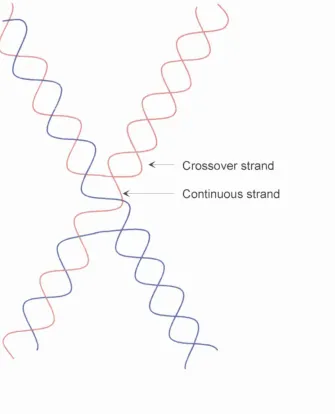
Figure 1.16.4. Schematic of a Holliday junction showing
the crossover and continuous strand. 44
Figure 1.16.6 RuvA as the Stator for the RuvB motor. 47
Figure 1.17.4 The double strand break repair model. 52
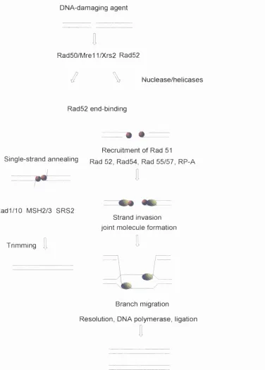
Figure 1.21.8. Schematic outline of protein functions in DSBR
by homologous recombination. 64
CHAPTER 2
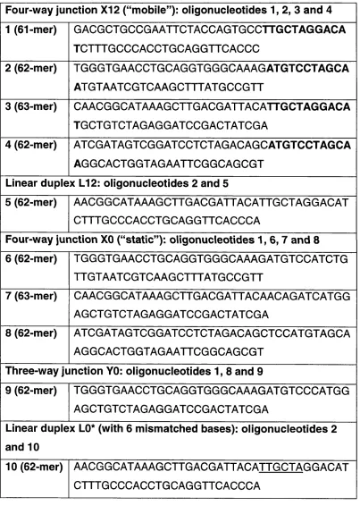
Table 2.8: Sequences of oligonucleotides used to make
synthetic substrates. 78
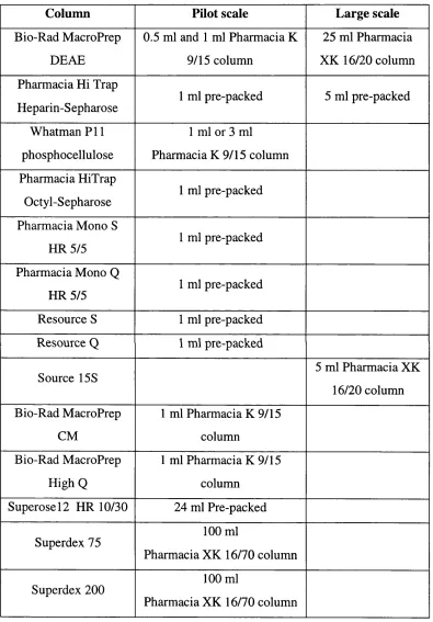
Table 2.11: Chromatography columns used. 89
CHAPTER 3
Figure 3.1. Synthetic Holliday junction constructed by annealing
four synthetic oligonucleotides. 98
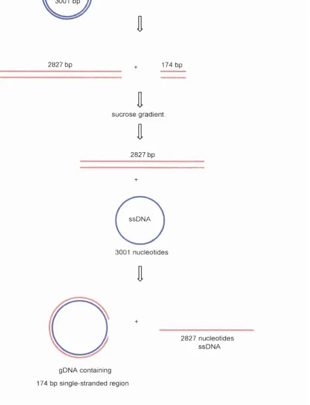
Figure 3.2.1. gDNA preparation. 103
Figure 3.2.2. Formation and processing of RecA-made
alpha structures. 104
Figure 3.3. Chi DNA formation. 107
Table 3.0. Possible sources of Holliday junction-processing activities. 97
Table 3.4. Holliday junction-processing activities identified. 109
CHAPTER 4
Figure 4.1. Processing of RecA-made alpha-structures by fractionated
cell-free extracts from S. pombe. I l l
Figure 4.2. Endonuclease activities of S. pombe are specific for a synthetic
four-way Holliday junction and Holliday junction-containing Chi DNA. 114
Figure 4.3 Sequence comparison of S. cerevisiae CCE1 and
S. pombe YDC2. 117
Figure 4.4. Cloning of the YCD2_SCHPO open reading frame
from S. pombe. 118
Figure 4.5. Purification of YDC2. 120
Figure 4.6.1. YDC2 and S. pombe fraction 3 cut each strand of junction
X12 at identical sites. 123
Figure 4.6.2. Shematic presentation of the central sequences of
junction X 12 cleaved by YDC2 125
Figure 4.6.3. Repair by DNA ligase of nicks produced by YDC2
and S. pombe cellular activity. 126
Figure 4.7. YDC2 binds specifically to the four-way junction X12. 129
Figure 4.8. YDC2 cleaves Holliday junctions more efficiently at
alkaline pH. 130
Figure 4.9. The effect of increasing protein concentration on
junction resolution. 132
Figure 4.10. YDC2 requires the presence of a specific divalent
cation for resolution. 134
Figure 4.11. The effect of temperature and salt concentration on
YDC2 Holliday junction resolution. 137
Figure 4.13. YDC2 resolution of junction X0. 141
CHAPTER 5
Figure 5.2. PCR analysis of S. cerevisiae ccelA strain SK20. 146
Figure 5.3.1. Scheme for partial purification of a resolvase activity from
haploid and diploid cells of S. cerevisiae ccelA strain S K I9, SK20, and
AK47. 150
Figure 5.3.2. Elution profile of fractionated extracts from SK20 cells
Figure 5.3.4. Comparison of activity from diploid AK47 cells
grown in supplemented and unsupplemented media. 155
Figure 5.4.1. Identification and initial fractionation of a resolvase activity
from diploid cells of S. cerevisiae ccelA strain AK47. 157
Figure 5.4.2. The activity from diploid S. cerevisiae ccelA cells shows
specificity and selectivity for resolving four-way junctions. 158
Figure 5.5.1. S. cerevisiae ccelA activity, CCE1 and YDC2 cut each
strand of junction X12 at sites symmetric to the point of crossover. 162
Figure 5.5.2. S. cerevisiae ccelA activity, CCE1 and YDC2 cut each
strand of junction X12 at sites symmetric to the point of crossover. 163
Figure 5.6. T4 DNA ligase repaires nicked DNA. 165
Figure 5.7 Activity from diploid S. cerevisiae ccelA cells cuts
and binds specifically to the four-way junction X I 2. 166
Figure 5.8. Determination of the molecular weight of the
Holliday junction specific endonuclease. 169
Figure 5.9. RuvB-mediated branch migration. 171
Table 5.2. Strain Genotype. 147
Table 5.3. Chromatography column performance. 148
CHAPTER 6
Figure 6.1 Rat testis cell-free extract fractionated on Resource S. 177
Figure 6.2 HeLa cell extract fractionated on phosphocellulose. 179
Figure 6.3. Dimer products form without the addition of ATP. 180
Figure 6.4.1. Fraction 12 incubated with labelled linear DNA. 183
Figure 6.4.2. Chi DNA reactions. 184
Figure 6.5.1 Rat testis cell-free extract fractionated by gel filtration
on Superdex 200. 187
Figure 6.6.1. Pairing reactions. 190
Figure 6.6.2. gDNA dependency of products. 191
Figure 6.7. Further fractionation of partially purified HeLa cell extract. 194
Figure 6.8. The presence of Rad51 in fractions from HeLa cells. 196
Figure 6.9. A possible model for double strand break repair. 198
CHAPTER 7
Table 7.2. Comparison of known Holliday junction resolvases
ABBREVIATIONS
ADP adenosine diphosphate
ATP adenosine triphosphate
ATPyS adenosine 5’-(y-thio) triphosphate
BCDP 5-bromo-4-chloro-3-indolyl phosphate
bp base pair(s)
BSA bovine serum albumin
CCE1 cruciform cutting enzyme 1
CIP calf intestinal alkaline phosphotase
Da dalton
dATP deoxyadenosine triphosphate
dsDNA double-stranded DNA
gDNA gapped circular duplex DNA
DTT dithiothreitol
GLB gel-loading buffer
EDTA ethylenediaminetetraacetic acid (disodium salt)
EGTA ethylene glycol-bis(p-aminoethyl ether)-N,N,N’,N’-tetraacetic
acid
EtOH ethanol
FACS fluorescence-activated cell sorter
FPLC fast protein liquid chromatography
HC1 hydrochloric acid
IPTG p-D-isopropyl-thiogalactopyranoside
kDa kilodalton
mtDNA mitochondrial DNA
NBT nitro blue tetrazolium
NER nucleotide excision repair
NHEJ non-homologous end-joining
PAGE polyacrylamide gel electrophoresis
PCR polymerase chain reaction
PEG polyethylene glycol
PMSF phenylmethylsulfonyl fluoride
PNK polynucleotide kinase
RNA ribonucleic acid
SDS sodium dodecyl sulphate
SSB single stranded DNA binding protein
ssDNA single-stranded DNA
TAE tris/acetate/EDT A
TBS tris buffered saline
TBE tris/borate/EDT A
TBST tris buffered saline tween
TE tris/EDTA
TEMED N,N ,N ’,N’-(tetramethylethylenediamine)
TLCK N-tosyl-L-lysine chloromethyl ketone
TNE tris/sodium chloride/EDTA
TPCK N-tosyl-L-phenylalanine chloromethyl ketone
Tris tris (hydroxymethyl) aminoethane
Tween 20 polyoxyethylenesorbitan monolaurate
UV ultraviolet
v/v volume:volume ratio
w/v weight:volume ratio
1. INTRODUCTION
The aim of the project was to provide evidence for the enzymatic processing of
Holliday junctions in eukaryotes, the central intermediate in homologous
recombination.
1.1 Homologous recombination
Homologous recombination is important for a number of fundamental processes
during a cell’s lifetime. These include double-strand break repair of damaged
DNA, repair of post-replication gaps, proper segregation of chromosomes
during meiosis and mitosis, maintenance of the genetic information and
sequence copy number, as well as creation of new alleles and generation of
genetic diversity. Homologous recombination is also thought to have a strong
interdependence with DNA replication (Kogoma, 1996; Kogoma et al., 1996)
and the rescue of stalled replication forks (Seigneur et al., 1998).
Understanding the molecular mechanisms of homologous recombination will
help to explain some fundamental features of DNA maintenance, but could also
lead to the successful targeting of gene sequences in higher eukaryotes and
perhaps be of use in designing efficient gene replacement strategies.
Understanding what controls gene integration and, conversely, what inhibits
gene integration, would help in addressing broader issues concerning cancer.
Genetic evidence now suggests that cancers can arise as a result of a defect in
repair processes involving homologous recombination (Matsuda M and
Yasutomi M, 1999).
The prerequisite for homologous recombination to occur, as the name implies, is
that the parental DNA molecules share extensive sequence homology. This is
different from site-specific recombination, which occurs between defined short
sequences. These specific target sequences are recognised by the proteins that
mediate the recombination process and the two recombining sites are brought
together through protein-protein interactions. This form of recombination needs
to be tightly regulated, as the outcome can be very disruptive producing
deletions and inversions, leading to gene inactivation and chromosomal
rearrangement. In prokaryotes site-specific recombination allows the integration
and excision of a bacteriophage genome into and out of the host bacterial
chromosome. The rearrangement of the immunoglobulin genes through V(D)J
recombination is a form of site-specific recombination in higher eukaryotes.
During illegitimate recombination, another type of recombination, no sequence
homology or specific sites are required. It occurs when certain genetic elements
move from one chromosome location to another. Strand slippage is also a form
of illegitimate recombination that can occur during replication. Often there is a
small loss or gain of DNA sequence and the process is usually very deleterious.
The classification of these reactions into homologous, site specific and
illegitimate is for understanding purposes, as it is often the case that more than
one reaction mechanism is involved.
Homologous recombination is a process whereby DNA molecules sharing
extensive regions of homology interact to form daughter DNA molecules that
are different from the parental DNA as a result of rearrangement and/or
exchange of genetic information. The Holliday model (Holliday, 1964),
formulated to explain gene conversion events in fungi, proposed that the
molecular basis for this process is the physical exchange of DNA strands
between the participating DNA molecules, leading to the formation of an
intermediate where two DNA duplexes are joined through a crossover (Figure
1.1). This intermediate became known as the Holliday junction. The principle
features of this model are central to other models proposed to explain
homologous recombination, which will be discussed later in this chapter. The
Holliday junction can branch migrate to extend the heteroduplex DNA, which
Figure 1.1 The Holliday model. (A)
Two parental homologous DNA
molecules are aligned.
(B)
Strand exchange of DNA leads to the
formation of a Holliday junction.
(C)
The Holliday junction can branch
migrate to extend the region of heteroduplex DNA.
(D)
The Holliday
junction can be cleaved in two possible directions A - B and C - D.
Resolution of the junction in the direction A - B or C - D can lead to the
production of splice
(E)
or
(F)
patch products.
A
1
3
B
1
\ 7
2 6
D
C B
4 8
cleavage A-B
\ S
cleavage C-D
splice products
Resolution of the junction by the introduction of matching nicks in the DNA
phosphodiester backbone in one of two possible orientations can subsequently
separate the two duplex DNA molecules and produce “splice” or “patch”
recombinant products (with or without exchange of flanking markers,
respectively).
1.2 Homologous recombination in
Escherichia coli
Homologous recombination has been studied extensively in E. coli (reviewed in
(Kowalczykowski et al., 1994; Shinagawa and Iwasaki, 1996; W est, 1992;
West, 1997). Many of the reactions involved have been characterised using
purified proteins. The process can be divided into three stages: pre-synapsis,
synapsis and post-synapsis. During synapsis homologous DNA molecules
become aligned together, exchange strands and become joined together by a
Holliday junction. Finally in post-synapsis the Holliday junction is cleaved to
release the two DNA molecules.
1.3 Initiation of homologous recombination by RecBCD
During conjugation when the bacterial chromosome (or part of it) is transferred
from one bacterium to another as a linear molecule, and in DNA double-strand
break repair, duplex DNA ends are processed to allow RecA protein to search
for homology. Because RecA requires the presence of a single-stranded region
to form an active nucleoprotein filament on DNA, the production of single
stranded DNA is the first stage of pre-synapsis. Initiation of homologous
recombination begins with the formation of DNA with a single-stranded region
and a 3’ tail. The primary initiator function during conjugation is carried out by
the heterotrimeric enzyme RecBCD, an ATP-dependent dsDNA and ssDNA
nuclease, and a DNA helicase (Kowalczykowski and Eggleston, 1994; Roman
and Kowalczykowski, 1989). RecBCD enters at the end of a linear duplex and
begins to unwind DNA. The unwinding activity of RecBCD is accompanied by
a simultaneous nuclease activity (Taylor and Smith, 1985). Nuclease activity
continues until RecBCD reaches a correctly orientated Chi site (5’gctggtgg3’),
where the enzyme pauses. This pause increases the probability of the enzyme
cleaving in the vicinity of the Chi site, resulting in nicks 4-6 nucleotides 3 ’ of
the Chi site. The interactions with the Chi site also results in the attenuation of
the 3’ - 5 ’nuclease activity of RecBCD, and upregulation of its 5 ’ - 3’ nuclease
activity. The overall result is the formation of a single-stranded 3 ’ tail
containing the Chi site (Anderson and Kowalczykowski, 1997). RecBCD can
then facilitate the loading of RecA onto the Chi-containing single-stranded
DNA (Anderson et al., 1997).
1.4 Other pathways for initiation of homologous recombination
The major recombination pathway in E. coli wild type cells depends on
RecBCD. In recBC mutants initiation of recombination depends on other genes.
This has led to the idea of multiple pathways of homologous recombination,
such as the RecE pathway, seen in recBC sbcA mutants (Barbour et al., 1970),
and the RecF pathway seen in recBC sbcBC mutants (Horii and Clark, 1973).
In the absence of RecBCD (or in addition to RecBCD), recombinogenic single
stranded DNA (ssDNA) could be formed by duplex DNA unwinding by
helicases, by strand-specific exonucleases or by combination of these activities.
The RecE (exonuclease VIII) and the RecJ protein are both single-strand specific
exonucleases, which act 5-3' and are known to play a role in recombination.
These nucleases may function in combination with DNA helicases such as RecQ
(Umezu and Nakayama, 1993) to generate single-stranded 3’ termini. DNA
helicase n , the uvrD gene product or DNA helicase IV, the helD gene product,
may also act in concert with RecJ, RecN, and RecE.
RecF binds ssDNA and dsDNA in the presence of ATP (Madiraju and Clark,
1991; Madiraju and Clark, 1992) and has been shown to form a complex in
vitro with RecO and RecR (Hegde et al., 1996). RecF, RecO and RecR are also
believed to be involved in repair of DNA damage in a mechanism other than
one involving recombination and may assist in replisome assembly (Discussed
in (Kogoma, 1997).
duplexes and strand exchange leading to the formation of a Holliday junction.
RecA is a structural protein and a catalyst of 37.8 kD. In the presence of ATP
RecA polymerises on ssDNA to form a right-handed presynaptic helical
nucleoprotein filament within which pairing occurs (reviewed in (West, 1992)).
The DNA can be partially single-stranded with a ssDNA region at the end of a
linear duplex or a gap in the middle of a linear or circular duplex DNA
molecule. RecA initiates filament formation at the region of ssDNA but
nucleoprotein filament formation can invade the entire duplex DNA molecule.
Accessory proteins may also facilitate filament assembly. SSB is believed to
remove secondary structure in ssDNA. The DNA within the filament increases
in pitch from 10 bp per turn to 18.6 bp per turn, as RecA imposes its own
helicity upon the DNA. One helical turn of this filament consists of 6.2 RecA
monomers. The RecA nucleoprotein filament forms a deep open groove where
a second naked duplex DNA can be bound to a secondary DNA binding site and
randomly searched for homology. When homology is established strand
exchange is initiated and proceeds with a defined polarity (5’ to 3’ relative to
the ssDNA strand within the nucleoprotein filament that stays with the
filament). The strand exchange process can also be described as switching of
base-pairing partners between two DNA molecules bound within the two
binding sites of the RecA nucleoprotein filament (West, 1992).
In order for DNA strands to be exchanged sequence homology between the two
strands must be established. This homology search does not require homologous
ends in any of the chains involved. The initial alignment of homology is poorly
understood. During the strand exchange reaction catalysed by RecA,
complementary base pairing provides the specificity of recognition between the
two DNA molecules.
Homologous alignment leads to strand exchange and the production of a
Holliday junction. The Holliday junction can be processed by the RuvAB and
RuvC proteins encoded by the ruv locus.
1.5 The
ruv
locus
The ruv locus located at minute 41 on the E. coli genetic map contains two
operons. The ruvA and ruvB operon is controlled by the LexA repressor and is
part of the SOS stress response regulon triggered by DNA damage. The second
operon consisting of ruvC and orf-26 is not under SOS control (Sharpies and
Lloyd, 1991; Takahagi et al., 1991). Mutations in all three ruv genes result in
increased sensitivity to UV light, ionising radiation and mitomycin C, phenotypes
indicating defects in DNA repair. The involvement of the ruv genes in
recombination was demonstrated by the fact that ruv mutants are severely
defective in recombination in a recBC-sbcA, recBC-sbcBC or recG background
(Lloyd, 1991; Lloyd et al., 1984; Lloyd et al., 1987). RuvA is a 22 kDa DNA-
binding protein that seems to target RuvB to the DNA (Parsons and West, 1993).
RuvB is a 37 kDa DNA-dependent ATPase that in the presence of ATP, Mg2+
(clOmM) and RuvA catalyses branch migration.
1.6 Branch migration of the Holliday junction is promoted by RuvA and
RuvB
RuvA and RuvB promote branch migration of the Holliday junction along the
two duplexes leading to the formation of heteroduplex DNA by the progressive
exchange of base-pairs (Parsons et al., 1992; Shiba et al., 1991; Tsaneva et al.,
1992). The heteroduplex DNA provides opportunity for transfer of genetic
information from one strand to the other.
RuvA shows a high affinity for binding to Holliday junctions but also binds
both ssDNA and duplex DNA (Iwasaki et al., 1992; Parsons et al., 1992). RuvA
acts as a specificity factor that targets RuvB to the junction (Parsons and West,
1993) where RuvB forms hexameric rings (Stasiak et al., 1994). Binding of
RuvA to DNA is structure-specific and sequence independent. In addition to
binding Holliday junctions, RuvA can bind Y structures (Hiom et al., 1996).
The interaction of RuvA with DNA is not dependent on ATP and is most stable
RuvB is a DNA-dependent ATPase and in the presence of ATP, M g2+ and
RuvA, catalyses branch migration. High concentrations of RuvB alone can
promote branch migration of recombination intermediates made by RecA. This
reaction requires high concentrations of Mg2+ (>15mM) which facilitates the
binding of RuvB to DNA (Muller et al., 1993; Tsaneva et al., 1993). In 2 mM
MgCl2 RuvB has a weak DNA-independent ATPase activity, but in 15 mM
M gCb the ATPase activity of RuvB can be stimulated with form I DNA (Iwasaki
et al., 1992; Mitchell and West, 1994; Muller et al., 1993; Tsaneva et al., 1992).
In the presence of ATP and Mg2+ RuvA and RuvB form a tripartite protein
complex (Hiom and West, 1995; Parsons et al., 1995) where RuvA binds to the
crossover and is sandwiched between two hexameric rings of RuvB. The
binding of RuvA leads to the Holliday junction within the complex adopting a
square planar conformation. The hexameric RuvB rings assemble on two
diametrically opposed arms of the open square junction (Parsons et al., 1995).
1.7 RecG
The 76 kDa protein RecG can also promote branch migration of the Holliday
junction in vitro. RecG does this in an ATP-dependent manner in the presence
2 ,
of Mg ions (Lloyd and Sharpies, 1993). The activities of RecG seem to
partially overlap with the Ruv proteins as ruv mutants are not recombination
deficient whereas double ruv recG mutants are severely deficient in conjugal
recombination (Lloyd, 1991; Lloyd and Buckman, 1991; Mandal et al., 1993).
RecG is a structure-specific helicase that can act on both R-loops and D-loops
and is implicated to function in processes other than recombination (Fukuoh et
al., 1997; McGlynn et al., 1997; Vincent et al., 1996). RecG does not fit into
either of the RecBCD, RecE, or RecF pathways of recombination. Probably the
pathways are dependent on the DNA substrate that initiates recombination and
the genetic background in which the recombination takes place.
1.8 RuvC
Finally the Holliday junction can be resolved by the 19 kDa protein RuvC
(Dunderdale et al., 1991; Iwasaki et al., 1991; Sharpies and Lloyd, 1991;
Takahagi et al., 1991). Firstly RuvC binds the junction as a dimer (Shah et al.,
1997). The DNA structure is modified by distortion of the DNA backbone
(Bennett and West, 1995) and then cleaved by the introduction of two
symmetrically related nicks in a pair of strands of like polarity, in the presence
of either M g2+ or M n2+ (Bennett et al., 1993; Shah et al., 1997). Cleavage of the
continuous (non-crossover) strands show sequence specificity for the sequence
5’W T T lS -3 ’ where W is A or T and S is G or C (Bennett and W est, 1995).
This specificity is relaxed by the presence of Mn ions. Cleavage produces 5’
phosphate and the 3 ’ OH termini that can be re-joined by ligation (Bennett et al.,
1993). In vitro studies have shown that RuvC will cleave 3 and 4 stranded
recombination intermediates at the junction crossover, but it fails to act on Y-
junctions, G/A mismatches heterologous loops or 2-stranded branched
junctions. Binding to three-stranded recombination intermediates is less stable
than binding to four-stranded intermediates. In an on-going RecA
recombination reaction RuvC will cleave a four-stranded intermediate but
cannot cut a three-stranded intermediate, suggesting that RecA blocks the
accessibility of RuvC to three-stranded junctions (Benson and West, 1994).
RuvC resolves Holliday junctions during genetic recombination and post-
replicational repair of DNA damage. After cleavage, ligation rejoins the
5 ’phosphate and the 3’ hydroxl (Bennett et al., 1993; Shah et al., 1997).
Cleavage of the Holliday junction and ligation lead to splice and patch products,
depending on the orientation of cleavage (Figure 1.1).
RuvC has been expressed in plant cells. RuvC was targeted to the nucleus of
the plant cell by fusion to a plant viral nuclear localisation signal and shown to
stimulate both genomic and extrachromosomal homologous recombination
To summarise, homologous recombination in E. coli is initiated at regions of
ssDNA such as gaps or 3’ single-stranded tails formed at double-strand breaks
by helicase and/or nuclease activities. The active RecA nucleoprotein filament
nucleated at the ssDNA searches for homology and promotes the pairing and
exchange of strands between homologous DNA molecules forming a Holliday
junction. Extension of the heteroduplex region by branch migration of the
Holliday junction can be catalysed by RuvAB or RecG. RuvC can then resolve
the Holliday junction. The fact that different proteins can perform similar
activities indicates that several pathways operate within a single cell.
1.9 Ruv ABC as a complex
Genetic analysis of the ruv locus has suggested that RuvABC co-operate in
processing the Holliday junction (Mahdi et al., 1996; Mandal et al., 1993;
Sharpies et al., 1990). Further in vitro evidence for coupled activity has come
from experiments that demonstrate that RuvB and RuvC can co-operate on
junctions and promote branch migration in the absence of RuvA. Using
junctions that contained a RuvC consensus sequence located in different
positions relative to the crossover, cleavage was shown to be stimulated in the
presence of RuvB (van Gool et al., 1998). The reduced cleavage of the junction
when the consensus sequence is located away from the point of strand exchange
suggests that, in vivo, the resol vase would be inefficient if the Holliday junction
did not have a correct sequence located at the point of crossover. The formation
of a functional RuvABC complex or “resolvasome” on the Holliday junction is
supported by experiments using monoclonal antibodies raised to the three Ruv
proteins. Recombination reactions in vitro showed that the resolution of
recombination intermediates by RuvC was inhibited by antibodies against RuvA
and RuvB, and all three proteins co-immunoprecipitated with synthetic Holliday
junctions (Davies and West, 1998; Eggleston et al., 1997). Recent evidence has
demonstrated that RuvC cleavage is more efficient in reactions that contain
RuvAB (Zerbib et al., 1998).
1.10 RuvAB and RuvC at the replication fork
Recent work has shown that strains that are rep, recBC ts mutants, accumulate
linear DNA due to the presence of stalled replication forks. These strains are
not viable at the restrictive temperature, however the lethality can be rescued by
inactivation of the ruvAB operon. Similar experiments using dnaB temperature
sensitive mutants showed that the accumulation of double-strand breaks was
suppressed by inactivation of both RuvAB and RuvC. These results strongly
suggest that RuvABC could act on stalled replication forks (Seigneur et al.,
1998).
The RuvABC proteins appear to be highly conserved and widely represented in
eubacteria. Homologues of the RuvA and RuvB proteins have been identified
in the genomes of all completely sequenced prokaryotic species, and most
contain an identifiable RuvC homologue. Other prokaryotic enzymes that cut
Holliday junctions have also been identified and characterised; such as RusA,
bacteriophage T4 endonuclease VII and T7 endonuclease I (reviewed in (White
et al., 1997)).
1.11 RusA
RusA is a 14 kDa protein that resolves four-way junction (Sharpies et al., 1994).
The protein is encoded for by the rusA gene of the defective lambdoid prophage
DLP12 and is constitutively repressed in E. coli (Mahdi et al., 1996)
1.12 T4 endonuclease VII
T4 endo VII was identified from T4 phage mutants that accumulated multi
branched DNA (Kemper and Janz, 1976). The 157 amino acid protein has a
molecular weight of 18 kDa and cleaves a variety of substrates including four
1991; Jensch and Kemper, 1986; Lilley and Kemper, 1984; Mizuuchi et al.,
1982).
1.13 T7 endonuclease I
Phage T7 endo I is encoded by gene 3. Mutants that lack T7 endo I accumulate
branched DNA intermediates (Tsujimoto and Ogawa, 1978) similar to the
inactivation of T4 endo VII. The gene has been cloned and the protein product
has been purified. The 149 amino acid endonuclease has a molecular weight of
17 kDa (de Massy et al., 1987). Similar to T4 endo VII, T7 endo I is also
promiscuous about its substrate and will cleave both three and four-way
junctions (de Massy et al., 1987; Dickie et al., 1987).
A common feature of all of these enzymes is that they bind to the junction as
dimers and manipulate the structure of the junction, as discussed later in this
chapter (reviewed in (White et al., 1997)).
Holliday junction resolvases have been categorised according to their structure
and sequence selectivity. Group one, which includes RuvC, RusA, and CCE1,
are structure and sequence selective. Group two, which includes T4 endo VII
and T7 endo I, are less selective about structure and sequence.
1.14 Archaea
A Holliday junction resolvase functionally similar to RuvC was recently
identified in the Archaea species Pyrococcus furiosus. The resolvase has a
predicted molecular weight of 13,766 Da and shows sequence identity of -30%
to open reading frames from other archea species. The activity was shown to
exist as a dimer in solution and cleaved a four-way junction with a strong
preference for the non-crossover strand. Cleavage of a three-way junction was
seen but at greatly reduced efficiency (Komori et al., 1999).
1.15 Holliday junction-specific endonucleases in eukaryotes
Our understanding of the molecular mechanisms of homologous recombination
in eukaryotes is still limited compared with that of prokaryotes. Despite the
significant progress in understanding the proteins that promote homologous
pairing and strand exchange in eukaryotes, which will be discussed later in this
chapter, (reviewed in (Baumann and West, 1998)), little is known about the
proteins that participate in the later stages of homologous recombination,
including the processing of Holliday junctions.
Holliday junction resolvase activities have been found in a number of eukaryotic
organisms (reviewed in (White et al., 1997)). However, most of these activities
have not been identified at the molecular level and no resolvase has been
unequivocally shown to function in the nucleus.
Yeast cells exhibit particularly high frequencies of homologous recombination
and are expected to have well expressed recombination enzymes. There are
several reports of Holliday junction-specific endonucleases in Saccharomyces
cerevisiae.
1.15.1 CCE1
One of the first eukaryotic resolvases characterised was the mitochondria-
specific protein, cruciform cutting enzyme 1 (CCE1) from S. cerevisiae (Kleff et
al., 1992). This resolvase is sequence selective in cleavage showing a
Lilley, 1996). The enzyme does not require homology in the arms of the
Holliday junction and will cleave a fixed junction providing the correct
sequence is present. CCE1 functions as a dimer (White and Lilley, 1996). The
resolvase has a predicted molecular mass of 41 kDa which is near to the
molecular mass of 40 kDa determined by FPLC Superose 12 gel filtration, and
also near to the 38 kDa as determined by SDS PAGE (Kupfer and Kemper,
1996). Resolvase activity has been tested on supercoiled and relaxed
cruciforms, both of which are cleaved by CCE1. These substrates are from
phage supercoiled replicative form-I DNA and contain inverted repeats cloned
into unique restriction sites. The palindrome sequences form a stable cruciform
structure.
S. cerevisiae ccel mutants were identified as temperature sensitive strains
lacking DNA cruciform cutting endonuclease activity and the CCE1 gene was
mapped to the left arm of chromosome XI (Kleff et al., 1992). ccel A mutant
cells show no defect in chromosomal meiotic or mitotic recombination, but have
a petite phenotype (Kleff et al., 1992), which is characteristic of non-functional
mitochondria (Ezekiel and Zassenhaus, 1993; Zweifel and Fangman, 1991).
CCE1 is a DNA junction-resolving enzyme involved in maintaining the stability
of the mitochondrial genome in S. cerevisiae. The accumulation of branched
mitochondrial DNA (mtDNA) structures, as a result of resolvase deficiency, can
account for an increased loss of mtDNA in vegetative ccel A cells, and the
altered pattern of mtDNA segregation from zygotes (Lockshon et al., 1995).
1.15.2 Other
S. cerevisiae
resolvase activities
Prior to the identification of CCE1 (Kleff et al., 1992), several groups reported
resolvase activities in S. cerevisiae (Evans and Kolodner, 1987; Jensch et al.,
1989; Parsons et al., 1989; Parsons and West, 1988; Symington and Kolodner,
1985; West and Korner, 1985; W est et al., 1987).
1.15.3 Endonuclease XI
Endo X I was identified in crude cell free extracts and was partially purified
from mechlorethamine-treated cells by column chromatography. The activity
was characterised using negatively supercoiled plasmids that contained inverted
repeats extruded into cruciforms. Its native molecular weight was 200,000 Da,
estimated by gel filtration (West and Komer, 1985; W est et al., 1987). Mapping
the sites of cleavage showed that when the arms of the cruciform were
homologous, cleavages were symmetric. The activity could also cleave
junctions asymmetrically if the arms contained heterologous sequences. The
enzyme would cleave the junction at 5’ A iC ^C vlG 3’ symmetrically where i
indicates a cleavage site. The activity was also shown to cleave 5 ’
C vl/riA iC vU j'lC 'lC G 3’ (Parsons et al., 1989; Parsons and West, 1988). These
cleavage sites are quite different to other prokaryotic and eukaryotic resolvases
characterised.
1.15.4 Endonuclease X2
The partial purification and characterisation of a second Holliday junction
resolvase was also reported (Evans and Kolodner, 1987; Symington and
Kolodner, 1985). After the identification of CCE1, this activity was verified as
CCE1 (Kleff et al., 1992).
1.15.5 Endonuclease X3
An activity was identified in S. cerevisiae that had similar characteristics to the
bacteriophage resolvase T4 endo VII (Jensch et al., 1989). The enzyme had
very similar cleavage specificity with regards to both sequence and structure.
The activity eluted from gel filtration chromatography with the same molecular
weight as T4 endo VII and interacted with antibodies raised against T4 endo
v n .
1.15.6 Endonuclease X4
The fact that some of the activities reported (Evans and Kolodner, 1987;
Symington and Kolodner, 1985) are now known to be the resolution activity of
CCE1, argues that the use of a strain where the CCE1 mitochondrial resolvase
gene has been deleted, would have a considerable advantage in identifying a
potential nuclear resolvase, compared to groups that published reports of
resolvase activity in CCE1+ cells. Residual cruciform-cutting activity was
previously observed in a ccelA strain (Kleff et al., 1992), but was never
pursued.
1.15.7 Mammalian resolvase activities
Mammalian resolvase activities have been reported by several groups and
characterised to varying degrees (Couture and Chow, 1992; Elborough and
West, 1990; Hyde et al., 1994; Jeyaseelan and Shanmugam, 1988; Waldman
and Liskay, 1988). Some of these activities are poorly characterised and were
never shown to cleave the junction symmetrically at or close to the point of
crossover in strands of like polarity (Couture and Chow, 1992; Jeyaseelan and
Shanmugam, 1988; Waldman and Liskay, 1988).
The mammalian activities partially purified from calf thymus and Chinese
hamster ovary cells were characterised in more detail. The cleavage sites were
mapped using a synthetic Holliday junction which contained a homologous core
of 12 bp. The mammalian enzyme was shown to cleave this junction on strands
of like polarity at sites that were symmetric. Symmetric cleavage did not
require homology as cleavage was also seen on a junction with a fixed crossover
point. Cleavage was specific for the junction and the enzyme would not cleave
a G/A mismatch, a heterologous loop or a Y junction (Elborough and West,
1990; Hyde et al., 1994).
The molecular identity of the Holliday junction resolvases described above
remains unknown. As there are no recognisable eukaryotic homologues of
RuvC, or of any of the other known Holliday-junction resolvases, including that
from Archaea, in the available eukaryotic sequence databases (Bult et al., 1996;
Goffeau et al., 1996; Klenk et al., 1997; Smith et al., 1997), a biochemical
approach is still needed to identify these enzymes.
S. cerevisiae is an excellent model organism to look for a nuclear Holliday
junction resolvase since it has efficient meiotic and mitotic homologous
recombination. For example, the predominant repair pathway for double-strand
breaks in S. cerevisiae is by homologous recombination, whereas mammalian
cells predominantly repair double-strand breaks by non-homologous end-joining
(Kanaar et al., 1998). Also, exogenous homologous sequences integrate into
chromosomal DNA predominantly via homologous recombination in S.
cerevisiae (Orr-Weaver et al., 1981). Although homologues of known
resolvases, including CCE1 have not been found in the completed S. cerevisiae
genome sequence, there is strong evidence for Holliday junction formation in S.
cerevisiae and hence for the requirement of a nuclear resolvase. Homologues of
the E. coli strand exchange protein RecA that can lead to Holliday junction
formation have been found in yeast and mammalian cells (Shinohara et al.,
1993), as discussed later in this chapter. Holliday junction intermediates in S.
cerevisiae were first detected in 2 |im plasmid DNA from meiotic cells, both on
Southern blots and by electron microscopy (Bell and Byers, 1979). The
existence of Holliday junctions resulting from interhomologue exchange
between chromosomes in S. cerevisiae was demonstrated physically using two-
dimensional gel electrophoresis, both in meiotic (Collins and Newlon, 1994;
Schwacha and Kleckner, 1995; Schwacha and Kleckner, 1994) and mitotic cells
(Zou and Rothstein, 1997).
1.16 Structure and function
The Holliday junction is well established as a central intermediate formed
during homologous recombination, site-specific recombination and double
strand break repair. These structures have been seen by electron microscopy of
recombng DNA extracts from virally infected E. coli (Benbow et al., 1975;
Doniger et al., 1973; Valenzuela and Inman, 1975) but not in recA' strains
products. Early studies of the kinetics of branch migration suggested that
spontaneous branch migration of double-stranded DNA would be fast enough to
allow for the production of heteroduplex DNA (Thompson et al., 1976).
However, more recent studies suggest branch migration needs to be catalysed as
a single mismatch, or deletion will impede spontaneous branch migration
(Johnson and Symington, 1993; Muller et al., 1992; Panyutin and Hsieh, 1993;
Panyutin and Hsieh, 1994). In addition the DNA sequence at the junction will
determine which isomeric form of a folded junction is formed (Duckett et al.,
1988). Therefore, spontaneous branch migration is unlikely because the
junction may have to switch isomeric forms at each step.
These results imply that the Holliday junction is processed by proteins that need
to interact specifically with this structure. To do this the proteins need to
recognise the structure of the junction.
1.16.1 The structure of the Holliday junction
Small synthetic four-way junctions annealed from four oligonucleotides are
widely used as model Holliday junctions. The structure adopted by these
synthetic Holliday junctions in solution has been studied extensively using a
variety of biochemical and biophysical methods. Full base-pairing of the four
arms is maintained right to the crossover in these junctions, showing no
evidence for single-stranded DNA at the point of strand exchange (Reviewed in
(Lilley and Clegg, 1993)
The Holliday junction can adopt different structural conformations depending
on the presence of metal ions. In the absence of added cations the junction has a
square planar conformation with four-fold rotational symmetry. The angles
between the arms of the junction are equal, and at the centre of the junction,
which is open, the bases are accessible. The four arms are fully extended and
unstacked. This conformation is energetically most favourable in the absence of
cations.
In the presence of divalent metal ions the junction folds to form a stacked “X ”
structure formed by pairwise coaxial stacking of helical arms (Lilley and Clegg,
1993; Lilley and Clegg, 1993; Seeman and Kallenbach, 1994). The structure
has a two-fold symmetry. The two exchanging strands pass between the two
helical stacks at the point of strand exchange and the two continuous strands
have continuous helical axis. The four strands, which contain different
sequences, are non-equivalent and stack to minimise steric and electrostatic
interactions. The pairwise stacking of the arms of the junction is accompanied
by rotation of the arms. The sequence of the junction affects the stacking
preference of the arms and therefore will determine the rotation of the arms into
an arrangement, that is the most thermodynamically favourable combination of
stacked arms. This is the likely structure in vivo.
The evidence for the structure of the four-way junction has come from a number
of approaches including gel electrophoresis, fluorescence resonance energy
transfer, and probing experiments.
Gel electrophoresis techniques have been used to compare the electrophoretic
mobility of junctions. Six isomeric junctions were formed by cleavage with
different restriction endonucleases to produce arms of different lengths. The
mobility of the junctions in the presence of magnesium suggested that the angle
subtended between the long arms of the junction were acute, obtuse and 180°.
These results suggest that the junction adopts a stacked conformation. A
different mobility pattern was seen in the absence of metal ions. Only two
different mobility species, compared to three previously, were observed for the
same restriction digested junctions. These results suggested a square planar
confirmation for the junction (Duckett et al., 1988).
Fluorescence resonance energy transfer experiments measure the acceptor
emission of two different fluorescent dyes conjugated to the 5 ’ end of two arms
different combinations for comparison. In the presence of magnesium two pairs
of arms had increased acceptor emissions compared to other combinations of
arms. These sets of results are consistent with the junction adopting a square
planar structure in the absence of magnesium ions and a stacked structure in the
presence of magnesium ions (Clegg et al., 1992; Murchie et al., 1989).
Chemical probing experiments using hydroxyl radicals also lead to similar
conclusions about the structure of the junction in the presence of metal ions.
The above mentioned experimental data also indicated that the stacked strands
were antiparallel with base pairs at the point of strand exchange presenting a
major and minor groove on each side of the junction (Duckett et al., 1988;
Murchie et al., 1989). Different types of groove presented at two sides of the
junction could effect protein binding. Cleavage sites produced by three
Holliday junction resolvases have been mapped to the minor-groove side of the
junction (Lilley and Clegg, 1993).
Cations present in solutions can localise to specific sites of the junction and can
form a general sea of ions. The presence of sodium, magnesium or calcium
cause the junction to form a stacked structure but only magnesium and calcium
protect the junction from hydroxyl attack (Duckett et al., 1990). The presence
of metal ions could function to overcome the charge densities at the site of
strand exchange (von Kitzing et al., 1990).
Junctions can stack in a parallel or antiparallel conformation. Free energy
calculations have shown that thermodynamically the junction will adopt an
antiparallel conformation. The antiparallel conformation can be changed by
protein interactions to the parallel conformation (Lu et al., 1990). However,
free energy is not related to kinetic activation energy, which may be quite large
to rotate a DNA helix.
The studies on junctions have been conducted using protein free junctions.
Proteins may mould the junction into a particular structure for catalysis.
Junctions constructed as mentioned will vary depending on sequence. Junctions
formed during homologous recombination will be able to branch migrate to
structures that are energetically more favourable.
1.16.2 Resolvase junction binding
The binding of different resolvases to synthetic four-way junctions has been
studied extensively. Gel electrophoretic mobility assays often show the presence
of two junction protein complexes. The heavier complex is usually competed
away by competitor DNA suggesting that the more retarded complex is not
junction specific (White et al., 1997).
The binding of a resolvase to a junction is independent of cations in all cases
examined. Most junction binding proteins, such as T4 endo VII, T7 endo I,
RuvC, CCE1, and RuvA, change the structure of the junction, as discussed
below.
Protein binding leads to the junction taking a specific novel conformation.
These confirmations have been determined by extending the gel electrophoretic
mobility assay using junctions with different length arms to study junction-
bound complexes (Duckett et al., 1988). As with the free junction, the mobility
of the junction complex depends on the angle between the two long arms of the
junction. Using this assay, protein-induced changes in the structure can be
revealed by comparing the patterns obtained with the free junction to that of the
protein-junction complexes. The results obtained have demonstrated that
resolvase binding can unfold the junction and impose different open
Figure 1.16.2 Schematic showing some conformations adopted by
synthetic Holliday junctions upon protein binding. (A)
Unbound
junction in the presence of magnesium ions has stacked folded
structure.
(B)
Junction adopts square planer conformation upon binding
of CCE1 or RuvA.
(C)
Junction structure imposed by binding of RuvC.
(Adapted from (White et al., 1997)).
In a complex with RuvC the global structure of the junction was altered from
stacked into an open conformation with a two-fold symmetry (Bennett and
West, 1995) (Figure 1.16.2.). CCE1 holds the junction in an open-square planar
conformation (White and Lilley, 1997). T4 endo VII (Giraud-Panis and Lilley,
1996; Pohler et al., 1996), RusA (Chan et al., 1998; Giraud-Panis and Lilley,
1998) and T7 endo I (Duckett et al., 1995) have also been shown to distort the
junction on binding. RuvA also holds the junction in an open square planar
conformation (Parsons et al., 1995). The structure of the junction in the
complex was determined by the bound protein and was independent of the
presence or absence of added metal ions (reviewed in (White et al., 1997)).
The particular structure of the junction adopted upon binding of the resolvase
may serve to facilitate close contact between the catalytic domain of the
resolvase and the particular groove of the DNA helix.
1.16.3 Resolvases function as dimers
The crystal structure of RuvC has been determined at 2.5 A resolution. Four
acidic residues were proposed to form a catalytic centre located at the bottom of
a putative DNA-binding cleft (Ariyoshi et al., 1994). Support for the catalytic
centre was obtained by constructing RuvC mutants that retained binding ability
but not resolution activity. The crystal structure was derived from a dimeric
molecule of RuvC.
Most Holliday junction resolvases form dimers in solution and bind the
junction as dimers. T4 endo VII (Pohler et al., 1996), T7 endo I (Parkinson and
Lilley, 1997), CCE1 (White and Lilley, 1996) and RuvC (Shah et al., 1997)
have all been shown to function as dimers (Shah et al., 1997). Mutants that
bind the junction but do not cleave the junction, have been used to show that a
heterodimeric resolvase will nick one strand of the Holliday junction (Giraud-
Panis and Lilley, 1997; Parkinson and Lilley, 1997). However, concerted
cleavage of a Holliday junction by a dimeric resolvase would be the most