o\r
the Peripheral Airways e f the Transplanted
StfVLluf(Lor uic I'enpiierai /Airways
Lung,
aa
Çjcipjk-'i.vu.^tTN-'i svv><^^
submitted by
Alyson Huntley
for the degree of PhD.1996
ProQuest Number: 10106529
All rights reserved
INFORMATION TO ALL USERS
The quality of this reproduction is dependent upon the quality of the copy submitted.
In the unlikely event that the author did not send a complete manuscript and there are missing pages, these will be noted. Also, if material had to be removed,
a note will indicate the deletion.
uest.
ProQuest 10106529
Published by ProQuest LLC(2016). Copyright of the Dissertation is held by the Author.
All rights reserved.
This work is protected against unauthorized copying under Title 17, United States Code. Microform Edition © ProQuest LLC.
ProQuest LLC
789 East Eisenhower Parkway P.O. Box 1346
Abstract.
Obliterative bronchiolitis (OB) of the peripheral airways remains the most
important long-term complication following lung transplantation. In the present
study, the rat unilateral lung transplant model has been used. The aims were to
describe the effects of preservation at 4^C , early reperfusion and
transplantation on the peripheral airway structure of the lung and to investigate
whether any changes persisted and might therefore be relevant to the
pathogenesis of OB. Quantitative light and electron-microscopic and cell culture
techniques were used.
Preservation of the rat lung resulted in significant damage to the peripheral
airways which increased after reperfusion. All intracellular damage had
resolved by 48 hours after transplantation, although basal cell hyperplasia was
present and persisted for up to 6 months after transplantation. This response was observed following both isogeneic and allogeneic transplants and was
influenced by the strain and the maturity of the transplanted lung and recipient.
It was also shown that peripheral airway size became greater than in age-
matched controls following transplantation but that airway structure and
composition were otherwise normal. This response occurred in both isogeneic
and allogeneic transplants, but was greatest following allogeneic transplantation
and was influenced by the maturity of the transplanted lung and recipient.
The persistent basal cell hyperplasia and the increase in airway size were also
observed in the recipient native non-transplanted lung but to a lesser degree,
suggesting an interaction between the two lungs, possibly via humoral factors.
Cultured porcine epithelial cells and fibroblasts which had been cooled to 4^C
and re warmed exhibited a greater proliferative response to fibroblast-
conditioned medium than those kept at 37^C.
In conclusion, single-lung transplantation led to long-term structural changes in
the peripheral airways of both the transplanted and native lung. Early damage
leading to basal cell hyperplasia may be important in the development of OB.
Contents.
Page.Title page.
i .
Abstract.
ii.
Contents.
iii.
Index of figures.
x .Index of tables.
xv.
Chapter 1: Introduction.
I) Pulmonary Transplantation.
2.
A) History of pulmonary transplantation.
2 .
B) Types of pulmonary transplantation.
3 .
C) Donor lung:
5 .
Lung preservation. Preservation fluids. Storage temperature.
Structural studies of preservation and reperfusion injury.
D) Reimplantation response. 1 0 .
E) Acute lung rejection. 1 2 .
F) Chronic rejection of the lung. 1 4 .
Pathology of obliterative bronchiolitis. Pathogenesis of obliterative bronchiolitis
Rejection. Infection.
Bronchial artery ligation. Denervation.
Role of growth factors in obliterative bronchiolitis. Treatment.
G) The future of pulmonary transplantation.
2
0.
II) Airway structure.
21.
A) Normal airway structure. 2 1 .
B) Pulmonary epithelium.
2 2.
C) Methods used to study the structure of the transplanted lung.
2
4.
III) Airway epithelial cell/fibroblast interactions.
25.
A) Proliferative response to lung injury.
2 5.
B) Lung epithelial/fibroblast interaction.
2
6 .C) Role of growth factors.
2
7.
D) Cell culture of pulmonary cells.
2
9.
Page.
Chapter 2: Materials and Methods.
I) The effect of transplantation on airway structure.
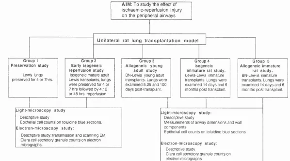
3 3 .A) Study Design. 3 3 .
Study Groups.
Operative procedure, collection and fixation of tissue.
B) Preparation of tissue for light and electron
microscopy. 3 8 .
Selection of blocks. Light microscopy (LM).
Processing and cutting of tissue for light microscopy. Staining Procedures.
Haematoxylin and Eosin stain (H+E) Elastic van-Gieson (EVG)
Transmission electron microscopy (TEM).
Processing and cutting of specimens for TEM. Staining of grids for TEM.
Electron microscopy of grids. Development of negatives. Printing from negatives.
Scanning electron microscopy (SEM).
Preparation of lung tissue for SEM. SEM of specimens.
Development of SEM negatives. Printing of SEM images.
C) Definition of terms. 4 1 .
Airway generations.
Conducting airways. Gas exchange region. Components of the airway wall. Epithelial cell types.
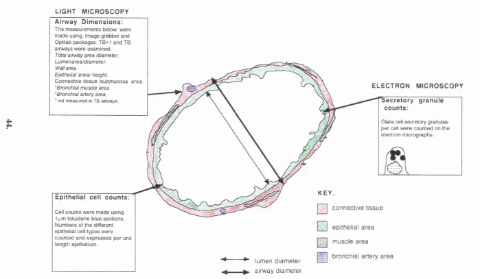
D) Quantitative analysis of airway structure
by light and electron-microscopy. 4 3 .
Analysis by light microscopy. Selection of airways.
Methods of measuring and calculating airway dimensions. Epithelial cell counts.
Analysis by electron microscopy. Selection of airways.
Counts of secretory granules in Clara cells.
E) Principles of statistical analysis. 4 5 .
Page.
II) In vitro studies on epithelial cell/fibroblast
interaction.
4 6
A) Preparation of materials. 4 6
Source and harvesting of cells. Plating of cells.
Production of fibroblast-conditioned medium (FCM). Preparation of fibroblasts on plastic.
Cooling and rewarming of fibroblasts and collection of medium.
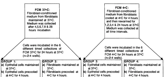
B) Experimental design. 4 8,
C) Experimental procedure. 48
Preparation of epithelial cells and fibroblasts. Incubation of epithelial cells and fibroblasts in
FCM/labelling with thymidine.
Measurement of thymidine incorporation
into cells by autoradiography.
Coating of coverslips with photographic emulsion. Development, fixation of coverslips
and counter-staining of cells. Method of counting labelled cells.
D) Statistical analysis of cell counts. 4 9.
Chapter 3: Effect of Preservation and Early Reperfusion on
the Peripheral Airways of the Isogeneic Transplanted Lung.
I) Qualitative Studies.
5 3 .Control airways.
A) Group 1 : Preservation study. 5 3 .
Appearance of the peripheral airways. Appearance of the alveolar region.
B) Group 2: Early reperfusion study after isogeneic transplantation. 5 4 .
Appearance of the peripheral airways. After 4 hours reperfusion. After 12 hours reperfusion. After 48 hours reperfusion Appearance of the alveolar region. After 4, 12 & 48 hours reperfusion.
II) Quantitative Studies.
7 7 .A) Counts of different epithelial cell types using light microscopy. 7 7 .
Total epithelial cell number. Clara cells.
Page.
B) Analysis of Clara cell secretory granules using electron-microscopy. 8 1 .
Ill) Summary of Chapter 3.
85.
Chapter 4: Long-Term Changes in Peripheral Airways After
Single Lung Transplantation (Allogeneic) in Adult Rats.
I) Qualitative Studies.
87.
Control airways.
Appearance, 5 days post-transplantation. Appearance, 25 days post-transplantation. Appearance, 100 days post-transplantation.
II) Quantitative Studies.
92.
A) Analysis of airway structure.
9 2.
Effect of age on lung dimensions and composition in normal rats. Control BN Lung.
TB+1 airways. TB airways.
Control Lewis Lung.
TB+1 and TB airways.
Effect of transplantation on airway dimensions and composition. Transplanted BN Lung.
TB+1 airways. TB airways.
Native Lewis Lung.
TB+1 airways. TB airways.
B) Counts of different epithelial cell types using light microscopy.
9 9.
Total epithelial cell number. Clara cells.
Ciliated cells. Basal cells.
C) Analysis of Clara cell secretory granules using electron-microscopy. 1 0 2 .
III) Summary of Chapter 4.
106.
Chapter 5: Long-Term Changes in Peripheral Airways
After Single-Lung Transplantation (Isogeneic & Allogeneic)
in Immature Rats.
Isogeneic transplants: Lewis-Lewis.
I) Qualitative studies.
113.
Appearance, 14 days post-transplantation. Appearance, 6 months post-transplantation.
Page.
II) Quantitative Studies.
116.
A) Analysis of airway structure. 1 1 6 .
Effect of age on airway dimensions and composition in normal Lewis rats. Control Lewis Lung.
TB+1 airways TB airways
Effect of transplantation on airway dimensions and composition. Transplanted Lewis Lung.
TB+I airways TB airways
Native Lewis Lung.
TB+1 airways. TB airways.
B) Counts of different epithelial cell types using light-microscopy. 1 2 3 .
Total epithelial cell number Clara cells
Ciliated cells Basal cells.
C) Analysis of Clara cell secretory granules using electron-microscopy. 1 2 7 .
Allogeneic transplants: BN-Lewis.
I) Qualitative studies.
130.
Appearance, 14 days post-transplantation. Appearance, 6 months post-transplantation.
II) Quantitative Studies.
133.
A) Analysis of airway structure. 1 3 3 .
Effect of age on airway dimensions and composition in normal BN rats. Control BN Lung.
TB+1 airways TB airways
Effect of transplantation on airway dimensions and composition. Transplanted BN Lung.
TB+1 airways TB airways
Native Lewis Lung.
TB+1 airways. TB airways.
B) Counts of different epithelial cell types using light-microscopy. 1 3 9 .
Total epithelial, Clara and ciliated cell number. Basal cells.
C) Analysis of Clara cell secretory granules using electron-microscopy. 1 3 9 .
P age.
Chapter 6: In-Vitro Studies of Bronchial
Epithelial Cell/Fibroblast Interactions.
Introduction.
I)
The effect of cooling and re warming on the response
of bronchial epithelial cells to fibroblast-conditioned
medium.
155.
Epithelial cell thymidine labelling in serum-free medium (SFM). The growth stimulatory effect of FCMbv^c on epithelial cells . The growth stimulatory effect of FCM^^c on epithelial cells . Comparison of the growth stimulatory effect of FCMgy^c and FCMt^c on epithelial cells maintained at 37®C. Comparison of the growth stimulatory effect of FCMgy^c and FCM4°c on cooled epithelial cells.
II)
The effect of cooling and rewarming on the
response of fibroblasts to fibroblast-conditioned
medium.
161.
Fibroblast thymidine labelling in serum-free medium (SFM). The growth stimulatory effect of FCMgy^c on fibroblasts. The growth stimulatory effect of FCM^^c on fibroblasts. Comparison of the growth stimulatory effect of FCMsv^c
and FCM4°c on fibroblasts maintained at 37®C.
Comparison of the growth stimulatory effect of FCMg^^c
and FCM4°c on cooled fibroblasts.
III)
Summary of Chapter 6.
165.
Chapter 7: Discussion.
I) Aims of study and principle findings.
169.
II) Methods used in transplantation studies.
170.
A) The model of unilateral lung transplantation in the rat. 1 7 0 .
B) Methods of investigation of lung tissue. 1 7 4 .
C) Methods of investigation using cell culture. 1 7 5 .
III) Effect of preservation and early reperfusion
on the peripheral airways.
177.
A) Preservation Injury. 1 7 7 .
B) Early Reperfusion Injury. 1 8 0 .
Page.
IV) Persistent epithelial hyperplasia following
182.
transplantation.
A) Epithelial hyperplasia in the transplanted lung. 1 8 2 .
B) Epithelial hyperplasia in the native Lewis lung. 1 8 4 .
C) Role of basal cells. 1 8 5 .
V)
Peripheral airway size and composition following
transplantation
186.
A) Peripheral airway size and composition in the transplanted lung. 1 8 7 .
B) Peripheral airway size and composition in the native Lewis lung. 1 8 8 .
VI) Possible mechanisms for peripheral airway changes
seen following transplantation.
189.
A) Epithelial cell/fibroblast interaction in the transplanted lung. 1 9 0 .
B) Immunological influences in the transplanted and native lung. 1 9 2 .
Immunological influences and epithelial hyperplasia. Immunological influences and peripheral airway size.
C) Influence of rat strain and age, 1 9 4 .
Influence of rat strain and age on epithelial cell hyperplasia.
Influence of rat strain and age on peripheral airway size.
D) Role of denervation, bronchial artery ligation, infection
and immunosupression. 1 9 5 .
VII) Relevance of findings to the development of OB
in the transplanted human lung.
197.
VIII) Future studies.
198.
IX) Conclusion.
199.
Appendices.
201.
A) Chapter 2
202.
B) Chapter 3
206.
C) Chapter 4
211.
D) Chapter 5
219.
Bibliography.
234.
Index of Figures.
Chapter 2:
Figure 2.1: Study design.
Figure 2.2: Quantitative analysis of airways.
Figure 2.3: Experimental design.
Figure 2.4: Experimental procedure to investigate the effect of fibroblast-conditioned
medium (FCM) on epithelial cells and fibroblasts.
Chapter 3:
Figure 3.1a: Light-micrograph of a bronchiole in the control Lewis lung.
Figure 3.1b: Transmission electron-micrograph of the bronchiolar epithelium in the
control Lewis lung
Figure 3.1c: Scanning electron-micrograph of the bronchiolar epithelial surface in the
control Lewis lung.
Figure 3.2a: Transmission electron-micrograph of a bronchiole in the preserved Lewis
lung.
Figure 3.2b: Transmission electron-micrograph of the intra-cellular bronchiolar
epithelial damage in the preserved Lewis lung.
Figure 3.2c: Scanning electron-micrograph of the epithelial surface of a bronchiole in
the preserved Lewis lung.
Figure 3.3a: Light-micrograph of the alveolar region of the control Lewis lung.
Figure 3.3b: Light-micrograph of the alveolar region of the preserved Lewis lung.
Figure 3.3c: Transmission electron-micrograph of the alveolar Type II cell in the
control Lewis lung.
Figure 3.3d: Transmission electron-micrograph of the alveolar Type II cell in the
preserved Lewis lung.
Figure 3.4a: Light-micrograph of the bronchiolar wall after 4 hours reperfusion in the
transplanted Lewis lung.
Figure 3.4b: Transmission electron-micrograph of the damaged bronchiolar epithelium
after 4 hours reperfusion in the transplanted Lewis lung.
Figure 3.4c: Transmission electron-micrograph of the damaged bronchiolar epithelium
after 4 hours reperfusion in the transplanted Lewis lung.
Figure 3.4d: Scanning electron-micrograph of the bronchiolar epithelial surface after 4
hours reperfusion in the transplanted Lewis lung.
Figure 3.5a: Transmission electron-micrograph of the damaged bronchiolar epithelium
after 12 hours reperfusion in the transplanted Lewis lung
Figure 3.5b: Scanning electron-micrograph of the bronchiolar epithelium surface after
12 hours reperfusion in the transplanted Lewis lung.
Figure 3.6a: Light-micrograph of a bronchiole after 48 hours reperfusion in the
transplanted Lewis lung.
Figure 3.6b: Transmission electron-micrograph of epithelial hyperplasia after 48 hours
reperfusion in the transplanted Lewis lung.
Figure 3.6c: Scanning electron-micrograph of the bronchiolar epithelial surface after
48 hours reperfusion in the transplanted Lewis lung.
Figure 3.7: Transmission electron-micrograph of the alveolar Type II cell after 48
hours reperfusion in the transplanted Lewis lung.
Figure 3.8a: Total number of cells per unit length of epithelium in the TB+1 airways.
Figure 3.8b: Total number of cells per unit length of epithelium in the TB airways.
Figure 3.9a: The number of Clara cells per unit length of epithelium in the TB+1
airways.
Figure 3.9b: The number of Clara cells per unit length of epithelium in the TB airways.
Figure 3.10a: The number of ciliated cells per unit length of epithelium in the TB+1
airways.
Figure 3.10b: The number of ciliated cells per unit length of epithelium in the TB
airways.
Figure 3.11a: The number of basal cells per unit length of epithelium in the TB+1
airways.
Figure 3.11b: The number of basal cells per unit length of epithelium in the TB
Figure 3.12a: The number of unidentifiable cells per unit length of epithelium in the
TB+1 airways.
Figure 3.12b: The number of unidentifiable cells per unit length of epithelium in the
TB airways.
Figure 3.13: The number of secretory granules per Clara cell after preservation and
reperfusion.
Chapter 4:
Figure 4.1a: Light-micrograph of a peripheral airway of the transplanted BN lung, 5
days post transplantation.
Figure 4.1b: Electron-micrograph of the peripheral airways of the transplanted BN
lung , 5 days post transplantation.
Figure 4.1c: Light micrograph of an acutely rejected peripheral airway following
allogeneic transplantation with no immunosuppression.
Figure 4 .Id: Electron-micrograph of the peripheral airways of the transplanted BN
lung , 100 days post transplantation.
Figure 4.2: TB+1 airways of the control and transplanted BN Lung.
Figure 4.3: TB airways of the control and transplanted BN Lung.
Figure 4.4: TB+1 airways of the control and native Lewis Lung.
Figure 4.5: TB airways of the control and native Lewis Lung.
Figure 4.6: Total number of cells per unit length of epithelium in the TB+1 airways.
Figure 4.7: Total number of cells per unit length of epithelium in the TB airways.
Figure 4.8: The number of Clara cells per unit length of epithelium in the TB+1
airways.
Figure 4.9: The number of Clara cells per unit length of epithelium in the TB airways.
Figure 4.10: The number of ciliated cells per unit length of epithelium in the TB+1
airways.
Figure 4.11: The number of ciliated cells per unit length of epithelium in the TB
airways.
Figure 4.12: The number of basal cells per unit length of epithelium in the TB+1
airways.
Figure 4.13: The number of basal cells per unit length of epithelium in the TB
airways.
Figure 4.14: The number of secretory granules per Clara cell in the TB+1 airways of
the transplanted BN lung.
Chapter 5:
Figure 5.1a: Electron-micrograph of the peripheral airways of the transplanted
immature Lewis lung, 14 days post transplantation.
Figure 5.1b: Electron-micrograph of the peripheral airways of the transplanted
immature Lewis lung, 6 months post transplantation.
Figure 5.2: TB+1 airways of the control and transplanted Lewis Lung.
Figure 5.3: TB airways of the control and transplanted Lewis Lung.
Figure 5.4: TB+1 airways of the control and native Lewis Lung.
Figure 5.5: TB airways of the control and native Lewis Lung.
Figure 5.6: Total epithelial cell number per unit length of epithelium in the TB+1
airways.
Figure 5.7: Total epithelial cell number per unit length of epithelium in the TB
airways.
Figure 5.8: The number of Clara cells per unit length of epithelium in the TB+1
airways.
Figure 5.9: The number of Clara cells per unit length of epithelium in the TB airways.
Figure 5.10: The number of ciliated cells per unit length of epithelium in the TB+1
airways.
Figure 5.11: The number of ciliated cells per unit length of epithelium in the TB
airways.
Figure 5.12: The number of basal cells per unit length of epithelium in the TB+1
Figure 5.13: The number of basal cells per unit length of epithelium in the TB
airways.
Figure 5.14: The number of secretory granules per Clara cell in the TB+1 airways of
the transplanted Lewis lung.
Figure 5.15a: Electron-micrograph of the peripheral airways of the transplanted
immature BN lung, 14 days post transplantation.
Figure 5.15b: Electron-micrograph of the peripheral airways of the transplanted
immature BN lung, 6 months post transplantation.
Figure 5.16: TB+1 airways of the control and transplanted BN Lung.
Figure 5.17: TB airways of the control and transplanted BN Lung.
Figure 5.18: TB+1 airways of the control and native Lewis Lung.
Figure 5.19: TB airways of the control and native Lewis Lung.
Figure 5.20: The number of basal cells per unit length of epithelium in the TB+1
airways.
Figure 5.21: The number of basal cells per unit length of epithelium in the TB
airways.
Figure 5.22: The number of secretory granules per Clara cell in the TB+1 airways of
the transplanted BN lung.
Chapter 6:
Figure 6.1a: Porcine bronchial epithelial cells 48 hours after initial isolation, stained
with AB-PAS.
Figure 6.1b: Porcine bronchial epithelial cells 7 days after initial isolation, stained with
AB-PAS.
Figure 6.1c: Cultured porcine bronchial epithelial cells labelled with tritiated
thymidine.
Figure 6.Id: Cultured porcine bronchial fibroblasts labelled with tritiated thymidine.
Figure 6.2: The growth stimulatory effect of FCMsv^c on epithelial cells either
maintained at 370C or cooled and rewarmed.
Figure 6.3: The growth stimulatory effect of FCM4®c on epithelial cells either
maintained at 370C or cooled and rewarmed.
Figure 6.4: Comparison of the percentage of labelled epithelial cells maintained at
37®C incubated in FCMg^^c and FCM^^c.
Figure 6.5: Comparison of the percentage of labelled cooled and rewarmed epithelial
cells incubated in FCMgy^c and FCM^^c.
Figure 6.6: The growth stimulatory effect of FCM sv^c on fibroblasts either
maintained at 3?0C or cooled and rewarmed.
Figure 6.7: The growth stimulatory effect of FCM4°c on fibroblasts either maintained
at 3 7 0c or cooled and rewarmed.
Figure 6.8: Comparison of the percentage of labelled fibroblasts maintained at 370C
incubated in FCMgy^c and FCM^^c.
Figure 6.9: Comparison of the percentage of labelled cooled and rewarmed fibroblasts
incubated in FCMgy^c and FCM4®c.
Index of Tables.
Chapter 2:
Table 2.1: Strain and body weight of donor, recipient and control rats.
Table 2.2: Fixed lung weights following isogeneic transplants between immature
Lewis rats (Group 4).
Table 2.3: Fixed lung weights following allogeneic transplants between immature BN
and Lewis rats (Group 5).
Chapter 3:
Table A3.1: ANOVA values of the epithelial counts of peripheral airways following
preservation and reperfusion in Lewis rats.
Table A3.2: ANOVA values for the effect of preservation and reperfusion on epithelial
cell counts in the TB+1 and TB airways.
Table A3.3: Epithelial cell counts per 100pm epithelium in the TB+1 airways of
controls and following preservation and early reperfusion.
Table A3.4: Epithelial cell counts per 100pm epithelium in the TB airways of controls
Table 4.1: Airway dimensions and component areas of the TB+1 airways in controls
and after transplantation.
Table 4.2: The airway components expressed as a percentage of the total airway area
in the TB+1 airways in controls and after transplantation.
Table 4.3: The airway components expressed as a percentage of the airway wall area
in the TB+1 airways in controls and after transplantation.
Table 4.4: Airway dimensions and component areas of the TB airways in controls and
after transplantation.
Table 4.5: The airway components expressed as a percentage of the total airway area
in the TB airways in controls and after transplantation.
Table 4.6: The airway components expressed as a percentage of the airway wall area
in the TB airways in controls and after transplantation.
Table A 4.7: ANOVA values for the effect of age and allogeneic transplantation on the
airway dimensions and component areas of TB+1 airways.
Table A4.8: ANOVA values for the effect of age and allogeneic transplantation on
airway component percentages of TB+1 airways.
Table A4.9: ANOVA values for the effect of age and allogeneic transplantation on the
airway dimensions and component areas of the TB airways.
Table A4.10: ANOVA values for the effect of age and allogeneic transplantation on
airway component percentages of TB airways.
Table A4.11: Epithelial cell counts per 100pm epithelium in the TB+1 airways of
controls and 5, 25 and 100 days post transplant.
Table A4.12: Epithelial cell counts per 100pm epithelium in the TB airways of
controls and 5, 25 and 100 days post transplant.
Table A4.13: ANOVA values for the effect of age and allogeneic transplantation on
epithelial cell counts in the TB+1 and TB airways.
Chapter 5:
Table 5.1: Airway dimensions and component areas of the TB+1 airways in controls
and after transplantation.
Table 5.2: The airway components expressed as a percentage of the total airway area
in the TB+1 airways in controls and after transplantation.
Table 5.3: The airway components expressed as a percentage of the airway wall area
in the TB+1 airways in controls and after transplantation.
Table 5.4: Airway dimensions and component areas of the TB airways in controls and
after transplantation.
Table 5.5: The airway components expressed as a percentage of the total airway area
in the TB airways in controls and after transplantation.
Table 5.6: The airway components expressed as a percentage of the airway wall area
in the TB airways in controls and after transplantation.
Table A5.7: ANOVA values for the effect of age and isogeneic transplantation on
airway dimensions and component areas of the TB+1 airways.
Table A5.8: ANOVA values for the effect of age and isogeneic transplantation on the
proportions of the TB+1 airways.
Table A5.9: ANOVA values for the effect of age and isogeneic transplantation on the
airway dimensions and component areas of TB airways.
Table A5.10: ANOVA values for the effect of age and isogeneic transplantation on the
airway component percentages of the TB airways.
Table A5.11: Epithelial cell counts per lOOjxm epithelium in the TB+1 airways of
controls and 14 days and 6 months post transplant.
Table A5.12: Epithelial cell counts per 100pm epithelium in the TB airways of
controls and 14 days and 6 months post transplant.
Table A5.13: ANOVA values for the effect of age and isogeneic transplantation on
epithelial cell types in the TB+1 and TB airways.
Table 5.14: Airway dimensions and component areas of the TB+1 airways in controls
and after transplantation.
Table 5.15: The airway components expressed as a percentage of the total airway area
Table 5.16: The airway components expressed as a percentage of the airway wall area
in the TB+1 airways in controls and after transplantation.
Table 5.17: Airway dimensions and component areas of the TB airways in controls
and after transplantation.
Table 5.18: The airway components expressed as a percentage of the total airway area
in the TB airways in controls and after transplantation.
Table 5.19: The airway components expressed as a percentage of the airway wall area
in the TB airways in controls and after transplantation.
Table A5.20: ANOVA values for the effect of age and allogeneic transplantation on the
airway dimensions and component areas of the TB+1 airways.
Table A5.21: ANOVA values for the effect of age and allogeneic transplantation in the
airway component percentages of the TB+1 airways.
Table A5.22: ANOVA values for the effect of age and allogeneic transplantation on the
airway dimensions and component areas of TB airways.
Table A5.23: ANOVA values for the effect of age and allogeneic transplantation on
airway component percentages on the TB airways.
Table A5.24: Epithelial cell counts per lOOpm epithelium in the TB+1 airways of
controls and 14 days and 6 months post transplant.
Table A5.25: Epithelial cell counts per lOOpm epithelium in the TB airways of
controls and 14 days and 6 months post transplant.
Table A5.26: ANOVA values for the effect of age and allogeneic transplantation on
epithelial cell counts in the TB+1 and TB airways.
Chapter 6:
Table 6.1: The effect of fibroblast conditioned medium from fibroblasts maintained at
37°C and from cooled and rewarmed fibroblasts on epithelial cells kept at either 37°C
or 4°C and rewarmed.
Table 6.2: The effect of fibroblast conditioned medium from fibroblasts maintained at
37°C and from cooled and rewarmed fibroblasts on fibroblasts kept at either 37®C or
4°C and rewarmed.
I. Pulmonary Transplantation.
A) History of pulmonary transplantation.
Pulmonary transplantation, either heart-lung, single lung or double lung has become a
therapeutic option for patients with end-stage pulmonary and cardiopulmonary
diseases. The first recorded interest in lung transplantation dates back to 1907 when
Charles Guthrie performed a heterotopic heart-lung transplant between a kitten and an
adult cat.^^ Demikhov, a Russian physiologist performed canine heart-lung transplants
in 1947. However, it was 15 years before his work was translated for the Western
w o r l d . M e t r a s in France described the technique of canine pulm onary
allotransplantation in 1950.
The first recorded attempt at lung transplantation in humans was performed by Hardy
and workers at the University of Missisipi in 1963.^^ The patient survived 18 days and
died of renal failure. The first reported heart-lung transplant was by Cooley and
workers in 1968.^^ Despite many attempts at isolated lung transplantation no long
term clinical success was achieved for some time.^^ Patients survived the post
operative period only to succumb to airway complications which ultimately lead to
their death. Immunosuppressive regimens at this time used high dose corticosteroids.
Animal experiments at the University of Toronto demonstrated that methylprednisone
hindered skin as well as bronchial anastomosis h e a l i n g . A new immunosuppressive
agent Cyclosporine (first known as Cyclosporine A) was discovered by Borel and
workers, in 1976. This powerful immunosuppressive agent inhibits T-lymphocyte
helper cells but does not impair wound-healing.^^ This meant that high dose steroids
could be avoided in the early post-operative period and thus wound healing was
improved.
Using this drug in combination with the newly developed "en bloc" heart-lung
transplant technique, Reitz and co-workers at Stanford were successful in the first
long-term survival of rhesus monkeys following heart-lung transplantation. Using
the "en bloc" method resulted in good healing of the tracheal anastomosis. This was
followed in 1981 by the first successful heart-lung transplant in a patient with end-
stage primary pulmonary hypertension.
Initially, most heart-lung recipients had advanced pulmonary vascular disease caused
by the Eisenmenger syndrome or primary pulmonary h y p e r t e n s i o n , b u t an
increasing number of patients with end-stage lung disease have now been successfully
treated by heart-lung transplantation.^^’ ^^^42
B) Types of pulmonary transplantation.
It was not until 1983 that the first successful unilateral lung transplantation was carried
out, in Toronto. Widespread use of heart-lung transplantation is limited by the
availability of suitable donors. Single-lung transplantation has several advantages. The
donor heart and lungs can be used for different recipients, the risk of intra-operative
and post-operative bleeding is lower since the surgical procedure is easier and it is
possible to avoid using cardiopulmonary b y p a s s . T h e disadvantages of single-lung
transplantation include complications related to the remaining diseased lung.^^ The
most im portant difference between single or double lung and heart-lung
transplantation is that the coronary to bronchial collaterals are not disrupted by heart-
lung transplantation and therefore the healing of the tracheal anastomoses is more
reliable. The problem of ischaemic disruption of the bronchial anastomosis in
unilateral lung transplantation was eased by the withdrawal of corticosteroids in the
early post-operative period and the omental pedicle being wrapped around the
bronchial anastomosis^^® to aid healing and bronchial revascularisation ^^^127
recent work by the Stanford group has disproved the need for the omental pedicle
wrap, instead telescoping of the bronchi and continuous sutures have been
single-lung as well as double-lung transplantation has now been used successfully for
transplant patients with pulmonary vascular disease.
The first successful "en-bloc" double-lung transplant in humans was carried out by the
Toronto Lung Transplant Group in 1988 in a woman with end-stage emphysema
secondary to alpha-1-antitrypsin d e f i c i e n c y . De s p i t e initial encouraging results the
"en-bloc" double-lung transplant procedure was plagued with problems related to
tracheal healing. To address the issue of ischaemic airway complications Noirclerc
and co-workers in Marseilles introduced the technique of bilateral bronchial
anastomoses. The next step was the so-called "bi-lateral lung transplant" described
by Pasque and co-workers from Washington University. The advantage of this latter
technique which involves sequential transplantation of the lungs, is that
cardiopulmonary bypass is not necessary and bleeding complications are substantially
ameliorated.
As the procedure of lung transplantation has become an accepted form of treatment for
patients with end-stage pulmonary parenchymal and vascular disease, lack of donor
organs has become the major limitation. This shortage is even greater in the paediatric
p o p u l a t i o n . Th e r e f o r e grafts of reduced size, lobes rather than whole lungs, have
been used in selected patients.^^ In initial animal experiments the smaller lung graft
has provided adequate pulmonary function. In clinical work, lobes have been
taken from cadavers and more controversially, from a living-related donor.
Bilateral lobar transplantation for patients with cystic fibrosis has involved lobes
donated from 2 relatives, the right lower lobe from one and the left lower lobe from
another. The immunological advantages of related-donor organs has not yet been
fully evaluated and so the recipients still receive triple-immunosupressive therapy. At
present, lobar transplantation seems to be best suited for children and small adults. The
main concern of this type of pulmonary transplantation is uncertainty about how the
mature lobe is going to cope as the young recipient grows. This question remains
unanswered in humans, although at Stanford a 12 year old girl with bronchopulmonary
dysplasia who received an adult upper lobe was still alive three years later, with no
physical handicap and increased lung function along with somatic growth.
Experimental work in rats has shown that the immature lung continues growing after
transplantation, the alveoli and airways increasing in size after single-lung
transplantation.^^ However, other studies have demonstrated abnormal vascular and
airway function in transplanted immature porcine lungs, possibly due to chronic
denervation or abnormal g r o w t h . W h e n the airway function of transplanted
porcine immature whole lungs was compared with that of transplanted mature lobes,
the mature lobes showed superior long-term airway function. It appears that mature
lobar transplants in immature animal recipients are capable of supporting life,^’^^’^^
although functional compensatory growth of these transplanted mature lobes may not
be adequate^®^’^®"^ and this may limit clinical effectiveness. At worst, mature lobar
transplants could be used as a bridge until the child is old enough to accept a larger
adult transplant.
C) Donor lung.
Lung preservation.
Good preservation of the donor lung is essential for early graft function and thus
survival. Adequate and prolonged preservation prior to transplantation is one of the
major problems of lung transplantation. Optimising the technique is complicated, not
only due to the delicate alveolar-capillary network of the lung but because our
knowledge of the pathogenic mechanisms involved in pulmonary ischaem ia-
reperfusion injury is incomplete. This has resulted in optimal preservation of the lung
lagging behind that of other o r g a n s . T h e important factors to be considered include
the method of preserving the lung, the type of preservation solution, the time and
temperature of preservation, whether to store the lung inflated or deflated and donor
pre-treatment. All these issues have been addressed in animal models. Clinical
methods used for lung preservation have included "immediate transplantation" (donor
artery with cold preservation fluid followed by hypothermic storage with and without
varying degrees of inflation, donor-core cooling on cardiopulmonary bypass, and
autoperfused heart-lung preparations at normothermia.^^ Currently the most widely
used methods involve some form of hypothermic pulmonary flush. Donor-core cooling
is used at some transplantation centres and has been used in over 50% of the world
experience in heart-lung transplantation.
Despite the Toronto lung transplantation group making a point in their early
experience of keeping the allograft deflated during procurement,^^ in recent years lung
inflation during hypothermic storage has become an issue in experimental lung
preservation models. Locke and workers showed that preservation with lung
inflation is superior to preservation after absorption atelectasis of the lung. The issue
as to whether 100% oxygen, room air or 100% nitrogen should be used for this
technique is still controversial and reports are conflicting.
Preservation fluids.
A wide variety of perfusates have been used in experimental lung preservation.^"^
Marshalls solution was described in 1976 by Ross and workers and was first used
for flush perfusing kidneys for subsequent transplantation. Using Marshalls, which is a
crystalloid fluid, successful kidney preservation was achieved for 72 hours.
Marshalls solution has never been used clinically for lung preservation. It has however
been used experimentally.
Modified EuroCollins kidney solution remains the most frequently used solution in
clinical lung t r a n s p l a n t a t i o n . I n 1986 Starkey and workers reported a successful 6
hour preservation of primate heart-lung allografts using cold modified Euro-Collins
solution to flush the pulmonary arteries followed by static hypothermic s t o r a g e . A t
this time it was realised that using a larger volume of perfusate flushed through the
pulmonary arteries over several minutes at a low pressure resulted in more uniform
cooling of the lungs than the lower flush volumes previously used. These advances
in pulmonary preservation provided sufficient protection to enable routine distant
donor procurement. Baldwin and workers reported the successful clinical application
of this technique.^ Since that time, modified Euro-Collins solution has become the
standard pulmonary flush solution for heart-lung and lung transplantation in most
centres. At the same time that Starkey was developing a preservation procedure using
Euro-Collins solution, the Papworth group were using an extra-cellular blood-based
solution containing prostacyclin for distant heart-lung procurement.^^ This solution
gave satisfactory lung preservation for up to 4 hours.
Intra-cellular crystalloid solutions have proved to be successful for lung preservation
and so in this study lungs were flush perfused and then stored in Marshalls solution at
4°C in the inflated state. The inability to preserve the lung safely for more than 4 to 6
hours still represents a major impediment to pulmonary transplantation. Better
alternatives to Euro-Collins solution have been sought. Euro-Collins solution may
cause pulmonary vasoconstriction due to its high potassium. Alternatives include an
extra-cellular, low potassium dextran flush solution which improved lung function
after 12 hours of preservation in the canine single-lung transplant, and in the rabbit
isolated lung model stored for 18 hours.
Further studies have not confirmed that the low potassium dextrin solution is superior
to modified Euro-Collins solution used in conjunction with prostaglandin (vasodilator)
treatment. University of Wisconsin solution is a colloidal solution which has
significantly prolonged the preservation of liver, kidney and pancreatic grafts.
UW solution is now used as a preservation solution in clinical practice.^^ An improved
UW solution has been developed with a low potassium content which results in
improved gas exchange and less oedema during reperfusion in the isolated rabbit lung
There is evidence that oxygenated fluorocarbon solution can be used as preservation
fluid for lungs. These solutions have been reported to enhance oxygenation, and to
improve perfusion and function of ischaemic myocardial t i s s u e . A study by Lehtola
and co-workers demonstrated better functional and morphologic preservation in the
porcine lung with an oxygenated fluorocarbon solution (FC-43) compared with
modified Euro-Collins solution.
Storage temperature.
Storage temperature is an important factor in lung preservation.^"^ Hypothermia
prolongs lung preservation and 4®C has been thought to be an acceptable
temperature^"^ and was used in the present preservation study. Some studies have
suggested that lO^C appears to be the optimal temperature for the long term
preservation of the lung.^^’^^^’^^® The traditional technique of lung storage after
procurement has been immersion in cold saline or iced slush. The problems with this
method are uneven cooling of the lung due to its buoyancy and portions of the graft
reaching a temperature below freezing point in iced slush, which results in cryogenic
lung i n j u r y . S t u d i e s into topical lung cooling by cold compressed air seemed to
suggest better oxygen transfer and less extra-vascular lung water than in grafts
immersed in an iced slush bath.^®^ More research into this type of storage is needed
before it can be applied clinically.
Structural studies of preservation and reperfusion injury.
Detailed structural and ultra-structural studies on the effect of preservation and
reperfusion on the lung are sparse and tend to concentrate on the blood vessels and the
blood-gas barrier. Rat studies have shown that during preservation of the donor lung,
capillary morphology changes r a p i d l y . B o t h endothelial cells and type 1
pneumocytes become thinner. Blebbing of endothelial cells can occur. Peri-capillary
oedema develops which involves the blood-gas barrier and basement membrane
thickness increases. This damage resolves on reperfusion. A study by Lehtola and
workers compared porcine lung tissue preservation with modified Euro-Collins and
fluorocarbon solutions. The study, using light microscopy, showed that preservation
induced dilation of the perivascular lymphatics and pleural oedema, while reperfusion
was followed by intra-alveolar bleeding, vascular congestion and a pleural
inflammatory reaction. These alterations were more pronounced in the Euro-Collins
group than in the fluorocarbon group. By electron-microscopy, the general appearance
was of well preserved lung tissue in both groups, although moderate oedema and
mitochondrial damage occurred in both groups. After reperfusion, the appearance of
the alveolar epithelial cells was significantly better following fluorocarbon rather than
Euro-collins preservation. The alveolar epithelial cells were slightly swollen and the
alveolar septae were also thickened. In poorly preserved areas of lungs in both groups,
alveoli totally denuded of epithelial cells were seen. After reperfusion, the alveolar
walls became more swollen and there was reduction and deformation of alveolar
spaces. Intra-alveolar fibrin nets, blood corpuscles, exfoliated epithelial cells,
desquamated or totally occluded alveoli and thrombosed vessels could be seen in
poorly preserved areas. In conclusion, the general appearance was that of well
preserved lung structures in both groups but preservation was thought to be better in
lungs preserved in fluorocarbon.
A study by Hidalgo and co-workers examined the morphological changes in rat single
lung isografts after 24 and 48 hours preservation in modified University of Wisconsin
solution which either a) mimicked the intra-cellular medium, high potassium and low
sodium or b) mimicked the extra-cellular medium, low potassium and high sodium.^^
Female inbred rats were used and lungs were examined by light and electron-
microscopy. Four weeks after transplantation, both lungs stored in a) and b) for 24
hours had the general appearance of well preserved pulmonary tissue although some
lungs presented areas of scarring, limited principally to the apical part of the lobes.
Smaller perivascular fibrotic areas were found but neither oedema nor haemorrhage
The structure of the lungs which had been stored for 48 hours in either solution were
considerably different from normal 4 weeks later. The lungs presented fibrotic
organisation with limited chronic inflammation of lymphocytes and plasma cells.
Small foci of scarring with focal mild inflammation in the pleura were observed.
Macrophages full of haemosiderin were occasionally seen in unscarred parenchymal
tissue. They concluded that extended preservation of the lungs was possible but that
the concentration of potassium and sodium in the preservation solution did not have a
significant impact in the long term.
Structural studies of lung preservation injury are obviously more scarce in human
transplantation. However, Fehrenbach and workers studied the preserved contra-lateral
donor lung in clinical single-lung transplantation.^^ This study used Euro-Collins as a
preservation solution, and good to excellent preservation of the lung tissue was
observed using light microscopy. Transmission electron microscopy however,
revealed some changes in the blood-gas barrier. In places, the capillary endothelium
showed small apical vacuoles, tentacle-like protrusions and double-membrane-bound
blisters. Most of the cells of the alveolar epithelium showed a normal appearance, but
swelling of both type I and II pneumonocytes was observed. The surfactant storing
multi-lamellar bodies were normal in appearance.
D) Reimplantation response.
The reimplantation response has been defined as "the morphologic, roentgenographic
and functional changes that occur in a lung transplant in the early post-operative
period as a result of surgical trauma, ischaemia, denervation, lymphatic interruption
and other injurious processes (exclusive of rejection)." Functionally, the pulmonary
reimplantation response impairs ventilation-perfusion relationships in the transplanted
lung.^^^ Siegelmann and w o r k e r s s h o w e d that the pulmonary reimplantation
response consists of alveolar oedema which is evident on histologic examination and
alveolar infiltrates which could be seen on chest roentgenograms. In mongrel dogs
these changes remained for three days after autotransplantation and then regressed
over one to three weeks. Radiographically, these changes were manifested by perihilar
air space disease with air bronchograms. These changes were maximal on the third
post-operative day and cleared between days 7 and 21 in dogs/^^ but persisted for as
long as 4-6 weeks in b a b o o n s . B i s h o p and colleagues have also shown that canine
unilateral lung ischaemia using a ballon occluder followed by reperfusion produces bi
lateral lung injury which is greatest in the ischaemic lung.^^ Histologically, marked
oedema and inflammatory infiltrates were observed in the reperfused lung using light
microscopy and to a lesser extent in the native lung. Electron microscopy
demonstrated lysis of both capillary endothelial and alveolar epithelial cells bilaterally
with the frequency of cell injury greater on the reperfused side. Since that work, a
study by Bryan and workers showed that both right and left lungs were affected by the
pulmonary reimplantation response following canine left lung allografts. Prop has
studied the reimplantation response in isogeneically transplanted rat lungs. On the day
of transplantation, light microscopy showed interstitial oedema throughout the lung,
mainly along the vessels and to a lesser degree along the bronchi. Foci of alveolar
oedema and haemorrhages were scattered throughout the lung. One day post
transplantation, the oedema around vessels and bronchi was still present but the
alveolar oedema now involved larger areas. Erythrocytes were present in many
alveolar spaces but not to an extent that indicated gross bleeding. On the fourth day
after transplantation these abnormalities were disappearing. From day seven the
transplanted lung looked almost normal. The native right lungs were normal except for
some perivascular oedema and alveolar haemorrhages during the first days after
transplantation.
Clinically, the reimplantation response is an early transient process which varies in its
manifestation on x-rays from a subtle perihilar haze to dense consolidation with air
bronchograms.^® These findings are non-specific and can be mistaken for those of
Herman and co -w o rk ersen co u n tered the reimplantation response in 13 patients.
Herman also observed^^ that the process was milder in patients who underwent heart-
lung transplantation than in those who underwent single-lung transplantation.
O'Donovan also showed that the re-implantation response was more impressive
radiographically in patients who received single-lung transplants for primary
pulmonary hypertension than in those who underwent lung transplantation for other
conditions.
Greater understanding of the reimplantation response and more appropriate pre- and
post- transplantation treatment will hopefully lead to superior early pulmonary
function and morphology in the future. The cause of early reperfusion lung damage is
unclear but is thought to be as a result of leukocyte and platelet activation, oxygen free
radical formation, the complement cascade, generation of inflammatory mediators and
arachidonic acid metabolites.^^
Platelet-activating factor (FAF) is synthesised and released by platelets, white blood
cells, macrophages and endothelial cells. FAF binds to platelets, causing activation
and aggregation. Cytotoxic oxygen-free radicals are produced after both warm
and cold lung ischaemia. Oxygen free radicals may originate from several sources
during ischaemia including activated neutrophils, dissociation of the intra-
mitochondrial electron-transport chain and the xanthine oxidase reaction."^^’^^^’^^^
E) Acute lung rejection.
The studies of Gibson and Medawar in the 1940s showed that rejection of skin grafts
had features of an inmiune response. Transplanted organs contain antigen foreign to
the organ recipient: human lymphocyte antigen (HLA); minor histocompatability
complexes and tissue-specific antigens. Donor antigen is recognised directly and
indirectly. Indirect recognition occurs by processed donor antigen being presented by
the host antigen-presenting cells (AFC) bound to self-major histocompatability
complex (MHC) molecules. Direct recognition occurs when the donor antigen is
recognised by the T-cell receptor (TCR) of the host's lymphocytes without the
intercession of APC.
HLA class II donor antigens are recognised by T helper/inducer cells (CD4 bearing
lymphocytes). HLA class I donor antigens are recognised by T suppressor/cytotoxic
cells (CDS bearing lymphocytes). Dendritic cells within the transplanted organ
constitutively bear HLA I and II class molecules and may function in antigen
presentation. Endothelial and epithelial cells within the transplanted organ can be
induced to express class II molecules under stress or cytokine exposure and may
therefore participate in antigen presentation.
Acute rejection of the lung is manifest on biopsy by a lymphocytic infiltrate which is
mainly perivascular but can involve other areas of the lung. The details of the
histopathological stages have been well described in the rat and human tissue
obtained at open lung biopsy or on trans-bronchial biopsy has shown similar
features. Using isolated rat lung allografts, the same experimental model as that
used in the present study, Prop described four phases of unmodified acute rejection.
In the latent phase, there is no evidence of acute rejection (immune response)
although a reimplantation response may well be seen. This is followed by the vascular
phase, characterised by the presence of first, perivenous and then peribronchial and
periarterial infiltrates of lymphocytes. Bronchus-associated lymphoid tissue (BALT)
also shows an increase in immunologic activity. The alveolar phase is defined by the
appearance of lymphocytes and increased numbers of macrophages in the alveolar
walls and spaces. Originally Prop described the alveolar phase ending with the
appearance of intra-alveolar oedema. This description defined the onset of the
destructive phase. However, as intra-alveolar oedema has been seen prior to
parenchymal necrosis in Prop's combined heart-lung transplantation rat study and
(oedematous) phase. The term destructive phase, is now reserved for actual alveolar
wall necrosis and, if it occurs, intra-alveolar haemorrhage.
Acute lung rejection may be an important factor in the subsequent development of
chronic rejection and obliterative b ro n ch io litis^T h e incidence and severity of acute
rejection episodes may make the transplanted lung more vulnerable to chronic disease
later on, possibly by priming lymphocytes. Much of our knowledge of the pathology
of lung transplantation comes from studying biopsy material. In 1990, the lung
rejection study group of the Registry of the International Study of Heart and Lung
Transplantation proposed a classification of lung rejection.^^"* This means that data can
be compared from different centres, knowledge shared and improvements made.
F) Chronic rejection of the lung.
Chronic rejection of the lungs with its principle features of obliterative bronchiolitis
(OB) and vascular occlusive disease occurs in 20-50% of adult lung transplant patients
who have survived the first year.^^’^^^ OB following adult pulmonary transplantation
was first reported by Burke and workers in 1984^^ and remains the single most
important complication in long term survivors of heart-lung and lung transplantation.
OB is also the most significant problem after heart-lung transplantation in children.
Symptoms of OB are those of obstructive lung disease and the radiographs reveal
predominantly basal honey-combed l e s i o n s . T h e flow-volume curve becomes curvi
linear, with the forced expiratory volume (FEV i) value falling by a greater degree than
the vital capacity (VC).^^ Diffuse bronchomalacia also occurs frequently after
pulmonary transplantation and worsens functional airflow obstruction, aiding decline.
Chronic lung rejection is characterised histologically by OB with variable pulmonary
vascular disease. It contrasts with chronic rejection of other transplanted organs in
which occlusive vascular disease leads to fibrosis and organ failure.^^’^^^
Pathology of obliterative bronchiolitis.
OB in humans develops through a sequence of epithelial injury that is initiated by an
active cellular phase of lymphocytic bronchiolitis with ulceration and denudation of
the mucosa. This may result in luminal ingrowth from the submucosa with
organisation of intralumenal plaques and the formation of a polyploid massive
granulation tissue (a Masson body).^^ A submucosal infiltration of lymphocytes and
histiocytes is often observed. Eccentric submucosal scars may form with luminal re-
epithelialisation and luminal narrowing, or the entire airway wall may become
completely replaced by dense scar tissue when the airway is identified only by its
location next to a pulmonary artery.
Many variations of bronchiolitis with subsequent scarring and obliteration are seen
during the course of the disease. The net result is bronchiectatic widening of the
peripheral bronchioli and mucous plugging. ^ ^ Diffuse interstitial fibrosis around the
damaged bronchioli can also be seen.^^’^^^ Both the arteries and the veins of the
pulmonary vasculature may show an accelerated form of arteriosclerosis consisting of
concentric intimai proliferation, often with an intact internal elastica. ^ ^ ^ ^ 8 2
Pathogenesis of obliterative bronchiolitis.
Suggested aetiologies for OB include rejection, infection, bronchial artery ligation and
denervation. Presently, it is thought that the incidence and severity of episodes of
acute rejection and infection, especially CMV infections are the main determinants of
the development of
Rejection: Scott and co-workers noted that chronic lung rejection is a likely outcome
in patients with early poorly controlled severe rejection. Yousem and workers in
1991, reported that the intensity and persistence of early acute rejection episodes
prime lymphocytes for subsequent respiratory epithelial injury and airway fibrosis late
in the post-operative period.^^^
Many immunological studies have been carried out in an attempt to discover whether
OB is a manifestation of chronic rejection. Both Uyama^^"^ and Sakiyama^^^ have
shown that late airway changes in long-term lung allografts are immunologically
mediated. Romanik and workers in 1990 showed that expression of Class II antigens
on the bronchial epithelium in rats is induced during early rejection and that this could
be suppressed by Cyclosporine t r e a t m e n t . C l a s s II positive epithelium might serve
as a rejection t a r g e t a n d indeed, heart-lung transplantation patients show enhanced
expression of certain class II MHC antigens on airway epithelial c e l l s . H o w e v e r this
phenomenon may not be specific to transplanted organs.^ Both Yousem and co-
workers^^^ and Uyama^^^’^^^ reported a significant increase in dentritic cells around
the airways in lung allografts. Dendritic cells are antigen-presenting cells and donor
dendritic cells are thought to play a role in acute rejection. Recipient dendritic cells
can also present graft antigens and it has been suggested that this might stimulate T
helper cells to release cytokines and thus cause local tissue damage by inducing a
vicious circle of increased MHC expression and attraction of dendritic cells, leading to
recruitment and activation of lymphocytes, which leads to relentless tissue damage and
The lymphocyte subsets associated with OB have been studied. Holland and workers
used immunohistochemistry to illustrate an increased presence of CD8-f- cells as
compared with CD4+ cells in the lung tissue and BAL. The CD8+ cells were
predominantly located in the peri-bronchial, peri-vascular and interstitial areas of the
tissue. A detailed study of the phenotypic profile of blood lymphocytes during OB has
also been d esc rib e d .H o w e v e r, it must be noted that lymphocyte profiles may be
different in the actual graft compared with those in the blood.
Infection: An experimental study in rats by Winter^^^ showed that a respiratory viral
infection aggravates the airway damage in rat lung allografts with chronic rejection,
suggesting a synergistic role for chronic rejection and infection in OB development. A
survey of 27 patients by the Pittsburgh group has correlated CMV post-operative
infections with an increased risk of developing chronic rejection.
Infectious complications, without necessarily involving rejection are an important
cause of morbidity and m o r t a l i t y . A n incidence of infection as high as 86% was
reported in two early series and 75% of all deaths have been related to infection in
the overall Pittsburgh experience.^^
In addition to immunosuppression, the tendency to develop infections is probably
associated with early post-operative ischaemic injury, impaired pulmonary drainage
and interrupted lymphatic drainage. The pathogens in heart-lung and lung
transplantation infection can be bacterial, viral, protozoal or f u n g a l . T h e transplanted
lung is particularly sensitive to bacterial infection and a prevalence of bacterial
pneumonia of greater than 60% has been reported.^^ CMV infection which frequently
occurs during the first months after surgery, may lead to a high morbidity and
mortality in lung allograft recipients. Patients with biopsy or cytology-proven
CMV pneumonia have an increased prevalence of chronic allograft rejection,
demonstrable by transbronchial biopsy, and a much worse overall s u r v i v a l . O t h e r
important herpes virus infections following pulmonary transplantation are herpes
simplex virus (HSV) and Epstein-Barr virus (EBV).
Bronchial artery ligation: Evidence for the role of bronchial artery ligation in the
pathogenesis of OB is less clear than that of infection. Following pulmonary
transplantation, one of the major sources of early morbidity and mortality is the
from bronchopulmonary collaterals in the initial post-operative period. Closset and co-
workers investigated the effect of bronchial artery severing on ischaemia of airways
and subsequent development of OB.^^ They saw no significant ischaemic lesions
occurring during reperfusion of the bronchiolar vascular bed and concluded that even
if ischemia does occur it is not a significant factor in OB. However, other studies have
shown that reimplantation of the bronchial artery results in significant improvement of
graft bronchial blood flow^ although restoration of perfusion to normal levels was not
achieved. It was suggested that this may be due to a defect in the microcirculation as a
result of endothelial damage on ischemia-reperfusion injury, as the patency of the
bronchial artery was confirmed by angiography. Bronchial revascularisation by graft
in pulmonary transplantation is being used in human patients and is associated with
improved airway healing.
Denervation: Heart-lung and single and double lung (singly or en bloc ) all result in
total extrinsic denervation of the transplanted organs and the long term effects of this
phenonenom are unclear. Denervation was not thought to be the cause of changes in
growth in the immature allogeneic and isogeneic transplanted rat lung. In this study,
these immature lungs grew with an increase in alveolar number, airway diameter and
total lung volume compared to control lungs and was thought to have a humoral basis.
Auto-transplantation of single porcine lobes has been shown to lead to small airway
flow obstruction in the absence of immune rejection and this was thought to be due
to denervation, rather than immunosupression or rejection in the immature pig lobe.^^
However again, further work by the same investigators showed transplantation of
the m ature porcine lobe did not result in abnormally small airways. Thus, they
concluded that abnormally small airways of the transplanted or reimplanted immature
porcine lung were likely to be due to impaired airway development and not to
bronchoconstriction caused by denervation.