Vibrio cholerae
at the Intersection of Immunity and the
Microbiome
Ana A. Weil,a,b*Rachel L. Becker,aJason B. Harrisa,c,d
aDivision of Infectious Diseases, Massachusetts General Hospital, Boston, Massachusetts, USA
bDepartment of Medicine, Harvard Medical School, Boston, Massachusetts, USA
cDepartment of Pediatrics, Harvard Medical School, Boston, Massachusetts, USA
dDivision of Pediatric Global Health, Massachusetts General Hospital, Boston, Massachusetts, USA
ABSTRACT Vibrio choleraeis a noninvasive pathogen that colonizes the small intes-tine and produces cholera toxin, causing severe secretory diarrhea. Cholera results in long lasting immunity, and recent studies have improved our understanding of the antigenic repertoire of V. cholerae. Interactions between the host, V. cholerae, and the intestinal microbiome are now recognized as factors which impact susceptibility to cholera and the ability to mount a successful immune response to vaccination. Here, we review recent data and corresponding models to describe immune re-sponses to V. cholerae infection and explain how the host microbiome may impact the pathogenesis of V. cholerae. In the ongoing battle against cholera, the intestinal microbiome represents a frontier for new approaches to intervention and preven-tion.
KEYWORDS Vibrio cholerae, cholera, immune response, innate immunity, microbiome, vaccine
V
ibrio choleraeis a highly motile, salt-tolerant bacterium. It was identified as thecause of cholera by Pacini in 1854, which was later confirmed in 1884 by Koch (1, 2). Further understanding of the pathogenesis of cholera stalled until the 1950s, when Nath De demonstrated the effects of cholera toxin (CT) by injecting rabbit ileal loops with cell extracts from culturedV. cholerae(3). While our understanding of the regu-lation and mechanisms of action of CT have advanced considerably, there are still significant gaps in our understanding of the pathogenesis of cholera. In this review, we highlight two such areas. First, how is protective immunity againstV. cholerae gener-ated? Second, how does the intestinal microbiome impact host-pathogen interactions
inV. choleraepathogenesis and immunity?
ETIOLOGY
V. cholerae is a facultative pathogen. It persists in aquatic reservoirs and forms
biofilms in association with plankton (4). Although environmentalV. choleraeis diverse, cholera is caused by a restricted subset of pandemicV. choleraestrains which cyclically emerge and replace their precursors. The current, seventh pandemic V. cholerae
biotype El Tor (7PET) lineage was first recognized as a cause of widespread cholera in 1961 and, within two decades, replaced the previous sixth pandemic classical biotype globally (5). The emergence of new dominant lineages is also apparent within the seventh pandemic, and genotyping of 7PET isolates reveals three distinct but overlap-ping waves of transmission, each associated with horizontal gene acquisitions (6). The SXT/R391 antibiotic resistance element was acquired during the second wave, and a new CT-encoding bacteriophage, similar to that associated with the previous sixth pandemic classical biotype, replaced the toxin encoding region during the third wave (6).
CitationWeil AA, Becker RL, Harris JB. 2019.
Vibrio choleraeat the intersection of immunity and the microbiome. mSphere 4:e00597-19.
https://doi.org/10.1128/mSphere.00597-19.
EditorChristopher J. Papasian, University of Missouri-Kansas City School of Medicine
Copyright© 2019 Weil et al. This is an open-access article distributed under the terms of theCreative Commons Attribution 4.0 International license.
Address correspondence to Jason B. Harris, [email protected].
*Present address: Ana A. Weil, Department of Medicine, University of Washington, Seattle, Washington, USA.
Vibrio cholerae at the intersection of immunity and the microbiome. @anaweilmd
Published
Host-Microbe Biology
27 November 2019
on September 8, 2020 by guest
http://msphere.asm.org/
More than 200 serogroups of environmental V. cholerae are defined by their O-antigen structure (7), but only serogroup O1 is associated with pandemic cholera (8). Other serogroups have caused sporadic cases or limited outbreaks. A unique exception thus far is theV. choleraeO139 serogroup, which caused epidemic cholera from 1992 to 2002 (9).V. choleraeO139 resulted from a single horizontal gene exchange of therfb
(O-antigen encoding) locus in the circulating 7PET O1 strain (10). After this serendipi-tous recombination event, it is possible that an increase in prevalence ofV. cholerae
O139 was then facilitated by the niche created by widespread existing immunity to the O1 serogroup and a corresponding lack of immunity to the emergent O139 serogroup. This is conceivable given thatV. choleraeO1 and O139 infections confer homologous immunity (against reinfection with the same serogroup) but not heterologous immu-nity (against infection with the other serogroup) (11). However, because other non-O1 strains do not typically cause cholera epidemics, it is likely that unknown constraints prevent their more frequent emergence.
V. cholerae serogroup O1 is divided into two serotypes, Inaba and Ogawa. The
difference between serotypes is the absence of a single methyl group in the terminal perosamine of the O-polysaccharide in Inaba, an alteration acquired through a lack of function mutation in the wbeTmethyltransferase (11). In areas of endemicity, either serotype may predominate for years (12). The prolonged serotype cycles can be explained by a high, but incomplete level of cross-protection between serotypes (13). This model also explains why there is a transient increase in the average age of patients with cholera that coincides with shifts in the dominant serotype (12). However, one longitudinal study in a cholera endemic area of Bangladesh suggests that cross-serotype immunity is asymmetric (14). WhileV. choleraeO1 Inaba infection conferred protection against both serotypes, there was no evidence of cross-protection against Inaba followingV. choleraeO1 Ogawa infection (14). This differs from human challenge studies that demonstrate protection following infection with either serotype for at least 3 years (15). Considering these results, the mechanisms which generate and maintain serotype-specific immunity and serotype cycling are not fully understood (16).
PATHOGENESIS
Cholera is a severe secretory diarrhea which can result in death within hours of the onset of symptoms (17). Fluid losses may exceed 1% of total body weight per hour (18). Infection usually requires ingestion of a large inoculum, and in North American adult volunteers, between 108 and 1011viable organisms are needed to produce disease consistently. This is because mostV. choleraeare killed in the acidic gastric environment (18) and the required inoculum is decreased in individuals with reduced gastric acidity (19).
OnceV. choleraereaches the intestine it is propelled by a single sheathed flagellum.
It then penetrates the mucus barrier to adhere to the small intestinal mucosal surface (20). Motility is required for successful colonization. In animal models of cholera, V.
choleraepreferentially colonizes the mid-small intestine to the distal small intestine,
where it forms clonal microcolonies in villous crypts (21). The presence of mucus, bile, and other external signals activate the ToxR regulon, a signaling hub which controls virulence through the expression of CT and the toxin-coregulated pilus (TCP) (22). All cholera-causing strains of V. choleraeharbor the ToxR regulon and the machinery to secrete both TCP and CT. TCP is a long, flexible type IV pilus that is required for colonization (23). It is made up of a repeating configuration of TcpA, its main structural subunit (24). TCP is also the receptor for the lysogenic bacteriophage CTX, which encodes CT, an AB5-subunit toxin (24). CT is composed of one enzymatically catalytic A subunit (CtxA) and a pentamer of B subunits (CtxB). The B subunit pentamer binds the monosialoganglioside GM1 via cell surface receptors on the apical surface of the epithelium (23, 25). The toxin is endocytosed, and CtxA escapes the endosome to ribosylate the G-protein-regulated adenylyl cyclase on the cell basolateral membrane (26). This results in chloride (Cl–) loss and massive fluid secretion into the small intestine,
on September 8, 2020 by guest
http://msphere.asm.org/
overwhelming the resorptive capacity of the large intestine and resulting in severe watery diarrhea (7).
The diarrhea produced byV. choleraeis a vehicle for transmission. OnceV. cholerae
populations in the small intestine grow to a high density, the organisms detach from the intestinal surface to escape from the host (27). In severe cholera, up to 109/viable organisms are excreted per ml of stool and vomitus (28), and without effective antibiotic treatment, the secretion of organisms continues for several days (29). Organ-isms remain highly infectious for up to 24 h after excretion from the host, and human-shed organisms have higher infectivity compared to aquatic V. cholerae. This increased infectivity may contribute to spread during cholera epidemics (30).
INNATE IMMUNITY
Unlike invasive intestinal bacterial pathogens likeShigellaandSalmonella,V. chol-eraeinfection does not cause clinically overt inflammation. Nonetheless, cholera dis-rupts the mucosal barrier at a microscopic level, resulting in widening of intracellular spaces, disruption of the apical junction, and an influx of neutrophils, macrophages, and other lymphocytes into the lamina propria (27, 31, 32). Cholera also triggers the production of innate effector molecules at the mucosal surface, such as lactoferrin, defensins, and oxidase (30, 31, 33, 34). In severe cholera, the disruption of intestinal homeostasis lasts for up to 6 months (33), much longer than the diarrheal symptoms of cholera, which usually resolve within an average of 2 to 4 days depending on antibiotic treatment (35).
V. choleraeinfection activates several hubs of innate immune signaling (30, 33). This
has been observed in biopsy specimens from patients with acute cholera and can be modeledin vitro(30, 33, 36, 37). Some of the major innate signaling pathways which are upregulated in response toV. choleraeare not typical of the innate immune response to bacterial infection. For example, both the NLRP3 inflammasome and type I interferon signaling pathways are highly activated in response to cholera, even though such responses are canonically associated with responses to viral infection (33).
Activation of the host-innate immune system in cholera may serve more than one purpose, but whose purpose; the host or the pathogen? Innate immunity serves as a first line of defense, but in severe cholera this initial defense is clearly ineffective. In the short term, the human-innate immune response may better serve the pathogen, since the production of innate effectors to whichV. choleraeis resistant may provide an edge in its competition with commensal organisms. For example, innate signaling pathways activated duringV. choleraeinfection induce the expression of proteins that generate reactive oxygen species. Dual oxidase 2 (DUOX2) and inducible nitric oxide synthase (iNOS) are both among the most upregulated proteins in duodenal tissue during cholera (30, 33). However, the antibacterial effects of the resulting reactive oxygen species may end up benefitting V. choleraein its competition with gut commensals, sinceV. choleraeemploys inducible resistance to oxidative stress during colonization (38, 39).
In addition to its role in immediate defense, the innate immune system also directs the development of subsequent adaptive immunity. The innate immune system senses pathogen and damage associated molecular patterns, then produces signals which direct T and B lymphocyte responses. This immune modulation is achieved through the production of cytokines, costimulatory molecules, and other signals. In cholera, the production of cytokines, including interleukin-1(IL-1), IL-6, and IL-17, are increased in response to acute infection (33, 40). This role of innate immunity in shaping the adaptive response may also explain why variations in the type I interferon and inflammasome signaling pathway which are associated with susceptibility to cholera have been under strong selection pressure in Bangladesh, an area where cholera has been historically endemic (36). This is akin to the finding that individuals with blood type O are more susceptible to severe cholera, an association that may account for the low prevalence of the O blood type in the Bengal delta (41, 42).
on September 8, 2020 by guest
http://msphere.asm.org/
ADAPTIVE IMMUNITY
Infection with V. cholerae results in long-lasting immunity (14, 15, 43). Human challenge studies suggest that complete protection lasts at least 3 years, which is the longest interval tested (15). Surveillance in Matlab, Bangladesh, from 1968 to 1977 showed that an episode of cholera resulted in 90% protection against subsequent disease over the entire follow-up period. In this cohort, there were only three repeat hospitalizations from cholera out of a predicted 30, all of which occurred in young children (43). Similarly, mathematical models based on decades of longitudinal surveil-lance in areas of endemicity suggest that the level of immunity remains stable for 5 years or more after infection (13).
ANTIGEN REPERTOIRE
V. choleraeproduces many potential antigens, but only a few appear to be dominant
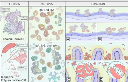
targets of human immunity (Fig. 1). This was demonstrated by a 2016 study which evaluated the antigenic repertoire of recombinant monoclonal antibodies (MAbs) generated from individually sorted plasmablasts from Bangladeshi patients recovering from cholera (44). Over 75% of the antibodies produced from clonally expanded plasmablasts bound to CT or the O-specific polysaccharide antigen (OSP) (44). Addi-tional screening of the MAbs using aV. choleraeproteome array demonstrated that the sialidase NanH (which facilitates toxin binding by converting higher-order cell surface gangliosides to GM1) was a third dominant antigen, though to a much lesser extent than CT and OSP (44). Other known antigens include TcpA, hemolysin A, and flagellar proteins (44, 45). In the case of TcpA, repeated exposure appears to be required to prime class-switched responses (46).
FIG 1 Cholera toxin (CT) and the O-specific polysaccharide (OSP) are the two dominantV. choleraeantigens. Remarkably, following infection in an area of endemicity, more than 75% of the antibodies derived from clonally expanded plasmablasts targeted either CT or the OSP. CT antibodies target both the A and B subunits and may block toxin binding (A) or activity (B), yet the persistence of circulating antitoxin antibodies does not appear to confer long lasting immunity to cholera. OSP-specific antibodies target the bacterial outer membrane and confer protection. Several mechanisms have been proposed, including agglutination (C) and motility inhibition (D) or other effector functions which may entrapV. choleraebefore it reaches the mucosal surface, such as activation of neutrophil extracellular traps.
on September 8, 2020 by guest
http://msphere.asm.org/
ANTITOXIN RESPONSES
CT is the primary virulence factor ofV. choleraeand ingestion of as little as 5g produces the symptoms of cholera (47). Despite this, antitoxin responses do not appear to mediate long-term protective immunity against cholera. One limitation of previous studies of antitoxin immunity has been their persistent focus on the GM1-binding CtxB subunit. Previous studies focused on CtxB, because it was found to be the immuno-dominant component of CT in animal models of cholera, and because this nonactive component of the toxin was an obvious vaccine-antigen candidate (48, 49). A study of responses in CT-immunized mice demonstrated that CtxB-specific responses were dominant and protective while CtxA-binding antibodies had no appreciable toxin-neutralizing capacity (50). However, in humans, CtxB-binding antibodies do not appear to confer durable protection. This is supported by the fact that circulating levels of CtxB-IgG antibodies and CtxB-specific memory B cells are not associated with protec-tive immunity in household contacts of patients with cholera, nor in human challenge studies (51–53). Although elevated circulating levels of CtxB-specific IgA are associated with protection, these wane within months after natural infection (53, 54).
These findings are in line with observations from cholera vaccine trials. In one major field trial, while the addition of CtxB to inactivated oral whole-cell vaccines briefly boosted the effectiveness of vaccination (compared to the inactivated vaccine without CtxB), it did not improve protection beyond the first 8 months. This suggests that CtxB provides only a short-term boost of immunity (55), and more concerningly, that overall protection with the CtxB containing vaccine appeared to drop to lower levels than the non-CtxB containing vaccine within 2 years after vaccination (56). This reduction in long-term immunity may be due to the immunomodulatory effects of CtxB (40).
Similarly, trials of oral vaccination with a glutaraldehyde-treated CT toxoid vaccine alone produced only limited short-term immunity (57). But why do anti-CT responses fail to provide lasting protection against a singular toxin mediated disease? High levels of CtxB-specific sIgA at the mucosal surface are present for months after infection and may provide protection in the short term. However, when exposure occurs more distantly from initial infection, there may be insufficient time to mobilize an effective anamnestic antitoxin response at the mucosal surface once bacterial toxin production is already established.
Still, it may be worth reevaluating the role of CtxA as a protective antigen. This is because recent studies show that unlike mice exposed in a laboratory, humans living in areas where cholera is endemic have prominent CtxA-specific antibody responses (44). More importantly, CtxA antibodies are capable of neutralizing toxin activity at very low concentrations. Because CtxA-antibodies are highly cross-reactive with the entero-toxigenicEscherichia coli(ETEC) heat-labile toxin (LT) (44) and because exposure to the ETEC LT toxin is a frequent occurrence for people living where cholera is endemic, it is likely that the CtxA response observed following human cholera is the result of the expansion of cross-reactive memory B cells derived from prior ETEC infection (44). This may explain why humans respond differently to CtxA than laboratory mice and is an excellent example of the limitations of animal models in studies of human adaptive immunity, where exposure to other infections and commensal organisms may drama-tically shape the immune response.
ANTIBACTERIAL RESPONSES
Unlike antitoxin responses, functional antibody responses directed at the bacterial outer membrane O-polysaccharide are more overtly important in protection against cholera. The serum vibriocidal antibody titer has been used to measure the functional humoral immune response toV. choleraefor decades; it remains the best seroepide-miologic marker of recent exposure (58), and the best-established immunologic corre-late of protection against cholera (53, 59, 60). The vibriocidal titer measures the lowest concentration of serum or plasma required for antibody-dependent complement me-diated bacterial killing, and vibriocidal antibodies almost exclusively target OSP (44, 61). Despite the utility of the vibriocidal antibody titer as a correlate of protection, it is
on September 8, 2020 by guest
http://msphere.asm.org/
unlikely that protection is mediated by a circulating, complement-fixing antibody. First, although increasing vibriocidal titers are associated with protection against cholera, there is no threshold titer at which 100% protection is achieved (62). Second, while complement mediated killing is essential in protecting against certain bloodborne pathogens, it is unclear whether there is enough complement at the mucosal surface to block colonization withV. cholerae. Third, at the intestinal surface, sIgA responses predominate, and sIgA does not induce complement activation. A caveat to this conclusion is that, in areas where cholera is endemic, class switched OSP responses also induce IgM and IgG memory B cell responses (44, 63). Thus, complement mediated killing is not completely excluded as a possible defense againstV. cholerae.
Given that motility is required for colonization, inhibition of bacterial motility has been proposed as a mechanism of protection against cholera, and OSP-targeted IgA antibodies can directly inhibit motility by interfering with flagellar function (64–66). Other proposed antibody-mediated mechanisms include bacterial trapping and clear-ance prior to penetration of the mucous barrier and colonization of the small intestine (16, 67, 68).
Another important question is how are OSP-specific antibody responses main-tained? One possibility is that immunity is maintained by long-lived plasma cells at the mucosal surface. However, basal levels of OSP-specific intestinal sIgA secretion drop quickly after recovery from cholera (69). Another possibility is that mucosal antibody-mediated immunity is maintained in the memory B cell compartment (16, 70). Memory B cells express antibodies at the cell surface but do not secrete them. Yet, memory B cells are capable of rapid differentiation into plasma cells and generation of anamnestic immune responses upon reexposure to antigen.
There is already evidence that the presence of circulating OSP-specific memory B cells is associated with protection against cholera, even in individuals with undetect-able levels of circulating vibriocidal antibodies (52, 71, 72). This association has been observed in household contacts of patients with cholera (71, 72) and in recipients of an attenuatedV. choleraevaccine who were challenged with wild-typeV. cholerae(52). In vaccinees, the magnitude of the initial vibriocidal antibody response was strongly predictive of their subsequent OSP memory B cell response (52). Interestingly, while mucosal O-antigen responses are thought to occur primarily through T-cell-independent IgA class switching pathways,V. choleraeO antigen responses in patients recovering from cholera are characterized by high levels of somatic hypermutation, affinity maturation, and cross-reactive recall responses of memory B cells from prior antigen exposure (44). These features of the OSP response are evidence that long-lasting memory B cell responses may play a role in maintaining immunity against cholera.
THE MICROBIOME AND CHOLERA
The microorganisms composing the complex and self-regulated community of the gut microbiota are increasingly recognized as an important factor in enteric infections. Advances in metagenomic profiling of microbial communities are beginning to reveal the physiologic mechanisms of microbiome-related effects on enteric infection that have been appreciated for decades. These include resistance to colonization and the anti-bacterial activity of commensal gut microbes. For example, in cholera, it has long been recognized that infection in animal models requires disruption of the commensal microbiota with antibiotics, because the undisturbed gut microbiota is otherwise protective againstV. choleraeinfection (73, 74).
Our understanding of how gut microbes impact host-pathogen interactions in V.
choleraeinfection is nascent. Because presence of specific members of the gut
micro-biota correlate with susceptibility to cholera, further studies investigating causality and mechanisms behind this association may identify new approaches to prophylaxis and treatment. Cutting-edge studies of how the microbiota modulate immune responses to oral cholera vaccination may also impact prevention.
on September 8, 2020 by guest
http://msphere.asm.org/
EFFECT OF CHOLERA ON THE MICROBIOME
Cholera essentially eradicates the normal gut microbiota (75). First, the massive efflux of water into the intestinal lumen washes away the protective mucus where much of the gut microbiota resides. Second, depletion of the microbiota is likely exacerbated by treatments for cholera, including ingestion of large amounts of oral rehydration solution and antibiotics that kill some gut bacterial species (75, 76). Based on DNA sequencing of all bacteria in the stool during the initial phase of infection,V.
cholerae itself makes up the majority of bacteria found in rice-water stool (75, 77).
Alterations in the microbiota extend beyond the duration of symptoms, and recovery after infection follows a distinct pattern (75). Colonizing gut microbes are scant immediately following infection. Then, in the first few days of recovery, aerobes and facultative anaerobes ingested from the environment and the oral cavity dominate the microbiota. These recolonizing organisms likely flourish due to the abundance of nutrients and oxygen that accumulates during the early recovery period while the flora is depleted. As aerobes proliferate, oxygen tension is lowered, and obligate anaerobes again colonize the gut. Several weeks after cholera, community profiling demonstrates a return to a near-baseline gut microbiota composition, when competition for nutrients and resources resumes (75). A similar pattern of recovery was also demonstrated in a study of gut microbiota post-ETEC infection in Bangladesh, although in an ETEC human challenge study, anaerobic species persisted during acute infection in some subjects (75, 78). The dissimilarity in patterns of microbial recovery in these two groups could be due to differences in baseline microbiota of American volunteers compared to Bangla-deshis. Alternatively, these differences may exist due to factors that we do not yet understand regarding underlying patterns of microbial recovery after acute diarrhea. The disruption to gut homeostasis after cholera and other forms of severe diarrhea is associated with enteric dysfunction and malnutrition in children living in areas of inadequate safe water sources (79, 80). While the gut microbiota likely mediates some of this pathology, this complex relationship and the physiology of enteric dysfunction remains poorly understood (81, 82).
THE MICROBIOME ANDV. CHOLERAEPATHOGENESIS
Household contacts of patients with cholera are at high risk for infection. In a prospective evaluation of household contacts of patients with cholera, there was a strong association between specific microbial groups at the time of exposure to V.
choleraeand infection during the following week. In fact, the composition of the gut
microbiome predicted infection as well as the known clinical, immunologic, and epidemiologic risk factors for cholera (83). Based on a machine learning model, bacterial taxa were ranked according to their association with either increased or decreased susceptibility to infection, and the top 100 bacterial taxa from this ranking successfully discriminated between clinical outcomes. However, the baseline alpha and beta diversity alone were not predictive of susceptibility to cholera. This contrasts with other enteric pathogens which are more likely to cause infection in humans with a lower-diversity microbiome (84, 85). Investigation of the specific bacterial groups correlated with susceptibility may provide further insight on relationships betweenV.
choleraeinfection, the gut microbiome, and clinical outcomes.
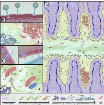
Mucins, bile acids, and T6SS. Sensing of the intestinal environment allows V.
choleraeto tailor the expression of virulence and compete with the host microbiota for
resources, including access to the epithelial surface. In colonizing the small intestine,V.
choleraeencounters a layer of highly glycosylated proteins (mucins) lining the intestinal
epithelium (Fig. 2). This mucus layer is also heavily colonized with commensal bacteria,
andV. choleraedetects the presence of these microbes and their antibacterial
metab-olites through various mechanisms. Sensing of mucin activates theV. choleraetype VI secretion system (T6SS), which operates as a molecular syringe, delivering toxic pro-teins to other bacteria (86, 87). In a study screening for genes critical to in vivo V.
choleraefitness, the T6SS apparatus was found to be vital for successful colonization,
highlighting one method by whichV. choleraeovercomes colonization resistance (88).
on September 8, 2020 by guest
http://msphere.asm.org/
In a study of mice infected withV. choleraeT6SS mutants compared to wild-type strains,
V. choleraecolonization was increased several fold in wild-type infections due to the
T6SS-mediated attack of host commensal organisms (89).
Another aspect of the small intestinal environment that impacts this interaction is cholic acid from bile, which can be processed by some gut microbes. V. cholerae
recognizes bile acids as a cue for the small intestinal environment, and upon exposure to bile acid, gene expression shifts to optimize locating the mucosal epithelium
FIG 2 Interactions between the gut microbiota, their metabolites,V. cholerae, and the small intestinal environment. (A) AfterV. cholerae
survives the acidic gastric environment, the pathogen enters the small intestinal gut lumen, where both bile (green) and mucus (yellow) signal toV. choleraeto express the virulence factors that cause symptomatic infection. Mucus coats the villi and acts as a diffusion barrier, andV. choleraeuses flagellar motion to traverse the inner and outer mucus layers. During this journeyV. choleraeencounters the resident gut microbes and their metabolites. After reaching the intestinal epithelial crypts,V. choleraeforms biofilms (shown as fibrous mats of organisms) to adhere to the epithelial surface. (B and C) When encountering the mucus layer, theV. choleraetype VI secretion system (T6SS) is activated. This system operates as a contractile organelle that extends fromV. choleraeto make contact with neighboring organisms to translocate toxic effectors. T6SS activity can be suppressed by metabolites of cholic acid formed by gut microbiota that process bile (green and red curved arrow). (D) Autoinducer AI-2 (yellow) is produced by some commensal gut microbes and can induce quorum-sensing responses inV. cholerae. The presence of autoinducers indicate toV. choleraea high density of organisms, resulting in reduced expression of virulence genes that enable colonization and cholera toxin production, and activation of genes that promote exit from the host, such as increased flagellar motion. (E) Bacteriophage specific toV. cholerae(vibriophage) can infect and lyse large numbers of organisms rapidly, drastically reducingV. choleraepopulations.
on September 8, 2020 by guest
http://msphere.asm.org/
through increased motility while repressing virulence factors CT and TCP (90). Some small intestinal microbes also dehydroxylate primary bile acids, and whenV. cholerae
senses the metabolic product deoxycholic acid, T6SS activity is suppressed (86). By metabolizing bile acids and masking the “sensing” of the small intestinal environment, these commensals may advance their own survival. Through the enterohepatic circu-lation in the liver, both primary and secondary bile acids can be conjugated, resulting in metabolites that enhance T6SS function (86). Sensing of bile and bile acid metabo-lites by V. cholerae impacts the infected host because CT and TCP expression vary depending on the specific form of bile acid present in the small intestine (91, 92). For further resolution on how V. cholerae fine-tunes virulence expression based on the intestinal environment, we need a better understanding of the gut microbes that participate in bile acid metabolism and howV. choleraevirulence is impacted by these metabolites.
Autoinducers.Small molecules produced by the gut microbiota and detected byV.
cholerae are an example of interspecies communication that impacts pathogenesis.
Two quorum-sensing molecules, autoinducer-2 (AI-2) and cholera autoinducer 1 (CAI-1) are sensed byV. choleraeusing histidine kinase receptors LuxQ and CqsS, respectively (93). Recognition of autoinducers allows for population-level coordinated activity byV.
cholerae. When V. cholerae is present at high density, a quorum is “sensed” and
autoinducers are produced (94). Autoinducer binding results in modulation of down-stream virulence factors, including a reduction in TcpA expression and production, which signals thatV. choleraeshould disassociate from the epithelial surface (93). Thus far, the autoinducer CAI-1 is known to be produced naturally only byVibriospecies. In an infant mouse model ofV. choleraeinfection, an engineered strain ofE. colimade to express CAI-1 resulted in an 80% reduction in CT binding at the intestinal surface, thereby preventingV. choleraecolonization (95).
In humans recovering from cholera, AI-2 production by commensal intestinal bac-teria was found to block V. cholerae virulence expression (77). In a 2014 study, a community of 14 gut microbes associated with recovery from human cholera was reconstituted in germ-free mice. Upon challenge withV. cholerae, one species,Blautia
obeum(formerlyRuminococcus obeum), was associated with a reduction inV. cholerae
colonization through a LuxS-based AI-2-dependent signaling pathway. This demon-strates one mechanism of host gut microbe colonization resistance through disruption
ofV. choleraequorum sensing via interspecies signaling (77). Because AI-2 is made by
numerous members of the gut microbiota, this may represent one of many examples of interspecies communication that impact virulence (96, 97). Studies in humans are needed to determine whether autoinducers from naturally occurring host gut microbes or engineered species could impact clinical outcomes.
Other gut microbe metabolites.Commensal bacteria have long been postulated
to influence behavior ofV. choleraeby secreting antimicrobial compounds, and these interactions have been studied by applying culture supernatant from commensals toin
vitro V. cholerae culture or in animal models of infection. Prior to the advances of
genomic analyses, plating of fecal samples from patients with cholera demonstrated restriction ofV. choleraegrowth in the presence ofLactobacilliandPeptostreptococcus
species due to unidentified “inhibitory diffusible compounds” (98). In a 2018 study,
Lactobacillusspecies from the stool of healthy children were screened to detect effects
on formation and dispersal of V. cholerae biofilms (99). Biofilms are an important virulence factor forV. choleraesurvival, facilitating adherence to the intestinal epithe-lium, protecting the pathogen from antibiotics and acid inactivation, and even pro-tecting against predation by other gut microbe species (100). Metabolites in the culture supernatant of sevenLactobacillusisolates inhibitedV. choleraebiofilm formation in a pH-dependent manner, although the structure and function of the antimicrobial com-pounds in these studies remain unknown (99).
Several other known microbial metabolites can alter the chemical environment, impactingV. choleraepathogenesis. In infant mice, ingestion of microbes that secrete
on September 8, 2020 by guest
http://msphere.asm.org/
lactic acid reduce V. cholerae growth and colonization (101). Like many bacteria, V.
choleraeis also sensitive to reactive oxygen species. Infant mice that ingest commensal
E. colistrains defective in ROS degradation prior toV. choleraechallenge had a higher
susceptibility to infection compared to those givenE. coliwith the ability to degrade reactive oxygen species (102). While these studies exemplify how a single metabolite or environmental shift can potentially alter the course of infection, the relevance of these findings in human cholera is not known. Based on the recognized mechanisms of microbial metabolites that interact withV. choleraeand the complexity of the bacterial community of the gut, the full scope of metabolites that impactV. cholerae pathogen-esis are likely heterogeneous in structure and mechanism.
Vibriophage. In addition to bacteria, the intestine harbors fungi, parasites, and viruses, including bacteriophage. While hundreds of bacteriophages are known to infectV. cholerae, few have been characterized (103). Vibriophage are found in humans recovering from cholera, and phage predation ofV. choleraehas been observedin vivo
during human infection, where it is associated with the rapid acquisition of intrahostV.
choleraemutations in phage receptors (104). This suggests that lytic vibriophage has
the potential to impact the course of disease in humans. In the preantibiotic era, two studies evaluated the efficacy of vibriophage administration for treatment of cholera in humans (105, 106). In India, a reduction in cholera cases was also observed after adding vibriophages into community drinking water on a weekly basis (106). However, because
V. cholerae resistance to lytic phage evolves rapidly (102, 104, 107), it is likely that
combinations of vibriophages would be required for effective treatment of clinicalV.
choleraestrains.
THE GUT MICROBIOME AND ORAL CHOLERA VACCINE RESPONSES
Both live-attenuated and killed oral cholera vaccines (OCV) are increasingly used to prevent cholera (108, 109), and killed OCVs are a critical component of the World Health Organization’s strategy to reduce the global threat of cholera by 2030 (110). Because OCVs are absorbed at the mucosal surface where the intestinal microbiota continually interfaces with immune cells conducting antigen surveillance, the gut microbiome could impact immune responses to vaccination. This has led to the hypothesis that the gut microbiota is a frequently unmeasured host factor that partially determines im-mune responses to OCV. Evidence supporting this theory dates to the 1990s when bacterial overgrowth was found to correlate with reduced response to an attenuated OCV (111).
While the gut microbiota’s impact on OCV responses has not yet been systematically studied, emerging data from other oral vaccines suggest relevant interactions (112). For example, the immunogenicity of oral live-attenuated typhoid vaccine in adults was correlated with more diverse gut microbial communities, and the abundance of several specific microbes differentiated multiphasic from late cell-mediated immune responses after vaccination (113). A stool analysis of pairs of infants from Ghana and the Neth-erlands compared discordant immune responses to oral rotavirus vaccination and showed that the gut microbiome of responders was similar regardless of country of residence, and that nonresponders from either country had lessStreptococcus bovisand increased organisms from the phylumBacteroidetes(114). Although little studied, the nonbacterial microbial factors impacting baseline immunoactivation or inflammation may also impact immune responses to OCVs. For example, children receiving antihel-minth treatment prior to vaccination had higher vibriocidal responses to OCVs com-pared to untreated children (115). However, antihelminth treatment did not improve immune responses of Bangladeshi children to Ty21a, an oral typhoid vaccine (116). Use of perivaccination antibiotics is an additional microbiota-altering intervention that could impact immune responses to OCVs, and this has yet to be studied systemically. Further investigation is needed to understand the dynamics and advantages of ad-junctive treatments that affect the gut microbiota at the time of vaccination.
To date, studies of oral vaccination in humans and the gut microbiota have not evaluated mechanistic hypotheses or explored causative relationships. Yet, based on
on September 8, 2020 by guest
http://msphere.asm.org/
current knowledge of how gut microbes can impact human immune responses in the gut, it is clear that specific metabolites of the gut microbiota have the potential to impact immune responses to OCVs. For example, short-chain fatty acids (SCFAs) are by-products of nondigestible carbohydrate metabolism by some members of the anaerobic microbiota and are known to impact the development of mucosal immune responses in other disease processes (117, 118). The effect of SCFAs on OCV responses has thus far only been tested in cell culture. When the SCFA butyrate was added to cultured gut epithelial cells and exposed to an attenuatedV. choleraevaccine, CCL20 production was increased (119), providing a pathway by which butyrate may accentu-ate mucosal immune responses by recruiting dendritic cells and lymphocytes. Specific SCFAs are also known to depress the effect of LPS-induced cytokines on the develop-ment of T cell subgroups, although these possibilities have not been investigated inV.
choleraeinfection or vaccination (120, 121). SCFAs produced in the normal gut
micro-biota in mouse models increased CT specific antibody responses, and these high-level antibody responses were restored to antibiotic-treated mice fed acetate and butyrate (122). However, it is not yet clear whether SCFAs impact responses to V. cholerae
antigens more closely associated with protection from cholera in humans, such as OSP. Further studies that include testing of mechanistic hypotheses are needed to advance the potential for microbes (or their metabolites) to be used as vaccine adjuvants.
Probiotic impact onV. choleraeinfection and OCVs.Probiotics have safely been coadministered with OCVs in two small, randomized placebo-controlled trials without a clear effect on vaccine immunogenicity (123, 124). These studies and others evalu-ating the effect of probiotics on human health are hampered by variation in choice of bacterial strain, purity, dose, and timing of administration, limiting comparisons be-tween studies (125). The challenge of interpreting cause and effect is further com-pounded in studies that do not account for human host factors known to impact immune responses to OCVs, such as age. In addition, whether a probiotic strain is just “passing through” or whether sustained colonization is necessary to effectively mod-ulate OCV responses is an important unanswered question. For further development of probiotics as OCV adjuvants, identification of an appropriate strain in addition to optimization of the dose and formulation are needed. If safety data are reassuring, testing of a probiotic OCV adjuvant should include young children and infants who are most in need of improved responses to OCV.
ON THE HORIZON
Expansive cholera outbreaks and the large burden of cholera in areas of endemicity represent ongoing public health crises. In Yemen alone, over 1.7 million cases of cholera have occurred since 2016 (126). A better understanding of immunity, including improved biomarkers of protective immunity, is required to develop and test improved OCV candidates. Key unanswered questions include how OSP-specific antibody re-sponses are maintained, and how the gut microbiome at the time of vaccination or infection may influence these long-term protective responses. An improved knowledge of host-pathogen interactions is needed to harness the natural phenomenon of colo-nization resistance and move new vaccine and therapeutic possibilities forward. Central to this goal is a need for mechanistic studies to understand how other microbes and their signals impactV. choleraevirulence expression and the development of protective immunity.
Innovative approaches for prevention may exploit colonization resistance through the administration of engineered commensal organisms. As discussed, genetic manip-ulation ofE. coliwith the ability to express CAI-1 was successful in preventing cholera in an animal model (95). In a 2016 study, a recombinantE. colistrain that expressed glycosyltransferases mimicking ganglioside GM1, the binding site for cholera toxin, absorbed cholera toxin with high avidity and resulted in survival of challenged infant mice (127). As a therapeutic possibility, vibriophages are interesting because bacteri-cidal activity is targeted, thereby limiting the potential for the emergence of antibiotic resistance. Historical data from the preantibiotic era suggests this approach could be
on September 8, 2020 by guest
http://msphere.asm.org/
promising, but modern trials and human data are lacking. While interest in bacterio-phage therapy has increased with the use of bacterio-phage against multidrug resistant bacterial infections (128), and a triple vibriophage cocktail was found to prevent colonization and limit the emergence of antibiotic resistance in an infant mouse model of cholera (107), there are no current human trials of vibriophage or engineered microbes for cholera treatment or prevention.
Perhaps the most promising new approach to prevention combines the long-term potential of live attenuated oral cholera vaccination with the immediate impact of colonization resistance. In a 2018 study, an attenuated V. cholerae strain protected against cholera in an infant rabbit model within 24 h of administration, prior to the development of an adaptive immune response (129). This attenuated strain conferred protection in colonized infant rabbits and prevented colonization of wild-type V.
cholerae, presumably by occupying the intestinal niche of the wild-type strain or by
dominating resources needed for colonization. Interestingly, the mechanism of this competitive exclusion remains unknown and was not due to one specific mutation generated in the attenuated strain (129).
Overall, these studies all demonstrate that manipulating the gut microbiota to alter the course of human cholera is within reach. Ultimately a successful approach may rely on attenuatedV. choleraevaccine strains or naturally occurring commensal organisms to either provide resistance to infection or boost the effectiveness of the cholera vaccines through interactions with the host immune system. Further mechanistic studies to explore the how the human microbiome impacts immune responses to both live attenuated and killed oral cholera vaccines are needed, and insights into the mechanisms of colonization resistance are also critical for driving these new transla-tional approaches forward.
REFERENCES
1. Lippi D, Gotuzzo E. 2014. The greatest steps towards the discovery of
Vibrio cholerae. Clin Microbiol Infect 20:191–195. https://doi.org/10 .1111/1469-0691.12390.
2. Koch R. 1884. An address on cholera and its bacillus. Br Med J 2:403– 407.https://doi.org/10.1136/bmj.2.1235.403.
3. De SN. 1959. Enterotoxicity of bacterium-free culture-filtrate of
Vibrio cholerae. Nature 183:1533–1534. https://doi.org/10.1038/ 1831533a0.
4. Colwell RR, Huq A. 1994. Environmental reservoir ofVibrio cholerae: the causative agent of cholera. Ann N Y Acad Sci 740:44 –54.https://doi .org/10.1111/j.1749-6632.1994.tb19852.x.
5. Harris JB, LaRocque RC, Qadri F, Ryan ET, Calderwood SB. 2012. Cholera. Lancet 379:2466 –2476. https://doi.org/10.1016/S0140 -6736(12)60436-X.
6. Mutreja A, Kim DW, Thomson NR, Connor TR, Lee JH, Kariuki S, Croucher NJ, Choi SY, Harris SR, Lebens M, Niyogi SK, Kim EJ, Rama-murthy T, Chun J, Wood JL, Clemens JD, Czerkinsky C, Nair GB, Hol-mgren J, Parkhill J, Dougan G. 2011. Evidence for several waves of global transmission in the seventh cholera pandemic. Nature 477: 462– 465.https://doi.org/10.1038/nature10392.
7. Kaper JB, Morris JG, Jr, Levine MM. 1995. Cholera. Clin Microbiol Rev 8:48 – 86.https://doi.org/10.1128/CMR.8.1.48.
8. Devault AM, Golding GB, Waglechner N, Enk JM, Kuch M, Tien JH, Shi M, Fisman DN, Dhody AN, Forrest S, Bos KI, Earn DJ, Holmes EC, Poinar HN. 2014. Second-pandemic strain ofVibrio choleraefrom the Philadelphia cholera outbreak of 1849. N Engl J Med 370:334 –340.https://doi.org/ 10.1056/NEJMoa1308663.
9. Albert MJ, Siddique AK, Islam MS, Faruque AS, Ansaruzzaman M, Faruque SM, Sack RB. 1993. Large outbreak of clinical cholera due to
Vibrio choleraenon-O1 in Bangladesh. Lancet 341:704.https://doi.org/ 10.1016/0140-6736(93)90481-u.
10. Stroeher UH, Jedani KE, Dredge BK, Morona R, Brown MH, Karageorgos LE, Albert MJ, Manning PA. 1995. Genetic rearrangements in therfb
regions of Vibrio choleraeO1 and O139. Proc Natl Acad Sci U S A 92:10374 –10378.https://doi.org/10.1073/pnas.92.22.10374.
11. Karlsson SL, Thomson N, Mutreja A, Connor T, Sur D, Ali M, Clemens J, Dougan G, Holmgren J, Lebens M. 2016. Retrospective analysis of
serotype switching ofVibrio choleraeO1 in a cholera endemic region shows it is a non-random process. PLoS Negl Trop Dis 10:e0005044.
https://doi.org/10.1371/journal.pntd.0005044.
12. Koelle K, Pascual M, Yunus M. 2006. Serotype cycles in cholera dynam-ics. Proc Biol Sci 273:2879 –2886. https://doi.org/10.1098/rspb.2006 .3668.
13. Koelle K, Rodo X, Pascual M, Yunus M, Mostafa G. 2005. Refractory periods and climate forcing in cholera dynamics. Nature 436:696 –700.
https://doi.org/10.1038/nature03820.
14. Ali M, Emch M, Park JK, Yunus M, Clemens J. 2011. Natural cholera infection-derived immunity in an endemic setting. J Infect Dis 204: 912–918.https://doi.org/10.1093/infdis/jir416.
15. Levine MM, Black RE, Clements ML, Cisneros L, Nalin DR, Young CR. 1981. Duration of infection-derived immunity to cholera. J Infect Dis 143:818 – 820.https://doi.org/10.1093/infdis/143.6.818.
16. Harris JB. 2018. Cholera: immunity and prospects in vaccine develop-ment. J Infect Dis 218(Suppl 3):S141–S146. https://doi.org/10.1093/ infdis/jiy414.
17. Anonymous. 2010. Update: outbreak of cholera—Haiti, 2010. MMWR Morb Mortal Wkly Rep 59:1586 –1590.
18. Cash RA, Music SI, Libonati JP, Snyder MJ, Wenzel RP, Hornick RB. 1974. Response of man to infection withVibrio cholerae. I. Clinical, serologic, and bacteriologic responses to a known inoculum. J Infect Dis 129: 45–52.https://doi.org/10.1093/infdis/129.1.45.
19. Nalin DR, Levine RJ, Levine MM, Hoover D, Bergquist E, McLaughlin J, Libonati J, Alam J, Hornick RB. 1978. Cholera, non-vibrio cholera, and stomach acid. Lancet 2:856 – 859.https://doi.org/10.1016/s0140-6736 (78)91568-4.
20. Freter R, O’Brien PC, Macsai MS. 1981. Role of chemotaxis in the association of motile bacteria with intestinal mucosa:in vivostudies. Infect Immun 34:234 –240.
21. Millet YA, Alvarez D, Ringgaard S, von Andrian UH, Davis BM, Waldor MK. 2014. Insights into Vibrio cholerae intestinal colonization from monitoring fluorescently labeled bacteria. PLoS Pathog 10:e1004405.
https://doi.org/10.1371/journal.ppat.1004405.
22. Peterson KM, Gellings PS. 2018. Multiple intraintestinal signals
on September 8, 2020 by guest
http://msphere.asm.org/
nate the regulation ofVibrio choleraevirulence determinants. Pathog Dis 76:.https://doi.org/10.1093/femspd/ftx126.
23. Holmgren J, Lonnroth I, Mansson J, Svennerholm L. 1975. Interaction of cholera toxin and membrane GM1 ganglioside of small intestine. Proc Natl Acad Sci U S A 72:2520 –2524.https://doi.org/10.1073/pnas.72.7 .2520.
24. Waldor MK, Mekalanos JJ. 1996. Lysogenic conversion by a filamentous phage encoding cholera toxin. Science 272:1910 –1914.https://doi.org/ 10.1126/science.272.5270.1910.
25. King CA, Van Heyningen WE. 1973. Deactivation of cholera toxin by a sialidase-resistant monosialosylganglioside. J Infect Dis 127:639 – 647.
https://doi.org/10.1093/infdis/127.6.639.
26. Cassel D, Pfeuffer T. 1978. Mechanism of cholera toxin action: covalent modification of the guanyl nucleotide-binding protein of the adenylate cyclase system. Proc Natl Acad Sci U S A 75:2669 –2673.https://doi.org/ 10.1073/pnas.75.6.2669.
27. Qadri F, Bhuiyan TR, Dutta KK, Raqib R, Alam MS, Alam NH, Svenner-holm AM, Mathan MM. 2004. Acute dehydrating disease caused by
Vibrio choleraeserogroups O1 and O139 induce increases in innate cells and inflammatory mediators at the mucosal surface of the gut. Gut 53:62– 69.https://doi.org/10.1136/gut.53.1.62.
28. Nelson EJ, Harris JB, Morris JG, Jr, Calderwood SB, Camilli A. 2009. Cholera transmission: the host, pathogen and bacteriophage dynamic. Nat Rev Microbiol 7:693–702.https://doi.org/10.1038/nrmicro2204. 29. Yen CH. 1964. A recent study of cholera with reference to an outbreak
in Taiwan in 1962. Bull World Health Organ 30:811– 825.
30. Ellis CN, LaRocque RC, Uddin T, Krastins B, Mayo-Smith LM, Sarracino D, Karlsson EK, Rahman A, Shirin T, Bhuiyan TR, Chowdhury F, Khan AI, Ryan ET, Calderwood SB, Qadri F, Harris JB. 2015. Comparative pro-teomic analysis reveals activation of mucosal innate immune signaling pathways during cholera. Infect Immun 83:1089 –1103.https://doi.org/ 10.1128/IAI.02765-14.
31. Flach CF, Qadri F, Bhuiyan TR, Alam NH, Jennische E, Lonnroth I, Holmgren J. 2007. Broad up-regulation of innate defense factors during acute cholera. Infect Immun 75:2343–2350.https://doi.org/10.1128/IAI .01900-06.
32. Mathan MM, Chandy G, Mathan VI. 1995. Ultrastructural changes in the upper small intestinal mucosa in patients with cholera. Gastroenterol-ogy 109:422– 430.https://doi.org/10.1016/0016-5085(95)90329-1. 33. Bourque DL, Bhuiyan TR, Genereux DP, Rashu R, Ellis CN, Chowdhury F,
Khan AI, Alam NH, Paul A, Hossain L, Mayo-Smith LM, Charles RC, Weil AA, LaRocque RC, Calderwood SB, Ryan ET, Karlsson EK, Qadri F, Harris JB. 2018. Analysis of the human mucosal response to cholera reveals sustained activation of innate immune signaling pathways. Infect Im-mun 86:e00594-17.
34. Shin OS, Uddin T, Citorik R, Wang JP, Della Pelle P, Kradin RL, Bingle CD, Bingle L, Camilli A, Bhuiyan TR, Shirin T, Ryan ET, Calderwood SB, Finberg RW, Qadri F, Larocque RC, Harris JB. 2011. LPLUNC1 modulates innate immune responses to Vibrio cholerae. J Infect Dis 204: 1349 –1357.https://doi.org/10.1093/infdis/jir544.
35. Saha D, Karim MM, Khan WA, Ahmed S, Salam MA, Bennish ML. 2006. Single-dose azithromycin for the treatment of cholera in adults. N Engl J Med 354:2452–2462.https://doi.org/10.1056/NEJMoa054493. 36. Karlsson EK, Harris JB, Tabrizi S, Rahman A, Shlyakhter I, Patterson N,
O’Dushlaine C, Schaffner SF, Gupta S, Chowdhury F, Sheikh A, Shin OS, Ellis C, Becker CE, Stuart LM, Calderwood SB, Ryan ET, Qadri F, Sabeti PC, Larocque RC. 2013. Natural selection in a bangladeshi population from the cholera-endemic Ganges river delta. Sci Transl Med 5https:// doi.org/10.1126/scitranslmed.3006338.
37. Weil AA, Ellis CN, Debela MD, Bhuiyan TR, Rashu R, Bourque DL, Khan AI, Chowdhury F, LaRocque RC, Charles RC, Ryan ET, Calderwood SB, Qadri F, Harris JB. 2019. Posttranslational regulation of IL-23 production distinguishes the innate immune responses to live toxigenic versus heat-inactivated Vibrio cholerae. mSphere 4 https://doi.org/10.1128/ mSphere.00206-19.
38. Wang H, Naseer N, Chen Y, Zhu AY, Kuai X, Galagedera N, Liu Z, Zhu J. 2017. OxyR2 modulates OxyR1 activity andVibrio choleraeoxidative stress response. Infect Immun 85:e00929-16.
39. Xia X, Larios-Valencia J, Liu Z, Xiang F, Kan B, Wang H, Zhu J. 2017. OxyR-activated expression of Dps is important forVibrio cholerae oxi-dative stress resistance and pathogenesis. PLoS One 12:e0171201.
https://doi.org/10.1371/journal.pone.0171201.
40. Kuchta A, Rahman T, Sennott EL, Bhuyian TR, Uddin T, Rashu R, Chow-dhury F, Kahn AI, Arifuzzaman M, Weil AA, Podolsky M, LaRocque RC,
Ryan ET, Calderwood SB, Qadri F, Harris JB. 2011.Vibrio choleraeO1 infection induces proinflammatory CD4⫹T-cell responses in blood and intestinal mucosa of infected humans. Clin Vaccine Immunol 18: 1371–1377.https://doi.org/10.1128/CVI.05088-11.
41. Glass RI, Holmgren J, Haley CE, Khan MR, Svennerholm AM, Stoll BJ, Belayet Hossain KM, Black RE, Yunus M, Barua D. 1985. Predisposition for cholera of individuals with O blood group: possible evolutionary significance. Am J Epidemiol 121:791–796. https://doi.org/10.1093/ oxfordjournals.aje.a114050.
42. Harris JB, LaRocque RC. 2016. Cholera and ABO blood group: under-standing an ancient association. Am J Trop Med Hyg 95:263–264.
https://doi.org/10.4269/ajtmh.16-0440.
43. Glass RI, Becker S, Huq MI, Stoll BJ, Khan MU, Merson MH, Lee JV, Black RE. 1982. Endemic cholera in rural Bangladesh, 1966 –1980. Am J Epidemiol 116:959 –970.https://doi.org/10.1093/oxfordjournals.aje.a113498. 44. Kauffman RC, Bhuiyan TR, Nakajima R, Mayo-Smith LM, Rashu R, Hoq
MR, Chowdhury F, Khan AI, Rahman A, Bhaumik SK, Harris L, O’Neal JT, Trost JF, Alam NH, Jasinskas A, Dotsey E, Kelly M, Charles RC, Xu P, Kovácˇ P, Calderwood SB, Ryan ET, Felgner PL, Qadri F, Wrammert J, Harris JB. 2016. Single-cell analysis of the plasmablast response to
Vibrio choleraedemonstrates expansion of cross-reactive memory B cells. mBio 7:e02021-16.https://doi.org/10.1128/mBio.02021-16. 45. Charles RC, Nakajima R, Liang L, Jasinskas A, Berger A, Leung DT, Kelly
M, Xu P, Kovac P, Giffen SR, Harbison JD, Chowdhury F, Khan AI, Calderwood SB, Bhuiyan TR, Harris JB, Felgner PL, Qadri F, Ryan ET. 2017. Plasma and mucosal immunoglobulin M, immunoglobulin A, and immunoglobulin G responses to theVibrio choleraeO1 protein immu-nome in adults with cholera in Bangladesh. J Infect Dis 216:125–134.
https://doi.org/10.1093/infdis/jix253.
46. Mayo-Smith LM, Simon JK, Chen WH, Haney D, Lock M, Lyon CE, Calderwood SB, Kirkpatrick BD, Cohen M, Levine MM, Gurwith M, Harris JB. 2017. The live attenuated cholera vaccine CVD 103-HgR primes responses to the toxin-coregulated pilus antigen TcpA in subjects challenged with wild-typeVibrio cholerae. Clin Vaccine Immunol 24: e00470-16.
47. Levine MM, Kaper JB, Black RE, Clements ML. 1983. New knowledge on pathogenesis of bacterial enteric infections as applied to vaccine de-velopment. Microbiol Rev 47:510 –550.
48. Holmgren J, Svennerholm AM. 1977. Mechanisms of disease and im-munity in cholera: a review. J Infect Dis 136(Suppl):S105–S102.https:// doi.org/10.1093/infdis/136.supplement.s105.
49. Peterson JW, Hejtmancik KE, Markel DE, Craig JP, Kurosky A. 1979. Antigenic specificity of neutralizing antibody to cholera toxin. Infect Immun 24:774 –779.
50. Lindholm L, Holmgren J, Wikstrom M, Karlsson U, Andersson K, Lycke N. 1983. Monoclonal antibodies to cholera toxin with special reference to cross-reactions withEscherichia coliheat-labile enterotoxin. Infect Im-mun 40:570 –576.
51. Levine MM, Nalin DR, Craig JP, Hoover D, Bergquist EJ, Waterman D, Holley HP, Hornick RB, Pierce NP, Libonati JP. 1979. Immunity of cholera in man: relative role of antibacterial versus antitoxic immunity. Trans R Soc Trop Med Hyg 73:3–9. https://doi.org/10.1016/0035-9203(79) 90119-6.
52. Haney DJ, Lock MD, Gurwith M, Simon JK, Ishioka G, Cohen MB, Kirkpatrick BD, Lyon CE, Chen WH, Sztein MB, Levine MM, Harris JB. 2018. Lipopolysaccharide-specific memory B cell responses to an at-tenuated live cholera vaccine are associated with protection against
Vibrio cholerae infection. Vaccine 36:2768 –2773. https://doi.org/10 .1016/j.vaccine.2018.04.011.
53. Harris JB, Larocque RC, Chowdhury F, Khan AI, Logvinenko T, Faruque AS, Ryan ET, Qadri F, Calderwood SB. 2008. Susceptibility to Vibrio choleraeinfection in a cohort of household contacts of patients with cholera in Bangladesh. PLoS Negl Trop Dis 2:e221.https://doi.org/10 .1371/journal.pntd.0000221.
54. Harris AM, Bhuiyan MS, Chowdhury F, Khan AI, Hossain A, Kendall EA, Rahman A, LaRocque RC, Wrammert J, Ryan ET, Qadri F, Calderwood SB, Harris JB. 2009. Antigen-specific memory B-cell responses to Vibrio cholerae O1 infection in Bangladesh. Infect Immun 77:3850 –3856.
https://doi.org/10.1128/IAI.00369-09.
55. Clemens JD, Sack DA, Harris JR, Van Loon F, Chakraborty J, Ahmed F, Rao MR, Khan MR, Yunus M, Huda N, Stanton BF, Kay BA, Walter S, Eeckels R, Svennerholm AM, Holmgren J. 1990. Field trial of oral cholera vaccines in Bangladesh: results from three-year follow-up. Lancet 335: 270 –273.https://doi.org/10.1016/0140-6736(90)90080-O.
on September 8, 2020 by guest
http://msphere.asm.org/
56. van Loon FP, Clemens JD, Chakraborty J, Rao MR, Kay BA, Sack DA, Yunus M, Ali M, Svennerholm AM, Holmgren J. 1996. Field trial of inactivated oral cholera vaccines in Bangladesh: results from 5 years of follow-up. Vaccine 14:162–166.https://doi.org/10.1016/0264-410x(95) 00122-h.
57. Curlin GL, Levine R, Aziz KMA, Rahman ASM, Verwey WF. Field trial of cholera toxoid, p 314 –329. 11th Joint Conference of the U.S.-Japan Cooperative Medical Science Program Symposium on Cholera. National Institute of Allergy and Infectious Diseases, Bethesda, MD.
58. Azman AS, Lessler J, Luquero FJ, Bhuiyan TR, Khan AI, Chowdhury F, Kabir A, Gurwith M, Weil AA, Harris JB, Calderwood SB, Ryan ET, Qadri F, Leung DT. 2019. Estimating cholera incidence with cross-sectional serology. Sci Transl Med 11:eaau6242.https://doi.org/10.1126/scitransl med.aau6242.
59. Chen WH, Cohen MB, Kirkpatrick BD, Brady RC, Galloway D, Gurwith M, Hall RH, Kessler RA, Lock M, Haney D, Lyon CE, Pasetti MF, Simon JK, Szabo F, Tennant S, Levine MM. 2016. Single-dose live oral cholera vaccine CVD 103-HgR protects against human experimental infection withVibrio choleraeO1 El Tor. Clin Infect Dis 62:1329 –1335.https://doi .org/10.1093/cid/ciw145.
60. Ritter AS, Chowdhury F, Franke MF, Becker RL, Bhuiyan TR, Khan AI, Saha NC, Ryan ET, Calderwood SB, LaRocque RC, Harris JB, Qadri F, Weil AA. 2019. Vibriocidal titer and protection from cholera in children. Open Forum Infect Dis 6:ofz057.https://doi.org/10.1093/ofid/ofz057. 61. Johnson RA, Uddin T, Aktar A, Mohasin M, Alam MM, Chowdhury F,
Harris JB, LaRocque RC, Bufano MK, Yu Y, Wu-Freeman Y, Leung DT, Sarracino D, Krastins B, Charles RC, Xu P, Kovac P, Calderwood SB, Qadri F, Ryan ET. 2012. Comparison of immune responses to the O-specific polysaccharide and lipopolysaccharide ofVibrio choleraeO1 in Bangla-deshi adult patients with cholera. Clin Vaccine Immunol 19:1712–1721.
https://doi.org/10.1128/CVI.00321-12.
62. Saha D, LaRocque RC, Khan AI, Harris JB, Begum YA, Akramuzzaman SM, Faruque AS, Ryan ET, Qadri F, Calderwood SB. 2004. Incomplete correlation of serum vibriocidal antibody titer with protection from
Vibrio cholerae infection in urban Bangladesh. J Infect Dis 189: 2318 –2322.https://doi.org/10.1086/421275.
63. Kendall EA, Tarique AA, Hossain A, Alam MM, Arifuzzaman M, Akhtar N, Chowdhury F, Khan AI, Larocque RC, Harris JB, Ryan ET, Qadri F, Calderwood SB. 2010. Development of immunoglobulin M memory to both a T-cell-independent and a T-cell-dependent antigen following infection with Vibrio cholerae O1 in Bangladesh. Infect Immun 78: 253–259.https://doi.org/10.1128/IAI.00868-09.
64. Wang Z, Lazinski DW, Camilli A. 2017. Immunity provided by an outer membrane vesicle cholera vaccine is due to O-antigen-specific anti-bodies inhibiting bacterial motility. Infect Immun 85:e00626-16. 65. Bishop AL, Schild S, Patimalla B, Klein B, Camilli A. 2010. Mucosal
immunization withVibrio choleraeouter membrane vesicles provides maternal protection mediated by antilipopolysaccharide antibodies that inhibit bacterial motility. Infect Immun 78:4402– 4420.https://doi .org/10.1128/IAI.00398-10.
66. Levinson KJ, Baranova DE, Mantis NJ. 2016. A monoclonal antibody that targets the conserved core/lipid A region of lipopolysaccharide affects motility and reduces intestinal colonization of both classical and El TorVibrio choleraebiotypes. Vaccine 34:5833–5836.https://doi.org/ 10.1016/j.vaccine.2016.10.023.
67. Queen J, Satchell KJ. 2013. Promotion of colonization and virulence by cholera toxin is dependent on neutrophils. Infect Immun 81: 3338 –3345.https://doi.org/10.1128/IAI.00422-13.
68. Bishop AL, Patimalla B, Camilli A. 2014.Vibrio cholerae-induced inflam-mation in the neonatal mouse cholera model. Infect Immun 82: 2434 –2447.https://doi.org/10.1128/IAI.00054-14.
69. Uddin T, Harris JB, Bhuiyan TR, Shirin T, Uddin MI, Khan AI, Chowdhury F, LaRocque RC, Alam NH, Ryan ET, Calderwood SB, Qadri F. 2011. Mucosal immunologic responses in cholera patients in Bangladesh. Clin Vaccine Immunol 18:506 –512.https://doi.org/10.1128/CVI.00481-10. 70. Falkard B, Charles RC, Matias WR, Mayo-Smith LM, Jerome JG, Offord ES,
Xu P, Kovac P, Ryan ET, Qadri F, Franke MF, Ivers LC, Harris JB. 2019. Bivalent oral cholera vaccination induces a memory B cell response to the V. cholerae O1-polysaccharide antigen in Haitian adults. PLoS Negl Trop Dis 13:e0007057.https://doi.org/10.1371/journal.pntd.0007057. 71. Patel SM, Rahman MA, Mohasin M, Riyadh MA, Leung DT, Alam MM,
Chowdhury F, Khan AI, Weil AA, Aktar A, Nazim M, LaRocque RC, Ryan ET, Calderwood SB, Qadri F, Harris JB. 2012. Memory B cell responses to
Vibrio choleraeO1 lipopolysaccharide are associated with protection
against infection from household contacts of patients with cholera in Bangladesh. Clin Vaccine Immunol 19:842– 848. https://doi.org/10 .1128/CVI.00037-12.
72. Aktar A, Rahman MA, Afrin S, Akter A, Uddin T, Yasmin T, Sami MIN, Dash P, Jahan SR, Chowdhury F, Khan AI, LaRocque RC, Charles RC, Bhuiyan TR, Mandlik A, Kelly M, Kovac P, Xu P, Calderwood SB, Harris JB, Qadri F, Ryan ET. 2018. Plasma and memory B cell responses targeting O-specific polysaccharide (OSP) are associated with protection against
Vibrio cholerae O1 infection among household contacts of cholera patients in Bangladesh. PLoS Negl Trop Dis 12:e0006399.https://doi .org/10.1371/journal.pntd.0006399.
73. Freter R. 1955. The fatal enteric cholera infection in the guinea pig, achieved by inhibition of normal enteric flora. J Infect Dis 97:57– 65.
https://doi.org/10.1093/infdis/97.1.57.
74. Richardson SH. 1994. Vibrio cholera and cholera: molecular to global perspectives. American Society of Microbiology, Washington, DC. 75. David LA, Weil A, Ryan ET, Calderwood SB, Harris JB, Chowdhury F,
Begum Y, Qadri F, LaRocque RC, Turnbaugh PJ. 2015. Gut microbial succession follows acute secretory diarrhea in humans. mBio 6.https:// doi.org/10.1128/mBio.00381-15.
76. Monira S, Nakamura S, Gotoh K, Izutsu K, Watanabe H, Alam NH, Nakaya T, Horii T, Ali SI, Iida T, Alam M. 2013. Metagenomic profile of gut microbiota in children during cholera and recovery. Gut Pathog 5:1.
https://doi.org/10.1186/1757-4749-5-1.
77. Hsiao A, Ahmed AM, Subramanian S, Griffin NW, Drewry LL, Petri WA, Jr, Haque R, Ahmed T, Gordon JI. 2014. Members of the human gut microbiota involved in recovery fromVibrio choleraeinfection. Nature 515:423– 426.https://doi.org/10.1038/nature13738.
78. Pop M, Paulson JN, Chakraborty S, Astrovskaya I, Lindsay BR, Li S, Bravo HC, Harro C, Parkhill J, Walker AW, Walker RI, Sack DA, Stine OC. 2016. Individual-specific changes in the human gut microbiota after chal-lenge with enterotoxigenicEscherichia coliand subsequent ciprofloxa-cin treatment. BMC Genomics 17:440.https://doi.org/10.1186/s12864 -016-2777-0.
79. Nataro JP, Guerrant RL. 2017. Chronic consequences on human health induced by microbial pathogens: growth faltering among children in developing countries. Vaccine 35:6807– 6812.https://doi.org/10.1016/j .vaccine.2017.05.035.
80. Tickell KD, Pavlinac PB, John-Stewart GC, Denno DM, Richardson BA, Naulikha JM, Kirera RK, Swierczewski BE, Singa BO, Walson JL. 2017. Impact of childhood nutritional status on pathogen prevalence and severity of acute diarrhea. Am J Trop Med Hyg 97:1337–1344.https:// doi.org/10.4269/ajtmh.17-0139.
81. Prendergast AJ, Kelly P. 2016. Interactions between intestinal patho-gens, enteropathy, and malnutrition in developing countries. Curr Opin Infect Dis 29:229 –236. https://doi.org/10.1097/QCO.00000000 00000261.
82. Schwarzer M, Makki K, Storelli G, Machuca-Gayet I, Srutkova D, Her-manova P, Martino ME, Balmand S, Hudcovic T, Heddi A, Rieusset J, Kozakova H, Vidal H, Leulier F. 2016. Lactobacillus plantarum strain maintains growth of infant mice during chronic undernutrition. Science 351:854 – 857.https://doi.org/10.1126/science.aad8588.
83. Midani FS, Weil AA, Chowdhury F, Begum YA, Khan AI, Debela MD, Durand HK, Reese AT, Nimmagadda SN, Silverman JD, Ellis CN, Ryan ET, Calderwood SB, Harris JB, Qadri F, David LA, LaRocque RC. 2018. Human gut microbiota predicts susceptibility to Vibrio cholerae infection. J Infect Dis 218:645– 653.https://doi.org/10.1093/infdis/jiy192. 84. Kampmann C, Dicksved J, Engstrand L, Rautelin H. 2016. Composition
of human faecal microbiota in resistance toCampylobacterinfection. Clin Microbiol Infect 22:e1– e8.
85. Britton RA, Young VB. 2014. Role of the intestinal microbiota in resis-tance to colonization by Clostridium difficile. Gastroenterology 146: 1547–1553.https://doi.org/10.1053/j.gastro.2014.01.059.
86. Bachmann V, Kostiuk B, Unterweger D, Diaz-Satizabal L, Ogg S, Pukatzki S. 2015. Bile salts modulate the mucin-activated type VI secretion system of pandemicVibrio cholerae. PLoS Negl Trop Dis 9:e0004031.
https://doi.org/10.1371/journal.pntd.0004031.
87. MacIntyre DL, Miyata ST, Kitaoka M, Pukatzki S. 2010. TheVibrio chol-eraetype VI secretion system displays antimicrobial properties. Proc Natl Acad Sci U S A 107:19520 –19524. https://doi.org/10.1073/pnas .1012931107.
88. Fu Y, Waldor MK, Mekalanos JJ. 2013. Tn-Seq analysis ofVibrio cholerae
intestinal colonization reveals a role for T6SS-mediated antibacterial