INTERNATIONAL RESEARCH JOURNAL OF PHARMACY
www.irjponline.com
ISSN 2230
–
8407
Research Article
A COMBINATION OF THRESHOLD BASED PARTICLE SWARM OPTIMIZATION AND FUZZY K-MEANS SEGMENTATION TECHNIQUES FOR MRI BRAIN TUMOR DETECTION
S. Gopinath 1* and D. Somasundareswari 2
1Department of Electronics and Communication Engineering, CSI College of Engineering, Ketti near Ooty, The Nilgiris, Tamil Nadu, India
2Department of Electrical and Electronics Engineering, Sriguru Institute of Technology, Coimbatore, Tamil Nadu, India
*Corresponding Author Email: [email protected]
Article Received on: 06/10/17 Approved for publication: 23/11/17
DOI: 10.7897/2230-8407.0811235
ABSTRACT
Segmenting the tumor from the brain Magnetic Resonance Imaging (MRI) is an important and demanding task in the recent days. Because, it helps for the medical experts during the disease diagnosis process. So, different image processing techniques are developed in the existing works for an efficient brain tumor detection and segmentation. But, it lacks some major issues such as, increased complexity, inaccurate segmentation, increased dimensionality, and over segmentation. Thus, this paper aims to design a new segmentation system based on the combination of Threshold based Particle Swarm Optimization (T-PSO), and Fuzzy K-Means (FKM) for an accurate tumor segmentation from brain MRIs. Initially, the given MRI brain is preprocessed to remove the noise in the image by implementing the Fuzzy Adaptive Median Filtering (FAMF) technique. After that, the features of the filtered image are extracted with the use of eXtented Center Symmetric – Local Derivative Pattern (XCS – LDP) technique. It efficiently extracts the features of the image for the better segmentation. Finally, the T-PSO and FKM techniques are applied to segment the tumor region based on the extracted features. The novelty of this paper is, it identifies the common pixels from the images that are segmented by T-PSO and FKM. So, it improves the accuracy of the proposed segmentation system. During experimentation, the performance results of existing and proposed brain tumor segmentation techniques are analyzed and compared by using various measures.
Keywords: Brain Tumor, Magnetic Resonance Imaging (MRI), Image Segmentation, Fuzzy Adaptive Median Filtering (FAMF), eXtended Center Symmetric – Local Derivative Pattern (XCS-LDP), Threshold based Particle Swarm Optimization (T-PSO), and Fuzzy K-Means (FKM).
INTRODUCTION
BRAIN tumor detection plays an important and essential role in many medical imaging applications (1-3). It is the main organ in the human body that contains the Gray Matter (GM), White Matter (WM), and Cerebrospinal Fluid (CSF) parts. The overall operations of the brain are performed in a controlled manner, because the cells in the human body has the capability to multiply them. When these cells are growth uncontrollably, the abnormality can be occurred, which is known brain tumor. It is a collection of abnormal cells that can be classified (4-6) into two types such as benign (non-cancerous), and malignant (cancerous). In which, the benign tumors grow in a regulated way, and it does not occupy the surrounding brain tissues. But, the malignant grow quickly, which spreads to the other parts of the brain. So, detecting these tumors are highly demanded in the recent days for disease diagnosis. For this purpose, different imaging modalities are used by the medical experts for proper treatment. The Magnetic Resonance Imaging (MRI) (7) is one of the widely used imaging modality for brain tumor segmentation and classification. The main advantages of using MRI are as follows: • It has the capability to accurately detect the abnormalities in
the soft tissue structures.
• It enables the visualization of the active parts in the brain. • It does not involve any kind of radiations.
Problem description
Objectives
Based on the problem identification, this research work has the following objectives:
• To preprocess the image by efficiently eliminating the noise from the given image, a Fuzzy Adaptive Median Filtering (FAMF) technique is developed.
• To extract the meaningful features for an efficient segmentation, an eXtented Center Symmetric – Local Derivative Pattern (XCS-LDP) technique is introduced. • To accurately segment the tumor region based on the
extracted features, two different segmentation techniques such as, thresholding and Fuzzy K-Means (FKM) are proposed.
Organization
The rest of the sections in the paper are organized as follows: In Section II, the image processing techniques that include preprocessing, feature extraction, feature selection, segmentation, and classification are surveyed with its advantages and disadvantages. In Section III, the clear description about the proposed methodology is presented. The performance results of both existing and proposed techniques are evaluated with respect to different parameter measures in Section IV. Finally, the overall summary of the paper and the future enhancement that will be implemented further are stated in Section V.
RELATED WORKS
In this section, the existing techniques and algorithms used for an efficient brain tumor detection and segmentation are surveyed. Also, it investigates the advantages and disadvantages of those works.
Preprocessing
Abdel-Maksoud, et al (8) recommended a hybrid clustering technique, namely, KIFCM by integrating the k-means and Fuzzy C-Means (FCM) algorithms for an efficient tumor segmentation. This work includes the following stages:
• Preprocessing • Clustering
• Feature extraction and contouring • Segmentation
Here, the median filtering technique was implemented to eliminate the noise from the image before processing it. The aim of preprocessing was to improve the quality of image and to remove the skull. After that, the KIFCM technique was implemented to cluster the image with reduced number of iteration and execution time. Then, the object was extracted from the background with the use of thresholding segmentation technique. Finally, the active contour level set was applied to segment the tumor region from the given image. However, this paper required to improve the accuracy of segmentation.
Rajeswari and Sharmila (9) surveyed some of the preprocessing techniques for improving the quality of brain MRIs. The filtering techniques that surveyed in this paper were,
• Average filter • Median filter
involves the processes of image acquisition, preprocessing, segmentation and tumor detection. The benefit of this work was, it has the capability to segment the tumor region from various MRIs. Also, it was studied that the image enhancement and noise removal were the main stages of tumor segmentation.
Feature extraction and feature selection
Kong, et al (11) developed an Information Theoretic Discriminative Segmentation (ITDS) technique for segmenting the tumor from brain MRIs by implementing the clustering based feature selection approach. Here, a Simple Linear Iterative Clustering (SLIC) technique was used to generate the 3D voxels from brain MRI. From that, it was analyzed that the suggested technique required minimum time consumption by efficiently extracting the meaningful features for segmentation. Bron, et al
(12) recommended different feature selection techniques such as direct approach and iterative approach for an efficient brain tumor segmentation. In this work, the feature selection techniques were categorized into the following types:
• Filter methods • Wrapper methods • Embedded methods
Also, the SVM technique was utilized to rank the features based on the weights of a statistical test. Nabizadeh and Kubat (13) designed a fully automatic image segmentation system based on the Gabor wavelet and statistical feature extraction techniques. The main aim of this paper was to solve the problems of computational complexity by implementing the fully automated system. Moreover, the single spectral MRIs were considered in this work for improving the computational efficiency of segmentation. Islam, et al (14) developed a multifractal feature based segmentation technique for an efficient brain tumor detection. Here, the tumor affected area was differentiated from the non-tumor area by using the Adaboost ensemble classifier. But, the suggested technique was not highly suitable, if the image has complex tumor regions.
Segmentation and classification
Adhikari, et al (18) implemented a conditional spatial Fuzzy C-Means (csFCM) technique for segmenting the MRI brain tumor with increased robustness. This work stated that the traditional FCM technique does not consider the correlation between the neighboring pixels and generated inaccurate clusters. So, the suggested work focused to solve these issues by integrating the local spatial information between the adjacent pixels. The merit of this paper was, it produced the better segmentation results, even if the image has noise and intensity homogeneity. But, it required to integrate the spatial and intensity inhomogeneity by modifying the membership functions of the suggested segmentation technique. Vishnuvarthanan, et al (19) identified the tumor and segmented the tissue part by using an unsupervised learning and clustering techniques. Here, a Self-Organizing Map (SOM) was integrated with the Fuzzy K-Means (FKM) technique to provide an accurate segmentation results. The aim of using SOM was to reduce the dimensionality of the features and, FKM was to group the prototypes by segmenting the tissue region. From the paper, it was analyzed that this integrated technique was not highly suitable for handling the large range of data. Demirhan, et al (20) suggested a Neural Network (NN) based classification technique to identify the tumor region from the given brain MRIs. This work includes the following stages: preprocessing, skull stripping, feature extraction, feature selection, and classification. Here, the SOM was integrated the Linear Vector Quantization (LVQ) method to analyze the tissue and diagnose the tumor in an efficient manner. This paper failed to improve the accuracy of segmentation by considering the features of knowledge, shape and model.
Kim, et al (21) recommended a mesh to volume registration approach for segmenting the tumor region from the brain MRIs. The main intention of this paper were as follows:
• It attained an increased accuracy and robustness during segmentation.
• It efficiently detects the shape differences between the healthy and disease regions with improved sensitivity.
Moreover, the authors developed a shape based model to encode the shape characteristics of the hippocampus. However, this technique required to reduce the complexity of segmentation.
Roy, et al (22) introduced a patch based tissue classification model for MRI brain tumor segmentation. Here, a new machine learning framework was developed with the atlas data for segmenting the image based on the patch based features. The relevant features were learned from the atlas by using a subject specific patch dictionary model. The drawback that analyzed from the paper was, it required to manually delineate some atlases from different sequences.
Kumari and Mehra (23) developed a hybrid method by combining the procedures of both Particle Swarm Optimization (PSO) and Support Vector Machine (SVM) for detecting brain neoplasms. Here, the histogram equalization was applied to enhance the quality of the image, then the Discrete Wavelet Transformation (DWT) technique was used to e
xtract the features of the image. Also, the best set of features were selected with the help of PSO, and the SVM was applied to detect
the tumor affected region. However, this work failed to prove its superiority by analyzing various performance measures. Preetha and Suresh (24) recommended a FCM technique for segmenting the brain tumor in an automated manner. The aim of this technique were, to increase the robustness and efficiency of image segmentation. But, the FCM has an increased computational complexity, due to this drawback, it was not highly suitable for a perfect image segmentation. Sridhar and Krishna (25) implemented both the Discrete Cosine Transform (DCT) and Probabilistic Neural Network (PNN) techniques for brain tumor detection and classification. The DCT was mainly used to reduce the dimensionality of the features and the PNN was used to accurately classify the tumor part based on the selected features. The benefits of this paper were, it has a high speed processing capability, and required low computational resources.
From the survey, the existing brain tumor segmentation system has both the advantages and disadvantages. But, it mainly lacks with some major drawbacks:
• Increased computational complexity • Less efficiency
• Not highly suitable for complex tumor segmentation • Required to improve the overall efficiency of segmentation • Not applicable for varying image modalities
In order to solve these issues, this paper aims to develop a new segmentation system for an accurate MRI brain tumor detection and segmentation.
PROPOSED METHOD
In this section, the detailed description about the proposed MRI brain tumor detection and segmentation system is presented. The aim of this paper is to accurately segment the tumor region by implementing various image processing techniques. The overall flow of the proposed system is shown in Figure 1, which includes the following stages:
• Preprocessing • Feature extraction • Segmentation
Figure 1. Flow of the proposed system
Preprocessing
The image preprocessing is an essential and demanding task for processing any medical images. Because, the original image contains noise and artifacts, which affects the quality of the image for further processing. Thus, image preprocessing is one of the important process, in which the irrelevant noise are eliminated by using the filtering techniques. In this work, a Fuzzy Adaptive Median Filtering (FAMF) technique is used to preprocess the image, which efficiently reduces the noise. The major objectives of using FAMF are as follows:
• It efficiently removes the level of noise compared than the traditional filtering techniques.
• It enhances the quality of image by increasing the smoothening effect.
• It is highly suitable for processing all kinds of medical images.
Due to these reasons, this paper used a FAMF technique image preprocessing, in which the pixels that is affected by the noise is determined. Then, each pixel in the image is compared with the neighboring pixels for identifying the pixel that is affected by the noise. Consequently, the noisy pixels are replaced with the value of median pixel, which enables the flexibility of the filter to change its size with the intensity of local noise. Here, the local weight operator is modified to design the filter based on the trans-conductance comparator. Furthermore, the intensity difference between the central pixel and the neighboring pixels are estimated in a sliding window. Figure 2 shows the original and preprocessed images.
Step 4: Check neighboring pixel variation; Step 5: S = sort (W);
Step 6: If S (1) < S (5) && S (5) < S (9) && 0 < S (5) && S (5) < 255 Y (i, j) = C;
End if;
Step 7: If S(1) >= S (5) || S (5) >= S (9) || S(5) == 255 && S(5) == 0 D (i, j) = D (i, j-1);
End if;
(a) (b)
Figure 2 (a). Original image, and (b). Normalized edges
Feature extraction
After preprocessing the image, the features are extracted for accurately spotting the tumor region. Feature extraction is also one of the essential stage in image processing, in which a set of useful features are extracted from the filtered image for making a decision. In this paper, an eXtented Center Symmetric Local Derivative Pattern (XCS – LDP) technique. This technique is enhanced from the base of standard Local Derivative Pattern (LDP) technique. In this algorithm, the filtered result is given as the input, in which the symmetrical diagonal features are extracted in a pixel wise order.
Algorithm II – Extended Center Symmetric Local Diagonal Pattern (XCS-LDP) Input: Filtered result Fitimage
Output: Extracted features Feares
Step 1: Initialize temporary matrix
[𝑚, 𝑛] = 𝑠𝑖𝑧𝑒 (𝐹𝑖𝑙𝑡𝑖𝑚𝑎𝑔𝑒)
𝑇𝑒𝑚𝑝𝑖𝑚1= 𝑧𝑒𝑟𝑜𝑠 (𝑚 + 2, 𝑛 + 2)
Step 2: Assign the original pixels into Tempim1 Step 3: 𝑇𝑒𝑚𝑝𝑖𝑚1(2: 𝑚 + 1,2: 𝑛 + 1) = 𝐹𝑖𝑙𝑡𝑖𝑚𝑎𝑔𝑒
Step 4: Initialize window size 𝑤𝑖𝑛 = 3
Symmetric diagonal features are extracted based on a pixel wise order;
𝑓𝑜𝑟 𝑖𝑖 = 1: 𝑚 𝑓𝑜𝑟 𝑗𝑗 = 1: 𝑛
𝑡𝑒𝑚𝑝 = 𝑇𝑒𝑚𝑝𝑖𝑚1(𝑖𝑖: 𝑖𝑖 + 2, 𝑗𝑗: 𝑗𝑗 + 2)
Convert block into vector form; 𝑡𝑒𝑚𝑝 1 = 𝑡𝑒𝑚𝑝 (: )
𝐺𝑐𝑒𝑛𝑡𝑒𝑟= 𝑡𝑒𝑚𝑝1(5);
𝐺0= 𝑡𝑒𝑚𝑝1(8);
𝐺1= 𝑡𝑒𝑚𝑝1(9);
𝐺2= 𝑡𝑒𝑚𝑝1(6);
𝐺3= 𝑡𝑒𝑚𝑝1(3);
𝐺4= 𝑡𝑒𝑚𝑝1(2);
𝐺5= 𝑡𝑒𝑚𝑝1(1);
𝐺6= 𝑡𝑒𝑚𝑝1(4);
𝐺7= 𝑡𝑒𝑚𝑝1(7);
𝑖𝑓 ((𝐺1− 𝐺5) + 𝐺𝑐𝑒𝑛𝑡𝑒𝑟+ (𝐺1− 𝐺𝑐𝑒𝑛𝑡𝑒𝑟) ∗ (𝐺5−
𝐺𝑐𝑒𝑛𝑡𝑒𝑟)) ≥ 0
𝑆𝑣𝑎𝑙1= 1 ∗ 20;
Else
𝑆𝑣𝑎𝑙1= 0 ∗ 20;
End if
𝑖𝑓((𝐺3− 𝐺7) + 𝐺𝑐𝑒𝑛𝑡𝑒𝑟+ (𝐺7− 𝐺𝑐𝑒𝑛𝑡𝑒𝑟)) ≥ 0
𝑆𝑣𝑎𝑙2= 1 ∗ 21;
Else
𝑆𝑣𝑎𝑙2= 0 ∗ 21;
End if
𝑖𝑓((𝐺5− 𝐺1) + 𝐺𝑐𝑒𝑛𝑡𝑒𝑟+ (𝐺5− 𝐺𝑐𝑒𝑛𝑡𝑒𝑟) ∗ (𝐺1−
𝐺𝑐𝑒𝑛𝑡𝑒𝑟)) ≥ 0
𝑆𝑣𝑎𝑙3= 1 ∗ 22;
Else
𝑆𝑣𝑎𝑙3= 0 ∗ 22;
End if
𝑖𝑓((𝐺7− 𝐺3) + 𝐺𝑐𝑒𝑛𝑡𝑒𝑟+ (𝐺7− 𝐺𝑐𝑒𝑛𝑡𝑒𝑟) ∗ (𝐺3−
𝑆𝑣𝑎𝑙4= 1 ∗ 23;
Else
𝑆𝑣𝑎𝑙4= 0 ∗ 23;
End if;
𝐹𝑒𝑎𝑟𝑒𝑠= 𝑆𝑣𝑎𝑙1+ 𝑆𝑣𝑎𝑙2+ 𝑆𝑣𝑎𝑙3+ 𝑆𝑣𝑎𝑙4;
End for; End for;
Segmentation
After extracting the features, the image is further segmented by using the combination of two techniques such as, Threshold based Particle Swarm Optimization (T-PSO), and Fuzzy K-Means (FKM). The main reason for using these techniques are, the common pixels that are segmented by the two techniques are considered for an efficient segmentation. Typically, image segmentation is defined as the process of splitting an image into sub-parts for further processing. The accuracy of image processing system is fully depend on the process of segmentation. Thresholding is one of the most widely used segmentation technique that separates the pixels of the image in varying classes based on the level of intensity. But, it cannot be applicable for processing the multichannel image.
Thresholding based particle swarm optimization
Thus, this work integrates the thresholding with the PSO technique, which functions based on the collaborative behavior of the bird flocking. In this technique, each particle represents the candidate solution that is identified based on the specific coordinates in the search space. For each particle, the fitness function is evaluated and compared with the previous value, based on this the best particle in the swarm is identified. After that, the velocity and position with respect to the best particle are updated. The outputs of threshold based PSO and its stretched result are depicted in Figure 3. The major advantages of T-PSO are as follows:
• Increased robustness and efficiency • Provides the best optimal solution • Highly flexible
• Simplicity
Algorithm III – Threshold based Segmentation using PSO Input: Feature result 〖Fea〗_res;
Output: Segmented output 〖Seg〗_PSO;
Step 1: Initialize population P, particle velocity 𝑃𝑣𝑒𝑙, particle position 𝑃𝑝𝑜𝑠𝑖𝑡𝑖𝑜𝑛 and 𝑃𝑏𝑒𝑠𝑡
𝑃 = 𝑜𝑛𝑒𝑠 (𝑠𝑖𝑧𝑒 (𝐹𝑒𝑎𝑟𝑒𝑠, 1), 1) ∗ 𝑟𝑎𝑛𝑑 (1,1);
𝑃𝑣𝑒𝑙= 𝑟𝑎𝑛𝑑(1, 𝑠𝑖𝑧𝑒(𝑃)), 𝑃𝑝𝑜𝑠𝑖𝑡𝑖𝑜𝑛= 𝑟𝑎𝑛𝑑(1, 𝑃);
𝑃𝑏𝑒𝑠𝑡= 𝑃𝑝𝑜𝑠𝑖𝑡𝑖𝑜𝑛;
Step 2: Calculate the objective function
𝑂𝑏𝑗𝑒𝑐𝑡𝑖𝑣𝑒𝑟𝑒𝑠= (𝐹𝑒𝑎𝑟𝑒𝑠(: ,1) − 20)2+ (𝐹𝑒𝑎𝑟𝑒𝑠(: ,2) − 25)2; Step 3: Compute global best location;
𝐼𝑓 𝑂𝑏𝑗𝑒𝑐𝑡𝑖𝑣𝑒𝑟𝑒𝑠≤ 𝑂𝑏𝑗𝑒𝑐𝑡𝑖𝑣𝑒𝑝𝑟𝑒𝑣−𝑟𝑒𝑠
𝑃𝑔𝑙𝑜𝑏𝑎𝑙−𝑏𝑒𝑠𝑡= 𝑃𝑏𝑒𝑠𝑡;
𝑂𝑏𝑗𝑒𝑐𝑡𝑖𝑣𝑒𝑝𝑟𝑒𝑣−𝑟𝑒𝑠= 𝑂𝑏𝑗𝑒𝑐𝑡𝑖𝑣𝑒𝑟𝑒𝑠; End if
Step 4: Iterate the loop for obtaining the global best solution
𝑊ℎ𝑖𝑙𝑒 𝐼𝑡𝑒𝑟 ≤ 𝐼𝑡𝑒𝑟𝑚𝑎𝑥 𝐹𝑜𝑟 𝑝𝑝 = 1: 𝑃
Update velocity;
𝑀𝑎𝑥𝑏𝑖𝑟𝑑= 𝑚𝑎𝑥( 𝐹𝑒𝑎𝑟𝑒𝑠) ;
𝑀𝑎𝑥𝑣𝑒𝑙𝑜= 𝑀𝑎𝑥𝑏𝑖𝑟𝑑∗ 𝑉𝑒𝑙𝑜𝑐𝑖𝑡𝑦𝑐𝑙𝑎𝑚𝑝; Where, 𝑉𝑒𝑙𝑜𝑐𝑖𝑡𝑦𝑐𝑙𝑎𝑚𝑝= 2;
𝑀𝑖𝑛𝑣𝑒𝑙𝑜= −𝑀𝑎𝑥𝑣𝑒𝑙𝑜;
𝑉𝑒𝑙𝑜𝑐𝑖𝑡𝑦 = 𝑀𝑖𝑛𝑣𝑒𝑙𝑜(𝑝𝑝) + (𝑀𝑎𝑥𝑣𝑒𝑙𝑜(𝑝𝑝) − 𝑀𝑖𝑛𝑣𝑒𝑙𝑜(𝑝𝑝)) ∗ 𝑟𝑎𝑛𝑑(𝑝𝑜𝑝𝑢𝑙𝑎𝑡𝑖𝑜𝑛, 1); Update 𝑃𝑝𝑜𝑠𝑖𝑡𝑖𝑜𝑛 based on the updated velocity;
𝑂𝑏𝑗𝑒𝑐𝑡𝑖𝑣𝑒𝑟𝑒𝑠= (𝐹𝑒𝑎𝑟𝑒𝑠(: ,1) − 20)2+ (𝐹𝑒𝑎𝑟𝑒𝑠(: ,2) − 25)2; Compute the global best location
If 𝑂𝑏𝑗𝑒𝑐𝑡𝑖𝑣𝑒𝑟𝑒𝑠≤ 𝑂𝑏𝑗𝑒𝑐𝑡𝑖𝑣𝑒𝑝𝑟𝑒𝑣−𝑟𝑒𝑠
𝑃𝑔𝑙𝑜𝑏𝑎𝑙−𝑏𝑒𝑠𝑡(𝑖) = 𝑃𝑏𝑒𝑠𝑡; 𝑂𝑏𝑗𝑒𝑐𝑡𝑖𝑣𝑒𝑝𝑟𝑒𝑣−𝑟𝑒𝑠=
𝑆𝑒𝑔𝑃𝑆𝑂= 𝑖𝑚2𝑏𝑤(𝐹𝑖𝑙𝑡𝑖𝑚𝑎𝑔𝑒, 𝐹𝑖𝑛𝑎𝑙𝑡ℎ𝑟𝑒𝑠ℎ);
(a) (b)
Figure 3 (a). Thresholding based PSO and (b). Stretched result
Fuzzy k-means segmentation
The FKM segmentation algorithm uses a continuous membership value for estimating the volume of the tumor. Also, it equalizes the population based on the weight and class of the image. It is an extension of traditional k-means algorithm, which identifies the varying number of classes for a given image. The main reasons for using this technique are, it provides an increased accuracy, and it integrates the advantages of both fuzzy and k-means segmentation techniques. The segmented output of FKM technique is shown in Figure 4.
Algorithm IV – Fuzzy K-Means based Segmentation
Input: Filtered result 𝐹𝑖𝑙𝑡𝑖𝑚𝑎𝑔𝑒; Output: Segmented output 𝑆𝑒𝑔𝑓𝑢𝑧𝑧𝑦𝑘; Step 1: Initialization
𝑑𝑎𝑡𝑎 = 𝐹𝑖𝑙𝑡𝑖𝑚𝑎𝑔𝑒(: );
𝑟𝑜𝑤 = 𝑠𝑖𝑧𝑒 (𝑑𝑎𝑡𝑎, 1);
𝑁𝑐𝑙𝑎𝑠𝑠= 3; // Where, 𝑁𝑐𝑙𝑎𝑠𝑠 represents the number of clustered class; Step 2: Initialize membership;
U=1 𝑁
𝑐𝑙𝑎𝑠𝑠∗ 𝑜𝑛𝑒𝑠(𝑟𝑜𝑤, 𝑁𝑐𝑙𝑎𝑠𝑠)
⁄ ;
U= U+( sca+ 𝑟𝑎𝑛𝑑(𝑟𝑜𝑤, 𝑁𝑐𝑙𝑎𝑠𝑠)); //Where, sca – scatter representation;
𝑈𝑖𝑛𝑖𝑡𝑖𝑎𝑙= 𝑈
𝑠𝑢𝑚(𝑈)∗ 𝑜𝑛𝑒𝑠(𝑁𝑐𝑙𝑎𝑠𝑠, 1);
Step 3: Perform morphological operation to cluster the image with initial membership; Step 4: Open the image for performing the morphological operation;
𝑂𝑝𝑒𝑛𝑖𝑚𝑎𝑔𝑒 = 𝑖𝑚𝑜𝑝𝑒𝑛(𝐹𝑖𝑙𝑡𝑖𝑚𝑎𝑔𝑒, 𝑆𝑒); Step 5: Form the segmented image;
𝑓𝑜𝑟 𝑥𝑥 = 1: 𝑠𝑖𝑧𝑒(𝐹𝑖𝑙𝑡𝑖𝑚𝑎𝑔𝑒, 1)
𝑓𝑜𝑟 𝑦𝑦 = 1: 𝑠𝑖𝑧𝑒(𝐹𝑖𝑙𝑡𝑖𝑚𝑎𝑔𝑒, 2)
𝐼𝑓 𝑂𝑝𝑒𝑛𝑖𝑚𝑎𝑔𝑒(𝑖𝑖, 𝑗𝑗) == 255
𝑆𝑒𝑔𝑓𝑢𝑧𝑧𝑦𝑘(𝑖𝑖, 𝑗𝑗) = 1; Else
𝑆𝑒𝑔𝑓𝑢𝑧𝑧𝑦𝑘(𝑖𝑖, 𝑗𝑗) = 0; End if;
End for;;
Figure 4. Segmented result using FKM
After segmenting the image using FKM, the segmented outputs of both T-PSO and FKM are compared. Then, the common regions segmented by these techniques are finalized as segmented output, which is shown in Figure 5.
Figure 5. Final segmented result
PERFORMANCE ANALYSIS
In this section, the experimental evaluation of existing and proposed segmentation techniques that are used for MRI brain images are validated. For this purpose, different performance measures are used, which includes sensitivity, specificity, accuracy, jaccard, dice, precision and recall. The dataset that used in this work is, BRATS 2016 obtained from the SICA medical imaging repository (26). In this analysis, varying tumor affected images (i.e. 100 MRIs) are used to evaluate the effectiveness of the proposed segmentation systems.
Sensitivity, specificity, and accuracy
𝑆𝑒𝑛𝑠𝑖𝑡𝑖𝑣𝑖𝑡𝑦 = 𝑇𝑃
𝑇𝑃+𝐹𝑁 (1)
𝑆𝑝𝑒𝑐𝑖𝑓𝑖𝑐𝑖𝑡𝑦 = 𝑇𝑁+𝐹𝑃𝑇𝑁 (2)
The accuracy of the segmentation technique is measured for identifying the correctness of the segmentation. It is determined based on the values of both sensitivity and specificity values, which is calculated as follows:
𝐴𝑐𝑐𝑢𝑟𝑎𝑐𝑦 = (𝑇𝑁+𝑇𝑃+𝐹𝑁+𝐹𝑃)𝑇𝑁+𝑇𝑃 (3)
Where, TP represents the True Positive, TN represents the True Negative, FP denotes the False Positive and FN indicates the False Negative. The terms that used to determine the sensitivity, specificity, accuracy are illustrated in Table 1.
Table 1. Terms used to define sensitivity, specificity and accuracy
Outcome of the diagnostic test
Condition determined by the standard of truth
Positive Negative Row Total
Positive TP FP TP + FP (Total number of subjects with positive result)
Negative FN TN FN + TN (Total number of subjects with negative test)
Column total TP + FN (Total number of subjects with given condition)
FP + TN (Total number of subjects without given condition)
N = TP + TN +FP + FN (Total number of subjects in study)
Figure 6 shows the sensitivity, specificity, and accuracy of the proposed T-PSO with FKM segmentation techniques. From the results, it is identified that the sensitivity is increased to 86%, the specificity is increased to 100%, and the accuracy is improved to 99%.
Figure 6. Sensitivity, specificity, and accuracy
Precision and recall
In many medical image processing applications, precision and recall are the mostly used measures for evaluating the effectiveness of image segmentation. Precision is defined as the positive predictive value that provides the results relevant to an accurate segmentation. Then, it is estimated based on the ratio of true positives and true positives plus false positives. Recall is also termed as sensitivity, which provides the most relevant results during tumor segmentation. Based on the values of TP, TN, FP and FN, the precision and recall are estimated as shown in the following equations:
𝑃𝑟𝑒𝑐𝑖𝑠𝑖𝑜𝑛 = 𝑇𝑃
(𝑇𝑃+𝐹𝑃) (4)
𝑅𝑒𝑐𝑎𝑙𝑙 = 𝑇𝑃 (5)
Figure 7. Precision and recall
Jaccard, dice and kappa coefficients
Jaccard, Dice and Kappa are the similarity coefficients that are mainly used for disease diagnosis purpose in medical image processing. In which, Jaccard is defined as the ratio of intersection and union of two objects, which varies from 0 to 1. If the value is 1, the two objects are identical and their sets have no common regions. Moreover, it finds the overlap between two labeled regions r in I1 and I2 over the union. It is calculated as follows:
𝐽𝑎𝑐𝑐𝑎𝑟𝑑 = |𝐴𝑟1∩𝐴𝑟2| |𝐴𝑟1∪𝐴𝑟2| (6)
Dice is mainly used to identify the similarity between two different images A1 and A2, which is calculated as follows:
𝐷𝑠=
2.|𝐴𝑟1∩𝐴𝑟2|
|𝐴𝑟1|+|𝐴𝑟2| (7)
similarity coefficients of the proposed segmentation system, from this, it is evident that the T-PSO with FKM technique provides the better results with increased accuracy.
Figure 8. Similarity coefficients
Rand index, global consistency error, and voi
Rand index is defined as the ratio of the computed segmentation and ground truth segmentation. Also, it measures the similarity between two labeled regions, which ranges from 0 to 1. Here, 0 indicates that the two data points are not in the same region, and 1 indicates that the data points are exactly in the same region. It is estimated as follows:
𝑅 = 𝑥+𝑦
𝑥+𝑦+𝑧+𝑎 (9)
Where, R indicates the rand index, x+y indicates the number of agreements, and z+a indicates the number of disagreements between two data points. Then, the Global Consistency Error (GCE) estimates the extent of one segment that can be viewed as a refinement of other segmentation. It is calculated as follows:
𝐺𝐶𝐸 = 1𝑛 {∑ (𝑋1, 𝑋2, 𝑝), ∑ 𝐸(𝑋2. 𝑋1. 𝑝)𝑖 𝑖 } (10)
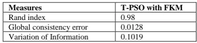
Where, p indicates the pixel, X1 and X2 indicates the segmentation inputs, which produces the output in the range of [0::1]. The Variation Of Information (VOI) finds the distance between two segmentations based on the average conditional entropy. Table 2 shows the rand index, GCE and VOI of the proposed T-PSO with FKM techniques, from this analysis, it is evaluated that the proposed segmentation technique provides the results in terms of increased rand index, and reduced GCE and VOI.
Table 2. Segmentation results of T-PSO with FKM
Measures T-PSO with FKM
Rand index 0.98 Global consistency error 0.0128 Variation of Information 0.1019
Comparative analysis
To prove the superiority of the proposed segmentation technique, it is compared with the existing techniques (27) based on the measures of dice, sensitivity, and specificity. During this analysis, the BRATS 2013 dataset is used to evaluate the results of the existing and proposed techniques. From this analysis, it is
observed that the proposed technique provides the better results, when compared to the existing technique.
Table 2. Comparison between existing and proposed techniques
Methods Dice Specificity Sensitivity T-PSO with FKM 0.91 0.9 0.94 Input Cascade CNN 0.88 0.89 0.87 Tustison 0.87 0.85 0.89 MFCascade CNN 0.86 0.92 0.81 Two Path CNN 0.85 0.93 0.8 LocalCascadeCNN 0.88 0.91 0.84 LocalPathCNN 0.85 0.91 0.8 Meier 0.82 0.76 0.92 Reza 0.83 0.82 0.86 Zhao 0.84 0.8 0.89 Cordier 0.84 0.88 0.81 Festa 0.72 0.77 0.72 Doyle 0.71 0.66 0.87
CONCLUSION AND FUTURE WORK
This paper presents a new segmentation system for segmenting the tumor region from the brain MRIs. For this purpose, a T-PSO with FKM techniques are developed in this work, in which the segmented results of both techniques are compared for identifying the common segmented region. Here, the FMAF filtering technique was used to preprocess the image by eliminating the noise and improving the quality. Then, the features of the preprocessed image are extracted by the use of XCS-LDP technique. The novel techniques such as T-PSO and FKM are implemented to segment the tumor portions from the image based on its features. Finally, the common pixels that are detected by these techniques are considered as a tumor portion. During evaluation, the segmentation results of the proposed system is evaluated and compared for improving the effectiveness of T-PSO and FKM techniques. The measures that are used to evaluate the results are accuracy, sensitivity, specificity, similarity coefficients, rand index, GCE, ROI, precision and recall. In this analysis, it is proved that the proposed T-PSO with FKM provides the better results, when compared to the existing segmentation technique.
In future, this work can be enhanced by implementing this segmentation technique for different types of imaging modalities.
REFERENCES
1. Gordillo N, Montseny E, Sobrevilla P. State of the art survey on MRI brain tumor segmentation. Magnetic resonance imaging. 2013;31(8):1426-38.
2. Joshi A, Charan V, Prince S, editors. A novel methodology for brain tumor detection based on two stage segmentation of MRI images. Advanced Computing and Communication Systems, 2015 International Conference on; 2015: IEEE. 3. Menze BH, Jakab A, Bauer S, Kalpathy-Cramer J, Farahani
K, Kirby J, et al. The multimodal brain tumor image segmentation benchmark (BRATS). IEEE transactions on medical imaging. 2015;34(10):1993-2024.
4. El-Dahshan E-SA, Mohsen HM, Revett K, Salem A-BM. Computer-aided diagnosis of human brain tumor through MRI: A survey and a new algorithm. Expert systems with Applications. 2014;41(11):5526-45.
5. Liu J, Li M, Wang J, Wu F, Liu T, Pan Y. A survey of MRI-based brain tumor segmentation methods. Tsinghua Science and Technology. 2014;19(6):578-95.
7. Nerurkar SN. Brain Tumor Detection using Image Segmentation. Brain. 2017;4(4).
8. Abdel-Maksoud E, Elmogy M, Al-Awadi R. Brain tumor segmentation based on a hybrid clustering technique. Egyptian Informatics Journal. 2015;16(1):71-81.
9. Rajeshwari S, Sharmila TS, editors. Efficient quality analysis of MRI image using preprocessing techniques. 2013 IEEE Conference on Information & Communication Technologies; 2013 11-12 April 2013.
10.Malathi R, AR DNK, editors. Brain Tumor Detection and Identification Using K-Means Clustering Technique. Proceedings of the UGC Sponsored National Conference on Advanced Networking and Applications; 2015.
11.Kong Y, Deng Y, Dai Q. Discriminative clustering and feature selection for brain MRI segmentation. IEEE Signal Processing Letters. 2015;22(5):573-7.
12.Bron EE, Smits M, Niessen WJ, Klein S. Feature selection based on the SVM weight vector for classification of dementia. IEEE journal of biomedical and health informatics. 2015;19(5):1617-26.
13.Nabizadeh N, Kubat M. Brain tumors detection and segmentation in MR images: Gabor wavelet vs. statistical features. Computers & Electrical Engineering. 2015;45:286-301.
14.Islam A, Reza SM, Iftekharuddin KM. Multifractal texture estimation for detection and segmentation of brain tumors. IEEE Transactions on Biomedical Engineering. 2013;60(11):3204-15.
15.Njeh I, Sallemi L, Ayed IB, Chtourou K, Lehericy S, Galanaud D, et al. 3D multimodal MRI brain glioma tumor and edema segmentation: a graph cut distribution matching approach. Computerized Medical Imaging and Graphics. 2015;40:108-19.
16.Moeskops P, Benders MJ, Chiţǎ SM, Kersbergen KJ, Groenendaal F, de Vries LS, et al. Automatic segmentation of MR brain images of preterm infants using supervised classification. NeuroImage. 2015;118:628-41.
17.Moreno JC, Prasath VS, Proenca H, Palaniappan K. Fast and globally convex multiphase active contours for brain MRI segmentation. Computer Vision and Image Understanding. 2014;125:237-50.
18.Adhikari SK, Sing JK, Basu DK, Nasipuri M. Conditional spatial fuzzy C-means clustering algorithm for segmentation of MRI images. Applied Soft Computing. 2015;34:758-69.
19.Vishnuvarthanan G, Rajasekaran MP, Subbaraj P, Vishnuvarthanan A. An unsupervised learning method with a clustering approach for tumor identification and tissue segmentation in magnetic resonance brain images. Applied Soft Computing. 2016;38:190-212.
20.Demirhan A, Törü M, Güler İ. Segmentation of tumor and edema along with healthy tissues of brain using wavelets and neural networks. IEEE journal of biomedical and health informatics. 2015;19(4):1451-8.
21.Kim J, Valdes-Hernandez MdC, Royle NA, Park J. Hippocampal shape modeling based on a progressive template surface deformation and its verification. IEEE transactions on medical imaging. 2015;34(6):1242-61. 22.Roy S, Carass A, Prince JL, Pham DL, editors. Subject
specific sparse dictionary learning for atlas based brain MRI segmentation. International Workshop on Machine Learning in Medical Imaging; 2014: Springer.
23.Kumari A, Mehra R. Design of hybrid method PSO and SVM for detection of brain neoplasm. International Journal of Engineering and Advanced Technology. 2014;3(4):262-6. 24.Preetha R, Suresh GR, editors. Performance Analysis of
Fuzzy C Means Algorithm in Automated Detection of Brain Tumor. 2014 World Congress on Computing and Communication Technologies; 2014 Feb. 27 2014-March 1 2014.
25.Sridhar D, Krishna IM, editors. Brain Tumor Classification using Discrete Cosine Transform and Probabilistic Neural Network. 2013 International Conference on Signal Processing , Image Processing & Pattern Recognition; 2013 7-8 Feb. 2013.
26.SICAS. BRATS: Brain Tumor Image Segmentation
Challenge 2016. Available from:
https://www.smir.ch/BRATS/Start2016.
27.Havaei M, Davy A, Warde-Farley D, Biard A, Courville A, Bengio Y, et al. Brain tumor segmentation with deep neural networks. Medical image analysis. 2017;35:18-31.
Cite this article as:
S. Gopinath and D. Somasundareswari. A combination of threshold based particle swarm optimization and fuzzy K-means segmentation techniques for MRI brain tumor detection. Int. Res. J. Pharm. 2017;8(11):153-162 http://dx.doi.org/10.7897/2230-8407.0811235
Source of support: Nil, Conflict of interest: None Declared